International Journal of
Molecular Sciences
Review
Natural Molecules and Neuroprotection: Kynurenic Acid, Pantethine and α -Lipoic Acid
Fanni Tóth1, Edina Katalin Cseh1and LászlóVécsei1,2,*
Citation:Tóth, F.; Cseh, E.K.; Vécsei, L. Natural Molecules and
Neuroprotection: Kynurenic Acid, Pantethine andα-Lipoic Acid.Int. J.
Mol. Sci.2021,22, 403. https://doi.
org/10.3390/ijms22010403
Received: 13 November 2020 Accepted: 29 December 2020 Published: 2 January 2021
Publisher’s Note: MDPI stays neu- tral with regard to jurisdictional clai- ms in published maps and institutio- nal affiliations.
Copyright:© 2021 by the authors. Li- censee MDPI, Basel, Switzerland.
This article is an open access article distributed under the terms and con- ditions of the Creative Commons At- tribution (CC BY) license (https://
creativecommons.org/licenses/by/
4.0/).
1 Department of Neurology, Interdisciplinary Excellence Centre, Faculty of Medicine, University of Szeged, Semmelweis Street 6, H-6725 Szeged, Hungary; toth.fanni@med.u-szeged.hu (F.T.);
csehedina.k@gmail.com (E.K.C.)
2 MTA-SZTE, Neuroscience Research Group, Semmelweis Street 6, H-6725 Szeged, Hungary
* Correspondence: vecsei.laszlo@med.u-szeged.hu; Tel.: +36-62-545-351
Abstract:The incidence of neurodegenerative diseases has increased greatly worldwide due to the rise in life expectancy. In spite of notable development in the understanding of these disorders, there has been limited success in the development of neuroprotective agents that can slow the progression of the disease and prevent neuronal death. Some natural products and molecules are very promising neuroprotective agents because of their structural diversity and wide variety of biological activities.
In addition to their neuroprotective effect, they are known for their antioxidant, anti-inflammatory and antiapoptotic effects and often serve as a starting point for drug discovery. In this review, the following natural molecules are discussed: firstly, kynurenic acid, the main neuroprotective agent formed via the kynurenine pathway of tryptophan metabolism, as it is known mainly for its role in glutamate excitotoxicity, secondly, the dietary supplement pantethine, that is many sided, well tolerated and safe, and the third molecule,α-lipoic acid is a universal antioxidant. As a conclusion, because of their beneficial properties, these molecules are potential candidates for neuroprotective therapies suitable in managing neurodegenerative diseases.
Keywords: neurodegenerative diseases; neuroprotection; kynurenine pathway; kynurenic acid;
pantethine;α-lipoic acid; Alzheimer’s disease; Parkinson’s disease; Huntington’s disease; multi- ple sclerosis
1. Introduction
Neuronal damage in the central nervous system (CNS) is universal in neurodegener- ative diseases (NDs) [1]. NDs are defined by the progressive loss of neurons in the CNS, which generates deficits in brain function [2]. The symptoms of NDs vary from memory and cognitive deficits to the deterioration of one’s capability to breath or move [3]. The most frequent NDs are Alzheimer’s disease (AD), Parkinson’s disease (PD), Huntington’s disease (HD), amyotrophic lateral sclerosis (ALS) and multiple sclerosis (MS). The inci- dence of NDs has increased greatly worldwide due to the rise in life expectancy, and this associates them with profound social and economic burdens [4].
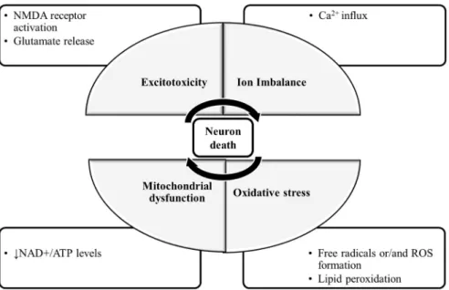
The molecular mechanisms of neuronal damage are mostly based on excitatory amino acid release and oxidative stress, causing mitochondrial dysfunction [5] (Figure1). Under physiological conditions, in the CNS, excitatory amino acids are crucial neurotransmitters and their release and uptake are very well controlled. Nonetheless, their accumulation can lead to brain damage [6]. In glutamate-induced excitotoxicity, glutamate activates N-methyl-D-aspartic acid receptors (NMDARs), leading to a Ca2+ overload [7]. This process is associated with increased reactive oxygen species (ROS), as well as mitochondrial dysfunction resulting in neuronal apoptosis [8,9]. The brain is more vulnerable to damage by oxidative stress due to its high content of polyunsaturated fatty acids [10], that are very prone to ROS attacks [11], which result in lipid peroxidation. Furthermore, the brain has a
Int. J. Mol. Sci.2021,22, 403. https://doi.org/10.3390/ijms22010403 https://www.mdpi.com/journal/ijms
Int. J. Mol. Sci.2021,22, 403 2 of 25
high rate of O2utilization and a low antioxidant defense, and the accumulated metals like copper or iron are capable of catalyzing the formation of hydroxyl radicals [12].
Int. J. Mol. Sci. 2021, 22, x FOR PEER REVIEW 2 of 25
the brain has a high rate of O
2utilization and a low antioxidant defense, and the accumu- lated metals like copper or iron are capable of catalyzing the formation of hydroxyl radi- cals [12].
Figure 1. The molecular mechanisms of neuronal damage. The mechanisms of neuronal death are focused on excitatory amino acid release, calcium influx and calcium overload, oxidative stress and mitochondrial dysfunction. During glutamate-induced excitotoxicity, glutamate activates NMDA receptors, leading to a Ca2+ influx and overload, which is associated with increased ROS formation and the damaged mitochondria resulting in neuronal death. Abbreviations: NMDA receptor: N- methyl-D-aspartic acid receptor; NAD+: nicotinamide adenine dinucleotide; ATP: adenosine triphos- phate; ROS: reactive oxygen species.
Neuroprotection denotes approaches that defend the CNS against neuronal injury and/or death while subjected to trauma or neurodegenerative disorders. It slows the pro- gression of the disease and prevents neuronal death [13]. Hence, neuroprotection is a cru- cial part of care for NDs [14].
Neuroprotection can be classified into three groups: pharmacological-, non-pharma- cological- and cellular and genetic approaches. Pharmacological approaches include anti- oxidants, neurotransmitter agonists/antagonists, anti-inflammatory drugs and natural products [15]. Non-pharmacological approaches include exercise that influences body metabolism [16], diet control to reduce risk factors such as hyperlipidemia [17] and acu- puncture, that can help adjust body metabolism and immunity [18]. Cellular and genetic approaches include growth/trophic factors [19].
Considering there are various changes that occur in the aging brain, it is implausible that targeting a single change is able to intervene in the complexity of the disease progres- sion. Hence, compounds with multiple biological activities affecting the different age-as- sociated factors that contribute to ND development and progression are extremely needed [20]. The existing therapies available for NDs only relieve symptoms [21].
Natural products (including natural molecules) are defined as organic compounds syn- thesized by living organisms. Some of them are very promising neuroprotective agents be- cause of their structural diversity and wide variety of biological activities [4]. The major neuroprotective targets of natural products and molecules are excitotoxicity, apoptosis, mi- tochondrial dysfunction, inflammation, oxidative stress and protein misfolding [22,23].
They have anti-neurodegenerative, antioxidant, anti-inflammatory and antiapoptotic effects [24,25]. Natural products and molecules are commonly used as starting points for drug dis- covery, from which synthetic analogs are synthetized to improve efficacy, potency and to
Figure 1.The molecular mechanisms of neuronal damage. The mechanisms of neuronal death are focused on excitatory amino acid release, calcium influx and calcium overload, oxidative stress and mitochondrial dysfunction. During glutamate-induced excitotoxicity, glutamate activates NMDA receptors, leading to a Ca2+influx and overload, which is associated with increased ROS forma- tion and the damaged mitochondria resulting in neuronal death. Abbreviations: NMDA receptor:
N-methyl-D-aspartic acid receptor; NAD+: nicotinamide adenine dinucleotide; ATP: adenosine triphosphate; ROS: reactive oxygen species.
Neuroprotection denotes approaches that defend the CNS against neuronal injury and/or death while subjected to trauma or neurodegenerative disorders. It slows the progression of the disease and prevents neuronal death [13]. Hence, neuroprotection is a crucial part of care for NDs [14].
Neuroprotection can be classified into three groups: pharmacological-, non-pharmacological- and cellular and genetic approaches. Pharmacological approaches include antioxidants, neu- rotransmitter agonists/antagonists, anti-inflammatory drugs and natural products [15]. Non- pharmacological approaches include exercise that influences body metabolism [16], diet control to reduce risk factors such as hyperlipidemia [17] and acupuncture, that can help adjust body metabolism and immunity [18]. Cellular and genetic approaches include growth/trophic fac- tors [19].
Considering there are various changes that occur in the aging brain, it is implausi- ble that targeting a single change is able to intervene in the complexity of the disease progression. Hence, compounds with multiple biological activities affecting the different age-associated factors that contribute to ND development and progression are extremely needed [20]. The existing therapies available for NDs only relieve symptoms [21].
Natural products (including natural molecules) are defined as organic compounds synthesized by living organisms. Some of them are very promising neuroprotective agents because of their structural diversity and wide variety of biological activities [4]. The major neuroprotective targets of natural products and molecules are excitotoxicity, apoptosis, mitochondrial dysfunction, inflammation, oxidative stress and protein misfolding [22,23].
They have anti-neurodegenerative, antioxidant, anti-inflammatory and antiapoptotic ef- fects [24,25]. Natural products and molecules are commonly used as starting points for drug discovery, from which synthetic analogs are synthetized to improve efficacy, potency and to reduce side effects and to increase bioavailability. A lot of the U.S. Food and Drug Administration (FDA)-approved drugs are prompted by natural products [26].
Int. J. Mol. Sci.2021,22, 403 3 of 25
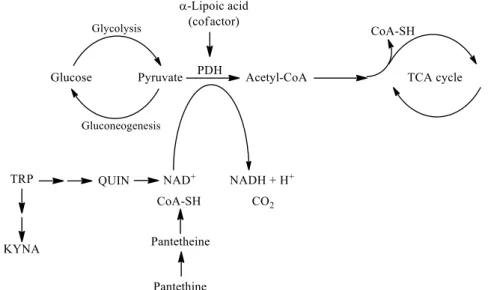
In this review, the following natural molecules are discussed: kynurenic acid, pan- tethine andα-lipoic acid. In the kynurenine pathway (KP) route of tryptophan (TRP) metabolism, neuroprotective kynurenic acid (KYNA) and the neurotoxic quinolinic acid (QUIN) are formed. QUIN can be further converted to nicotinamide adenine dinucleotide (NAD+) [27], which has a cardinal role in energy metabolism [28]. Kynurenines, the metabo- lites of KP, often have pro- and antioxidant properties with the aromatic hydroxyl acting as an electron acceptor [29]. Pantethine is a neuroprotective reducing agent, it is a precursor in the formation of coenzyme A (CoA). CoA functions as an acetyl carrier. It enables the transfer of acetyl groups from pyruvate to oxaloacetate, initiating the tricarboxylic acid (TCA) cycle [30]. Pyruvate dehydrogenase (PDH) is a mitochondrial matrix multienzyme complex that provides the link between glycolysis and the TCA cycle by catalyzing the conversion of pyruvate to acetyl-CoA [31].α-lipoic acid (LA) has a redox active disulfide group and in the mitochondria it functions as a cofactor for PDH E2 subunit [32] (Figure 2.). Common features of all three natural molecules include that they are (i) neuroprotec- tive (ii) antioxidant (iii) reducing agents, which have important roles in glycolysis and the TCA cycle. This makes them potential candidates for neuroprotective therapies for managing NDs.
Int. J. Mol. Sci. 2021, 22, x FOR PEER REVIEW 3 of 25
reduce side effects and to increase bioavailability. A lot of the U.S. Food and Drug Admin- istration (FDA)-approved drugs are prompted by natural products [26].
In this review, the following natural molecules are discussed: kynurenic acid, pan- tethine and α-lipoic acid. In the kynurenine pathway (KP) route of tryptophan (TRP) me- tabolism, neuroprotective kynurenic acid (KYNA) and the neurotoxic quinolinic acid (QUIN) are formed. QUIN can be further converted to nicotinamide adenine dinucleotide (NAD
+) [27], which has a cardinal role in energy metabolism [28]. Kynurenines, the me- tabolites of KP, often have pro- and antioxidant properties with the aromatic hydroxyl acting as an electron acceptor [29]. Pantethine is a neuroprotective reducing agent, it is a precursor in the formation of coenzyme A (CoA). CoA functions as an acetyl carrier. It enables the transfer of acetyl groups from pyruvate to oxaloacetate, initiating the tricar- boxylic acid (TCA) cycle [30]. Pyruvate dehydrogenase (PDH) is a mitochondrial matrix multienzyme complex that provides the link between glycolysis and the TCA cycle by catalyzing the conversion of pyruvate to acetyl-CoA [31]. α-lipoic acid (LA) has a redox active disulfide group and in the mitochondria it functions as a cofactor for PDH E2 sub- unit [32] (Figure 2.). Common features of all three natural molecules include that they are (i) neuroprotective (ii) antioxidant (iii) reducing agents, which have important roles in glycolysis and the TCA cycle. This makes them potential candidates for neuroprotective therapies for managing NDs.
Figure 2. The roles of kynurenine pathway metabolites, pantethine and α-lipoic acid in glycolysis and TCA cycle. PDH is the link between glycolysis and the TCA cycle. α-lipoic acid functions as a cofactor for pyruvate dehydrogenase. The kynurenine pathway of TRP metabolism ultimately leads to the formation of NAD+, which will be reduced to NADH. Pantethine is a precursor in the for- mation of CoA, which functions as an acetyl carrier. It transfers acetyl groups from pyruvate to oxaloacetate, initiating the TCA cycle. Abbreviations: TRP: tryptophan; KYNA: kynurenic acid;
QUIN: quinolinic acid; NAD+: nicotinamide adenine dinucleotide; CoA: coenzyme A; TCA: tricar- boxylic acid; PDH: pyruvate dehydrogenase.
2. Kynurenine Pathway, with Focus on Kynurenic Acid
Kynurenines are considered a hot topic nowadays, as in the last 20 years (2000–2020) more than 4600 articles have been published on the topic [33].
TRP is an essential amino acid, a building block for protein synthesis and also a pre- cursor for the synthesis of serotonin, KYNA and NAD
+. The main metabolic route of TRP degradation is through the KP. More than 95% of TRP is metabolized through this route, and only 5% is degraded through the methoxy-indole pathway [34]. The KP is activated by free radicals, interferons and cytokines, which induce the activity of indoleamine 2,3-
Figure 2.The roles of kynurenine pathway metabolites, pantethine andα-lipoic acid in glycolysis and TCA cycle. PDH is the link between glycolysis and the TCA cycle.α-lipoic acid functions as a cofactor for pyruvate dehydrogenase. The kynurenine pathway of TRP metabolism ultimately leads to the formation of NAD+, which will be reduced to NADH. Pantethine is a precursor in the formation of CoA, which functions as an acetyl carrier. It transfers acetyl groups from pyruvate to oxaloacetate, initiating the TCA cycle. Abbreviations: TRP: tryptophan; KYNA: kynurenic acid; QUIN: quinolinic acid; NAD+: nicotinamide adenine dinucleotide; CoA: coenzyme A; TCA: tricarboxylic acid; PDH:pyruvate dehydrogenase.
2. Kynurenine Pathway, with Focus on Kynurenic Acid
Kynurenines are considered a hot topic nowadays, as in the last 20 years (2000–2020) more than 4600 articles have been published on the topic [33].
TRP is an essential amino acid, a building block for protein synthesis and also a precursor for the synthesis of serotonin, KYNA and NAD+. The main metabolic route of TRP degradation is through the KP. More than 95% of TRP is metabolized through this route, and only 5% is degraded through the methoxy-indole pathway [34]. The KP is activated by free radicals, interferons and cytokines, which induce the activity of indoleamine 2,3-dioxygenase (IDO) [35] and tryptophan 2,3-dioxygenase (TDO) enzymes [36], the rate-limiting enzymes of the pathway [37].
The KP is comprised of several enzymatic steps (Figure3), which ultimately lead to the formation of NAD+, which has a pivotal role in different cellular functions (energy
Int. J. Mol. Sci.2021,22, 403 4 of 25
metabolism, gene expression, cell death and regulation of calcium homeostasis) [28]. KP’s center compound is L-kynurenine (KYN) [38], which can be further degraded through three different routes, resulting in several neuroactive metabolites. KYNA, the main neuroprotective agent is formed after KYN is catalyzed by the enzyme kynurenine amino- transferase (KAT) [39], whereas QUIN [27] and 3-hydroxy-L-kynurenine (3HK) [40] both show neurotoxic properties.
Int. J. Mol. Sci. 2021, 22, x FOR PEER REVIEW 4 of 25
dioxygenase (IDO) [35] and tryptophan 2,3-dioxygenase (TDO) enzymes [36], the rate- limiting enzymes of the pathway [37].
The KP is comprised of several enzymatic steps (Figure 3), which ultimately lead to the formation of NAD
+, which has a pivotal role in different cellular functions (energy metabolism, gene expression, cell death and regulation of calcium homeostasis) [28]. KP’s center compound is L-kynurenine (KYN) [38], which can be further degraded through three different routes, resulting in several neuroactive metabolites. KYNA, the main neu- roprotective agent is formed after KYN is catalyzed by the enzyme kynurenine ami- notransferase (KAT) [39], whereas QUIN [27] and 3-hydroxy-L-kynurenine (3HK) [40]
both show neurotoxic properties.
Figure 3. The kynurenine pathway. More than 95% of TRP is metabolized through the KP [34].
The L-tryptophan converting IDO and TDO depict the rate-limiting enzymes of the pathway [37]. KP’s center metabolite is L-kynurenine [38] which can be further degraded through three distinct routes to form several neuroactive metabolites: kynurenic acid, 3-hydroxy- L-kynurenine and quinolinic acid, which are cardinal in the CNS. Kynurenic acid is formed from L-kynurenine in astrocytes and neurons by KATs [39]. Quinolinic acid and 3-hydroxy- L-kynurenine are synthesized by infiltrating macrophages and microglia [27]. Neurotoxicity is mediated by quinolinic acid and 3-hydroxy-L-kynurenine (color red) via NMDA receptor ag- onism and free radical production [40], while neuroprotection can be exerted by kynurenic acid (color green) by acting as an antagonist at the NMDA receptor [41]. Abbreviations: TRP:
tryptophan; KP: kynurenine pathway; CNS: central nervous system; NMDA receptor: N-me- thyl-D-aspartic acid receptor; 3HAO: 3-hydroxyanthranilate oxidase; ACMSD: 2-amino-3-car- boxymuconate-semialdehyde decarboxylase; AMSDH: 2-aminomuconate-6-semialdehyde de- hydrogenase; IDO/TDO: indoleamine 2,3-dioxygenase/tryptophan 2,3-dioxygenase; KAT:
Figure 3.The kynurenine pathway. More than 95% of TRP is metabolized through the KP [34]. The L-tryptophan converting IDO and TDO depict the rate-limiting enzymes of the pathway [37]. KP’s center metabolite is L-kynurenine [38] which can be further degraded through three distinct routes to form several neuroactive metabolites: kynurenic acid, 3-hydroxy-L- kynurenine and quinolinic acid, which are cardinal in the CNS. Kynurenic acid is formed from L-kynurenine in astrocytes and neurons by KATs [39]. Quinolinic acid and 3-hydroxy-L-kynurenine are synthesized by infiltrating macrophages and microglia [27]. Neurotoxicity is mediated by quinolinic acid and 3-hydroxy-L-kynurenine (color red) via NMDA receptor agonism and free radical production [40], while neuroprotection can be exerted by kynurenic acid (color green) by acting as an antagonist at the NMDA receptor [41]. Abbreviations: TRP: tryptophan; KP: kynurenine pathway; CNS: central nervous system; NMDA receptor: N-methyl-D-aspartic acid receptor; 3HAO: 3-hydroxyanthranilate oxidase; ACMSD: 2-amino-3- carboxymuconate-semialdehyde decarboxylase; AMSDH: 2-aminomuconate-6-semialdehyde dehydrogenase; IDO/TDO:
indoleamine 2,3-dioxygenase/tryptophan 2,3-dioxygenase; KAT: kynurenine aminotransferase; KMO: kynurenine 3- monooxygenase; NAD+: nicotinamide adenine dinucleotide; QPRT: quinolinic acid phosphoribosyltransferase.
In the CNS, KYNA acts on multiple receptors. KYNA is an endogenous competitive antagonist with high affinity at the strychnine-insensitive glycine-binding NR1 site of NM-
Int. J. Mol. Sci.2021,22, 403 5 of 25
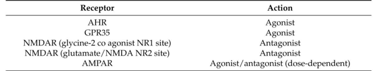
DARs, it exerts antidepressant and psychotomimetic effects [41,42]. KYNA can bind to the NMDA recognition NR2 site of the receptor as well, albeit with a weaker affinity [43,44]. It also acts uponα-amino-3-hydroxy-5-methyl-4-isoxazole-propionic acid (AMPA) receptors via two distinct mechanisms: at low (nanomolar to micromolar) concentrations, it facilitates AMPA receptor responses, whereas at high (millimolar) concentrations, it competitively antagonizes glutamate receptors [45,46]. KYNA exerts an endogenous agonistic effect on the orphan G protein-coupled receptor (GPR35) [47]. Additionally, KYNA is an endoge- nous agonist at the aryl hydrocarbon receptor (AHR), expressed in immune cells and in tumor cells [48,49] (Table1). In 2001, it was suggested by Hilmas et al. that KYNA is a noncompetitive inhibitor of theα7 nicotinic acetylcholine receptor (α-7nAChR) [50]; how- ever, this hypothesis is much debated. The current standpoint is that it does not directly affect nicotinic receptors and results concerning it should only be explained by KYNA’s confirmed sites of action [51].
Table 1.Main binding sites of kynurenic acid.
Receptor Action
AHR Agonist
GPR35 Agonist
NMDAR (glycine-2 co agonist NR1 site) Antagonist
NMDAR (glutamate/NMDA NR2 site) Antagonist
AMPAR Agonist/antagonist (dose-dependent)
Abbreviations: GPR35: G protein-coupled receptor 35; AHR: aryl hydrocarbon receptor; NMDAR:N-methyl-D- aspartic acid receptor; AMPAR:α-amino-3-hydroxy-5-methyl-4-isoxazolepropionic acid receptor.
Since KYNA acts upon multiple receptors, an abnormal decrease or increase in its level may disrupt the equilibrium of neurotransmitter systems, as it can be seen in vari- ous neurodegenerative- and neuropsychiatric disorders. KYNA could have therapeutic importance for neurological disorders [52], but since its ability to cross the blood–brain barrier (BBB) is limited [53], its use as a neuroprotective agent is somewhat limited. One option to prevent neurodegenerative diseases includes the influence of the metabolism towards the neuroprotective branch of the KP. Three potential therapeutic strategies for drug development are known: (i) KYNA analogs with better bioavailability and higher affinity to the binding sites of excitatory receptors; (ii) prodrugs of KYNA, which easily cross the BBB combined with an organic acid transport inhibitor to increase brain KYNA levels; and (iii) inhibitors of enzymes of the KP [54].
2.1. Kynurenic Acid and Alzheimer’s Disease
AD is a progressive irreversible neurodegenerative disorder, with age-related memory impairments and personality changes. It is known to be the most common cause of dementia [55]. In the pathogenesis of AD, senile plaques which are formed by extracellular deposits of amyloidβpeptides (Aβ) and neurofibrillary tangles containing an intracellular assemblage of hyperphosphorylated tau protein play crucial roles [56]. Aβoligomerization leads to oxidative stress, glutamate excitotoxicity and neuroinflammation, which are all connected to the KP [57]. Alterations in KP leading to a switch towards the production of neurotoxic QUIN have contributed to the pathogenesis of AD. Hence, KP is a potential target for AD therapy [51]. In animal models of AD, the application of 4-Cl-KYN, the BBB- penetrant pro-drug of 7-Cl-KYNA, mitigated hippocampal toxicity caused by QUIN [58].
Another way to enhance the bioavailability of KYNA is to improve its penetration through the BBB [59]. A BBB-permeable KYNA analog was found to be neuroprotective in a Caenorhabditis elegansmodel of AD [60]. Alterations causing elevated KYNA levels in the brain may be associated with cognitive impairments and behavioral alterations [61]. In mice, KAT-II enzyme knock out lead to the improvement of cognitive functions [62].
Regarding human studies, Gulaj et al. [63] found lower TRP and KYNA concentrations and a significant increase in QUIN in AD patient’s plasma compared to controls.
Int. J. Mol. Sci.2021,22, 403 6 of 25
In AD patients, positive correlations between cognitive function tests and plasma KYNA levels were observed. In contrast, inversely association was found between plasma QUIN levels and cognitive function tests in patients with AD.
In the hippocampus and neocortex of AD patients, high IDO and QUIN expression was found [64]. A correlation between the KYN/TRP ratio and cognitive dysfunction was demonstrated too [65]. In AD patients, lower KYNA levels were found in the serum and erythrocytes, [66] and in the lumbar cerebrospinal fluid (CSF), but no alteration in QUIN levels was found [67] (Table2).
Aβ1-42 stimulates IDO expression in the human microglia and macrophages where it induces QUIN production [68]. Aβ1-42 and QUIN stimulate cytokine production [69] and increases the hyperphosphorylation of tau proteins via NMDA receptor overactivation, leading to glutamate excitotoxicity in patients with AD [70] and it plays a pivotal role in lipid peroxidation and ROS production, in an NMDAR-dependent or -independent manner, assisting to the pathogenesis of AD [71,72].
Overall, the treatment of AD via the modulation of KP seems to be a logical target of investigation.
2.2. Kynurenic Acid and Parkinson’s Disease
PD is a chronic progressive neurodegenerative disorder with a complex dysfunction of the motor network. The pathological characteristic of the disease is the loss of dopaminergic neurons in the substantia nigra pars compacta (SNpc), the development of Lewy bodies [73]
and the generation of local inflammation. As inflammatory processes lead to the activation of microglia, the KP is activated, generating QUIN, which leads to the excitotoxic cell death of neurons [74]. Other hallmarks of the pathogenesis of the disorder is mitochondrial dysfunction, which results in oxidative stress and cell energy insufficiency. Immune mechanisms also lead to oxidative stress and apoptosis. Abnormal protein aggregation and glutamate excitotoxicity are also involved in neuronal cell death [75].
Alterations in the KP were seen in animal models of PD. After 1-methyl-4-phenyl- 1,2,3,6-tetrahydropyridine (MPTP) treatment, a reduction in the KAT-I activity in the SNpc of mice was observed [76]. The administration of 6-hydroxydopamine (6-OHDA) substantially diminishes KAT-I immunoreactivity in the SNpc neurons [77]. Moreover, 1- methyl-4-phenylpyridinium ion (MPP+) caused a decrease in KAT-II activity in rat cerebral cortical slices, leading to the depletion of KYNA [78]. These studies demonstrated the shift of the KP pathway towards 3HK and QUIN, leading to a decline in KYNA levels, generating neurotoxicity and cell death [79]. A pharmacological approach in the treatment of PD could be the modulation of enzymes involved in the KP to increase the level of neuroprotective intermediates and to reduce the neurotoxic ones [80]. KYNA levels are increased by nicotinylalanine, kynureninase and kynurenine 3-monooxygenase (KMO) inhibitor, which protects against NMDA and QUIN toxicity [81]. One approach for increasing KYNA levels in the brain is to prevent the excretion of KYNA from the brain by probenecid [82].
Dopaminergic toxicity was decreased by the co-administration of KYN and probenecid in rats treated with 6-OHDA [83]. Another approach is the use of KYNA analogs or pro-drugs of KYNA. Moreover, 7-Cl-KYNA, a synthetic derivative of KYNA, protected against neurotoxicity caused by QUIN [84]. Glucosamine–kynurenic acid had a similar effect as KYNA, and its effects suggest it might cross the BBB [85]. In cynomolgus monkeys treated with MPTP, a prolonged systemic administration of Ro61-8048, a KMO inhibitor, increased serum KYN and KYNA levels and decreased the development of levodopa (L-dopa)-induced dyskinesias, but it did not influence the anti-Parkinsonian efficacy of L-dopa [86].
Regarding the human studies, an altered KP has been found in PD patients [87]. In human post-mortem examinations of PD patients who had taken L-dopa, KYNA levels were significantly reduced in the frontal cortex compared to the control group, whereas KYNA showed lower concentrations in the putamen compared to PD patients who were not treated with L-dopa. In both groups, the KYN levels were reduced in the frontal cortex,
Int. J. Mol. Sci.2021,22, 403 7 of 25
in the putamen and in the SNpc compared to controls, this level was found to be the lowest in patients with L-dopa [88]. An increased KYN/TRP ratio was demonstrated in the serum and CSF of PD patients, compared to healthy controls [87]. In the serum of PD patients KAT I and KAT II activities are reduced, with a decreased plasma KYNA level [89]
(Table2). The low level of KYNA has a reduced ability to limit excitotoxicity, through NMDARs, which are induced by QUIN and/or glutamate excess [90]. In PD patients, elevated 3-OH-KYN concentrations were demonstrated in the frontal cortex, putamen and SNpc [88]. In the therapy of PD, some problems remain unsolved: motor and non-motor issues due to therapy, and the need for neuroprotective therapies [91]. The modulation of the KP could possibly be a successful therapeutic strategy for solving these issues.
2.3. Kynurenic Acid and Huntington’s Disease
HD is an autosomal dominantly inherited neurodegenerative disorder with motor, cognitive, and psychiatric symptoms [92]. It is caused by a mutation in the gene coding for the huntingtin protein [93]. The mutant huntingtin protein can sensitize the NMDA receptors [94] and QUIN can act upon the NMDARs and exert damaging effects [95].
Glutamate-induced excitotoxicity plays a crucial role in Huntington’s disease develop- ment [96], which can be influenced by kynurenines [97].
In animal models of HD, alterations were observed in the KP. Genetic inhibition of TDO and KMO leads to a neuroprotective shift toward KYNA synthesis and amelio- rates neurodegeneration in aDrosophila melanogaster model of HD [98]. In a study by Mazarei et al., the inhibition of IDO-1 is likely neuroprotective in HD [99]. A novel KYNA amide compound, which was synthesized in collaboration with our group,N-(2-N,N- dimethylaminoethyl)-4-oxo-1H-quinoline-2-carboxamide hydrochloride, had protective effects in the N171-82Q transgenic HD mouse model where it increased survival, mitigated their hypolocomotion, stopped weight loss and prevented striatal neurons atrophy [100]. At the neuroprotective dose, the KYNA analog did not show any significant side effects, it did not modify the working memory performance, or the long-lasting, consolidated reference memory as opposed to the side effects seen following KYN administration [101,102].
In human studies, IDO-1 activity and KYN levels are increased in the blood of HD pa- tients [103], whereas in their striatum KYNA levels and KAT activity are reduced [104,105].
KYNA levels show a significant decrease in the CSF and in cortex [106,107] (Table2), whereas 3HK and QUIN levels are increased in the brain of HD patients [108]. The inhibi- tion of KMO ameliorates neurodegeneration in the mice model of HD [109].
Overall, the KP has therefore become an obvious therapeutic target for the treatment of HD [27].
2.4. Kynurenic Acid and Amyotrophic Lateral Sclerosis
ALS is a fatal, neurodegenerative disorder that affects the human motor system.
Neuroinflammation is important in ALS, causing the death of motor neurons, and it is also associated with excitotoxicity [110], ROS generation, oxidative stress and lipid peroxidation, the latter one being an important feature in QUIN toxicity [111].
In ALS patients, significantly increased levels of CSF TRP, KYN and QUIN and decreased levels of serum picolinic acid (PIC) were found [54,112]. In ALS patients with bulbar onset, the CSF KYNA levels were higher compared to controls and patients with limb onset, also in patients with severe clinical status, the CSF KYNA concentrations were elevated compared to the controls, demonstrating KYNA’s neuroprotective role against excitotoxicity [113] (Table2). At present, the drugs approved for ALS treatment are riluzole, that targets glutamate-mediated excitotoxicity, increasing life expectancy by 2–3 months [114] and edaravone, which is effective in halting ALS progression during the early stages [115]. In identifying candidate drugs for ALS treatment, agents targeting the KP may yield a novel treatment strategy [116].
Int. J. Mol. Sci.2021,22, 403 8 of 25
2.5. Kynurenic Acid and Multiple Sclerosis
MS is a progressive, inflammatory, demyelinating disease of the CNS that causes a chronic neurological disability. According to Raine et al. [117], autoreactive T cells and macrophages infiltrate the CNS and attack oligodendrocytes, which myelinate axons, to be the trigger for acute MS lesions. The experimental autoimmune encephalomyelitis (EAE) model is histologically similar to human MS. In the EAE model, numerous disturbances in the KP have been described, i.e., in EAE-induced mice IDO-1 inhibition upon disease induction significantly worsened the disease severity [118], in EAE induced rats increased activity of KMO was observed, whereas KMO inhibition by Ro61-8048 decreased QUIN levels in the spinal cord [119].
In MS patients there is an imbalance in neuroactive and neurotoxic KP metabolites in MS disease pathogenesis [120]. A significant reduction in TRP levels in serum and CSF was observed, indicating the activation of KP in MS [121]. Furthermore, KP activation is a result of cytokines interferon-γ(IFN-γ) and tumor necrosis factor (TNF)-αproduction, causing IDO-1 expression [122]. In MS patients’ red blood cells, the KAT I and II activities were significantly higher compared to the controls [120]. This rise was correlated with an increase in plasma KYNA levels, which suggests a counterbalancing protective mechanism against neurotoxicity [120]. In the CSF of MS patients during acute relapse, increased KYNA levels were found [123]. In contrast, during the inactive chronic phase a decrease in KYNA concentration was seen [124] (Table2). Lim et al. [125] have created a predictive model for the disease subtypes using six predictors and metabolomic analysis to profile the KP from the serum of MS patients. The model analyzes the levels of KYNA, QUIN, TRP, PIC, fibroblast growth-factor, and TNF-αto project the disease course with an 85–91%
sensitivity. In the future metabolic profiling of the KP may possibly predict clinical course and disease severity [126].
Table 2.Kynurenic acid and kynurenine aminotransferase alterations in neurological diseases.
Alzheimer’s disease
Plasma KYNA↓[63]
Serum, erythrocytes KYNA↓[66]
CSF KYNA↓[67]
Parkinson’s disease
Frontal cortex, putamen (L-dopa treatment) KYNA↓[88]
Plasma KYNA↓[89]
Serum KAT I, KAT II↓[89]
Huntington’s disease
Cortex KYNA↓[107]
Striatum KYNA↓, KAT↓[104,105]
CSF KYNA↓[106]
Amyotrophic lateral sclerosis
CSF (patients with bulbar onset or severe
clinical status) KYNA↑[113]
Multiple sclerosis
Plasma KYNA↑[120]
Erythrocytes KAT I, KAT II↑[120]
CSF (patients with acute relapse) KYNA↑[123]
CSF (patients with chronic remission) KYNA↓[124]
Abbreviations:↓: decrease in level;↑: increase in level; KYNA: kynurenic acid; CSF: cerebrospinal fluid; KAT:
kynurenine aminotransferase.
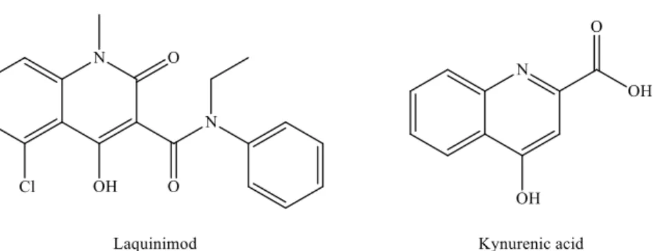
The available MS treatments are all anti-inflammatory, but there is a need for neuropro- tective agents or for drugs that facilitate remyelination. In the search for novel therapeutic candidates by modulating the KP pathway, laquinimod is of particular interest. Laquin-
Int. J. Mol. Sci.2021,22, 403 9 of 25
imod [127,128] displays a remarkable structural similarity to KYNA (Figure4), and it ameliorated inflammatory demyelination, metabolic oligodendrocyte injury, with further anti-inflammatory effects in the cuprizone-treated mice, a model of MS [129].
Int. J. Mol. Sci. 2021, 22, x FOR PEER REVIEW 8 of 25
bulbar onset, the CSF KYNA levels were higher compared to controls and patients with limb onset, also in patients with severe clinical status, the CSF KYNA concentrations were elevated compared to the controls, demonstrating KYNA’s neuroprotective role against excitotoxicity [113] (Table 2). At present, the drugs approved for ALS treatment are rilu- zole, that targets glutamate-mediated excitotoxicity, increasing life expectancy by 2–3 months [114] and edaravone, which is effective in halting ALS progression during the early stages [115]. In identifying candidate drugs for ALS treatment, agents targeting the KP may yield a novel treatment strategy [116].
2.5. Kynurenic Acid and Multiple Sclerosis
MS is a progressive, inflammatory, demyelinating disease of the CNS that causes a chronic neurological disability. According to Raine et al. [117], autoreactive T cells and macrophages infiltrate the CNS and attack oligodendrocytes, which myelinate axons, to be the trigger for acute MS lesions. The experimental autoimmune encephalomyelitis (EAE) model is histologically similar to human MS. In the EAE model, numerous disturb- ances in the KP have been described, i.e., in EAE-induced mice IDO-1 inhibition upon disease induction significantly worsened the disease severity [118], in EAE induced rats increased activity of KMO was observed, whereas KMO inhibition by Ro61-8048 de- creased QUIN levels in the spinal cord [119].
In MS patients there is an imbalance in neuroactive and neurotoxic KP metabolites in MS disease pathogenesis [120]. A significant reduction in TRP levels in serum and CSF was observed, indicating the activation of KP in MS [121]. Furthermore, KP activation is a result of cytokines interferon-γ (IFN-γ) and tumor necrosis factor (TNF)-α production, causing IDO-1 expression [122]. In MS patients’ red blood cells, the KAT I and II activities were significantly higher compared to the controls [120]. This rise was correlated with an increase in plasma KYNA levels, which suggests a counterbalancing protective mecha- nism against neurotoxicity [120]. In the CSF of MS patients during acute relapse, increased KYNA levels were found [123]. In contrast, during the inactive chronic phase a decrease in KYNA concentration was seen [124] (Table 2). Lim et al. [125] have created a predictive model for the disease subtypes using six predictors and metabolomic analysis to profile the KP from the serum of MS patients. The model analyzes the levels of KYNA, QUIN, TRP, PIC, fibroblast growth-factor, and TNF-α to project the disease course with an 85–
91% sensitivity. In the future metabolic profiling of the KP may possibly predict clinical course and disease severity [126].
The available MS treatments are all anti-inflammatory, but there is a need for neuro- protective agents or for drugs that facilitate remyelination. In the search for novel thera- peutic candidates by modulating the KP pathway, laquinimod is of particular interest.
Laquinimod [127,128] displays a remarkable structural similarity to KYNA (Figure 4), and it ameliorated inflammatory demyelination, metabolic oligodendrocyte injury, with fur- ther anti-inflammatory effects in the cuprizone-treated mice, a model of MS [129].
Figure 4. Structural similarities between laquinimod and kynurenic acid. Figure 4.Structural similarities between laquinimod and kynurenic acid.
It has further shown immunomodulatory and neuroprotective features, rather than immunosuppressive effects in relapsing-remitting MS (RR-MS) patients [130]. Laquinimod was not approved by the Committee for Medicinal Products for Human Use (CHMP) because of severe adverse effects. As reported by the CHMP, in animal studies, a higher occurrence of malignancy was found after long-term exposure to laquinimod. Even though in clinical trials no treatment-related cancer was detected, the CHMP concluded that after laquinimod treatment, long-term cancer risk could not be ruled out. Additionally, in animal studies, laquinimod has a possible teratogenic effect. Because laquinimod only had a modest effect in clinical trials, the CHMP came to the conclusion that the possible risk of long-term laquinimod treatment outweighs its advantageous effect [128].
3. Pantethine
A common feature of pantethine and tryptophan metabolism is that they have a metabolite somehow connected to the TCA cycle, i.e., in the KP route of TRP metabolism, the neurotoxic compound QUIN will lead to the formation of NAD+, whereas pantethine is responsible for the formation of CoA, important in the delivery of the acetyl-group to the TCA cycle. Pantethine’s importance was revealed in 1949, when a new compound, namedLactobacillus bulgaricusfactor (LBF) was discovered due to its capability to promote the growth of Lactobacillus bulgaricus. LBF was universally distributed in the natural materials [131]. LBF was shown to be a fragment of CoA and in the essential growth factor, mercaptoamine was combined with pantothenic acid (Vitamin B5) as an amide [132]. This substance occurs in two forms: pantetheine and pantethine.
Pantetheine is the cysteamine amide analog of pantothenic acid (vitamin B5) and it is an intermediate in the synthesis of CoA. In pantethine, two molecules of pantetheine are linked by a disulfide bridge. This forms the active part of the CoA molecule (Figure5).
Most plants and microorganisms can enzymatically combine pantoic acid withβ- alanine to produce pantothenic acid. Mammals are not able to synthesize pantothenic acid since they lack the enzyme. Different foods contain CoA, pantethine, pantetheine and pantothenic acid, so the endogenous synthesis of CoA can begin with pantothenic acid.
Regarding the metabolism of pantethine, following oral or intravenous intake, pan- tethine is immediately hydrolyzed to pantetheine in the small intestine membranes and in blood. Pantetheine can then be phosphorylated to 40-phosphopantetheine, which is later converted to dephospho-CoA-SH, and finally to CoA-SH in the mitochondria. Pantetheine is transformed to pantothenic acid and cysteamine in hepatocytes. Cysteamine is subse- quently metabolized to taurine or is reused to form pantetheine. Pantothenic acid cannot be further degraded in the liver [30,133] (Figure6).
Int. J. Mol. Sci.Int. J. Mol. Sci. 2021, 22, x FOR PEER REVIEW 2021,22, 403 10 of 2510 of 25
Figure 5. The structure of pantethine and pantetheine.
Most plants and microorganisms can enzymatically combine pantoic acid with β-al- anine to produce pantothenic acid. Mammals are not able to synthesize pantothenic acid since they lack the enzyme. Different foods contain CoA, pantethine, pantetheine and pan- tothenic acid, so the endogenous synthesis of CoA can begin with pantothenic acid.
Regarding the metabolism of pantethine, following oral or intravenous intake, pan- tethine is immediately hydrolyzed to pantetheine in the small intestine membranes and in blood. Pantetheine can then be phosphorylated to 4′-phosphopantetheine, which is later converted to dephospho-CoA-SH, and finally to CoA-SH in the mitochondria. Pan- tetheine is transformed to pantothenic acid and cysteamine in hepatocytes. Cysteamine is subsequently metabolized to taurine or is reused to form pantetheine. Pantothenic acid cannot be further degraded in the liver [30,133] (Figure 6).
Figure 6. The metabolism of pantethine. Abbreviations: CoA: coenzyme-A.
Since cysteamine in high doses depletes somatostatin and prolactin in different organs, the effect of pantethine was investigated on the level of these hormones in different tissues.
Pantethine significantly reduced somatostatin concentrations in the following tissues: duo- denal mucosa, gastric mucosa, pancreas, cerebral cortex and hypothalamus [134]. Prolactin was also markedly reduced in the pituitary and in plasma by pantethine, so it can be con- sidered for the management of hyperprolactinemia [135]. In rats, peripherally injected cys- teamine and to a lesser extent pantethine reduced noradrenaline and increased dopamine
Figure 5.The structure of pantethine and pantetheine.Int. J. Mol. Sci. 2021, 22, x FOR PEER REVIEW 10 of 25
Figure 5. The structure of pantethine and pantetheine.
Most plants and microorganisms can enzymatically combine pantoic acid with β-al- anine to produce pantothenic acid. Mammals are not able to synthesize pantothenic acid since they lack the enzyme. Different foods contain CoA, pantethine, pantetheine and pan- tothenic acid, so the endogenous synthesis of CoA can begin with pantothenic acid.
Regarding the metabolism of pantethine, following oral or intravenous intake, pan- tethine is immediately hydrolyzed to pantetheine in the small intestine membranes and in blood. Pantetheine can then be phosphorylated to 4′-phosphopantetheine, which is later converted to dephospho-CoA-SH, and finally to CoA-SH in the mitochondria. Pan- tetheine is transformed to pantothenic acid and cysteamine in hepatocytes. Cysteamine is subsequently metabolized to taurine or is reused to form pantetheine. Pantothenic acid cannot be further degraded in the liver [30,133] (Figure 6).
Figure 6. The metabolism of pantethine. Abbreviations: CoA: coenzyme-A.
Since cysteamine in high doses depletes somatostatin and prolactin in different organs, the effect of pantethine was investigated on the level of these hormones in different tissues.
Pantethine significantly reduced somatostatin concentrations in the following tissues: duo- denal mucosa, gastric mucosa, pancreas, cerebral cortex and hypothalamus [134]. Prolactin was also markedly reduced in the pituitary and in plasma by pantethine, so it can be con- sidered for the management of hyperprolactinemia [135]. In rats, peripherally injected cys- teamine and to a lesser extent pantethine reduced noradrenaline and increased dopamine
Figure 6.The metabolism of pantethine. Abbreviations: CoA: coenzyme-A.Since cysteamine in high doses depletes somatostatin and prolactin in different organs, the effect of pantethine was investigated on the level of these hormones in different tissues.
Pantethine significantly reduced somatostatin concentrations in the following tissues: duo- denal mucosa, gastric mucosa, pancreas, cerebral cortex and hypothalamus [134]. Prolactin was also markedly reduced in the pituitary and in plasma by pantethine, so it can be con- sidered for the management of hyperprolactinemia [135]. In rats, peripherally injected cys- teamine and to a lesser extent pantethine reduced noradrenaline and increased dopamine and 3,4-dihydroxyphenylacetic acid (DOPAC) hypothalamic concentrations [136]. Carbon tetrachloride-induced hepatotoxicity in rats is protected by pantethine, pantothenic acid and cystamine. Pantethine provided the greatest protection [137].
3.1. Pantethine and Alzheimer’s Disease
In AD, treatments are mainly based on the reduction in Tau hyperphosphorylation or Aβ.
Hence, currently available therapeutic strategies show only moderate symptomatic effects.
In primary cultured astrocytes of 5XFAD mice, pantethine mitigates metabolic dys- functions and decreases astrogliosis and IL-1βproduction [138]. These results associated with pantethine lead to the investigation of its effects in vivo in the 5XFAD (Tg) mouse model of AD. Long-term pantethine treatment significantly reduced glial reactivity and Aβaccumulation and modulated the aggressive attitude of Tg mice. Furthermore, the ex- pression of AD-related genes that are differentially expressed in Tg mice, were significantly
Int. J. Mol. Sci.2021,22, 403 11 of 25
mitigated. The expression of a great number of genes involved in the regulation of Aβ processing and synaptic activities, that are downregulated in Tg mice, were recovered after pantethine treatment.
Based on these results, pantethine could be contemplated as a possible therapeutic option for preventing, slowing, or halting AD progression [139].
3.2. Pantethine and Parkinson’s Disease
The fact that there is a deficiency in the activity of complex I in the substantia nigra of PD patients establishes the link between PD and mitochondria [140]. In the MPTP- mouse model of PD, pantethine reduced MPTP-induced neurotoxicity in treated mice, by enhancing fatty acidβ-oxidation, which causes an increase in the levels of circulating ketone bodies (KB) and an improvement of mitochondrial function [141]. Pantethine protects from MPTP-induced BBB leakage and significantly mitigates clinical scores [142].
If TCA cycle activity is low, acetyl-CoA can be used for the biosynthesis of KB via β-hydroxy-methylglutaryl-CoA (HMG-CoA) synthesis. The primary KB are D-β- hydroxybutyrate (DβHB) and acetoacetate (ACA) generated by hepatocytes and they are transported to the tissues, including the brain [142]. β-OHB and ACA are protective in a wide range of cerebral injuries and diseases and in vitro they maintain neuronal cell integrity and stability [143]. In another MPTP mouse model, different parameters were studied, such as changes in fatty acidβ-oxidation, the L-3-hydroxybutyryl-CoA dehydro- genase activity and the circulating ketone body (KB) levels, which all showed a decrease, however, these alterations were restored by pantethine treatment with improvement of dopaminergic neuron loss and motility disorders. Pantethine’s protective effect was due to the increase in glutathione (GSH) synthesis, the recovery of mitochondrial complex I activity, adenosine triphosphate (ATP) synthesis and oxygen consumption, resulting in neuroprotection against dopaminergic injury [141]. Pantethine has the same effects as KB administration and ketogenic diets, but with multiple advantages, including the prevention of the damaging effect of the long-term administration of high-fat diets, due to its hypolipidemic properties [144].
This natural compound should also be considered as a potential therapy against PD.
3.3. Pantethine and Major Depressive Disorder
Major depressive disorder (MDD) is a common psychiatric disorder [145] and it is treated with antidepressants. Unfortunately, current antidepressants in patients have about a 60% response rate [146]. Currently available main classes of antidepressants are tricyclic antidepressants, monoamine oxidase inhibitors, selective serotonin reuptake inhibitors and serotonin-noradrenalin reuptake inhibitors; which all increase the concentration of monoamines in the synaptic cleft [147]. Antidepressants increase central brain-derived neurotrophic factor (BDNF) levels and the activation of the BDNF-signaling pathway might contribute to their therapeutic mechanism [148].
The neuroprotective effects of cystamine and cysteamine were previously described [149], but due to serious side-effects, including seizures [150] and hepatic vein thrombosis [151], cysteamine-related agents should be further explored in the treatment of MDD. Among these agents, pantethine may be one of the most promising agents, as it is a naturally occurring sub- stance that can be administered orally with hardly any side effects, and it further metabolizes to cysteamine. Another advantage of pantethine is an anti-arteriosclerotic medicine sold by some pharmaceutical companies [152], and many geriatric depression patients may have an arteriosclerotic etiology [153].
3.4. Pantethine and Pantothenate Kinase-Associated Neurodegeneration Syndrome
Pantothenate kinase-associated neurodegeneration (PKAN) syndrome is the most common form of a group of genetic disorders, called neurodegeneration with brain iron accumulation, which are characterized by iron overload in the brain and are diagnosed by radiological and histopathological examinations [154]. PKAN is an autosomal recessive
Int. J. Mol. Sci.2021,22, 403 12 of 25
disease, with common features such as dystonia, dysarthria, rigidity, pigmentary retinal degeneration and brain iron accumulation [155]. PKAN is a result of mutations in the PANK2 gene that codes the mitochondrial enzyme pantothenate kinase 2. This enzyme is required for the de novo synthesis of CoA, as it is involved in the phosphorylation of pantothenate [156]. Reduced PANK2 enzymatic activity is proposed to be responsi- ble for the accumulation of cysteine, that can chelate iron, leading to the formation of free radicals [157]. In addition, CoA deficiency and, as a result, defects in phospholipid metabolism may impair the membranes and cause increased oxidative stress, altering iron homeostasis [158]. Unfortunately, to this day the pathophysiology of PKAN is not fully understood, and there is still no cure to halt or reverse the symptoms.
In a PKAN Drosophila model, pantothenate kinase deficiency caused a neurode- generative phenotype and a reduced lifespan. This Drosophila model revealed that the impairment of pantothenate kinase is linked to decreased levels of CoA, mitochondrial dysfunction and increased protein oxidation. The rescue of the phenotype found in the hy- pomorph mutant dPANK/fbl is obtained by pantethine feeding, which recovers CoA levels, ameliorates mitochondrial function, rescues brain degeneration, and improves locomotor abilities, and extends lifespan [159]. The zebrafish orthologue of hPANK2 can be found on chromosome 13. The downregulation of pank2 can cause a lack of CoA in zebrafish embryos in specific cells and tissues. Compensation of the wild type phenotype can be ob- tained by exposing P2-MO-injected embryos to 30µM pantethine [160]. A Pank2 knockout mouse model did not exactly repeat the human disorder but it showed azoospermia and mitochondrial dysfunctions. This mouse model was challenged with a ketogenic diet to stimulate mitochondrialβ-oxidation lipid use. The ketogenic diet could cause a general damage of bioenergetic metabolism in lack of CoA. Only the low glucose and high lipid content diet-fed Pank2 knockout mice developed a PKAN-like syndrome distinguished by significantly altered mitochondria, serious motor dysfunction, neurodegeneration in the CNS and peripheral nervous system. Pank2 knockout mice had structural alteration of muscle morphology, which was similar with that observed in PKAN patients. Pantethine administration was effective in ameliorating the onset of the neuromuscular phenotype observed in Pank2 knockout mice, which were fed a ketogenic diet [155].
Overall, these data indicate that pantethine administration to PKAN patients should be contemplated as a potential, safe and non-toxic therapeutic approach.
3.5. Other Properties of Pantethine
Pantethine is also effective in alcoholism, hyperlipoproteinemias and dyslipoproteine- mias, cystinosis and cataracts.
The oxidation of ethanol yields acetaldehyde, which is involved in the pathogenesis of alcoholic liver disease [161] and alcohol addiction [162]. Pantethine did not cause side effects at clinical doses of 30–600 mg/day. However, it significantly decreased the acetalde- hyde levels in blood in healthy (non-flushing) subjects. In contrast, this effect was not found in flushing (alcohol-sensitive) subjects [163]. Chronic ethanol treatment diminished acetyl-CoA availability by inhibiting pantothenic acid incorporation into CoA [164]. In chronic alcoholics there is an association between the diminished Ach level in brain, due to ethanol consumption, and the cognitive and memory impairment. The inhibitory effect of ethanol on brain Ach synthesis can be reversed or prevented by pantothenic acid [165].
Clinical investigations with acute and chronic alcohol intoxicated patients are necessary to clarify the therapeutic effects of pantethine in alcoholism.
There are effective drugs for the management of hyperlipoproteinemias and dys- lipoproteinemias available, but their long-term toxicity and hepatopathy may limit their clinical use. Moreover, these diseases require an almost life-long drug administration; so natural products, such as pantethine, have been studied to possibly replace the synthetic drugs. Pantethine has anticatabolic properties and it stimulates fatty acid oxidation. It can normalize dyslipoproteinemia, lower serum lipid levels, and increase the concentration of high-density lipoprotein (HDL) associated cholesterol (Chol) and apo-lipoprotein A-I
Int. J. Mol. Sci.2021,22, 403 13 of 25
(Apo A-I) [30]. The total CoA content is increased in perfused rat liver and in liver ho- mogenate by pantethine [166]. The increased availability of CoA leads to an enhancement of the TCA cycle, and so that it stimulates acetate oxidation at the cost of fatty acid and Chol synthesis. Apolipoprotein B (Apo B) is protected against peroxidation in vitro by the sulfhydryl (−SH) containing antioxidative pantethine [167], decreasing the atherogenic Chol concentration in blood [168]. Clinical trials demonstrated that the administration of pantethine at doses ranging between 300 and 600 mg twice daily was successful in the management of patients with familial or sporadic hyperlipidemia [168,169]. Pantethine is significantly more effective than diet in lowering the serum lipid content (triglyceride (TG), total Chol and low-density lipoprotein cholesterol), with an increase in the HDL-Chol level [170]. As a conclusion, pantethine can be an effective treatment of patients with total serum Chol levels > 200 mg/dL and/or serum triacylglycerol levels > 150 mg/dL [30].
Cystinosis is an autosomal recessive genetic disorder with an abnormal accumulation of cystine in the lysosomes of the cells, ultimately leading to the intracellular crystal formation. It is caused by a mutation on chromosome 17 in the CTNS gene that codes for cystinosin, the lysosomal membrane-specific transporter for cysteine. There are three types of cystinosis: infantile cystinosis, intermediate cystinosis, and non-nephropathic (also known as ocular) cystinosis [171]. Therapy has been aimed at decreasing intralysosomal cystine accumulation. Cysteamine can be prescribed for the treatment of cystinosis, but it has a bad taste and odor, side-effects and a low therapeutic index. Pantethine depleted cystine as effectively as cysteamine in cystinotic fibroblast cell cultures, so pantethine can be an alternative for cystinosis treatment [172].
A cataract is an opacification of the lens of the eye, which causes impaired vision.
Cataracts form when the proteins in the lens of the eye clump together. In the selenite model for cataract, pantethine inhibited lens opacification during cataract formation [173].
Overall, pantethine is a many sided and well tolerated therapeutic agent that appears to deserve much more attention than it has recently received.
4.α-Lipoic Acid
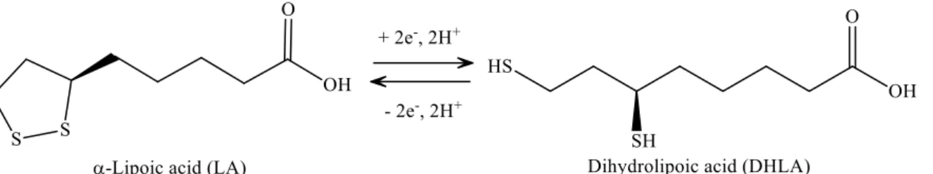
LA has a redox active disulfide group and it is found naturally in mitochondria as the coenzyme for pyruvate dehydrogenase andα-ketoglutarate dehydrogenase. Small amounts of LA are found in foods (spinach and broccoli) and it is also synthesized in the liver [174]. LA was first isolated in 1951 from bovine liver [175]. Dihydrolipoic acid (DHLA), which is the reduced form of LA, interacts with ROS and reactive nitrogen species (RONS) [176]. Both LA and DHLA have antioxidant effects [177]. LA easily penetrates the BBB [178], after which it is quickly internalized by cells and tissues and is reduced to DHLA [179]. LA is active in aqueous or lipophilic [176] environments. Its conjugate base, lipoate, is more soluble and under physiological conditions it is the most common form of LA. It has a highly negative redox potential of−0.32 V [180], therefore, the redox couple LA/DHLA is a “universal antioxidant” (Figure7.) [181].
Int. J. Mol. Sci. 2021, 22, x FOR PEER REVIEW 14 of 25
DHLA [179]. LA is active in aqueous or lipophilic [176] environments. Its conjugate base, lipoate, is more soluble and under physiological conditions it is the most common form of LA. It has a highly negative redox potential of −0.32 V [180], therefore, the redox couple LA/DHLA is a “universal antioxidant” (Figure 7.) [181].
Figure 7. Structure of α-lipoic acid and dihydrolipoic acid.
LA has antioxidant and anti-inflammatory effects [182]. LA supplementation is effec- tive in animal models in obesity and cardiometabolic disorders. LA produces a decrease in body weight [183]. This effect can be clarified by the suppression of protein kinase 5′ aden- osine monophosphate-activated protein kinase (AMPK), its hypothalamic action is pivotal for the regulation of food intake and energy expenditure elevation [184]. In animal models, LA supplementation generated the attenuation of ROS and RONS [185], which are both as- sociated with the reduction in lifespan. The oral supplementation of LA can be a potential supplement in cancer treatment, as it improved survival [186] and reduced unwanted effects of chemotherapy [187]. LA is important in combating inflammation and pain. Positive data on rheumatoid arthritis [188], chronic pain [189], neuropathy [190], migraines [191], ulcera- tive colitis [192] were published.
In various clinical trials which investigated the therapeutic potential of LA, it was concluded that moderate doses (up to 1800 mg/day) were considered safe. Meanwhile, high doses or intraperitoneally administered LA, at a dosage of 5 to 10 g/day, can elevate hydroperoxide levels in the blood [193].
4.1. α-Lipoic Acid and Alzheimer’s Disease
AD’s characteristic features include oxidative stress and energy depletion therefore antioxidants should have positive effects in AD patients. Cultured hippocampal neurons are protected from Aβ-induced neurotoxicity by LA [194], and LA prevents Aβ fibril for- mation too [195].
In an open clinical study, 600 mg LA was given daily to nine patients with AD (re- ceiving a standard treatment with acetylcholinesterase inhibitors) to examine the influ- ence of LA on the progression of AD. LA treatment stabilized cognitive functions in the patients, shown by constant scores in neuropsychological tests (mini-mental state exami- nation, AD assessment scale and cognitive subscale) [196].
LA has the ability to intervene with pathogenic principles of AD, as it stimulates ac- etylcholine (ACh) production by activating choline acetyltransferase and elevating glu- cose uptake, as a result providing more acetyl-CoA for ACh production [197]. LA might represent a potential neuroprotective therapy for AD.
4.2. α-Lipoic Acid and Parkinson’s Disease
The activation of microglia, and the accompanying oxidative stress and neuroinflam- mation play a pivotal role in the pathogenesis of PD [198,199]. In experimental models of PD, lipopolysaccharide (LPS) can be used to activate glial cells [200]. Nasal LPS-induced PD is completely inflammation-driven, and it effectively replicates the chronic, progres- sive PD pathology [201]. LA can block the LPS-induced inflammatory process [202]. LA administration ameliorated motor dysfunction, preserved dopaminergic neurons and re-
Figure 7.Structure ofα-lipoic acid and dihydrolipoic acid.
LA has antioxidant and anti-inflammatory effects [182]. LA supplementation is effec- tive in animal models in obesity and cardiometabolic disorders. LA produces a decrease in body weight [183]. This effect can be clarified by the suppression of protein kinase 50 adenosine monophosphate-activated protein kinase (AMPK), its hypothalamic action is
Int. J. Mol. Sci.2021,22, 403 14 of 25
pivotal for the regulation of food intake and energy expenditure elevation [184]. In animal models, LA supplementation generated the attenuation of ROS and RONS [185], which are both associated with the reduction in lifespan. The oral supplementation of LA can be a potential supplement in cancer treatment, as it improved survival [186] and reduced unwanted effects of chemotherapy [187]. LA is important in combating inflammation and pain. Positive data on rheumatoid arthritis [188], chronic pain [189], neuropathy [190], migraines [191], ulcerative colitis [192] were published.
In various clinical trials which investigated the therapeutic potential of LA, it was concluded that moderate doses (up to 1800 mg/day) were considered safe. Meanwhile, high doses or intraperitoneally administered LA, at a dosage of 5 to 10 g/day, can elevate hydroperoxide levels in the blood [193].
4.1.α-Lipoic Acid and Alzheimer’s Disease
AD’s characteristic features include oxidative stress and energy depletion therefore antioxidants should have positive effects in AD patients. Cultured hippocampal neurons are protected from Aβ-induced neurotoxicity by LA [194], and LA prevents Aβfibril formation too [195].
In an open clinical study, 600 mg LA was given daily to nine patients with AD (receiv- ing a standard treatment with acetylcholinesterase inhibitors) to examine the influence of LA on the progression of AD. LA treatment stabilized cognitive functions in the patients, shown by constant scores in neuropsychological tests (mini-mental state examination, AD assessment scale and cognitive subscale) [196].
LA has the ability to intervene with pathogenic principles of AD, as it stimulates acetylcholine (ACh) production by activating choline acetyltransferase and elevating glu- cose uptake, as a result providing more acetyl-CoA for ACh production [197]. LA might represent a potential neuroprotective therapy for AD.
4.2.α-Lipoic Acid and Parkinson’s Disease
The activation of microglia, and the accompanying oxidative stress and neuroinflam- mation play a pivotal role in the pathogenesis of PD [198,199]. In experimental models of PD, lipopolysaccharide (LPS) can be used to activate glial cells [200]. Nasal LPS-induced PD is completely inflammation-driven, and it effectively replicates the chronic, progressive PD pathology [201]. LA can block the LPS-induced inflammatory process [202]. LA admin- istration ameliorated motor dysfunction, preserved dopaminergic neurons and reduced SN α-synuclein accumulation. In M1 microglia, LA blocked nuclear factor-κB activation and the expression of pro-inflammatory molecules. Further neuroprotective action of LA was studied in an experimental model of PD, induced in male Wistar rats by the intrastriatal injection of 6-OHDA. LA improved learning and memory performance and neuromuscular coordination. LA significantly reduced lipid peroxidation levels and recovered the catalase activity and dopamine levels that were damaged by 6-OHDA administration all which lead to a reduction in oxidative stress [203]. It can be concluded that LA displays significant antiparkinsonian effects.
4.3.α-Lipoic Acid and Huntington’s Disease
There is a link between HD pathogenesis and the mitochondrial energetic defect. In HD post-mortem brain sections, respiratory chain deficits were found and this resulted in the use of mitochondrial complex-II (succinate dehydrogenase, SDH) inhibitors to create toxicity models that reproduced HD striatal pathology in vivo [204]. By using toxin 3-nitropropionic acid (3-NP), an irreversible inhibitor of SDH, it was concluded that mitochondrial dysfunctions contribute to the pathogenesis of HD [205]. 3-NP induces neuropathological changes similar to those observed in HD. In the 3-NP rat model of HD, the neuroprotective effect of LA and acetyl-L-carnitine (ALCAR) on 3-NP-induced alterations in the mitochondrial structure, lipid composition, and memory functions was investigated. The combined supplementation of LA + ALCAR improved mitochondrial
Int. J. Mol. Sci.2021,22, 403 15 of 25
lipid composition, blocked mitochondrial structural changes, and mitigated cognitive deficits in 3-NP-treated animals. Thus, a combined supplementation of LA + ALCAR can be a possible therapeutic strategy in HD management [206].
Excitotoxicity and oxidative damage are involved in the pathogenesis of HD. LA dietary supplementation increases unbound lipoic acid, its antioxidant effect ameliorates oxidative stress in vitro and in vivo [179]. It was examined whether LA exerted neuropro- tective effects in transgenic mouse models of HD. LA generated significant increases in survival in R6/2 and N171-82Q transgenic mouse models of HD.
These results indicate that LA may have valuable effects in HD patients [207].
4.4.α-Lipoic Acid and Multiple Sclerosis
In MS, oxidative stress, and the excess of ROS or RONS are among the contributors to neuronal- and axonal injury [208–210]. Animal model studies showed LA treatment decreased matrix metalloproteinase (MMP), IFN-γ, and interleukin-4 (IL-4) [211] and the reduced expression of soluble cell adhesion molecule (ICAM) in spinal cord tissue [212].
Two animal studies showed that LA had dose-dependent effects, with higher dosages (100µg/mL versus 25µg/mL) being more effective and oral administration being less effective than injections [211,212].
Studies investigating the effects of LA on demyelination and axonal damage in optic nerve, spinal cord, and brain reported that LA-treated EAE animals had reduced damage in the CNS, which was timing- and route of administration-dependent [211,213,214]. LA administration by intraperitoneal (i.p.) injection seven days or directly after immunization protected axons from demyelination and damage [211,213]. Delayed LA administration also decreased damage to the optic nerve but not as profoundly as the immediate treat- ment [213]. Oral administration was only protective immediately, but not delayed after EAE immunization [211]. LA treatment led to a reduced disease severity in EAE model animals [215].
Concerning the effects of LA on anti-oxidant/inflammatory mediators, a reduction in T cell infiltration into the CNS was found after LA treatment in the spinal cord [214], optic nerve [213], and cerebellum [216]. In terms of the effects of LA on mediators of inflammation, like MMP-9 or ICAM, in MS patients they observed varying results. In the serum of MS patients, 1200 mg LA administered daily for 14 days did not alter the serum levels of MMP-9, a tissue inhibitor of metalloproteinases (TIMP-1), or ICAM [217].
In contrast, another group after 12 weeks of AL treatment, observed marked reductions in IFN-γ, ICAM-1, TGF-β, and IL [218].
The use of LA is desirable in numerous areas of health. LA has various beneficial effects on the aging process and neurological disorders. More evaluation is needed to better instruct health professionals on the safety of prescribing LA as a supplement.
5. Conclusions
In conclusion, the extensive research and development of natural products including KYNA, pantethine andα-lipoic acid, will lead to information which will potentially enable novel drug discovery. The neuroprotective property of these compounds makes them worthy of much more attention and contemplates them as a possible therapeutic option for several neurological diseases.
Author Contributions:Conceptualization, F.T.; writing—original draft preparation, F.T.; writing—
review and editing, F.T., L.V., E.K.C.; visualization, F.T.; project administration, F.T.; supervision, L.V.;
figures, E.K.C. All authors have read and agreed to the published version of the manuscript.
Funding:This research was funded by GINOP 2.3.2-15-2016-00034, NKFIH-1279-2/2020 TKP 2020 Thematic Excellence Programme and University of Szeged Open Access Fund, Grant number 5043.
Acknowledgments: We would like to thank the University of Szeged Institutional Open Access Program for funding the article processing charge.
Conflicts of Interest:The authors declare no conflict of interest.
![Figure 3. The kynurenine pathway. More than 95% of TRP is metabolized through the KP [34]](https://thumb-eu.123doks.com/thumbv2/9dokorg/963465.57018/4.892.89.811.267.876/figure-kynurenine-pathway-trp-metabolized-kp.webp)