XII. POLYSACCHARIDES (7) Parti
General Aspects and Phyto and Microbial Polysaccharides
R O Y L. WHISTLER AND W. M. CORBETT
Polysaccharides are high-molecular-weight carbohydrates. They may be viewed as condensation polymers in which monosaccharides (or their deriv- atives such as the uronic acids and aminosugars) have been glycosidically joined with the elimination of water according to the empirical equation:
n C6H12Oe -+ [CeH10O6]„ + (n - 1)H20
From the reverse direction this equation states that polysaccharides, on complete hydrolysis, yield only simple sugars (or their derivatives). Poly- saccharides and oligosaccharides (Chapter IX) are thus, to this extent, defined similarly. However, the term polysaccharide is limited generally to those monosaccharide condensation polymers which contain ten or more monosaccharide residues. As was seen in Chapter IX, oligosaccharides contain two to nine sugar residues. Obviously this limitation of the two terms is completely arbitrary. It is justified on the basis that, as found in nature, most oligosaccharides contain two or three monosaccharide units in contrast to most polysaccharides which contain a hundred to several thousand monosaccharide units. Carbohydrates containing 5 to 15 sugar residues have not been found in nature although they surely occur, at least in small amounts. A few natural polysaccharides contain 30 to 100 sugar residues but most contain more.
1. NOMENCLATURE AND CLASSIFICATION
In the early periods of carbohydrate chemistry no systematic nomen- clature existed. A polysaccharide name coined at that time usually re- flected the origin of the polysaccharide or sometimes emphasized some property of the isolated substance. Illustrative examples of such poly- saccharide nomenclature are found in the terms cellulose, the principal
1. For a comprehensive review of polysaccharide chemistry see R. L. Whistler and C. L. Smart, "Polysaccharide Chemistry." Academic Press, New York, 1953.
641
component of cell walls in plants, and starch, a name derived from the Anglo-Saxon "stercan" meaning to stiffen. Names like these were not de- signed with a view toward their being put into a systematic nomenclature.
Yet they have been used so extensively that they must be accepted in spite of their deficiencies.
Progress toward a systematic nomenclature has produced the significant ending -an to designate that a substance is a polysaccharide. (The ending -osan signifies a simple sugar anhydride (see Chapter VII).) Thus, another word for polysaccharide is the generic term glycan. This term is evolved from the generic word glycose, meaning a simple sugar, and the ending -an, signifying a sugar polymer. Although not all older polysaccharide names employ this ending, it is found generally in names of polysaccharides composed of one or two sugar types: araban for an arabinose polymer, xylan for polymers of xylose, mannan for those of mannose, galactan for those of galactose, and galactomannans for galactose-mannose combina- tions. Until recently certain polymers of fructose were called fructosans.
The name of the fructose polymers has been correctly shortened to fruc- tans.
Persistent because of its wide use in the literature is the term pentosan, which designates polysaccharides composed of pentose sugar residues. This obviously unsystematic term has so far not been shortened to pentan or glycopentan. Cellulosan as a term for polysaccharides closely associated with cellulose is little used in modern carbohydrate literature.
Many polysaccharide names ending with the unsystematic and un- desirable -in ending have been changed to end in -an as a step toward uniformity. Such terms are laminaran, carrageenan, lichenan, isolichenan, asparagan, senistran, graminan, tritican, kritesan, phlean, secalan, poan, pyrosan, and irisan. Names which have not been changed because of their long-standing and wide use are: pectin, amylopectin, inulin, chitin, heparin, and chondroitin.
The systematic name glucan does not refer to a specific polysaccharide but signifies only that the polysaccharide is composed of glucose residues.
The manner of Unkage and the arrangement are not specified. The name is a group name only and applies as well to cellulose as to glycogen, laminaran, or other glucose polymers. The polysaccharide can be defined more specifi- cally if a source designation is also employed as part of the name. Thus, a more definite polysaccharide is specified in each case by the designation beechwood xylan, yeast mannan, or peanut araban.
Polysaccharides of the same type differ at least slightly from one source to another. Sometimes the differences are quite marked, as with starches.
There is a well-known and readily apparent difference among starches
xii. POLYSACCHARIDES: PART I 643
from different plants. A particular starch is meant, however, by banana starch or corn starch.
Ideally, the polysaccharides should be classified according to their chemical composition and structure as has been suggested by Whistler (1).
In such a classification, polysaccharides hydrolyzing to only one mono- saccharide type are termed homoglycans while polysaccharides hydrolyz- ing to two or three or more monosaccharide types are termed heteroglycans with prefixes of di-, tri-, and so on to designate the number of different types of sugar units. At present there is no proof that more than five or six types of sugar units occur in a single polysaccharide. The number of types of sugar units contained in a polysaccharide can easily be determined by Chromatographie examination of the hydrolyzate, provided, of course, that the polysaccharide is pure. In this structural classification the first logical subclassification separates polysaccharides as to whether they are linear or branched. This separation can readily be made by performing several simple tests. The easiest test is that of film formation. An aqueous polysaccharide solution when spread on a glass plate and dried will be brittle if a branched polysaccharide is present. Films from linear molecules will be strong, undergo folding without breaking and when plasticized can be stretched with the development of birefringence and a detectable "fi- brous" X-ray pattern. Linear polysaccharides also show streaming bire- fringence when their solutions are stirred and viewed between crossed polarizing plates. Methylation studies may further reveal whether a mole- cule is branched.
Unfortunately, many polysaccharides have not been examined in rigor- ously pure conditions, and often examination has not extended to charac- terization of films. Therefore, at times, it is useful to classify polysaccharides according to source.
In subsequent discussion, the polysaccharides will be discussed from the standpoint of their sources as:
Phytopolysaccharides (Phytoglycans) Bacterial and Fungal Polysaccharides
Zoöpolysaccharides (Zoöglycans) and Conjugates (Glycoproteins and Glycolipides)
The phytopolysaccharides and the bacterial and fungal polysaccharides will be considered in Part I of this chapter, and the zoöpolysaccharides in Part II. This classification, however, has the definite disadvantage that some polysaccharides of identical or similar structure are separated ac- cording to their origin. Thus, amylopectin (plants) and glycogen (animals) are separated. Chitin is found in lower animals, microorganisms, and fungi.
Cellulose, although considered typical of plants, is also produced by some bacteria and lower animals.
2. THE STRUCTURES OF POLYSACCHARIDES
A. GENERAL CONCEPTS
Though polysaccharides may be viewed as condensation polymers formed by the combination of monosaccharides with the elimination of the elements of water, the naturally occurring polysaccharides are far less complicated than would occur if the combination of monomers took place in random fashion. In fact many simplifying features are apparent on careful examination of all known polysaccharide structures. The basic reasons for such simplified and ordered arrangements stem from the action of those specific synthesizing enzymes which produce the monosaccharides and those enzymes which connect the monosaccharides, by various and sometimes complex routes, to polymer structures. Methods by which enzymes produce polysaccharides are given on page 703.
In the condensation of monosaccharides to form natural polymers, the hydroxyl on the anomeric carbon always participates in the condensation.
Since most polysaccharides are composed of aldose sugar units, this dis- cussion will be confined to such units. The hydroxyl on carbon atom 1 (anomeric carbon) may condense with any hydroxyl other than that at C-l on an adjoining monosaccharide unit. In this way, a linear chain can be formed with a free C-l hydroxyl group at one end. A complete random- ness of linkage with the various hydroxyl groups has never been found in nature. Most frequently, a particular mode of linkage is repeated uniformly through the chain. Even the stereoconfiguration of C-l remains constant in most observed cases. Thus, in amylose, there is a uniform α-Ό-1—»4 link- age, in cellulose a uniform β-Ό-1—*4 linkage, and in laminaran an essen- tially uniform β-D-l—>3 linkage.
A further and highly simplifying fact in polysaccharide chemistry is that of the multitude of stereoisomeric monosaccharides only a very few are found in natural polysaccharides. Of the hexoses there are D-glucose, D-man- nose, D-fructose, D-galactose, and infrequently L-galactose, and possibly D-idose or L-altrose. Of the pentoses there are D-xylose, L-arabinose, and infrequently D-arabinose. Of the modified simple sugars there are D-glucos- amine, D-galactosamine, D-glucuronic acid, D-galacturonic acid, D-man- nuronic acid, L-fucose, and L-rhamnose. Even these monosaccharides do not occur at random in polysaccharides but rather are found in a systematic arrangement.
Frequently a polysaccharide consists of but a single type of sugar unit.
The most abundant polysaccharides are of this type. Paramount, as an example, is cellulose, which is present in the world in a quantity equal to or greater than the quantity of all other polysaccharides. Yet cellulose consists of a chain of D-glucopyranose units linked uniformly together by
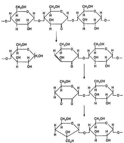
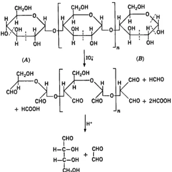
XII. POLYSACCHARIDES: PART I 645 β-D-l—A bonds. The presence of another linkage, once in some 700 links, is not ruled out. Essentially cellulose is a linear chain represented by A in Fig. 1.
Sometimes the hydroxyl groups of C-1 from two sugars have apparently condensed with two hydroxyls other than C-1 on a third sugar unit in a polysaccharide. When this occurs a branch point is produced in the mole- cule. The molecule may contain a single branch as in B of Fig. 1 or it may contain numerous branch points. Sometimes the branch may be but a single sugar unit in length. The molecule then is a substituted linear polysaccharide with sugar units acting as the substituents. Such a structure is C in Fig. 1. In other instances a branch-on-branch structure may occur which may be likened to a bush; a small section is depicted in D of Fig. 1.
In no known instance do polysaccharides occur as a cage or three- dimensional net structure. They are either linear, cyclic, or branched. It is apparent that when a branch point is introduced the glycosidic bond connects different positions from those connected in the linear portions between branches. It is common to find the same kind of glycosidic linkage at all branch points in a homoglycan. If more than one type of sugar unit
OOOOOOOOOOOOO
A
OOOOOOOOOOOOO
^DOOOOO
B
is present it is usual for all units of the same sugar to be linked in the chain by the same sort of glycosidic bond.
Even in a linear homoglycan it is possible for more than one type of glycosidic linkage to be prevalent. In such a molecule the linkages do not occur randomly but are usually in an ordered arrangement.
If two or more types of sugars occur in a polysaccharide, the sugar units, generally, seem to be in an ordered arrangment. Thus, in linear dihetero- glycans, polysaccharides composed of two kinds of sugars, the units seem- ingly are arranged in an alternating and regular fashion. Some dihetero- g]yeans have the structural arrangment illustrated by C of Fig. 1. In this structure the principal chain may be composed of one type of sugar linked uniformly throughout, while the branches are composed of a second type of sugar which may be connected to the main chain by identical glycosidic bonds.
When more than two types of sugars are combined to produce a poly- saccharide, they usually form a branch-on-branch structure exemplified by D in Fig. 1. Even here, some semblance of a simplifying order seems to exist. Thus, it is common to find hexose sugars and perhaps uronic acids in the main or central branches while the pentose sugars, D-xylose and L-arabinose, are in the side branches.
There are, then, in polysaccharides certain naturally imposed simplifica- tions which greatly facilitate their understanding and theii; structural characterization. However, it is possibly correct to say that no polysac- charide is completely uniform. Even such seemingly regular molecules as cellulose and amylose appear to have irregularities or "anomalous links"
in their structures. The irregular linkages are rare and may have a fre- quency of only 1 in each 700 linkages as seems to be true in some celluloses.
Such rare irregularities are explainable on the basis that the various pos- sible glycosidic bonds do not differ greatly in energy; hence, within the enzyme-substrate system there may at times occur a brief change, influen- tial in causing an irregularity to develop in the chain growth. The irregular- ity may be due to a brief abnormal action on the part of the principal chain- synthesizing enzyme or may be due to the interference or the usurping action of a second enzyme. If enzymes are beyond reproach in the forma- tion of irregular bonds, then quite conceivably such infrequent bonds could be produced by chance chemical synthesis.
B. PURIFICATION AND PROOF OF STRUCTURE
Since polysaccharides do not occur pure, it is essential that they be purified before their structures can be determined. Unfortunately, much careful structural work, particularly methylations, has been done on
XII. POLYSACCHARIDES: PART I 647 products of unknown purity. Sometimes impurities may be easily removed, as in the case of cotton cellulose. In other instances, involving the simul- taneous extraction of more than one polysaccharide, tedious procedures are necessary to effect separation and even then purification may be in- complete. Care must be taken when isolating polysaccharides from biologi- cal material to ensure that enzymic degradation does not occur. Enzyme action can be stopped by immersing the fresh tissue in hot ethanol.
Unfortunately, there are no specific tests for absolute purity of poly- saccharides. A polysaccharide may be regarded as free from other poly- saccharides if it may be separated by at least two suitable procedures into fractions each of which have the same physical and chemical properties.
The principal methods for the fractionation of polymeric compounds are listed below. Most of these are applicable to polysaccharides or glyco- proteins.
I. Solubility methods (2, 2a).
A. Fractional precipitation (by adding precipitant or by cooling).
B. Fractional solution (by solvents of varying composition and at varying temperatures).
C. Distribution between immiscible solvents (inclusive of Craig countercurrent distribution).
II. Ultracentrifugation (2, 2b).
III. Ultrafiltration through graded membranes (dialysis and electro- dialysis) (2} 2a).
IV. Methods depending on electrical charge (applicable to polymers which can be ionized or converted to ionic complexes) (2, 2c).
A. Electrophoresis (inclusive of "mobility spectra" and "isoelectric spectra").
B. Electroconvection.
C. Ionography (electrophoresis in stabilized media).
V. Chromatographie adsorption, ion exchange, and partition chroma- tography (inclusive of paper chromatography) (2, 2a, 2d).
VI. Molecular distillation (2, 2a).
2. L. H. Cragg and H. Hammerschlag, Chem. Revs. 39, 79 (1946) ; A. C. Corcoran, ed., "Methods in Medical Research," Vol. 5. Year Book Publishers, Chicago, 1952.
2a. A. Weissberger, ed., "Techniques of Organic Chemistry," 2nd ed., Inter- science, New York, 1954.
2b. A. E. Alexander and P. Johnson, "Colloid Science." Oxford U. P., New York, 1949.
2c. A. Kolin, Proc. Natl. Acad. Sei. (U. S.) 41, 101 (1955); S. Raymond, Proc. Soc.
Exptl. Biol. Med. 81, 278 (1952) ; M. E. Adams, M. L. Karon, and R. E. Reeves, J. Am.
Chem. Soc. 73, 2350 (1951); H. J. McDonald, "Ionography." Year Book Publishers, Chicago, 1955.
2d. E. Lederer and M. Lederer, "Chromatography." Elsevier, New York, 1953.
a. Hydroxyl Groups
The main reactive group present in any polysaccharide is, of course, the hydroxyl group. However, there may be other groups of importance which may occur naturally or be produced by the process of isolation or any other treatment. The average number of hydroxyl groups per sugar residue is usually measured by the determination of the acetyl or methyl groups introduced by acetylation or methylation of the polysaccharide. An estimate of the number of free primary hydroxyl groups may be obtained by tosyla- tion (p-toluenesulfonation) or tritylation (triphenylmethylation). Primary hydroxyl groups are generally tosylated some 20 times faster than secon- dary hydroxyl groups, and under suitable conditions esterification of primary hydroxyl groups only may be achieved. Estimation of the primary tosyl groups is made by reaction of the ester with sodium iodide whereby only primary tosyl groups are replaced by iodine. Primary hydroxyl groups can also be determined by measurement of the number of trityl groups introduced on reaction of the polysaccharide with trityl chloride. This reaction combined with carbanilation may be used to measure the number of both primary and secondary hydroxyl groups (8), Ultraviolet light absorption is used to estimate the number of carbanilate and trityl groups present.
Secondary hydroxyl groups cannot be determined directly. However, when adjacent hydroxyl groups (a-glycol groups) occur in the molecule, as in 1 —■> 4 glucans, they may be determined by oxidation with periodate or lead tetraacetate (see p. 699). Lead tetraacetate oxidations are restricted to nonaqueous solutions, whereas periodate is used for aqueous solutions.
One mole of oxidant is consumed for each carbon bond broken.
b. Uronic Acids
Almost half of the known types of polysaccharides contain uronic acid units. Uronic acid residues may be determined by decarboxylation with 12% hydrochloric acid and the evolved carbon dioxide determined by absorption (4) or by manometric measurement (5). A micro-quantitative colorimetric method employing carbazole has been developed for uronic acids (6), (See also Part II of this chapter and Chapter XL)
8. C. J. Malm, L. J. Tanghe, B. C. Laird, and G. D. Smith, Anal. Chem. 26, 188 (1954).
4. R. L. Whistler, A. R. Martin, and M. Harris, J. Research Natl. Bur. Standards 24, 13 (1940).
5. M. V. Tracey, Biochem. J. 43, 185 (1948).
6. Z. Dische, / . Biol. Chem. 167, 189 (1947); 183, 489 (1950).
XII. POLYSACCHARIDES: PART I 649 c. Other Acidic Groups
Native polysaccharides with acid groups other than the uronic type are not very common except for the sulfate esters. Total acidity may be esti- mated by direct titration, but erroneous results are obtained if the poly- saecharide is alkali-labile as is the case with many oxidized polysaccharides.
Addition of calcium acetate (7, 8) or sodium bromide (8, 9) to the poly- saecharide solution increases the accuracy of the titration. Other methods for the estimation of carboxyl and other acidic groups involve determina- tion of the amount of méthylène blue absorbed, or determination of the amount of silver salt formed by exchange from a solution which contains silver in combination with a very weak acid. The sulfate content of poly- saecharide sulfates, such as agar, is obtained by ordinary sulfate analysis of the completely hydrolyzed or ashed polysaecharide.
d. Carbonyl Groups
Although many methods have been devised for the estimation of car- bonyl groups in polysaccharides, a simple stoichiometric method is not available. The number of aldehyde groups in a polysaecharide is usually measured with alkaline iodine (10). Chlorous acid is also useful (11). The oxidation can be followed by the consumption of oxidant or amount of acid formed. In one colorimetric method, 2,5-dinitrosalicylic acid in al- kaline solution (12) is used. In another, the ferricyanide reduced by car- bonyl groups is measured after conversion to Prussian blue (12a). Two principal sources of difficulty with these methods must be guarded against, over-oxidation and/or alkali-lability of the polysaecharide. Frequent empirical use has been made in cellulose chemistry of the copper number
(a measure of copper reduction) and in starch chemistry of the alkali number (a measure of the alkali consumed in saccharinic acid formation).
Saccharinic acid formation begins at the reducing terminal and progresses through successive removals of sugar moieties (see p. 653).
Several methods have been devised for the determination of total carbonyl groups under neutral conditions. In one method the polysaecharide
7. E. C. Yackel and W. O. Kenyon, J. Am. Chem. Soc. 64, 121 (1942).
8. A. C. Ellington and C. B. Purves, Can. J. Chem. 31, 801 (1953).
8a. A Schwebel, H. S. Isbell, and J. V. Karabinos, Science 113, 465 (1951).
9. C. C. Unruh, P. A. McGee, W. F. Fowler, Jr., and W. O. Kenyon, J. Am. Chem.
Soc. 69, 349 (1947).
10. A. R. Martin, L. Smith, R. L. Whistler, and M. Harris, J. Research Nail. Bur.
Standards 27,449 (1941).
11. H. F. Launer, W. K. Wilson, and J. H. Flynn, J. Research Natl. Bur. Standards 51, 237 (1953).
12. K. H. Meyer, G. Noelting, and P. Bernfeld, Helv. Chim. Ada 31, 103 (1948).
12a. S. Nussenbaum and W. Z. Hassid, Anal. Chem. 24, 501 (1952).
is reacted with phenylhydrazine acetate and the combined nitrogen deter- mined; in another, the polysaccharide is reacted with hydroxylamine hydrochloride or O-methylhydroxylamine hydrochloride and the amount of liberated hydrochloric acid measured. Still another procedure involves the reaction of the carbonyl groups with hydrogen cyanide; the number of combined groups is estimated by measurement of the ammonia liberated on hydrolysis of the cyanohydrins (8), or, if radioactive hydrogen cyanide is used, by the activity of the final polysaccharide (8a).
e. Other Groups
Methyl groups are frequently found as esters of uronic acids and some- times as ethers of sugar residues. Ether-linked methyl groups and total methoxyl are determined by the Zeisel method (13). Methyl esters or glyco- sides may be differentiated from methyl ethers by the action of alkali and acid, respectively, which will saponify the ester or glycoside but have no effect on the ether. The ester or glycosidic methoxyl can be distilled as methanol and be determined colorimetrically after oxidation to formal- dehyde and condensation with Schiff reagent (14)·
Glucosamine and galactosamine in polysaccarides are measured usually by the method of Elson and Morgan after hydrolysis. The aminosugar is condensed with acetylacetone followed by p-dimethylaminobenzaldehyde (Ehrliche reagent) (15). Aminopolysaccharides frequently occur as the ΛΓ-acetates. In general, iV-acetyl groups are more resistant to hydrolysis than O-acetyl groups, being hydrolyzed only by hot, aqueous, strong acids or bases (15d). (See also Chapter VIII and Sialic Acid, Part II.)
/. Nonreducing End Groups
The ratio of nonreducing to reducing end groups in a polysaccharide gives a measure of the extent of branching, for there will be one branch for each nonreducing end unit found above one. The ratio of glycosidic units to nonreducing end units gives the average chain length of the branches.
Periodate oxidation is useful for end-group assay (16-18). A linear
IS. See F. J. Bates and associates, Nail. Bur. Standards Cire. C440, 509 (1942).
14. C. L. Hoffpauir and R. E. Reeves, Anal. Chem. 21, 815 (1949).
15. See E. A. Kabat and M. M. Mayer, "Experimental Immunochemistry," p. 312.
C. C Thomas, Springfield, 111., 1948; N. F. Boas, J. Biol. Chem. 204, 553 (1953).
15a. A. Chaney and M. L. Wolfrom, Abstr. papers, Am. Chem. Soc, Atlantic City p. 4D, 1956.
16. F. Brown, S. Dunstan, T. G. Halsall, E. L. Hirst, and J. K. N. Jones, Nature 156,785 (1945).
17. T. G. Halsall, E. L. Hirst, and J. K. N. Jones, J. Chem. Soc. p. 1427 (1947).
18. A. L. Potter and W. Z. Hassid, / . Am. Chem. Soc. 70, 3488 (1948).
XII. POLYSACCHARIDES: PART J 651 polysaccharide yields definite quantities of formic acid from the nonreducing terminal sugar and of formic acid and formaldehyde from the reducing terminal sugar (p. 700). The formaldehyde is produced in small yield, and, if precautions are not taken, is liable to be further oxidized. Formic acid may be titrated directly. Excess oxidant can be removed by ethylene glycol.
The formic acid liberated from the reducing end groups of a highly branched 1 —> 2, 1 —> 3, or 1 —> 4 polysaccharide will be negligible to that liberated from the nonreducing units, and the total formic acid produced may be taken as a measure of the branch length. This method is not applicable to 1 —> 6 hexans, because each of the sugar units will liberate one mole of formic acid.
The original methylation method introduced by Haworth (19) is still of importance. Paper-chromatographic modifications have made the method applicable at a micro-scale. The nonreducing terminal group of a linear polysaccharide contains one more hydroxyl group than the preceding units, and, if branching occurs, the branch unit will contain one less hy- droxyl group than the adjacent sugar units. These groups may be esti- mated by methylation of the polysaccharide followed by hydrolysis or methanolysis to give the methylated monosaccharides or glycosides. It is important that methylation be complete and that no degradation occur.
Hydrolysis or methanolysis also must be carefully controlled to keep demethylation to a minimum (20).
Originally, quantitative separation of the various methylated glyco- sides was achieved by fractional distillation in high vacuum (19). In the case of amylopectin, methyl 2,3,4,6-tetra-O-methyl- and 2,3,6-tri-O- methylglucosides were isolated as well as a small quantity of a mixture of methyl di-O-methylglucosides. The yield of di-O-methylglucoside, derived from the branching points, should be equal to the yield of the tetra-O- methylglucoside, from the nonreducing terminal groups, but in practice it is greater. This is due to incomplete methylation and to demethylation during methanolysis.
Other methods have been described for the separation of methylated sugars. These include partition between an organic solvent and water (21, 22) and separation on columns of alumina (22), silica gel (23), or charcoal (2J(). A micro method now generally adopted has come into being
19. W. N. Haworth and H. Machemer, J. Chem. Soc. p. 2270 (1932).
20. K. Freudenberg and H. Boppel, Ber. 73, 609 (1940).
21. J. Y. Macdonald, / . Am. Chem. Soc. 57, 771 (1935).
22. F. Brown and J. K. N. Jones, / . Chem. Soc. p. 1344 (1947).
23. D. J. Bell and A. Palmer, Nature 163, 846 (1949).
24. B. Lindberg and B. Wickberg, Acta Chem. Scand. 8, 569 (1954); W. J. Whelan and K. Morgan, Chemistry & Industry p. 78 (1954).
with the application of paper chromatography to carbohydrates {25). The positions of the various methylated monosaccharides which have been separated on sheets of filter paper by development with a butanol solvent are revealed by spraying with suitable reagents. Such components may be partially identified by the distance traveled on the paper and by the color which they produce with various reagents. Quantitative estimation of the methylated sugars is obtained by elution of each from unsprayed sheets of filter paper and oxidation with alkaline iodine or periodate.
Analysis of a mixture of methylated sugars may be obtained by spraying the papers to reveal the location of the various components and then measuring the amount of reflectance or light transmission of the various spots (26). (See also Chapter XI.) Only a very small percentage of tetra- O-methylhexose will be obtained on hydrolysis of a methylated polysac- charide of high molecular weight. Here greater accuracy in end-group determination may be obtained by concentration of the tetra-O-methyl- hexose, relative to the tri-O-methylhexose, by extracting the former from an aqueous solution with chloroform. By paper-sheet or cellulose-column chromatography of methylated polysaccharide hydrolyzates, sufficient amounts of the individual methylated monosaccharides may be obtained to prepare crystalline derivatives for identification (27).
g. Identification of Monosaccharides (see also Chapter XI)
Mixtures of sugars, such as occur in the hydrolyzates of polysaccharides, are frequently difficult to separate into the component sugars. Crystalliza- tions, selective precipitations with chemical reagents, and fermentations have been used when applicable. A more general tool is chromatography, particularly when only small amounts of material are available for study.
Some combinations are easily separated chromatographically and some, for example, fructose and mannose, only with considerable difficulty.
Silicates and activated carbon are particularly useful for adsorption chro- matography and cellulose (powder or paper) for partition chromatography of the sugars.
Paper chromatography provides a rapid, sensitive micro method often enabling trace components to be detected. On paper, artifacts have been detected which were produced by exposure of the sugar to alkali during neutralization of the hydrolyzates. Care should be taken to avoid even brief exposures of the sugars to alkalies. Neutralization through electro- dialysis has been used to avoid this danger. Information as to the identity of the sugar is obtained from the distance of its movement and from the
25. E. L. Hirst, L. Hough, and J. K. N. Jones, J. Chem. Soc. p. 928 (1949).
26. E. F. McFarren, K. Brand, and H. R. Rutkowski, Anal. Chem. 23,1146 (1951).
27. L. Hough, J. K. N. Jones, and W. H. Wadman, J. Chem. Soc. p. 2511 (1949).
xii. POLYSACCHARIDES: PART I 653 colors produced by different sprays (28). However, since many sugars move at the same rate even in several different solvents, confirmatory evidence is necessary. After elution, the monosaccharide at each location on a filter-paper chromatogram may be estimated by micro methods such as those with alkaline iodine (25), Somogyi's reagent (29), or periodate.
The sugars may also be estimated by the density of the spots produced by various sprays (26). Unequivocal identification, however, depends on the recovery of a sufficient quantity of the pure sugar for the determination of its properties and those of its derivatives.
Monosaccharides may be separated by the addition of borax to form negatively charged complexes which are absorbed on columns of basic ion-exchange resins, from which the sugar complexes are preferentially eluted with dilute solutions of sodium borate (30). The complexes may also be separated by ionophoresis because of differences in dissociation constants (81). Individual sugars may be determined by selective fermen- tation or enzymic degradations.
h. Nature of the Glycosidic Linkages
Information on the nature of the glycosidic links present in a polysac- charide may be obtained by investigation of the products formed by the action of alkali, especially lime water. Thus, 1 —» 4 hexans (e.g., hydro- cellulose) produce isosaccharinic acids (82) and 1 —» 3 hexans (laminaran) produce metasaccharinic acids (88).
Use has been made of the relationship between the optical rotation of D-glucans in water and cuprammonium hydroxide solution and that of the methyl mono-O-methyl-D-glucoside which is methylated on the same hydroxyl group as that involved in the glucosidic link of the glucan (84).
See Table I.
The methods most used in identifying the nature of the glycosidic bridge are periodate oxidation and examination of hydrolysis products from the fully methylated polysaccharides. Identification of the O-methylsugars from the hydrolyzed O-methylpolysaccharides suggest the location of glycosidic linkages and acetal rings in the partially methylated monosac- charides. Determination of the amount of formic acid and formaldehyde
28. L. Hough, J. K. N. Jones, and W. H. Wadman, J. Chem. Soc. p. 1702 (1950).
29. A. E. Flood, E. L. Hirst, and J. K. N. Jones, J. Chem. Soc. p. 1679 (1948).
80. J. X. Khym and L. P. Zill, J. Am. Chem. Soc. 73, 2399 (1951).
81. H. Michl, Monatsh. 83, 737 (1952) ; R. Consden and W. M. Stanier, Nature 169, 783 (1952).
32. J. J. Murumow, J. Sack, and B. Tollens, Ber. 34, 1427 (1901).
88. W. M. Corbett, J. Kenner, and G. N. Richards, Chemistry & Industry p. 462 (1953); p. 1483 (1954).
84. R. E. Reeves, J. Biol. Chem. 154, 49 (1944).
TABLE I
SHIFT IN OPTICAL ROTATION D U E TO COMPLEX FORMATION
Substance
Methyl 2-0-methyl-j8-D-glucoside Methyl 3-0-methyl-a-D-glucoside Laminaran
Methyl 4-0 -methyl -0-D-glucoside Cellulose
Soluble starch Glycogen
Methyl 6-0-methyl-j8-D-glucoside
α In 1:1 mixture of water and triton B
M
Water
-69°
-46°
-29°
-36°
-46°*
+375°
+366°
-48°
25 1 436
Cupram- monium hydroxide
+985°
-86°
+34°
-1008°
-1200°
-715°
-597°
+ 161°
Difference
+ 1054°
-40°
+63°
-972°
-1154°
-1090°
-963°
-209°
produced during periodate oxidation sometimes easily identifies the hy- droxyl groups which are not involved in glycosidic linkage or ring forma- tion (p. 700) (86, 86).
Additional information can be gained by reduction and hydrolysis of the oxidation products resulting from the action of periodate. Information concerning the position of the linkages in dextrans was derived by analyti- cal determinations of glycerol, erythritol, and glucose, which result, re- spectively, in this way from 1—>6, 1—>4, and 1—»3 linked glucopyranoses (87).
Final proof of the nature of the glycosidic linkages is obtainable only by partial hydrolysis (or acetolysis) of polysaccharide derivatives to low- molecular-weight oligosaccharides, whose structures, if not already known, may be unambiguously determined (for method see p. 701). This procedure is particularly essential for establishing the order of linkages in a homo- glycan containing different linkages and for establishing the order of sugar residues in a heteroglycan.
Establishment of the stereoconfiguration of the glycosidic bond is more complicated. Specific a- and 0-glycosidase enzymes have been used with considerable success, and application of infrared spectrophotometry seems to be useful (88). In the latter method the presence of a-linked D-glucose
85. J. C. Rankin and A. Jeanes, / . Am. Chem. Soc. 76, 4435 (1954).
86. J. J. Conell, E. L. Hirst, and E. G. V. Percival, / . Chem. Soc. p. 3494 (1950).
87. J. W. Sloan, B. H. Alexander, R. L. Lohmar, I. A. Wolff, and C. E. Rist, / . Am.
Chem. Soc. 76, 4429 (1954).
88. S. A. Barker, E. J. Bourne, M. Stacey, and D. H. Whiffen, J. Chem. Soc. p. 171 (1954).
XII. POLYSACCHARIDES : PART I 655 units is said to be indicated by an absorption peak at 844 db 8 cm.-1, whereas 0-linked residues are said to absorb at 891 ± 7 cm.-1. In the case of a-glucans it may also be possible to determine the position of linkages by their characteristic absorption peaks.
Type of a-glucan linkage 1 - » 2
1 ->3 1 ->4 1 ^ 6
Absorption (cm.""1) Not determined 793 ± 3 930 ± 4:758 =fc 2 917 ± 2:768 ± 1
C. MOLECULAR SIZE AND SHAPE (88a)
Determination of molecular weights is possible by a variety of methods.
Association of the molecules in solution gives rise to complications which may be minimized by the use of suitable solvents, low concentrations, and extrapolating data to infinite dilution. Since all polysaccharides possess a molecular-weight distribution, it is the average molecular weight which is usually measured. Physical and chemical methods based on osmotic pres- sure, depression of freezing point, and end-group assay count the molecules and give a number-average molecular weight, whereas light scattering and certain types of sedimentation methods give a weight average. In addition, sedimentation data give a Z-average molecular weight (89) and viscosity measurements a viscosity-average molecular weight (40). These may be defined as:
Σ(η,ΑΓί) Number-average, Af» =
Weight-average, Mw =
Z-average, Mz =
Viscosity-average, Mv
Σ(η{Μ*) Σ(η<Μ<») Σ(η*ΜΛ
L Σ(η
{Μ<) J
where n< is the number of molecules of molecular weight Mi, and β is a constant equal to unity if Staudinger's law is obeyed.
MJMW is a measure of the heterogeneity of the polysaccharide. For a perfectly homogeneous polymer the ratio would be unity, but the value
88a. See C. T. Greenwood, Advances in Carbohydrate Chem. 7, 289 (1952).
89. W. D. Lansing and E. O. Kraemer, J. Am. Chem. Soc. 67, 1369 (1935); I. Jul- lander, Arkiv. Kemi Mineral Geol. 21A, No. 8 (1945).
40. P. J. Flory, J. Am. Chem. Soc. 65, 372 (1943).
decreases as the heterogeneity of the polymer increases. Molecular weights determined by equilibrium methods, such as by osmotic pressure, are independent of the shape and flexibility of the molecule. On the other hand, kinetic measurements (as for example, determinations of viscosity), which are based on the properties of molecules in motion, may give rise to information concerning the shape and flexibility of the molecules.
a. Osmotic Pressure (2a, 2b)
Molecular weights of polysaccharides may be determined by measuring the osmotic pressure of dilute solutions. The osmotic pressure at infinite dilution, obtained by extrapolating a number of measurements at various low concentrations, is a function of the molecular weight as shown by the modified van't Hoff formula.
7Γ » cRT/M + Bcn
where π is the osmotic pressure in grams per cm.2, c is the concentration of solute in grams per ml., M is the gram-molecular weight, B and n are constants, T is the absolute temperature, and R the gas constant. This method is limited to polysaccharides with molecular weights in the range 10,000 to 500,000 owing to diffusion through the membrane of molecules with molecular weights lower than 10,000, and to the small pressures produced by molecules with a molecular weight greater than 500,000.
b. Sedimentation Velocity (2, 2a, 2b)
A solution of macromolecules, when subjected to centrifugal forces many times greater than gravity, undergoes sedimentation in the plane of rota- tion. Disturbing effects caused by convection currents are minimized by the use of correctly shaped cells. The velocity of sedimentation is followed by observing the changes of light absorption or refractive index. By the use of high angular velocities, initial sedimentation may occur without diffusion effects becoming important. Solutions of homogeneous molecules produce sharp sedimentation boundaries, whereas a mixture of several components differing appreciably in average molecular weight will give separate boundaries for each component. However, if the individual molec- ular weights of a mixture of polymers spread over a wide range, a diffuse boundary occurs and the derived molecular weights will be only an approxi- mation. Measurement of the position of the boundary at various times gives the velocity of sedimentation from which, by dividing it by the centrif- ugal acceleration, is obtained the sedimentation constant (£). This constant extrapolated to zero concentration at standard conditions is S0. The molec- ular weight is derived from the equation:
« SpRT
~ Z>„(1 - VP)
X I I . POLYSACCHARIDES: PART I 657 where V is the partial specific volume of the solute, p is the density of the solution, So is the sedimentation constant at zero concentration and stand- ard conditions, and D0 is the diffusion constant.
The diffusion constant is independently evaluated by observing the changes in concentration of a solution in a stationary tube as the solute diffuses into pure solvent with which the solution forms a liquid boundary.
From this can be calculated the molar frictional coefficient (/).
E TB MQ. - Vp)
f Do So
By comparison of the value with that calculated for a spherical particle of the same weight and density, information may be obtained as to the polymer's molecular configuration.
c. Sedimentation Equilibrium (2, 2a, 2b)
Smaller centrifugal forces are required for this method, but several days of continuous running are necessary to reach an equilibrium between sedimentation and diffusion. The molecular weight is calculated from the equation:
_ 2RT In (C2/d)
(1 - Vp)o>Kx22 - Xi2)
where the terms have the same meaning as above, cx and c2 are the concen- trations of solute at distances Xi and xi, respectively, from the axis of rotation, and ω is the angular velocity. If the plot of c against x2 deviates from a straight line, the polymer is heterogeneous. Measurement of the concentration by fight absorption gives the weight-average molecular weight, whereas refractive methods give the Z-average molecular weight.
d. Viscosity (2a, 2b)
The viscosity of a solution increases with the molecular weight of the solute. By making measurements on solutions of low concentrations, Staudinger derived the following empirical equation:
Vap = KMc or ^ = KM c
where
^solution n
Vsp = 1 = Vr — 1
^solvent
V»P = specific viscosity, K = constant for a particular homologous series, η, = relative viscosity, M = molecular weight of solute, c = concentration
of solute. The constant K shows reasonable constancy for a given homol- ogous series but varies for different series, temperatures, and solvents. The reduced viscosity f — J varies with the concentration, and more accurate results are obtained if the intrinsic viscosity [η] is used. This term is ob- tained by extrapolating the reduced viscosity to infinite dilution.
Originally the value of K for a particular series was derived by studying several lower members of the series. However, the value obtained did not hold for higher members and considerable error was introduced. A more desirable method is to evaluate the constant with the use of members of the series whose molecular weights have been obtained by osmotic pressure or ultracentrifuge methods. The more convenient viscosity method is thereafter used for determining the molecular weights of other members of the same series.
Deviation from Staudinger's equation occurs for molecular weights above 10,000, and a modified equation has been introduced:
fol = KMa
The constant a appears to be a function of the shape and solvation of the molecules. Theoretical calculations predict a to be 0.5 for a matted coil, 1.0 for a randomly linked chain, and 2.0 for a stiff chain. Experimental values vary from 0.53 for polystyrene in butanone to 1.5 for amylose in ethylenediamine. For polymers of molecular weight 100,000 to 1,000,000, the equation is further modified to give
M - KxMa - KM*
where K2 <C K\ and is significant only for large molecules.
e. Light Scattering (2a, 2b)
A beam of light on passage through a solution is partially scattered. This is due mainly to the radiation of light by loosely bound electrons of the solute molecules. The amount of light scattered by each particle is depend- ent on its particle size and may be related to the molecular weight by the equation:
He 1 2Bc T " M + RT where
„ 32rW (μ - μοY H =
ϋΫλΓ V ~ V 7
c = concentration μο = refractive index of solvent r = turbidity, defined by I/Io — e~rl μ — refractive index of solution M = molecular weight (above) λ = wavelength of light
B = constant, identical with that of the N = Avogadro's number modified van't Hoff equation
XII. POLYSACCHARIDES: PART I 659 This equation only holds when the particle size is less than 3^o of the wavelength of the primary light. With particles greater than this critical size (e.g., a glucan with a D.P. greater than 50-60), scattering takes place from more than one point of the molecule to give interference and a dis- symmetry in the angular distribution of scattered light. Dissymmetry is measured by comparing the scattered light intensity at two angles sym- metrical to the 90° position. If the molecular configuration is known, a correction factor for the measured turbidity may be obtained and the molecular weight obtained.
/. Streaming Birefringence (2a, 2b)
In a solution subjected to a velocity gradient, there is a tendency for the particles to align themselves with their long axis parallel to the direc- tion of flow. This results in a difference of refractive index in two directions at right angles. Although it is possible to obtain the molecular weight of polymers by the difference between the two refractive indices, streaming- birefringence measurements are more often used to obtain information on molecular shape. The apparatus for producing the velocity gradient con- sists of two concentric cylinders, one being*stationary while the other rotates.
g. Other Methods
Cryoscopic methods for determination of molecular weights of high polymers are of no practical value because of the small depression of tem- perature, the sensitiveness of the measurements to low-molecular-weight impurities, and the tendency of the solute molecules to associate. Ultrafil- tration through membranes of known pore size and various adaptations of the isopiestic method have been described but they are of limited applica- tion.
3. PHYTOPOLYSACCHARIDES (PHYTOGLYCANS)
A. PLANT CELL WALLS
Polysaccharides are components of almost all living things. They are present in greatest quantity in the higher orders of plants where they constitute approximately three-quarters of the dry weight. The majority of plant polysaccharides are components of the cell walls. In a typical tissue from either an annual or perennial plant, the cell walls consist of three morphologically distinct layers; namely, the intercellular cement, or middle lamella, the primary wall, and the secondary wall, as shown (41) in Fig. 2.
The middle lamella or intercellular layer contains cellulose, xylans, uronic acid - containing polysaccharides, and sometimes a mannan. Poly-
pi. From H. W. Giertz, World Paper Trade Rev. 138, 1451 (1952).
Side view—walls cut away. Transverse section
0. Middle lamella; 1. Primary or cam- 2. Secondary wall: outer layer; 3. Sec- bial wall ondary wall: middle layer; 4. Secondary
wall: inner spiral layer FIG. 2. Cell wall and intercellular layer
saccharides composed of pentose sugar units are the most prevalent but still constitute less than 15% of the layer. The most abundant middle lamellar substance is lignin which constitutes more than 70 % of the layer.
Lignin (/ft) is a three-dimensional, or net, plastic which is rich in aromatic rings and contains such groups as methoxyl, aromatic and aliphatic hy- droxyls, and several types of unsaturation. The material may be a polymer in which the basic unit is a phenylpropyl radical:
4 ^—CH2—CH2—CH2—
The origin of lignin is not known. One hypothesis is that in the aging tissue lignin is built up directly from simple sugars such as sucrose (43, 44)- Its postulated formation from pectin or other polysaccharides has never been experimentally verified. (See also p. 548).
The primary wall is a continuous, fairly pliable membrane forming the outside surface of the plant cell. It is about 0.5 micron in thickness and, thus, is only a small part of the cell. It is heavily lignified but is predomi- nantly carbohydrate, with pectin, uronic acid - containing polysaccharides, and xylan or mannan in abundance and with cellulose in lesser amounts.
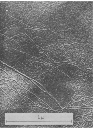
For the most part the very long cellulose molecules are grouped into many fine threads (Fig. 3) which form a meshlike but coherent arrangement.
They are intricately woven and mixed with the other polysaccharides, 42. F. E. Brauns, "The Chemistry of Lignin." Academic Press, New York, 1952.
48. M. Phillips and M. J. Gross, J. Agr. Research 51, 301 (1935).
44- M. Phillips, M. J. Gross, B. L. Davis, and H. Stevens, J. Agr. Research 59, 319 (1939).
XII. POLYSACCHARIDES: PART I 661
FIG. 3. Cellulose threads from Swedish spruce {45)
lignin, protein, organic extractives (tannins, terpenes, alkaloids, fats, sterols, and other substances removable from the plant tissue by hot ethanol and benzene), and inorganic salts. The primary wall expands as the cell grows, but with the attainment of final cell size the primary wall may further thicken with the deposition of cellulose to form a laminated structure. In most seeds and many other organs the wall may attain con- siderable thickness and may contain large amounts of xylan, pectin, and uronic acid containing polysaccharides.
Fibrous and other highly differentiated cells, after their mature size and shape are fully attained, undergo formation of a massive inner or secondary wall. In a few instances, the wall is of a compound nature such as that shown in Fig. 2. Cellulose is the principal component of the secondary wall, but there is present in small amount the other components found in the primary wall. Especially in wood fibers, the secondary wall consists of three layers with the center layer the most massive. From superficial
45. B. G. Rânby, Svensk Papperstidn. 55, 115 (1952).
examination, the massive center secondary wall may appear homogeneous.
Closer inspection shows that it consists of a number of very thin concentric lamellae which in turn consist mainly of closely packed cellulose threads arranged more or less parallel and wound at a spiral angle around the fiber axis. Alternate layers are wound in opposite directions and in different spiral angles.
The major nonprotein fibers of commerce—cotton, wood, ramie, flax, hemp, and jute—are all cellulosic fibers. They differ in the amount of lignin, the degree of polymerization and crystallinity of the polysaccharides, and the shape of the fiber (see next section).
a. Cellulose (46, 47)
The individual threads making up the concentric lamellae or ultra-small layers in the cell wall have a diameter of about 77 A. in wood and about 88 A. in cotton (48). The tiny threads constituting the bulk of the cell wall consist predominantly of cellulose molecules which are themselves threadlike. The molecules are chains of D-glucose units linked uniformly by 1—>4 ß-D-glycosidic bonds.
In undegraded molecules of cotton cellulose some 3000 or more D-glucose units may be combined to form a chain with an extended length of 15,700 A. and upward but with a cross-section of only about 4 A. X 8 A. These very long thin molecules can be coiled and twisted in numerous ways, but, because of the spacial arrangement between D-glucose units produced by the ß-D-1—>4 linkage, the chain is somewhat stiff and tends to remain more or less extended. An additional contribution to rigidity may be made by hydrogen-bonding between the oxygen atoms attached to carbon atoms 6 and 2 of the adjacent glucose residues. During their biosynthesis in the plant, the chains are grouped together to form the strings or threads visible with the electron microscope.
As the cellulose molecules are laid together, there are places where they are woven amongst each other in a random fashion, whereas a little farther on they are fitted together in perfectly ordered, crystalline arrangment.
The tiny threads are, therefore, composed mainly of cellulose molecules lying roughly parallel but with disordered or amorphous regions mixed with ordered or crystalline regions. On X-ray analysis a pattern is obtained which shows definite arcs from the crystalline regions and a halo from the amorphous regions. The appearance of arcs demonstrates that the crystal-
46. See E. Ott, H. M. Spurlin, and M. W. Grafflin, eds., "Cellulose and Cellulose Derivatives." Interscience, New York, 1954.
47. See: L. E. Wise and E. C. Jahn, eds, "Wood Chemistry," 2nd ed. Reinhold, New York, 1952; A. G. Norman, "The Biochemistry of Cellulose, the Polyuronides, Lignin &c." Oxford U. P., New York, 1937.
A8. B. G. Rânby, Ph.D Dissertation, Uppsala, 1952.
XII. POLYSACCHARIDES: PART I 663 line regions are more or less oriented along the long axis of the plant cells.
In the crystalline regions, the chains are directed alternately with "heads,"
in opposite directions. The dimensions of the unit cell are 10.3 A. in the linear chain direction and 8.35 Â. and 7.9 A. in the other directions. The unit cell has a length of two D-glucose units or one cellobiose unit.
The mixture of crystalline and amorphous regions give plant fibers their noteworthy physical properties. Within the crystalline regions, the closely packed chains are held together by numerous secondary forces, especially van der Waals' (ca. 8000 cal. per mole glucose unit) and hydrogen bonds (ca. 15,000 cal. per mole glucose unit). Although each individual hydrogen bond is relatively weak, three such bonds theoretically are possible per glucose unit. In the chain directions, primary valence forces hold the molecule together, the weakest being the carbon-oxygen glucosidic bonds with energies of the order of 50,000 cal. per mole. Plant fibers are conse- quently immensely strong with the tensile strengths of high grade steel.
Even so, the strength of cellulose fibers can be increased by mercerization (treatment with caustic) under tension, a process which causes a reorienta- tion of the chains toward each other.
The secondary bonding forces exert a peculiarly important influence on the reactions of cellulose. Because of the relative stiffness and the dense, tightly bound crystalline regions of cellulose fibers, they can be dissolved only by very energetic solvents, such as cuprammonium solution (copper hydroxide in ammonium hydroxide), which forms a soluble complex with cellulose, or acids such as 42 % hydrochloric acid, strong sulfuric or phos- phoric acids. Other than in the amorphous regions, cellulose is not very reactive chemically because the reagent molecules only slowly penetrate the crystalline portions. However, under some conditions the crystalline areas can be entered and soluble ethers or esters prepared (see p. 691).
Cellulose can be made more reactive by precipitation from solution in an amorphous form or by swelling with alkali or concentrated solutions of certain salts. A thread made by spinning cellulose as it is reprecipitated from solution in cuprammonium is known as cuprammonium rayon or
"Bemberg."
Cellulose is decomposed by cellulase enzymes found in germinating seeds, in fungal and bacterial extracts, and in the digestive juices of snails, Crus- tacea, and certain fish {49). Enzymes for the utilization of cellulose by termites and ruminants are provided by the microflora of the digestive tracts. In the well-studied cases of cellulose decomposition, the initial degradation has been shown to be a hydrolytic cleavage of the cellulose chains, but further attack of the sugars by living organisms often leads 49. See W. Pigman, in "The Enzymes" (J. B. Sumner and K. Myrbäck, eds.), Vol.
I, part 2, p. 725. Academic Press, New York, 1952.
to carbon dioxide and acidic materials. Cotton, wood, and other native celluloses exhibit a much greater resistance to enzymic hydrolysis than swollen or regenerated celluloses. For this reason, it has been common to use treated celluloses for measurements of cellulase activity, since the sensitivity is greatly increased {49). Karrer used cuprammonium rayon;
Pringsheim swelled filter paper with lithium chloride or calcium thio- cyanate; Walseth used cotton linters swollen with phosphoric acid; and Helferich and Goerdeler chose cellophane. Soluble, partially substituted celluloses are also being used, although an additional problem is introduced in the effect of the substituent group.
Enzymes, like acids, attack cellulose most rapidly in the amorphous areas and the reaction slows down as these areas are depleted. Striking differences, however, are observed in the courses of the degradations. A considerable portion of the cellulose sample can be degraded by enzymes to soluble fragments with the retention of a relatively high degree of poly- merization. For example, Walseth {50) found that the degree of polymeri- zation of a swollen cotton cellulose was reduced by enzymes only to about 1000 although there was a 38 % loss of weight. The same sample was re- duced by acid to a degree of polymerization of 80 with only an 8.3 % loss of weight. The attack of each reactant is restricted to accessible areas.
Presumably, the smaller molecules (the acid) can penetrate deeper into the cellulose structure. Another dramatic demonstration of the nature of enzymic attack is available in the attack of cotton cloth by mildew. A cotton cloth subjected to the action of the fungus Chaetomium globosum did not show a decrease in cuprammonium fluidity (a measure of D.P.), although a loss of 80% of its tensile strength occurred {49). In either case, the solubilized portion is rapidly converted to glucose or, in some cases with enzymes, to cellobiose:
Cellulose — > Cellodextrins —> Cellobiose — > D-Glucose
With crude enzymes from Aspergillus niger under optimal conditions (for this enzyme, pH 4.5 at 47°C), Walseth {50) was able to degrade a particu- larly reactive cellulose (preswollen with phosphoric acid) to the extent of 95 % and to account for the loss of weight as D-glucose (by reducing-sugar determinations).
During the reaction brought about by some enzymes which have been separated from the crude extracts by fractional precipitation or by Chroma- tographie adsorption, cellobiose rather than glucose accumulates {49, 51).
60. C. S. Walseth, Tappi 35, 228, 233 (1952).
61. D. R. Whitaker, Arch. Biochem. and Biophys. 43, 253 (1953); R. L. Whistler and C. L. Smart, J. Am. Chem. Soc. 76, 1916 (1953); K. Nisizawa and T. Kotayaski, J. Agr. Chem. Soc. Japan 27, 239 (1953) ; P. Kooiman, P. A. Roelofsen, and S. Sweeris, Enzymologia 16, 237 (1953).
x i i . POLYSACCHARIDES: PART I 665 Hence, the presence of glucose as the final product arises in some instances from the presence of a cellobiase, distinct from the cellulase, in the crude preparation. Evidence has been offered also for a random attack at any position in the chain rather than a splitting-off of successive small units from the end of the chains {52). Reese and co-workers (58) suggest that several enzymes take part in the degradation, the first of which is demon- strated, in the case of cotton, by its ability to attack the primary wall and, thus, to make the cellulose chains more available to hydrolytic attack (54).
In the decomposition of wood by enzymes, the reaction may proceed as an attack on the lignin or on the carbohydrate fraction. The so-called
"white rots" of wood are believed to arise from fungal attack mainly on the lignin component, whereas the "brown rots" may result when the principal attack takes place on the carbohydrate fraction (49, 55).
b. Hemicelluloses and Other Cell-Wall Polysaccharides (46)
Interlaced with cellulose in the primary and secondary walls are a num- ber of other polysaccharides. Most of them are more soluble than cellulose even though they are held in the complex cellulose matrix by abundant secondary forces and by mechanical entanglements and entrapments.
Polysaccharides extractable from the cell walls by alkaline solutions, such as 17.5% sodium hydroxide solution, are called hemicelluloses. (Cellulose is swollen, but not dissolved, by strong alkali.) The name hemicellulose was proposed in 1891 by Schulze (56), who was examining products ex- tracted from leguminous seeds, brans, and green tissues. It was assumed that the easily extractable polysaccharides were destined for conversion to cellulose, and, thus, the name hemicellulose seemed appropriate. Today it is known that these polysaccharides are not precursors of cellulose. They are a group of unrelated polysaccharides which vary in amount and kind from plant to plant and even from tissue to tissue within the same plant.
They consist of acidic and neutral molecules, some of low molecular weight and some of such high molecular weight that they are not easily extracted by strongly alkaline solutions. It is perhaps unfortunate that such a di-
52. See B. Norkrans, Physiol. Plantarum 3,75 (1950) ; Symbolae Botan. Upsalienses 11, 1 (1950); J. H. Hash and K. W. King, Science 120, 1033 (1954).
58. E. T. Reese and W. Gilligan, Textile Research J. 24,663 (1954) ; W. Gilligan and E. T. Reese, Can. J. Microbiol. 1, 90 (1954) ; E. T. Reese and H. S. Levinson, Physiol.
Plantarum 5, 345 (1952); E. T. Reese, W. Gilligan, and B. Norkrans, Physiol. Plan- tarum 5, 379 (1952) ; H. S. Levinson, G. R. Mandels, and E. T. Reese, Arch. Biochem.
and Biophys. 31, 351 (1951).
64. See also: P. B. Marsh, K. Bollenbacher, M. L. Butler, and L. R. Guthrie, Textile Research J. 23, 878 (1953).
65. See: G. Fâhraeus, R. Nilsson, and G. Nilsson, Svensk Botan. Tidskr. 43, 343 (1949).
56. E. Schulze, Ber. 24, 2277 (1891); Z. physiol. Chem. 16, 387 (1892).
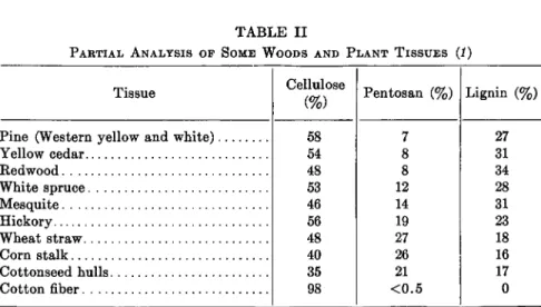
TABLE II
PARTIAL ANALYSIS OF SOME WOODS AND PLANT TISSUES (1)
Tissue
Pine (Western yellow and white) Yellow cedar
Redwood White spruce Mesquite Hickory Wheat straw Corn stalk Cottonseed hulls Cotton fiber
Cellulose (%)
58 54 48 53 46 56 48 40 35 98
Pentosan (%)
7 8 8 12 14 19 27 26 21
<0.5
Lignin (%)
27 31 34 28 31 23 18 16 17 0
vergent mixture of polysaccharides are classified together, and it is entirely likely that the term hemicellulose will eventually be dropped, particularly once the individual polysaccharides are isolated and clearly identified.
Some of the pentosans from wastes which accumulate in the processing of agricultural products are converted by acids to furfural, a raw material of increasing industrial value and interest. These and other waste hemi- celluloses such as those obtained from the pulping of wood represents a rich, almost untapped, source of raw material, much of which could be converted from a nuisance to a source of profit. (See also p. 799.)
Xylans. By far the most abundant polysaccharides in the hemicellulose group are the xylans (hemicellulose-A). These pentosans are composed either entirely or almost entirely of D-xylose units. Several types of xylans are extractable from different plant sources. Some are linear molecules, some contain one or more branches, and in some are combined one or more L-arabinose or D-glucuronic acid units. Xylans occur in practically all land plants and are present in some marine algae (57, 58). They are most abundant in annual crops, particularly in agricultural residues such as corncobs, corn stalks, grain hulls, and stems. Here they occur in amounts ranging from 15 to 30%. Hardwoods contain 20 to 25% xylans whereas softwoods contain 7 to 12 %. Spring wood has more pentosan than summer wood. Since xylans are the most abundant of the pentosans, their distribu- tion is indicated by pentosan analysis. Although cellulose, pentosans, and lignin are the most abundant substances in plants, their relative amounts vary greatly from one plant tissue to another (Table II).
Xylan is precipitated on neutralization of an alkaline plant extract.
57. V. C. Barry and T. Dillon, Nature 146, 620 (1940).
58. E. G. V. Percival and S. K. Chanda, Nature 166, 787 (1950).