Salmonella typhimurium and Escherichia coli
Neurotoxins
LYDIA MESROBEANU AND I. MESROBEANU
I. Introduction and History 301 II. Salmonella typhimurium Neurotoxin 3 0 4
A. Isolation of Toxins 305 B. Role of Some Physicochemical Factors 307
C. Serological Specificity 309
D. Toxicity 312 E. Cytopathogenicity 313
F. Bactericidal Activity 315 G. Resistance Induced in Mice by S. typhimurium S and R
Neurotoxin Vaccination 316
H. Purification 318 III. Escherichia coli Neurotoxins 3 2 0
A. Chemical Structure and Toxicity 3 2 0
B. Serological Specificity 322 C. Thermolabile Neurotoxins from E. coli, Proteus, and Pseudomonas
pyocyanea J ZO
D . Soluble Thermolabile Neurotoxins in Urine
Infected by Gram-Negative Organisms 327
IV. Concluding Remarks 333
References 3 3 4 I. I n t r o d u c t i o n a n d H i s t o r y
Vincent ( 1 9 2 5 , 1 9 2 9 ) was the first to point out that Escherichia coli produced two toxins. According to this author, this occurs in "all strains and varieties" grown on simple broth at pH 7 . 6 , at a temperature of 3 8 ° C . Under these growth conditions, a thermolabile exotoxin appears in the me
dium from 2 3 hours to 5 days after incubation; it is inactivated at 7 5 ° C but resists heating 5 6 ° C for 1 hour. The second toxin appears much later, usu
ally in filtrates of old cultures ( 1 5 - 2 0 days) in which bacterial lysis is ad
vanced and the pH exceeds 10. This toxin, an enterotoxin, is thermostable. The occurrence of these toxins at different phases of growth, and the possibility of separating them by heat inactivation of the thermolabile toxin, enabled Vincent to carry out a series of investigations which led to the setting up of a serotherapeutic regimen in man ( 1 9 2 5 ,
1 9 2 8 ) .
These toxins produce different clinical pictures when injected in ani
mals. Thus, according to Vincent, the intravenous injection of the ther-
3 0 1
302 L. MESROBEANU AND I. MESROBEANU
molabile exotoxin into laboratory animals, particularly in rabbits, pro
duced the following neurotropic symptoms: paralysis, first of the hind quarters, and, subsequently, of the front legs; bulbar paralysis; and muscle flaccidity.
Investigating the 12 exotoxins isolated from various strains in the rab
bit, Vincent (1925) observed that, irrespective of the toxin-producing strain, the injected animals presented the same symptoms, which included apathy, anorexia, and hypothermia, occasionally coma and early, severe diarrhea, with pronounced weakness. These first symptoms lasted 1-3 days; a number of animals that apparently had recovered died later. Out of 12 rabbits, 8 died with progressive paralysis, incontinence of the urine and feces, and respiratory center paralysis; for some of these animals, death occurred late, within 12-46 days. The slow evolution of late and lethal paralyses led Vincent to ask whether some human Landry-type paralyses were not due to a colon bacilli infection.
The enterotoxin, which affects the stomach and intestines, may at present be considered homologous to the Boivin endotoxin. Vincent showed that this toxin can produce antibodies which precipitate the ho
mologous toxin in vitro, yet do not protect the animals against the exo
toxin. Vincent prepared an antimicrobial serum by adding both toxins to the bacterial suspension used in immunization. This serum gave excellent clinical results, according to the accounts of the author himself and of other clinicians who used it.
Besides the neurological disorders found in animals intoxicated with E.
coli neurotoxin, Vincent (1933) described the role played by colon bacilli intoxication in the genesis of some mental disorders in man; these condi
tions improved subsequent to the above-mentioned serotherapy. The symptoms of a mental nature were studied by Baruk and his psychiatric school (1938). The studies first focused on experimental investigations of various animal species — rabbit, cat, mouse, guinea pig, pigeon, fishes, and reptiles. Their neurological and psychic disorders were accurately re
corded.
In mammals (mice, guinea pigs, and cats) the subcutaneously adminis
tered toxin induced such characteristic symptoms as narcolepsy, polyp
nea, torpor, paralysis, coma, a clear cataleptic state, and extreme hyperkinesia ("leaps, jumps, violent hits against the cage, with the animal becoming insensible for a few moments and dying"). The cataleptic ani
mals could be awakened by the slightest touch. Belated deaths (after some weeks) are recorded with cachexia and amyotrophy. Pigeons displayed a catatonia, with impulsive reactions. Fishes, lizards, and frogs died the day after injection. The general characteristics of the colon bacilli-induced catatonia included such signs as "active negativism" with periods of "vio-
lent opposition," then hyperkinesia, organovegetative disturbances, and catalepsy.
From his laboratory investigations on E. coli neurotoxin, Baruk (1938) disagreed with Vincent as to the occurrence of E. coli strains capable of producing neurotoxin. He found that some are unable to do so, while others rapidly exhaust their capability and that the toxin loses its neuro
toxic activity upon preservation, the clinical symptoms induced becoming less severe until they disappear.
Baruk also pointed out that the animals, in experimental intoxication, must be followed over a long period of time, since the action of the toxin may be very slow and the cataleptic and catatonic phases transient.
Therefore, intravenous inoculation is not recommended for the study of acute intoxication with neurotoxin, since death occurs before the entire range of intoxication-induced clinical manifestations appear.
Baruk used serotherapy with colon bacilli antecedents in severe cases of mental diseases with an occasional "spectacular" result. Baruk points out, however, that prolonged serum treatment is necessary to produce favorable results.
The failure of serotherapy could now be explained by the fact that, to be effective, the serological type of the human colon bacilli causing the infection should have been the same as that of the toxin used in the prepa
ration of the serum.
Extending his investigations and observations, Baruk introduced the concept of "colibacillary psychoses" and, clinically, the idea of "toxi- infectious psychoses," induced by the action of bacterial neurotoxins on the nervous system.
Tomesco et al. (1937) described catatonia in animals induced by the urine of a mental patient with a colon bacilli infection; this patient was only slightly helped by the serotherapy recommended in Romania by Dr.
Magheru.
It is worth mentioning that, in the Dr. I. Cantacuzino Institute, Dr.
Magheru et al. (1944) studied a series of strains of colon bacilli and found neurotoxin in the culture broth filtrates 24-48 hours after incubation. This neurotoxin was inactivated by heating 1 hour at 75°C. It was neurotropic and antigenic and could produce a serum which neutralized a lethal dose of neurotoxin. Retezeanu et al. (1961) reported two cases of stuporous confusion with significant alterations in muscle tonus in mental patients with colon bacilli urinary infections who were cured subsequently by anti
biotics.
Following the initial investigations of Vincent and Baruk, a number of scientists, including Hohorst (1953), attempted to reproduce intoxication phenomena with E. coli neurotoxin. However, they were not able to stan-
304 L. M E S R O B E A N U A N D I. M E S R O B E A N U
dardize the neurotoxin accurately, because of its lability and the lack of criteria for its production.
Vincent (1942), in studying over 200 Salmonella typhosa strains culti
vated in collodion bags inserted in guinea pig peritoneum, reached the conclusion that after 5-7 passages, approximately 7% of the strains se
creted a soluble, very active, thermolabile and oxygen labile toxin. The toxin, inoculated in experimental animals, was neurotropic, inducing the nervous symptoms characteristic of typhoid. After a series of investiga
tions, confirmed by clinical data on these infections, Vincent (1942, 1945) concluded that typhoid fever is not a genuine septicemia but an infection with toxic manifestations.
It is worth mentioning that the technique used by Vincent for demon
strating the ability of S. typhosa to secrete neurotoxins was applied by this author at the end of the last century (1898) to potentiate the virulence of some saprophyte microorganisms (Bacillus megaterium and B. mesen- tericus). Both species, cultivated in collodion bags inserted in guinea pig peritoneum, became virulent, but B. mesentericus vulgatus was found to secrete a thermolabile toxin which produced paralysis, a series of other nervous disorders, and hypothermic death in mice.
Early investigations on gram-negative bacterial thermolabile neuro
toxins involved S. typhimurium and continued with E. coli. The results are presented in this chapter. Some brief additional observations on neu
rotoxins isolated from the urine of subjects with colon bacilli infections, and a few cases with Proteus and Pseudomonas pyocyanea infections are also presented, as such observations seem to agree with Vincent's views and might contribute to a reasonable interpretation of the clinical aspects of gram-negative bacterial infections.
I I . Salmonella typhimurium N e u r o t o x i n
In their studies on chloroform autolyzates of some gram-negative bac
teria (S. typhimurium, E. coli, Proteus vulgaris, Salmonella berlin, Sal
monella weslaco, etc.). L. Mesrobeanu (1961; L. Mesrobeanu et al., 1962a,b, 1966) found an antigenic thermolabile toxin in the supernate which, upon inoculation into mice and rabbits, induced symptoms remi
niscent of those caused by Shigella dysenteriae neurotoxin. This influ
enced the authors to call this thermolabile toxin a neurotoxin. This toxin may be extracted from both the S and R forms, but, in contradistinction to S. dysenteriae neurotoxin which is much more active in the R form, in the bacteria studied by us (Salmonella, Shigella flexneri, Shigella schmitzii, Shigella sonnei, and E. coli) the S variant-derived preparations proved more toxic and immunogenic.
A . ISOLATION OF TOXINS 1. ISOLATION M E T H O D S
Isolation of these toxins was conducted by the following technique:
after 3-days of maceration at room temperature, the chloroform auto- lyzates of bacteria cultured for 18 hours were centrifuged, and the supernate, containing a mixture of thermostable endotoxin and thermo
labile toxin, was brought to pH 3.5 with cold trichloroacetic acid. The acid-insoluble thermolabile toxin precipitated, thus effecting its separa
tion from the acid-soluble endotoxin. The acid-insoluble toxin was immediately redissolved in alkaline solution (pH 8.5) and preserved under sterile conditions; it represented the crude neurotoxin which, by gel precipitation, electrophoresis, and ultracentrifugation was shown to be nonhomogeneous. Ultracentrifugation produced 4 delineated fractions, whereas acrylamide-agarose gel electrophoresis (Uriel, 1966) produced
7-9 fractions.
During our work on the S. typhimurium thermolabile toxin, various other extraction methods were used, e.g., chloroform autolysis of bacteria supplemented with 1 % sodium deoxycolate, autolysis in the presence of veronal followed by phosphate buffer extraction in hypertonic solution, and the citrate-sodium chloride method (Raynaud and Digeon, 1949); in every case thermolabile toxic substances were obtained from gram-nega
tive bacteria, but the yield was lower than that obtained by simple chloro
form autolysis.
We also used the method recommended by van Heyningen and Glad
stone (1953) for extraction and purification of S. dysenteriae neurotoxin, in which bacteria killed by 56°C heat are dissolved in potassium hy
droxide at pH 11. The supernate is treated with calcium phosphate at pH 7.6, the resulting precipitate removed, and the preparation dialyzed. With S. dysenteriae neurotoxin, the toxin precipitated and was purified subse
quently. Since 5. typhimurium S and R neurotoxins do not precipitate during dialysis, the dialyzed preparation was used for toxicity, specificity, and yield studies, and was compared with the material isolated by the method suggested by us above.
Boivin et ai (1940) have demonstrated that, in the case of S. dysenter
iae, heating the bacterial suspension for 1 hour at 56°C results in the re
lease of a quantity (30% of the amount obtained by autolysis) of the neu
rotoxin into the supernate. The same phenomenon is seen with 5.
typhimurium S neurotoxin when approximately 20% of the thermolabile toxin passes into the medium. In this procedure, the toxin is then precipi
tated with trichloroacetic acid at pH 3.5, redissolved, and inoculated in mice; the observed death rate was similar to that seen with the prepara
tion obtained by chloroform autolysis.
306 L. MESROBEANU AND I. MESROBEANU 2. CULTURE M E D I U M
Since we found variations in yield, neurotoxic characteristics, and bac
tericidal activity for different batches of toxin, we used several culture media formulas for S. typhimurium to find the most suitable one for stabi
lizing these characteristics. The following were tested:
(1) Agar + meat infusion
(2) Agar + meat infusion + 1 % soluble starch (3) Agar + meat infusion + 10% creamed milk
(4) Hottinger medium -1-7% agar in a 2 0 % C 02 atmosphere (5) A s ( 4 ) + 1 % starch
(6) A s (4) + 10% creamed milk in a 2 0 % C 02 atmosphere (7) Yeast infusion + agar
(8) Meat infusion + horse serum or blood
In the selection of these media, we took into account the findings of both Vincent (1942), who described the oxygen lability of typhoid neuro
toxin, and Jordan and Burrows (1935), who obtained soluble toxic sub
stances from 5. typhimurium, Proteus, and E. coli cultures in Burnet medium and carbon dioxide atmosphere (Burnet, 1929, 1930), supple
mented with starch or cream. The 5. typhimurium S strain cultured on these media did not yield more toxin, nor were neurotoxic or bactericidal characteristics restored for strains in which these characteristics had been lost or attenuated.
The single positive finding in these studies was the following: when S.
typhimurium S strain is grown on Dorset medium, the bactericidal charac
teristics of the extracted neurotoxin last a long time. As a matter of fact, this medium is used in our institute to maintain some S. typhimurium
strains which induce keratoconjunctivitis in guinea pigs.
3. T O X I N Y I E L D
Chloroform autolysis yields approximately 3 % S. typhimurium S neu
rotoxin and 5% R variant, by dry bacterial weight. The van Heyningen method gives yields which are roughly 3-fold lower, as may be seen in Table I.
If chloroform autolysis is prolonged, the yield of S. typhimurium S neu
rotoxin decreases as its nitrogen content decreases, although toxicity in mice by intraperitoneal injection as measured by neurotoxin nitrogen con
tent remains constant.
Extraction of the acid-soluble endotoxin follows a similar pattern. It has been found, however, that with endotoxin extracted by the classic Boivin-Mesrobeanu method (1933), the chloroform autolysis yield is higher though it decreases when autolysis is prolonged.
T A B L E I
Salmonella typhimurium S A N D R N E U R O T O X I N Y I E L D Yield" after
Neurotoxin 3 days autolysis
S neurotoxin —chloroform autolysis 2.94 R neurotoxin —chloroform autolysis 5.05 S neurotoxin (van Heyningen method) 0.80 R neurotoxin (van Heyningen method) 1.40
"Percent dry weight of bacteria.
B. R O L E OF SOME PHYSICOCHEMICAL FACTORS
1. TEMPERATURE
Acid-insoluble toxin can be differentiated from acid-soluble endotoxin by heating. Neurotoxins are thermolabile, and their degree of thermosta
bility seems to depend upon the amount of the residual polysaccharide which remain attached to the lipoprotein fraction during autolysis. Thus, a hydrolyzed crude preparation with a reducing substance content of 5% or less is inactivated at 80°C in approximately30 minutes. If the reducing substance content is higher than 5%, inactivation occurs only after heating for 1 hour at 100°C; sometimes the toxin must be autoclaved for 30 minutes at 115°C for inactivation to occur. Under the same conditions, the toxicity of Boivin endotoxin in neutral solutions is maintained.
It is worth mentioning that preparations with the highest biological ac
tivity were obtained when the amount of reducing substance was below 10% of the dry extract. The presence of large amounts of reducing sub
stance shows that the autolytic processes, in which the endotoxin is de
graded to liberate toxic lipoprotein, are incomplete. It has been found that fractions with various amounts of reducing substances occur. In some of them, the reducing substance content is over 15%; these fractions are acid insoluble, possess a high degree of thermostability, and, at the same time, are no longer neurotoxic.
When this occurs, the S. typhimurium culture must be replaced. The most prominent sign of loss of neurotoxic activity is the fact that the chlo
roform autolyzate at pH 3.5 precipitates slowly and has a gelatinous as
pect when collected by centrifugation. The same situation was described by Baruk (1938) for E. coli neurotoxin; this author showed that the yield of this toxin was variable and inconstant and that the toxin disappeared after frequent subculture of the strain. He found that recently isolated cul
tures were constantly required for toxin production.
308 L. M E S R O B E A N U A N D I. M E S R O B E A N U
We must, however, point out that our first strain was 5. typhimurium S , used in 1932-1933 by Boivin and Mesrobeanu for the endotoxin O isola
tion. Our results with this strain confirm those of Baruk, that is, frequent subculture adversely affected the yield and production of the acid-insol
uble toxin and altered other biological characteristics.
2. FORMALDEHYDE
Formaldehyde partially detoxifies neurotoxin preparations, while pre
serving the specific and immunogenic characteristics; it has no effect on endotoxin activity. We did not obtain complete detoxification; a similar partial detoxification has been recorded for S. dysenteriae neurotoxin.
A S. typhimurium S neurotoxin which initially had a MLD of 0.006 mg N in mice, was lethal at a 1.08 mg N after a 27-day exposure to 0.8%
formaldehyde at 37°C, followed by dialysis. Under the same conditions, the S. typhimurium R variant, with an initial M L D of 0.02 mg N, had a final MLD of 1.62 m g N .
No systematic studies have been carried out, but Raynaud (personal communication) suggests that lysine be added to induce complete detoxi
fication. It is known that this amino acid stabilizes diphtheria toxin during detoxification (Linggood et aL, 1963).
3. ROLE OF IRON
Dubos and Geiger (1946) and van Heyningen and Gladstone (1953) pointed out that as the iron concentration of the culture medium in
creases, the neurotoxin yield of S. dysenteriae decreases. The concentra
tion of iron in the medium has been increased (1-10 fig Fe/ml), and an optimal concentration of 2 /xg Fe/ml determined. At this point, the S. ty
phimurium S neurotoxin yield was 20% higher than the yield in an iron- free medium. Although this result shows that toxin production is influ
enced by iron concentration, we could not reproduce the results obtained by van Heyningen with S. dysenteriae neurotoxin, where the yield was found to vary with the iron concentration.
4. STABILITY OF NEUROTOXIN SOLUTIONS
The stability of S. typhimurium S neurotoxin solutions has been thor
oughly investigated. It has been observed that neurotoxin solutions of concentrations between 1 and 3 mg N/ml become turbid after several days at 4°C, and that later a precipitate appears leaving a clear supernate.
This supernate shows the same toxic and specific characteristics as the original solution. The precipitate is mainly lipid (70%); lipid depletion modifies the structure of the neurotoxin, increasing the content of ni
trogen and reducing substance, as expressed by percent of dry extract weight.
T A B L E II
P R E C I P I T A T I O N T I T E R S W I T H M I C R O B I A L ( S A N D R ) A N T I N E U R O T O X I N S S A N D R A N D A N T I E N D O T O X I N O (B O I V I N ) A N T I S E R A
Antiserum
S. typhi S. typhi
murium s murium R Endotoxin
Antigens neurotoxin neurotoxin O
S. typhimurium S microbial suspen
sions 1/2500 1/2 1/1600
S. typhimurium R microbial suspen
sions 1/2 1/1600 0
Endotoxin O anti
serum 1/1600 1/2 1/2500
S neurotoxin
antiserum 1/1600 1/5 1/1000
R neurotoxin
antiserum 1/4 1/600 0
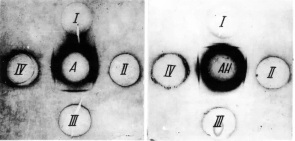
Results show that immune sera prepared with microbial suspensions and S and R neurotoxins display cross reactions with only negligible ti
ters. Moreover, the endotoxin antiserum precipitates only with S neuro
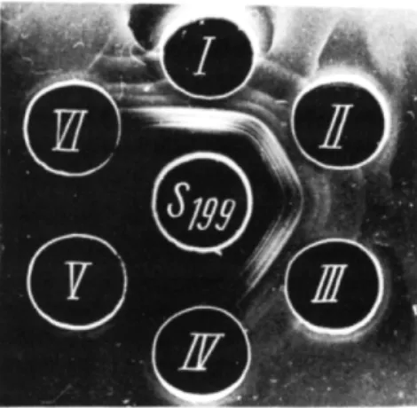
toxin and not with the R variant. Where the same sera were used in agar qualitative precipitation, identical results were obtained (Figs. 1-5).
It may be concluded from the above data that no cross reactions oc
curred with the R and the S neurotoxin, however, it has been observed that the S neurotoxin antiserum precipitates with the glycolipid polypep
tide endotoxin but not with a phenol-prepared lipopolysaccharide (West- The bactericidal activity of S. typhimurium neurotoxin solutions dimin
ishes with storage; after being preserved for 2 months at 4 ° C , the inhibi
tion zone, which had an initial diameter of 2 - 3 cm, shrinks to 2 - 3 mm or disappears completely.
C . SEROLOGICAL SPECIFICITY
The serological specificities of toxins extracted from S. typhimurium S
and R were analyzed, using endotoxin O and thermolabile S and R toxin antisera. These sera were prepared by immunization of common rabbits;
in hyperimmunization experiments, New Zealand rabbits were used. Pre
cipitation was carried out with both the classic quantitative method and the Ouchterlony qualitative method ( 1 9 4 9 ) in agar. Quantitative precipi
tations are shown in Table II.
F I G S . 1-5. A = Inactivated S. typhimurium S suspension. B = Inactivated S. typhimurium R suspension. C = S. typhimurium endotoxin solution 1 mg dry weight/ml. D = S. typhimu
rium S neurotoxin solution 2 mg N / m l . E = S. typhimurium R neurotoxin solution 3 mg N / m l . 1 = 5 . typhimurium S microbial antiserum. II = S. typhimurium R microbial anti
serum. I l l = S. typhimurium S neurotoxin antiserum. I V = S. typhimurium R neurotoxin antiserum. V = 5 . typhimurium endotoxin antiserum.
310
phal et aL, 1952) nor with a polysaccharide acidic hapten prepared by mild acid hydrolysis of the endotoxin.
Using antibody saturation, we were able to demonstrate that the glycolipid-polypeptide endotoxin complex induces the formation of two antibodies in immunized animals, i.e., one for the lipopolysaccharide and the other for the neurotoxin. Figure 6 shows reactions with a S. typhimu
rium S endotoxin antiserum exhausted with neurotoxin S, which does not precipitate with neurotoxin (I) but continues to react with the endotoxin, the polysaccharide hapten, and an inactivated homologous microbial sus
pension. In this case, positive reactions were ascribed to the presence of an antibody specific for the polysaccharide fractions.
Figure 7 shows reactions with the glycolipid polypeptide endotoxin antiserum exhausted with the polysaccharide hapten. It can be seen that this serum does not precipitate with the hapten, but it continues to precip
itate with the endotoxin and the S neurotoxin, both processes being as
cribed to the presence of the polypeptide fraction specific antibody.
A Boivin endotoxin antiserum sample successively exhausted with S neurotoxin and polysaccharide does not precipitate with either of the anti
gens used in this experiment or with the microbial suspension.
By a mild and short acid hydrolysis (1 % acetic acid, heated for 30 min
utes at 100°C), we obtained from the 5. typhimurium S endotoxin a poly- peptide-rich fraction which completely absorbed the antibodies of the S neurotoxin antiserum. By prolonging the time of autolysis to 8-10 days, and, particularly, by using rabbit hyperimmune sera so that at the end of a
F I G S . 6 and 7 . A = Boivin endotoxin antiserum depleted by cross reaction with S neuro
toxin. A H = Boivin endotoxin antiserum depleted by cross reaction with polysaccharidic hapten. 1 = 5 . typhimurium S neurotoxin. II = Boivin endotoxin. I l l = Polysaccharide. I V = 5 . typhimurium S inactivated microbial suspension (Azocarmin B-stained plates).
312 L. M E S R O B E A N U A N D I. M E S R O B E A N U
3-month period of immunization animals might tolerate 0.5 mg N and 1 mg S and R neurotoxin, respectively, we obtained serological fractions common to both S and R forms, and similar preparations from other Sal
monella chemotypes.
Our investigations confirm the studies of Raynaud et al. (1964) that in order to obtain sera that can precipitate the various antigenic constituents common to Salmonella, it is particularly important to prepare antisera by prolonged hyperimmunization.
D. TOXICITY
After inoculation, mice are in a state of stupor, with labored respiration, moderate diarrhea, and, frequently, a posterior limb and tail paralysis. In their recent studies on mice, Olinesco and Olinesco (1970) showed that 17% of the inoculated animals were paralyzed; these authors never ob
served paralysis in mice intoxicated with the homologous Boivin S. typhi
murium endotoxin. Another feature of neurotoxin intoxication is that in
testinal symptoms are less pronounced than with endotoxin intoxication.
The neurotoxin-intoxicated mice and guinea pigs die in a hypothermic state. These authors observed only quantitative differences in the patho
logical hematology in intoxication with S. typhimurium S neurotoxin and with endotoxin.
Rabbits are very sensitive to S. typhimurium neurotoxin. Inoculations, even in sublethal doses, induce an extreme weakness in animals and many of them die during the immunization procedure. Sometimes limb paralysis occurs, and we have also observed transient phenomena of paralysis; the rabbits which live through this phase continue to display anorexia, and they grow weak and die in cachexia.
In our laboratory, a similar situation was found in New Zealand rabbits immunized with various Salmonella chemotype-extracted neurotoxins. It is worth mentioning, however, that not all rabbits in a homogeneous popu
lation inoculated with the same toxin were paralyzed. The same observa
tion was made for S. dysenteriae neurotoxin.
A L D50 of 0.014 mg N for S. typhimurium S neurotoxin, and 0.04 mg N for the R variant, was obtained in mice injected intraperitoneally. It was observed that the intravenous route was approximately 4 times more effi
cient than the intraperitoneal one.
Preparations of S. typhimurium S and R neurotoxins obtained by the van Heyningen technique were injected intraperitoneally in mice, and the toxicity was observed to be lower than that for preparations obtained by chloroform autolysis.
E . CYTOPATHOGENICITY
The cytopathogenic activity of the thermolabile S. typhimurium S and R toxin preparations was investigated in our laboratories (I. Mesrobeanu et al, 1960, 1962, 1964) on HeLa and KB tumor cell cultures and on hu
man embryo and monkey testis normal cell cultures. Under our working conditions, the thermostable endotoxin did not have a cytotoxic effect on tumor cell cultures; in normal cell cultures a moderate, delayed cytopatho
genic effect was seen with concentrations above 0.5-1 mg. The S and R neurotoxins produced irreversible changes in tumor and normal cell cul
tures. It is worth mentioning, however, that the R neurotoxins have less effect than the S toxins, in contradistinction to the S. dysenteriae R neuro
toxin which proved the most active. Vicari et al. (1960) were the first workers to demonstrate the high cytopathogenic activity of this neuro
toxin on various cell strains; it was found that its effect was neutralized by its antitoxic serum.
We have been particularly interested in determining the minimum expo
sure time required to induce cytotoxic effects in human embryo cultures by S. typhimurium S neurotoxin. Three concentrations of toxin were used, at exposure times of 1, 4, and 20 hours. After exposure, the culture medium was removed, and a maintenance medium was added. The ob
served cytopathogenic effect was compared with controls and cultures with toxin incorporated into their maintenance medium. Results are shown in Table III.
The general conclusions drawn from these experiments were as fol
lows:
1. Human embryo cultures exposed to 0.4 mg N S. typhimurium S neurotoxin for 4 hours showed clear cytopathogenic changes which oc
curred approximately 4 hours later and gradually increased over a period of days. The effect of exposure to 0.2 mg N toxin was less pronounced and appeared 3 days after the maintenance medium was replaced.
2. Cultures which were exposed to the toxin for 20 hours showed the same changes as did those cultures which were continuously exposed to the toxin.
3. No cytopathogenic effect was observed upon exposure to 0.1 mg of toxin which we estimate to be one-half the cytopathogenic dose (V2 C D5 0) .
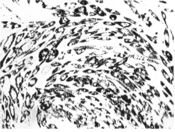
4. Cells exposed to S. typhimurium S neurotoxin show the following morphological changes: (1) At the first signs of cytopathogenicity, the in
dividual cells assume an elliptical shape within the monocellular layer (Fig. 8). (2) Nuclei become pyknotic, the cytoplasmic mass retracts, and
314 L. M E S R O B E A N U A N D I. M E S R O B E A N U T A B L E III
C Y T O T O X I C E F F E C T IN T I S S U E C U L T U R E S AS A F U N C T I O N O F E X P O S U R E TO S. typhimurium S TOXIN
Duration of con
tact between Observation Effect" at the following amounts (mg N ) of toxin cells and toxins time inoculated per culture tube:
(hours) (days) 0.1 0.2 0.4
1 1/24 - - +
1 - - +
2 - - +
3 - - +
4 - - +
4 1/6 - - +
1 - - +
2 - - +
3 - 4- + +
4 - + - H - f
20 1 - + + + +
2 - H H - + + + 3 + + + + M M 4 + M M M M
Permanently 1 - + + + +
2 - + + + M M 3 - + + + M M 4 - + + + + + + + +
"Key: — = no effect; + = discrete cytotoxic effect —rounding off of cells; - H - = clear-cut cytotoxic effect; M I = intense cytotoxic effect; M M = cytotoxic effect with detaching of cells from tube walls.
a great number of granules and vacuoles appear; these tend toward a di
polar localization with regard to the nucleus. (3) Staining with Sudan III demonstrates the occurrence of a very prominent fatty degeneration. (4) The cells tend to detach only at high toxin concentrations and at expo
sures exceeding 20 hours.
5. Injuries to the cell are not as severe as those described by Vicari et al. for 5. dysenteriae R neurotoxin which we were able to confirm (I.
Mesrobeanu et al., 1962). Indeed, at concentrations of only 0.00003 mg N, S. dysenteriae R neurotoxin causes injuries which progress until the fixed cells on the culture-tube wall are detached.
Brandes (1961, 1967; Brandes and Radola, 1964) demonstrated that several-day-old culture filtrates of 5. typhimurium, E. coli, and Shigella sonnei, flexneri, and boydii contain thermolabile materials, specific and toxic for both cultured tissue and the intact animal (mouse). By the use of ultrafiltration and ultracentrifugation, the author was able to separate mi- cromolecular fractions (2S) with cytotoxic characteristics from macromo-
FIG. 8. S. typhimurium S neurotoxin 0.2 mg N — i n o c u l a t e d human embryo cell culture after a 48-hour contact. A great number of granules are observed in the cytoplasm (x 2 0 0 ) .
lecular fractions without cytopathogenicity; the latter fraction still was antigenic and toxic to mice.
Chermann (1967; Chermann et ai, 1967), using our neurotoxin extrac
tion technique, also obtained from a S. typhi R variant thermolabile neu
rotoxic substances which exerted a cytotoxic effect on tissue cultures.
Using the same technique with Brucella, Tuszkiewicz and Gaico (1966) isolated thermolabile, protein-like cytotoxic substances, particularly from B. melitensis. The endotoxins extracted with trichloroacetic acid from the same microorganisms exhibited a late, very slight cytotoxic effect.
F. BACTERICIDAL ACTIVITY
Salmonella typhimurium neurotoxins act to inhibit growth of homolo
gous microorganisms. In our preliminary investigations (L. Mesrobeanu et al., 1966) we were surprised by the broad spectrum of activity shown by these preparations. Our results suggest that the element which is re
sponsible for this characteristic is related to the protein fraction of the endotoxin; hence these studies confirm the data of Goebel et al. (1945;
Goebel and Barry, 1958; Goebel, 1962) on colicines.
Initially, in agreement with Goebel, we observed that not every mi
crobial preparation displayed bactericidal activity, though other biological features were maintained. For a long time, even in its lyophilized state, the S. typhimurium S strain used by us did not produce thermolabile
316 L. M E S R O B E A N U A N D I. M E S R O B E A N U
toxins which were bactericidal. Procedures carried out during this period, such as ultraviolet irradiation, modification of culture media by addition of proteins, sugars, vitamins, etc., did not result in bactericidal activity.
However, after adopting the Dorset medium to preserve the S. typhi
murium culture, bactericidal activity of the neurotoxin preparation to
ward its own strain was observed, but with a low spectrum of activity. At a lower titer, the acid-soluble Boivin endotoxin which remains in the au- tolyzate supernate after the removal of the neurotoxin at pH 3.5 also dis
played bactericidal activity.
By heating the S. typhimurium S suspension at 56°C for 1 hour, we were able to isolate a thermolabile toxin, which maintained its bactericidal activity toward its own strain, from the supernate. This factor is undialyz-
able and is precipitated at 66 % saturation with ammonium sulfate. Precip
itation with alcohol and acetone causes these bactericidal neurotoxins to lose all bactericidal activity. This phenomenon is being investigated.
G. RESISTANCE INDUCED IN M I C E BY S. typhimurium S AND R NEUROTOXIN VACCINATION
Taking into account the immunological results described, we attempted to induce resistance in mice by means of S. typhimurium S neurotoxin, R neurotoxin, and S endotoxin vaccination. The responses of the immu
nized animals to lethal doses of toxins were observed, in order to ascertain:
(1) Whether vaccination with S and R neurotoxin produces mutual resist
ance; (2) if vaccination with the S neurotoxin produces a degree of resist
ance to the Boivin endotoxin, taking into account the fact that the lipo- polysaccharide fraction of the glycolipid-polypeptide complex was highly toxic; (3) if vaccination with Boivin endotoxin produces complete resist
ance to S neurotoxin, since, according to its immunological behavior, it is a fraction of the glycolipid-polypeptide endotoxin.
Escherichia coli S neurotoxin was also injected to determine whether any resistance observed was specific to the endotoxins used for immuni
zation, since it is known that endotoxins may induce a nonspecific resist
ance. Immunization of 16-18 gm mice was by the intraperitoneal route;
starting with V2 MLD, at 6-7 day intervals, the animals were given a total of 12 injections. When immunization of the experimental mice was com
plete, 25 control mice were injected with the toxin, in amounts equal to the last dose given the experimental mice. The toxin was injected in a 0.5 ml solution containg 40 fig streptomycin and 80 U penicillin per inoc
ulum. Resistance tests were performed 7 days after the last injection.
The 5. typhimurium S neurotoxin was injected in 440 mice; the initial
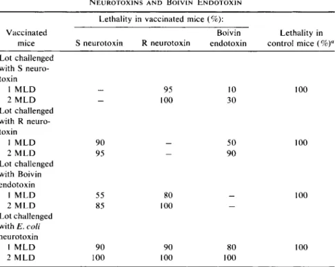
TABLE IV
R E S I S T A N C E I N D U C E D I N V A C C I N A T E D M I C E W I T H S. typhimurium S A N D R N E U R O T O X I N S A N D B O I V I N E N D O T O X I N
Lethality in vaccinated mice (%):
Vaccinated Boivin Lethality in
mice S neurotoxin R neurotoxin endotoxin control mice ( % )a
Lot challenged with S neuro
toxin
1 M L D — 95 10 100
2 M L D — 100 30
Lot challenged with R neuro
toxin
1 M L D 90 — 50 100
2 M L D 95 — 90
Lot challenged with Boivin endotoxin
1 M L D 55 80 — 100
2 M L D 85 100 —
Lot challenged with E. coli neurotoxin
1 M L D 90 90 80 100
2 M L D 100 100 100
"Twenty-five animals were used as controls for each toxin.
dose was 0.002 mg N which was gradually increased to 0.25 mg N . During immunization, 30% of the mice died; the remaining mice were di
vided into 3 groups and challenged with R neurotoxin, glycolipid- polypeptide neurotoxin, and heterologous E. coli neurotoxin.
Salmonella typhimurium R neurotoxin was injected intraperitoneally in 300 mice, the initial dose was 0.05 mg N which was gradually increased to 0.5 mg. Lethality was 18%. The mice were divided into 3 groups and challenged with S. typhimurium S neurotoxin, glycolipid-polypeptide endotoxin, and the heterologous E. coli neurotoxin.
The S. typhimurium S Boivin endotoxin was injected into 250 mice;
after 12 inoculations, the mice were immune to an intraperitoneal injec
tion of 0.5 mg endotoxin, which represented a dose of 10 MLD. After immunization, the mice were challenged with S. typhimurium S and R neurotoxins and the heterologous E. coli neurotoxin.
The general results are summarized in Table IV.
318 L. MESROBEANU AND I. MESROBEANU
The general conclusions drawn from these experiments are as follows:
1. Vaccination with S. typhimurium S and R neurotoxin induced immunity in mice.
2. Vaccination with R neurotoxin did not induce immunity against S.
typhimurium S neurotoxin and Boivin endotoxin.
3. Vaccination with S neurotoxin did not induce immunity against R neurotoxin; it did induce partial immunity against thermostable Boivin endotoxin.
4. Vaccination with Boivin endotoxin induced immunity against S neurotoxin but not against the R variant.
5. Vaccination with Boivin endotoxin and S and R neurotoxin did not induce immunity against heterologous E. coli neurotoxin in mice.
H . PURIFICATION
Since it was desirable to obtain homogeneous fractions for ultracen
trifugation and agar gel precipitation tests, we undertook the development of purification procedures.
In an earlier study, in collaboration with Marx (L. Mesrobeanu and Marx, 1964), we showed that the amino acid compositions of the glycolipid-polypeptide endotoxin and the S. typhimurium S and R neuro
toxins, were similar, with only quantitative differences observed. Using qualitative paper chromatography, we isolated 17 amino acids and gluco
samine, and, with sulfonate polystyrene resin chromatography, 13 amino acids and glucosamine.
As seen from Table V, S. typhimurium S and R neurotoxin contain the same amino acids; there are quantitative differences as compared with Boivin endotoxin. In S and R neurotoxins, glucosamine is present in trace amounts, whereas in endotoxin it is found in higher amounts. These data support the hypothesis of Westphal (1960) who suggests that the polysac
charide of the glycolipid-polypeptide complex is linked to the lipid by means of a hexosamine, a fact which could explain the high content of glucosamine found by us in the Boivin endotoxin hydrolyzate of S. typhi
murium S. According to Westphal, linkage between the lipid and polypep
tide could be effected by means of a dicarboxylic amino acid. The large amounts of aspartic and glutamic acids which occur in the S and R neuro
toxin hydrolyzates give added support to this hypothesis.
The following purification technique, consisting of three steps, was fol
lowed:
1. The S. typhimurium S neurotoxin solution was saturated with a 3 5 % ammonium sulfate solution and heated for 1 hour at 37°C. The re
sulting precipitate was dissolved and passed through a G25 Sephadex column to obtain three fractions. A fourth fraction, representing only
T A B L E V
S A N D R N E U R O T O X I N S A N D B O I V I N E N D O T O X I N A M I N O A C I D C O N T E N T "
Amino acid S neurotoxin R neurotoxin Boivin endotoxin
Aspartic acid 4.4 3.3 1.4
Threonine 0.9 1.3 0.5
Serine 1.3 1.4 0.4
Glutamic acid 4.0 3.8 1.3
Glycine 2.0 1.2 1.0
Alanine 2.7 2.2 0.6
Valine 1.8 1.3 0.5
Isoleucine 1.3 1.0 0.5
Leucine 2.8 2.6 0.6
Tyrosine 1.2 1.1 0.2
Phenylalanine 1.9 1.6 1.0
Lysine 2.3 1.9 0.8
Glucosamine Trace Trace 2.1
Arginine 1.5 1.0 0.4
"Milligram percent dry weight of extract.
2 - 3 % of the preparation, could not be isolated. The yields of these three fractions are the following: fraction I = 58%, fraction 11 = 3 1 % , fraction 111 = 8%.
2. Each fraction was passed through a G50 Sephadex column. The major fraction formed two perfectly delineated zones, one very concen
trated and one weak; the weak one was not studied. The fractions were concentrated and shaken for 30 minutes with a 10% suspension of D E A E Sephadex. Supernates were reserved for further treatment.
3. The fractions were passed through a G200 Sephadex column and analyzed. As seen in Table VI, fraction Ic is almost pure protein with a 1.3% lipid content. This preparation is 6 times less toxic than the crude neurotoxin. The last fraction was similar in composition to a glycolipid-polypeptide endotoxin and showed high levels of reducing substance and lipid.
T A B L E V I
F R A C T I O N A T I O N O F S. typhimurium S N E U R O T O X I N
Reducing
S. typhi N substance Lipids M L D I V
murium S ( % d r y ( % d r y ( % d r y in mice
neurotoxin weight) weight) weight) (mg N )
Crude neurotoxin 11.5 9.2 18.8 0.01
Fraction Ic 15.7 0.9 1.3 0.06
Fraction l i e 9.0 15.8 27.2 0.1
Fraction I l i e 4.4 34.5 37.8 0.2
320 L. M E S R O B E A N U A N D I. M E S R O B E A N U
All three fractions displayed bactericidal activity toward a homologous strain; fraction Ic was the most active. The three fractions of S. typhimu
rium neurotoxin give, with the crude neurotoxin antiserum, a single pre
cipitation band as shown in Fig. 9.
I I I . Escherichia coli N e u r o t o x i n s
This part of this chapter is concerned with the neurotoxins of the coli- form bacilli. We undertook these studies to see if, with our technique, we could demonstrate protein, thermolabile substances, with neurotoxic ac
tivity, in the coliform bacilli autolyzate, not in the culture medium.
We investigated 21 E. coli strains, 11 of which were recently isolated from individuals with urinary infections. The others were from our stock cultures, among which were 8 isolated from infantile toxic diarrhea. The bacteria were cultivated for 18 hours on solid media and then autolyzed;
the neurotoxin was isolated by the same method as described for the S.
typhimurium neurotoxin.
A. CHEMICAL STRUCTURE AND TOXICITY
Crude preparations of thermolabile E. coli toxin were analyzed for ni
trogen, lipid, and reducing substance content (after acid hydrolysis); also, the intraperitoneal M L D in mice was determined.
The crude preparations isolated from coliform bacilli had a nitrogen
F I G . 9 Sn = Crude neurotoxin antiserum. Lots A and C = bactericidal lots of 5 . typhimu
rium neurotoxin. Lot B = a nonbactericidal S. typhimurium neurotoxin. T h e last lot (C) served to isolate fractions Ic, l i e , and I l i e .
content of 10.5-13.5%, a lipid content of 5-10%, and a reducing sub
stance content of 5 -12 %.
It was observed that the products from the first E. coli group, which we isolated from urine, were more toxic than those from infantile toxic diar
rhea-isolated strains from our stock (L. Mesrobeanu et aL, 1962b).
When E. coli toxin solutions were aged at 4°C, a precipitate similar to that obtained from S. typhimurium neurotoxin, which contained approxi
mately 7 0 % lipid, was formed. Toxin analysis before and after aging showed that structural modifications had occurred after removal of the precipitate. When most of the lipid was removed, the nitrogen and the reducing substance content per unit dry weight increased, but the intra
peritoneal toxicity in mice and the serological specificity remained the same.
The toxicity of the toxins varies greatly among the E. coli strains, so that we were unable to compare values for the S or R variants. Thus, an E. coli27 strain isolated from urine proved to be a R variant, being, never
theless, able to produce a very active toxin with a M L D of 0.007 mg N when injected intraperitoneally in mice. However, the R E. coli Bruxelles strains, maintained in the Cantacuzino Institute Collection since 1921, yielded a preparation with an intraperitoneal M L D of 1.75 mg N in mice.
Intraperitoneal injections in mice with these two coliform bacilli toxins did not induce the death with limb paralysis that occurred with Salmonella typhimurium and Shigella dysenteriae neurotoxins. The animals, however, were very ill, in a state of torpor with labored respiration, and frequent diarrhea. Death with hypothermia occurred within 24-48 hours after the injection, but seldom later.
The intravenous injection of E. coli LM toxin induced in some mice hind leg paralysis and belated death at 72 hours with a M L D of 0.006 mg N or, by intraperitoneal inoculation, a MLD of 0.025 mg N.
These toxins were more heat resistant than the S. typhimurium neuro
toxins. Results of thermolability experiments carried out with E. coli27 toxin are shown in Table VII.
It was found that dilute solutions were more thermolabile than concen-
T A B L E VII
T H E R M O L A B I L I T Y O F E. coli21 T O X I N Neurotoxin dilution
(mg N/ml)
T o x i c i t y0 after heating 1 hour at:
75° C 80° C 85° C
0.5
1/5 3/5
4/5 5/5
5/5 5/5
"Dead mice/injected mice.
322 L. MESROBEANU AND I. MESROBEANU
trated solutions. We assumed that the increased thermal resistance of these concentrated toxin solutions was due to their higher polysaccharide content, resulting from an incomplete breakdown of Boivin endotoxin. At the same time, some of the strains investigated gave chloroform autoly- zates that, after clarification and precipitation at pH 3.5 with trichloro
acetic acid, became milky, but did not visibly precipitate. When 5 % so
dium chloride (w/v) was added, and the resulting solution was kept for 30 minutes to 1 hour at 4°C, a gelatinous deposit was obtained by centrifuga
tion at 10,000 rpm. Usually these preparations contain large amounts of polysaccharide which represent actual intermediate compounds between lipoprotein neurotoxins and Boivin endotoxins.
We prepared, from E. coli strain LM, Boivin endotoxin, lipopolysac
charide, and S neurotoxin; the neurotoxin was obtained by both chloro
form autolysis and by van Heyningen's method. Its structure is shown in Table VIII.
B. SEROLOGICAL SPECIFICITY
The E. coli LM strain was also used for extraction of fractions (namely Boivin endotoxin, S neurotoxin, and lipopolysaccharide) used in rabbit immunization. In order to obtain serological reactions, the antilipopoly- saccharide serum had to be concentrated 6 times since it is very poor in antibodies. Results are listed in Table IX.
It can be seen from the above data that the E. coli antineurotoxin serum does not cross react with the lipopolysaccharide or vice versa. The glycolipid-polypeptide endotoxin complex induces two antibodies, one for the lipopolysaccharide fraction and the other for the lipoprotein frac
tion.
We also studied an extreme R variant of E. coli. The antineurotoxin serum obtained with this variant agglutinated homologous bacteria at a
T A B L E VIII
C H E M I C A L C O M P O S I T I O N O F E. coli L M E N D O T O X I N , L I P O P O L Y S A C C H A R I D E , A N D S N E U R O T O X I N
mg N Lipids Reducing
( % d r y ( % d r y substances
Substance weight) weight) (% dry weight)
O endotoxin 3.2 21.2 38.7
Lipopolysaccharide (Westphal) 4.1 42.5 27.2
S neurotoxin 12.2 11.5 7.4
S neurotoxin (van Heyningen) 14.2 3.2 12.0
T A B L E I X
A G G L U T I N A T I O N A N D P R E C I P I T A T I O N R E A C T I O N S W I T H E. coli L M A N T I T O X I C S E R A
Antiserum
Aggluti
nation reaction
Precipitation reactions
Antiserum
Aggluti
nation reaction
O endotoxin (1 mg/ml)
S neurotoxin (3.2 mg N/ml)
Lipopoly
saccharide (1 mg/ml)
O endotoxin 1/3200 + 1/3000 + 1/1000 + 1/2500
S neurotoxin 1/1600 + 1/500 + 1/2500 ± 1/5
Lipopolysaccharide 1/3200 + 1/2500 ± 1/5 + 1/3000
dilution of 1/2000; with homologous toxin (3.5 mg N/ml), tube precipita
tion occurred up to a dilution of 1/500.
No serological cross reactions were obtained with E. coli LM and E.
coli27 bacteria or with the corresponding neurotoxins; cross reactions were found with E. coli Bruxelles antineurotoxin serum and E. coli 05 5B5 bacteria, which is agglutinated up to a titer of 1/2000 and which precipi
tates the E. coli 05 5B5 neurotoxin to a dilution of 1/800. Escherichia coli
O 5 5 B 5 antineurotoxin serum agglutinates the homologous neurotoxin up to a titer of 1 /1600 and E. coli Bruxelles up to 1 /800.
Qualitative agar gel precipitation (Ouchterlony method) confirmed our quantitative results as well as the results obtained with S. typhimurium.
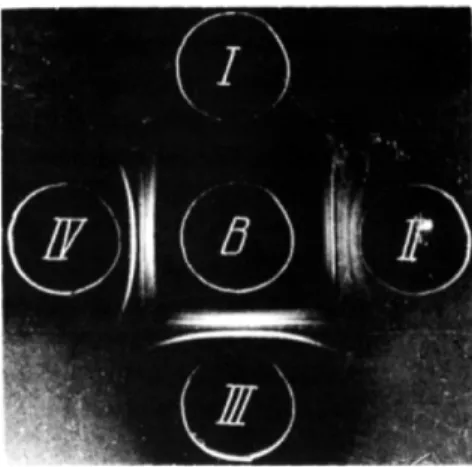
Thus, it is obvious, from Fig. 10, that E. coli LM antineurotoxin serum precipitates with the glycolipid-polypeptide endotoxin and lipoprotein
F I G . 10. A = E. coli LM antineurotoxin serum. I = E. coli LM neurotoxin. 11 = E. coli LM acid polysaccharide hapten. I l l =E. coli LM lipopolysaccharide. I V = Boivin endotoxin.
324 L. M E S R O B E A N U A N D I. M E S R O B E A N U
F I G . 11. B = E. coli L M antilipopolysaccharide serum. I = E. coli L M neurotoxin.
II = E. coli L M acid polysaccharide hapten. I l l = E. coli L M lipopolysaccharide. IV = Boivin endotoxin.
and gives no reactions with the polysaccharide prepared by acid hydrol
ysis or the Westphal lipopolysaccharide.
Figure 11 shows the results of a precipitation carried out with a 6 times concentrated E. coli LM antilipopolysaccharide serum where the lack of a precipitation reaction with the lipoprotein neurotoxin is obvious. Finally, in Fig. 12, an E. coli LM antiendotoxin serum can be seen to cross react with both polysaccharide and lipoprotein fractions.
A toxic lipoprotein was prepared by acid hydrolysis of the endotoxin.
For this, a Boivin endotoxin was hydrolyzed with 2 % acetic acid at 100°C. When a precipitate was obtained after 20 minutes, hydrolysis was
F I G . 12. C — E. coli L M antiendotoxin serum. I =E. coli L M neurotoxin. II =E. coli L M acid polysaccharide hapten. I l l =E. coli L M lipopolysaccharide. IV = Boivin endotoxin.
F I G . 13. A = E. coli LM antineurotoxin serum. I = E. coli LM neurotoxin. II = E. coli LM neurotoxin (van Heyningen). I l l = Boivin endotoxin. IV = Lipoprotein (acid cleavage of endotoxin, 2 % acetic acid). V = Polysaccharide (acid cleavage of endotoxin). V I = E.
coli21 heterologous neurotoxin.
interrupted and the precipitate was collected and redissolved in weak al
kaline solution. This lipopolypeptide solution reacts with E. coli LM anti
neurotoxin serum. An E. coli LM neurotoxin, prepared according to the van Heyningen method (first purification step), was investigated as well, and a precipitation spectrum similar to that of the neurotoxin prepared by chloroform autolysis was found (Fig. 13). The lipoprotein isolated from endotoxin (P) (Fig. 14) precipitates with an antiendotoxin serum; it does not react with concentrated antilipopolysaccharide serum.
F I G . 14. P = E. coli LM lipoprotein prepared from Boivin endotoxin. SE = Boivin anti
endotoxin serum. S L = Antilipopolysaccharide serum (concentrated 6 times).
326 L. MESROBEANU AND I. MESROBEANU
The lipoproteins prepared by a short, mild acid hydrolysis of E. coli LM Boivin endotoxin were analyzed for toxicity as well as for their pro
tein, lipid, and reducing substance content. Analysis of the 6 preparations obtained from different batches of Boivin endotoxin gave the following results: 40-45% protein, 44-60% lipid, and 3.2-9.2% reducing sub
stance, after hydrolysis. Intraperitoneal toxicity in mice expressed as MLD for the various preparations was in the range of 0.08 to 0.3 mg N .
Attempts to separate protein from lipid completely resulted in products which were no longer toxic; however, one cannot conclude from this that loss of toxicity is due to lipid removal or to protein degradation during the chemical treatment for isolation. If acid hydrolysis of the Boivin endotoxin is extended past 20 minutes, a nontoxic lipoprotein fraction is obtained.
It is possible to isolate from E. coli LM Boivin endotoxin a lipoprotein, which gave serological cross reactions with the preparation obtained by chloroform autolysis. It has been observed that the lipoprotein obtained by acid cleavage of the endotoxin is antigenic and yields a serum which does not precipitate the Westphal lipopolysaccharide (Fig. 15). We also observed, in a series of neurotoxins, some that gave cross reactions.
Results from serological investigations show that the thermolabile lipo
protein fraction is antigenic, toxic, and serologically identical with the endotoxin-isolated lipoprotein.
C . THERMOLABILE NEUROTOXINS FROM E. coli, Proteus, AND Pseudomonas pyocyanea ISOLATED FROM U R I N E
Using recently isolated strains from 52 patients with gram-negative bacterial urinary infections (E. coli, 21 cases; Proteus, 14 cases; Pseu
domonas pyocyaneus, 17 cases), we studied the lipoproteic toxin, pro
duction (L. Mesrobeanu et ai, 1962b, 1963, 1964).
In all cases, we were able to extract substances with various degrees of toxicity and thermolability irrespective of the R or S character of the strain.
For E. coli the intraperitoneal toxicity of lipoproteins in mice, ex
pressed as MLD, varied between 0.007 and 0.25 mg N ; for Proteus prep
arations between 0.05 and 0.5 mg N ; and for P. pyocyanea between 0.06 and 0.5 mg N .
However, in P. pyocyanea, if the chloroform autolyslate is adjusted to pH 3.5 by the addition of trichloroacetic acid, a mucopolysaccharide mu
cilaginous material also precipitates. Therefore, another extraction tech
nique was used, namely, the 18-hour bacterial suspension (50 mg dry weight/ml) was heated for 1 hour at 60°C, and the supernate was brought to pH 3.5 without chloroform autolysis. The products thus obtained were less toxic.
F I G . 15. D = E. coli antilipoprotein serum (lipoprotein was obtained from endotoxin).
I = Lipopolysaccharide. II = Neurotoxin (chloroform autolysis). I l l = Lipoprotein (acid hydrolysis of endotoxin). IV = Boivin endotoxin.
We conclude from these investigations that gram-negative bacteria iso
lated from the urine of patients with urinary infections elaborate thermo
labile toxins.
D . SOLUBLE THERMOLABILE NEUROTOXINS IN U R I N E I N F E C T E D BY G R A M - N E G A T I V E ORGANISMS
Since the pH of the urine from patients massively infected with gram- negative bacteria (E. coli, Proteus, P. pyocyanea) is alkaline, the ques
tion arose as to whether the bacteria autolyzed to liberate neurotoxins in the urine. With that end in view we studied 22 urines from patients in
fected with E. coli (11 cases), P. pyocyanea (3 cases), and Proteus (8 cases).
Urines were collected in the morning upon arising; thus it could be as
sumed that several hours of incubation had occurred in the bladder. These patients had not been treated with antibiotics or other drugs. Urines con
taining appreciable amounts of blood or albumin of nonmicrobial origin were discarded. The samples were immediately centrifuged at 10,000 rpm in the cold, and the toxin was isolated from the supernate as follows: 10 gm of sodium chloride were dissolved in eachlOO-ml urine aliquot, and trichloroacetic acid was added to bring the pH to 3.5. The mixture was chilled 30 minutes and then centrifuged for 1 hour at 10,000 rpm. The resulting gelatinous deposit was redissolved in a volume of water (at pH 8.6) equal to 1/10-1/50 of the initial volume of urine. If, at pH 3.5, the amount of precipitate was very low, it was dissolved in a few millimeters
328 L. M E S R O B E A N U A N D I. M E S R O B E A N U
of alkaline solution (pH 8.5) to determine the nitrogen content and to test its toxicity.
A comparative study was made on both urine-extracted and bacteria- extracted neurotoxins for all three bacteria. The results of these analyses are listed in Table X, in which the similarity between the urine- and bac
teria-extracted toxins is demonstrated.
T A B L E X
C H E M I C A L C O M P O S I T I O N O F N E U R O T O X I N S E X T R A C T E D F R O M U R I N E A N D FROM B A C T E R I A
Composition (% dry weight)
Reducing
Total nitrogen Lipids substances
From From From From From From
Bacteria urine bacteria urine bacteria urine bacteria E. coli 11-13 10-13 2 - 4 . 5 5 - 1 2 1 2 - 1 6 7 - 1 2 P. vulgaris 11-13 11-13.5 3 - 7 6 - 9 1 0 - 1 4 9 - 1 3 P. pyocyanea 1 2 - 1 4 1 2 - 1 4 5 - 8 6 - 1 2 12.5-13.5 8 - 1 3
Each substance produced toxic effects in mice and rabbits and in tissue cultures. The MLD of E. coli neurotoxin given intraperitoneally in mice was 0.1-0.05 mg N; that of Proteus and P. pyocyanea toxins was about 0.1 mg N.
The MLD of E. coli toxin injected intravenously in rabbits was 0.005 mg N. Limb paralysis was common, and, at higher doses, death some
times occurred within 24 hours. Diarrhea and an initial hyperthermia fol
lowed by hypothermia was observed. Many animals inoculated with small amounts of toxin displayed a transient paralysis and died in cachexia three weeks later. Toxin heated at 80°C for 60 minutes was used for injec
tion into control animals; no toxicity was observed.
We simaltaneously studied normal urine extracts which, in 5 cases out of 20, gave a small precipitate at pH 3.5. Albumin extracted from urines collected from renal patients without urinary infection constituted an
other control group. These preparations were nontoxic for laboratory animals, and their chemical composition differed from that of thermolabile endotoxins.
Although our investigations were almost always limited by the small amount of material present, in most cases we were able to carry out nitro
gen, lipid, and total reducing substance, as well as toxicity, tests on the same preparation. Cytopathogenicity tests and agar gel precipitation reac-