Send Orders for Reprints to reprints@benthamscience.ae
Current Medicinal Chemistry, 2018, 25, 1-18 1
REVIEW ARTICLE
0929-8673/18 $58.00+.00 © 2018 Bentham Science Publishers
The Therapeutic Impact of New Migraine Discoveries
Melinda Lukács
1, János Tajti
1, Ferenc Fülöp
2, József Toldi
3, Lars Edvinsson
4,5, and László Vécsei
1,6,*1
Department of Neurology, University of Szeged, Szeged, Hungary;
2Institute of Pharmaceutical Chemistry and MTA-SZTE Research Group for Stereochemistry, University of Szeged, Szeged, Hungary;
3Department of Physiology, Anatomy and Neuroscience, University of Szeged, Szeged, Hungary;
4Department of Clinical Sciences, Division of Experimental Vascular Research, Lund University, Lund, Sweden;
5Department of Clinical Experimental Research, Copenhagen University, Glostrup Hospital, Copenhagen, Denmark;
6MTA- SZTE Neuroscience Research Group, Szeged, Hungary
A R T I C L E H I S T O R Y Received: October 02, 2017 Revised: April 18, 2018 Accepted: May 03, 2018 DOI:
10.2174/0929867325666180530114534
Abstract: Background: Migraine is one of the most disabling neurological conditions and associated with high socio-economic costs. Though certain aspects of the pathomechanism of migraine are still incompletely understood, the leading hypothesis implicates the role of the activation of the trigeminovascular system. Triptans are considered to be the current gold standard therapy for migraine attacks; however, their use in clinical practice is limited. Pro- phylactic treatment includes non-specific approaches for migraine prevention. All these sup- port the need for future studies in order to develop innovative anti-migraine drugs.
Objective: The present study is a review of the current literature regarding new therapeutic lines in migraine research.
Method: A systematic literature search in the database of PUBMED was conducted concern- ing therapeutic strategies in a migraine published until July 2017.
Results: Ongoing clinical trials with 5-HT1F receptor agonists and glutamate receptor antago- nists offer promising new aspects for acute migraine treatment. Monoclonal antibodies against CGRP and the CGRP receptor are revolutionary in preventive treatment; however, further long-term studies are needed to test their tolerability. Preclinical studies show positive results with PACAP- and kynurenic acid-related treatments. Other promising therapeutic strategies (such as those targeting TRPV1, substance P, NOS, or orexin) have failed to show efficacy in clinical trials.
Conclusion: Due to their side-effects, current therapeutic approaches are not suitable for all migraine patients. Especially frequent episodic and chronic migraine represents a therapeutic challenge for researchers. Clinical and preclinical studies are needed to untangle the patho- physiology of migraine in order to develop new and migraine-specific therapies.
Keywords: Calcitonin gene-related peptide, 5-HT
1Freceptor agonist, glutamate, pituitary adenylate cyclase- activating polypeptide, kynurenic acid, trigeminovascular system.
1. INTRODUCTION 1.1. Migraine
Migraine is a severe neurological condition, ranked as the sixth most disabling condition of all illnesses and
*Address correspondence to this author at the Department of Neu- rology, University of Szeged, Szeged, Hungary; Tel: +3662545384;
Fax: +3662545597; E-mail: vecsei.laszlo@med.u-szeged.hu
the most disabling condition of neurological diseases,
based on the results of the Global Burden of Disease
Study [1]. Clinically, the earliest signs of a migraine
attack include non-specific premonitory symptoms
such as tiredness, concentrating difficulty and depres-
sion, symptoms related mainly to the activation of the
hypothalamus [2]. Visual aura can precede or even ac-
company a headache, represented predominantly by a
blind or scintillating scotoma [3-4]. Other non-visual
auras (e.g., sensory, olfactory, or temporary motor symptoms) might also occur [5-6]. In the headache phase, the pain is usually unilateral, throbbing, severe or moderate in intensity, aggravated by physical activ- ity, which is often accompanied by nausea, vomiting, and photophobia [7]. Postdrome symptoms are consis- tent with those in the premonitory phase, including physical and mental tiredness, depressed mood and muscle stiffness [8-9].
Despite numerous studies that have tried to shed light on the pathomechanism of migraine, several as- pects are still unclear. The leading hypothesis impli- cates the role of the activation of the trigeminovascular system. Dural perivascular nerve endings that originate from the neurons of the trigeminal ganglion (TG) rep- resent the primary sensory neurons of the pathway. The neuronal cell bodies within the TG are surrounded by satellite glial cells. The second-order neurons are lo- cated in the trigeminal nucleus caudalis (TNC) and C
1- C
2region of the spinal cord [10]. They connect pain signals to the thalamus and the cerebral cortex [10-11].
Structural and functional brain imaging studies have revealed a number of other brain areas that become activated during migraine attacks, such as the nucleus raphe magnus (NRM), the nucleus raphe dorsalis (DG), the periaqueductal grey matter (PAG), and the locus ceruleus (LC) [12-13]. Structural alterations of the brain have been noted in areas involved in pain proc- essing, such as the anterior cingulate cortex or the tri- geminal system [14]. The origin of migraine pain is still a question of debate. Imaging studies showed no vasodilation of intracranial and extracerebral arteries, rendering the vascular theory of Wolff obsolete [15].
One of the theories postulates that the above mentioned brainstem nuclei are responsible for the initiation of migraine pain; therefore, they are sometimes referred to as ‘migraine generators’. It is still a question whether activation of these brain areas generates the pain sensa- tion or they become activated secondarily. Neurogenic inflammation is hypothesized to be an important factor in migraine pathophysiology. It is thought to induce a state of hyperexcitability, as nociceptive signals are transported ortho- and antidromically, leading to the release of various cytokines and neuronal messenger molecules (such as calcitonin gene-related peptide (CGRP), substance P (SP), neuropeptide Y (NPY), and nitric oxide (NO)). These molecules are presumed to induce the activation of immune cells, mast cells, and astrocytes and lead to vascular changes that might evoke blood-brain barrier (BBB) dysfunction [16-17].
Descending neurons of the CNS might aggravate the
inflammatory responses, resulting in long-term poten- tiation (LTP) [17-18]. Another phenomenon that has been under investigation as an initiating component of the migraine pain process is cortical spreading depres- sion (CSD). CSD is a depolarization wave that moves across the cortex from the occipital lobe towards the frontal areas, and has been suggested to represent the electrophysiological correlate of the aura phase of mi- graine; however, CSD alone is neither sufficient nor necessary to trigger migraine attacks [19-20]. Although extensive efforts have been made to elucidate the pos- sible mechanisms that play pathogenic roles in mi- graine, certainly much is yet to be unveiled to correctly interpret this disease. The most relevant contemporary concept postulates that migraine is a neurovascular dis- order. We hypothesize that pain originates in the cen- tral nervous system (CNS), resulting in hypersensitivity of the perivascular nociceptive afferent nerve fibers, which play an essential role in the pathogenesis.
1.2. Current Treatments in Migraine
Regarding migraine treatment, analgesics (NSAIDs), antiemetics and triptans are the drugs to be chosen in the case of a migraine attack. The current gold standard therapy is the use of triptans, drugs with serotonin receptor (5-HT
1B/1D) agonist properties. The efficacy of triptans in migraine attacks has been proven in large placebo-controlled clinical trials [21]. They have proven efficacy in 60% of migraine attacks [22].
In clinical practice, the use of triptans has some limita- tions: clinical trials showed pain relief in only 28-59%
of the patients [23]. They should be taken in the early phase, which leads to frequent drug intake and thus increased risk of chronification [24]. Frequent use of analgesics or triptans might lead to medication overuse headache (MOH). Other important requirements are related to the side-effects, to the safety and tolerability profiles [25]. One of the most important problems with triptans is related to their side effects. Following suma- triptan therapy, severe cardiovascular adverse events (such as stroke, myocardial infarction, and cardiac ar- rhythmias) have been reported to occur with an inci- dence of 1:1.000.000 [26-27]. Prophylactic therapy of migraine includes beta-adrenergic receptor blockers, calcium ion channel blockers, antiepileptic drugs, and antidepressants. Chronic migraine represents a thera- peutic challenge because triptans can be used only 9 days/month due to high risk of chronification [24].
Lately, botulinum toxin A (BoNTA) injected intramus-
cularly into the muscles of face and head has proven to
be efficient in chronic migraine [28-30].
The future goal should be the development of migraine-specific drugs with good safety and tolerabil- ity profile [31]. Due to the individual pain sensation of the patients, therapy should aim at personalized medi- cine. The difficulty of developing new migraine- specific drugs is represented by the lack of adequate animal models and specific biomarkers [25].
2. NEW THERAPEUTIC TARGETS IN MIGRAINE
In the present article, we have aimed to assess the current and novel therapeutic strategies related to dif- ferent neuropeptides and molecules putatively involved in the pathomechanisms of migraine. A systematic lit- erature review was performed in the database of PUB- MED until July 2017. We used the following search strings: "migraine", "migraine treatment", and "clinical studies in migraine".
2.1. 5-hydroxytryptamine (5-HT, Serotonin)
5-HT is a metabolite of tryptophan that has proven to play a pivotal role in migraine because elevated lev- els of its metabolite, 5-hydroxyindoleacetic acid (5- HIAA) were detected early in the plasma of migraine patients during attacks [32]. Numerous studies related to 5-HT led to the discovery of triptans, which at pre- sent represent the gold standard therapy in the acute treatment. Early studies suggested a vasoconstrictor effect on the cranial arteries [33-36]; however, it has been demonstrated lately that triptans are not only cerebral and dural vasoconstrictors but they also exert neuronal effects. Indeed, triptans act on neurogenic
inflammation in the dura mater [37-38] and they also modulate the activity of trigeminal neurons [39]. 5-HT receptors represent one of the most complex families of neurotransmitter receptors, having seven subfamilies and a lot of subtypes. Except for 5-HT
3, all forms are parts of the G protein-coupled receptor (GPCR) super- family [40]. Several hundreds of genes encode different receptors for neurotransmitters, and post-translational modifications result in different proteins [41]. More than one hundred cloned orphan GPCRs have so far been identified, and it is still unknown how many of them belong to the 5-HT receptor family. In an attempt to reduce the cardiovascular side effects of triptans, researchers focused on finding potent and selective ligands for the different receptor subtypes. Sumatriptan and naratriptan have been shown to bind with high af- finity to the 5-HT
1Freceptor [42], a subtype of the 5- HT
1receptor class consisting of 366 amino acids and having 7 transmembrane domains (TMD) [43-44]. 5- HT
1Freceptors have been shown to be expressed in glu- tamatergic neurons of the trigeminal system and also in the cerebral vessels, with no vasoconstrictor effect [45- 46]. The ability of 5-HT
1Freceptors to modulate tri- geminal responses without any effect on the vascular tone made 5-HT
1Fagonists promising tools as innova- tive anti-migraine drugs [47-48]. 4-Fluoro-N-[3-(1- methyl-4-piperidinyl)-1H-indol-5-yl]-benzamide (LY334370) has been shown to be a high-affinity ago- nist of the 5-HT
1Freceptor [49]. Among other 5-HT receptors, LY334370 shows the next highest affinity for 5-HT
1Aand 5-HT
1E, binding only a small fraction of these receptors [50]. In preclinical studies, LY334370
Table 1. Pharmacological data for triptans (source: Tajti et al., 2015). IUPAC- International Union of Pure and Ap-plied Chemistry, p.o.-per oral, s.c-subcutaneous [25].
Drug name Sumatriptan Eletriptan Zolmitriptan Rizatriptan
Systematic (IUPAC) name
1-[3-(2-
Dimethylaminoethyl)-1H- indol-5-yl]-N-methyl- methanesulfonamide
(R)-3-[(-1- methylpyrrolidin-2- yl)merhyl]-5-(2- phenylsulfonethyl)-1H- indole
(S)-4-({3-[2-
(dimetylamino)ethyl]-1H- indol-5-yl}metyl-1,3- oxazolidin-2-one
N,N-dimethyl-2-[5-(1H- 1,2,4-triazol-1-ylmetyl)- 1H-indol-3-
yl]ethanesulfonamide Route of ad-
ministration p.o., s.c., nasal, rectal,
transdermal p.o. p.o., nasal p.o.
Cmax 54 ng/ml (50-100 mg tab-
let) 188-234 ng/ml (80 mg
tablet) 5.6-9 ng/ml (5mg tablet) 15.7 ng/ml (10 mg tablet) Tmax 1.5 h (50-100 mg tablet) 1.8-2.5 h (80 mg tablet) 1.5 h (5mg tablet) 2.3 h (10 mg tablet) T1/2 2.3 h (50-100 mg tablet) 4-7 h (80 mg tablet) 2.7 h (5mg tablet) 3.2 (10 mg tablet)
Metabolism Hepatic-MAO Hepatic-CYP-34A Hepatic-CYP1A2 Hepatic-MAO
Excretion Renal, fecal Hepatic Renal, fecal Renal, fecal
has been proven to block neurogenic inflammation [49]
and diminished c-fos immunopositivity in the TNC, without any vasomotor effect on cerebral vessels [51- 52]. In clinical studies, LY334370 was tested in three different doses. The 60 mg and 120 mg doses showed superiority over placebo at all three endpoints (2 h re- sponse, 2 h pain-free, sustained effect) with no cardio- vascular side effects [53]. Although the study proved the efficacy of LY334370 for acute treatment, the stud- ies were cancelled due to liver toxicity following long- term use in animal models.
2,4,6-Trifluoro-N-[6-[(1-methylpiperidin-4-yl)carb- onyl]pyridin-2yl]benzamide (lasmiditan) represents a new generation of 5-HT
1Fagonists. Lasmiditan does not contain the indole core that is present in 5-HT and triptans, as the indole core is replaced by a pyridinoyl- piperidine scaffold. Lasmiditan has higher selectivity to the 5-HT
1Freceptor than LY334370. Following oral lasmiditan administration, decreased c-fos activation was detected in TNC in a model of electrical stimula- tion of rat TG and no vasoconstrictive effect was noted [54]. Furthermore, lasmiditan, in contrast with triptans, can penetrate the BBB, suggesting to be able to act on central mechanisms [55]. Phase 1 and phase 2 clinical trials have been carried out to test the intravenous and oral efficacy of lasmiditan [56-57]. Lasmiditan has proven superiority over placebo in the primary end- point of headache response, with a linear dose-response relationship [56]. Oral administration in a dose of 400 mg showed higher therapeutic gain compared to the intravenous dose of 20 mg [57]. The studies show rapid absorption and a bioavailability of 40% in the case of oral administration [58]. Two ongoing phase 3 and a long-term open-label trial were started in 2015 to test lasmiditan in episodic, disabling migraine [55].
Due to its different chemical structure and the selective action on the 5-HT
1Freceptor, the side effects of las- miditan are completely different from those of triptans.
Dizziness, paresthesia, and vertigo were the most common adverse events reported, predominantly at- tributable to the presence of 5-HT
1Freceptors in the cerebellum and the vestibular nuclei, and also because of the high BBB penetration of lasmiditan [55, 58]. On the basis of these, CNS-related side effects can be an- ticipated following a long-term use of lasmiditan, which might limit its clinical use and delay further studies [59].
NXN-188 is an oral drug developed with a dual mechanism of action: acting on neuronal NO synthase (nNOS) enzyme and having a high affinity for the 5- HT
1B/1Dreceptor. Preclinical studies have demonstrated
the ability of this molecule to inhibit CGRP release in animal models using capsaicin or an electrical stimula- tion of the trigeminovascular system. NXN-188 itself did not induce any vasoconstriction in the middle men- ingeal artery (MMA) but blocked capsaicin-induced vasodilation. GR127935, a 5-HT
1B/1Dreceptor antago- nist was able to block the effect of sumatriptan on the MMA and did not influence the effect of NXN-188, suggesting that the novel compound acts partially on nNOS, being a promising future therapeutic approach for migraine prophylaxis [60]. Regarding clinical stud- ies, five phase 1 trials have been carried out in order to test the safety and assess the pharmacokinetics of NXN-188 in healthy volunteers. The compound had two absorption phases: the first peaked at 1 h, and the second at 4-5 h. Initial elimination from the plasma was rapid (plasma concentration decreased with 70-90% of the C
maxwithin 24 h) followed by a prolonged elimina- tion (with 1-5% of the C
max) for several weeks. NXN- 188 was well tolerated by the participants, the reported adverse events were dizziness, headache, and somno- lence [61]). Unfortunately, phase 2 clinical trials showed no statistically significant effect on migraine with aura compared to placebo [62].
NH N
CH3
HN
O F
LY-334370
N NH O
F F
F O
N CH3
Lasmitidan
Fig. (1). Chemical structure of LY-334370 and Lasmiditan.
2.2. Glutamate (Glu)
Glu, the major excitatory neurotransmitter in the CNS, has been shown to play crucial roles in the pathophysiology of migraine. Elevated levels of Glu were detected in the serum of migraine patients [63]
and in the cerebrospinal fluid (CSF) of patient affected by chronic migraine [64]. Glu mediates the CSD phe- nomenon [65] and has been shown to be co-released with CGRP from the neurons of the TG upon activation [66]. In animal models of trigeminovascular activation, increased Glu activity was detected [67-68]. Glu acts on ionotropic and metabotropic receptors as well [69].
The ligand-gated ion channels are the N-methyl-D-
aspartate (NMDA) receptors, the α-amino-3-hydroxí-5- methyl-4-isoxazelopropionic acid (AMPA) receptors and the kainate receptors (that can be divided into sev- eral subtypes), which mediate fast synaptic transport [70]. AMPA receptors have four semiautonomous do- mains: the amino-terminal domain, the extracellular ligand-binding domain (LBD), the TMD, and the intra- cellular carboxyl-terminal domain (CTD) [70-71].
NMDA receptors contain three subunits: NR1 is com- mon in all cell types; NR2 has four subtypes differen- tially expressed in different cell types and in various CNS structures during certain development processes;
and NR3 with modulatory function on the receptor. For optimal function, both the NR1 and the NR2 subunits need to be expressed. The NR1 subunit contains the glycine (Gly)-binding site. Gly acts as a coagonist of Glu and is essential for the activation of the receptor. In inactive form at the resting potential, the channel is blocked by Mg
2+. Following activation by binding of both Glu and Gly to the receptor, Mg
2+dissociates from the receptor and opens the channel, resulting in the flow of Na
+and Ca
2+into and K
+out of the cell. The Ca
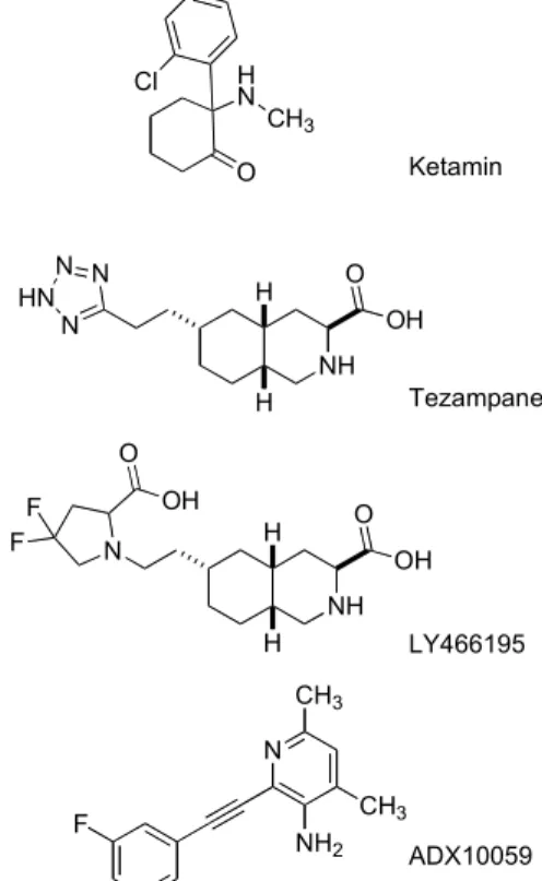
2+signal then activates various intracellular path- ways that lead to exocytotic effects [72-73]. All three ionotropic receptors play pivotal roles in migraine, act- ing on neuronal activation and signal transmission in the trigeminal system, but the only receptor linked to CSD and thus the aura phenomenon is the NMDA re- ceptor [74]. Metabotropic Glu receptors (mGluRs) are GPCRs and contain at least eight receptor subtypes [75]. The N-terminal domain, called Venus Flytrap Domain, contains two lobes. The binding of the ligand leads to the closure of the two lobes, leading to the ac- tivation of the receptors [76]. The seven TMD of mGluRs is rich in cysteine, which forms disulfide bonds in order to increase the stability of the domain [77]. The CTD is close to the cell membrane and inter- acts with signaling pathways [75]. In a small study on familial hemiplegic migraine (FHM), intranasal ad- ministration of ketamine, an NMDA receptor antago- nist that acts on the Glu-binding site, reduced the inten- sity and duration of motor symptoms in almost half of the patients [78]. In a phase 2 study with migraine pa- tients suffering from prolonged aura, intranasal keta- mine was able to reduce the severity of aura but did not influence its duration [79]. The AMPA/kainate receptor antagonist, tezampanel (LY293558), and a kainate re- ceptor antagonist, LY 466195, were shown to decrease c-fos activity in the rat TNC following electrical stimu- lation of the TG; whereas the AMPA receptor antago- nist LY300178 did not influence c-fos activation [80].
This leads to the conclusion that the effect of tezam-
panel might be mediated through the kainate rather than the AMPA receptor. A phase 2 clinical trial dem- onstrated that intravenous tezampanel was superior to placebo in all endpoints (sustained relief of pain and other associated symptoms) and no cardiovascular side effects were noted. Adverse events were mainly CNS- related, including dizziness and somnolence [81]. For LY466195, intravenous doses of 1 and 3 mg were compared with placebo. The 1 mg dose was not effec- tive whereas the 3 mg dose showed superiority com- pared to placebo. As adverse effects, visual distur- bances were reported in 21% of the patients [82]. ADX 10059, a negative allosteric modulator of the mGluR5, was also tested in phase 2 clinical trials for acute mi- graine therapy. Following oral administration of ADX 10059, pain relief was significantly higher than in the placebo group at the primary endpoint (2 h), but this effect was not proven to be sustained. As side effects, dizziness, vertigo, and visual disturbances were re- ported [74, 83]. All these data suggest that modulation of glutamatergic neurotransmission might represent an important mechanism for innovative drugs in both the acute and the prophylactic treatments of migraine. Fol- lowing the contemporary concept, kainate receptor an- tagonist might represent an effective therapeutic mo- dalities for acute treatment, whereas specific NMDA receptor antagonists might be promising therapeutic tools for aura phenomena.
Ketamin
Tezampanel
LY466195
ADX10059 HN
O CH3 Cl
NH OH O N N
HN N
H
H
NH OH O
N H
H F
F O
OH
N CH3
CH3 NH2 F
Fig. (2). Chemical structure of Glu related therapeutic tar- gets.
2.3. Calcitonin Gene-related Peptide (CGRP) The pivotal role of CGRP in migraine has been sug- gested for decades [84-85]. CGRP is a 37-amino-acid peptide that has two isoforms, αCGRP and βCGRP, with similar chemical structures and biological func- tions, encoded by two different genes on chromosome 11 in humans [86]. αCGRP, expressed predominantly in the nervous system, is encoded by the CALCI gene (together with calcitonin) by means of an alternative splicing mechanism. Expression of exons 5 and 6 leads to the production of αCGRP mRNA, which is trans- lated to a prohormone of 121 amino acids and cleaved secondarily to yield the mature protein [87]. βCGRP is expressed in the enteric nervous system and is tran- scribed from the CALCII gene [88]. The CGRP recep- tor is part of the GPCR superfamily and consists of a 7- TMD, called calcitonin receptor-like receptor (CLR), and a single-transmembrane spanning protein, called receptor activity-modifying protein 1 (RAMP1), which is necessary for the binding of CGRP to the receptor [89]. The third component, called receptor component protein (RCP), does not affect the binding of CGRP to the receptor but it is necessary for signal transduction [90]. Previous studies have demonstrated that CGRP is a key mediator in migraine, as elevated levels of CGRP were found in the plasma, CSF and saliva of migraine patients [91-93]. Subsequently, mapping of CGRP and its receptors in different structures of the rat and human brain demonstrated their role in nociceptive transmis- sion [94-96]. In animal models of trigeminovascular activation, increased CGRP activity was detected [97- 99]. All these findings support a well-established posi- tion of CGRP in migraine pathophysiology and current efforts pursue the identification of potent anti-migraine drugs targeting CGRP and its receptors. The most comprehensively studied compounds were the ‘gep- ants’, acting on CGRP receptors. Olcegepant (BIBN 4096BS, 1-[3,5-Dibromo-N-[[4-(1,4-dihydro-2-oxo- 3(2H)-quinazoline)-1-piperidinyl]carbonyl]-D-tyrosyl- L-lysyl]-4-(4-pyridyl)-piperazine) was the first CGRP receptor antagonist, binding with high selectivity to the CGRP receptors. Its relatively high molecular weight (Mw = 870 Da) and low oral bioavailability necessi- tated an intravenous route of administration. In animal models, olcegepant has proven to decrease CGRP- mediated trigeminal activation [100]. In phase 1 clini- cal study, the maximal concentration was dose- proportional (C
max= 0.87 mg/ml), the biological half- life (T
1/2) was 2.5 h and no serious adverse events were noted (the most common included fatigue and pares- thesia) [101]. Although phase 2 clinical trials supported the efficacy of olcegepant and, apart from paresthesia,
no severe adverse events were noted, [102] the intrave- nous route limited its use in the daily clinical practice [103]. Therefore, an oral formula was developed, re- ferred to as telcagepant (MK-0974, N-((3R,6S)-6-(2,3- difluorophenyl)-2-oxo-1-(2,2,2-trifluoroethyl)azepan- 3-yl)-4-(2-oxo-2,3-dihydro-1H-imidazo[4,5-b]pyridin- 1-yl)piperidine-1-carboxamide). In animal models, tel- cagepant did not show any effect on the diameter of cranial arteries, but blocked the effect of CGRP [104].
Human studies showed rapid absorption of telcagepant, with a T
1/2of 6 h and C
maxof 0.55 mg/ml. In phase 2 clinical trials, telcagepant in 300-600-mg doses showed superiority to placebo and the same efficacy as zol- mitriptan (5 mg). Unfortunately, the development of telcagepant and its successor, MK-3207, was stopped due to liver toxicity and elevated gamma- glutamyltransferase (GGT) levels developed after long- term and frequent administration [105]. It has been suggested that hepatotoxicity might be a side effect related to one of the metabolites of the compounds [106]. Rimagepant (BMS-927711, (5S,6S,9R)-5- amino-6-(2,3-difluorophenyl)-6,7,8,9-tetrahydro-5H- cycloheptapyridin-9-yl 4-(2-oxo-2,3-dihydro-1H- imidazo[4,5-b]pyridin-1-yl)piperidine-1-carboxylate) and BI 44370 TA (4-[[(7R)-8-cyclopentyl-7-ethyl-5- methyl-6-oxo-7H-pteridin-2-yl]amino]-3-methoxy-N- (1-methylpiperidin-4-yl)benzamide) have proven effi- cacy compared to placebo in phase 2 clinical studies without any adverse event related to liver toxicity;
however, the one-dose design of the trials offered lim- ited information regarding the safety profile in case of repeated treatment [107-108]. A newly designed com- pound, ubrogepant, (MK-1602, (3'S)-N-((3S,5S,6R)-6- methyl-2-oxo-5-phenyl-1-(2,2,2-trifluoroethyl)piperi- din-3-yl)-2'-oxo-1',2',5,7-tetrahydrospiro(cyclopenta(b) pyridine-6,3'-pyrrolo(2,3-b)pyridine)-3-carboxamide) has recently been developed for acute treatment of mi- graine and has demonstrated superiority to placebo with a good safety and efficacy profile [109]. A phase 3 clinical study is currently recruiting participants. We await further studies providing detailed assessment of the tolerability profile during long-term use.
New classes of biological therapies, monoclonal an-
tibodies acting on CGRP or its receptors have been de-
veloped in recent years. The mechanism of action of
these molecules is still not fully understood; however,
they have been shown to prevent repeated activation of
the trigeminovascular system induced by CGRP, reduc-
ing headache frequency [110]. The pharmacokinetic
and pharmacodynamic properties of these molecules
differ completely from those of the gepants. Due to
their high molecular weight (150 kDa), their instability
Olcegepant
Telcagepant
BMS-927711 Rimegepant N
N N
O
NH O NH2
HN N
O
N HN Br O
OH Br
N HN N
O
N N H OO N
F F
F
F F
N N NH
O O N
O N
H2N
F F
Fig. (3). Chemical structure of CGRP receptor antagonists.
in the gastrointestinal tract, and the low permeability through cell membranes, these molecules can only be administrated subcutaneously or intravenously. These drugs are also not able to cross the BBB [111]. These facts collectively lead to the conclusion that these molecules act on peripheral targets such as the TG [112], as TG was proven to be placed outside the BBB [94]. Their plasma half-life is extended to days and weeks. This enables longer dosing intervals, which is rather favorable in terms of patient compliance [113- 114]. As these molecules are metabolized to small pep- tides and amino acids, they do not affect hepatic or re- nal enzymes and the incidence of drug-drug interac- tions is very low [115]. They are potential immuno- gens; therefore, they might evoke various immunologi- cal adverse events such as hypersensitivity, immuno- suppression or autoimmune processes [116-117]. Al- though the studies performed with CGRP-related monoclonal antibodies have not revealed serious im- munotoxic effects so far, a small percentage of the pa- tients were positive for anti-drug antibodies [111].
These antibodies are of great importance as they might reduce future therapeutic efficacy and might aid the development of allergic reactions [111, 118]. Three of
the monoclonal antibodies tested act on CGRP:
ALD403 (eptinezumab), TEV-48125 (fremanezumab), and LY2951742 (galcanezumab) whereas AMG334 (erenumab) targets the CGRP receptor. All four anti- bodies have demonstrated positive effects in migraine prevention in phase 1 and phase 2 clinical trials, with no significant adverse reactions reported [119-122].
TEV-48125 might represent a promising target in chronic migraine as well [123]. There are small differ- ences between the association/dissociation rates of the four antibodies that might have an impact on their effi- cacy: ALD403 attaches to the target twice as rapidly as TEV-48125. LY2951742 takes effect as an incomplete agonist with fast binding to the target and rapid disso- ciation [111].
Primary results of phase 3 clinical trials were pre- sented on the International Headache Congress 2017 (IHC 2017), in Vancouver. PROMISE-1 study was de- signed to evaluate the effect of i.v. eptinezumab in the prevention of frequent, episodic migraine. Adult pa- tients were included, receiving 300mg, 100mg, 30mg eptinezumab and placebo. Significant reduction in mi- graine days was reported in case of eptinezumab, main- tained at similar levels for long-term (12 weeks). Two phase 3 studies (EVOLVE-1 and EVOLVE-2) for s.c.
use of galcanezumab were performed. Doses of 120mg and 240mg were tested and both doses proved superior- ity for overall mean change in migraine headache days.
The REGAIN trial was a 3-month study, performed for the same doses of s.c. galcanezumab in patients with a chronic migraine. Both doses were superior to placebo regarding the reduction of migraine headache days.
Also, treatment with self-administrated galcanezumab was proven to be safe and well-tolerated. The STRIVE trial was designed to test the effect of s.c. use of erenumab, for a dose of 140mg and 70mg. Over 24 weeks significant reduction was reported in the impact of migraine on physical, social and emotional function- ing of episodic migraine patients (Cephalalgia, Volume 37, Issue 1_suppl, September 2017).
Although additional long-term clinical studies are
needed, it might be reasonably postulated that these
anti-CGRP monoclonal antibodies represent a new and
effective therapeutic line in migraine prevention. They
can be considered revolutionary in the pharmaceutical
treatment of migraine, especially in case of chronic
migraine. The only obstacle in their way towards a
wide clinical use might be their cost/benefit ratio, as
the cost related to the manufacturing of monoclonal
antibodies is rather high. It should also be considered,
however, that migraine is among the priciest neurologi-
cal diseases in Europe [124] and an effective treatment might lead to reduced health care costs.
3. PROMISING FUTURE THERAPEUTIC TAR- GETS IN PRECLINICAL PHASE
3.1. Pituitary Adenylate Cyclase-activating Polypep- tide (PACAP)
PACAP is a neuropeptide isolated from the hypo- thalamus in 1989 [125]. Two bioactive forms of PACAP are known: PACAP1-38 and PACAP1-27.
PACAP1-38 is formed from a precursor (prepro- PACAP) by convertase enzyme. Two Arg residues are split by a carboxypeptidase and the remaining Gly resi- dues are used to amidate the Lys residue to yield PACAP1-38. Another series of Gly Lys Arg of PACAP 1-38 allows further reactions, providing PACAP1-27 [126]. The two active forms of PACAP cross the BBB in different ways: PACAP1-27 enters via transmem- brane diffusion attributable to its lipophilic property, whereas PACAP1-38 uses a carrier-mediated peptide transport mechanism [127]. The role of PACAP1-38 has been suggested in the ethiopathology of migraine, though some aspects are still yet to be revealed [128- 130]. PACAP1-38 was detected in the TG [131], in the TNC [132], and also in the parasympathetic otic and sphenopalatine ganglia [133-134]. Elevated levels of PACAP were found in the interictal phase compared to the ictal phase in migraine patients and also in cluster headache attacks [135-136]. Intravenous infusion of PACAP was able to induce headache in healthy volun- teers and evoke migraine-like attacks in patients suffer- ing from migraine without aura [137-138]. Vasoactive intestinal peptide (VIP) and PACAP1-38 share the same receptors: VPAC1 and VPAC2, whereas PAC1 has higher affinity for PACAP1-38 [139-140]. The vasodilatory effect of PACAP1-38 on dural vessels has been proven to be mediated via VPAC2 receptors, whereas neurogenic dural vasodilation induced by elec- trical stimulation of the trigeminal nerve terminals is mediated predominantly by PAC1 [138, 141]. Central PAC1 receptors are suggested to play key roles in cen- tral trigeminal activation [142], as PACAP1-38 (but not VIP) has been shown to cause delayed sensitization of the trigeminal system. In an animal model of dural electrical stimulation, a BBB-impermeable PAC1 re- ceptor antagonist was tested: in the case of peripheral administration, the substance was not able to prevent neuronal firing of the TNC, whereas the central (i.e.
intraventricular) administration was able to decrease the neuronal activity [141]. To our knowledge, no PAC1 receptor antagonists are available that can pene-
trate the BBB. It is a question whether the peripheral action of these PAC1 receptor antagonists will prove sufficient to prevent migraine attacks or the develop- ment of novel small molecules will be needed with op- timized BBB permeability. Another possibility to pre- vent PACAP1-38-initiated migraine attacks might be the use of monoclonal antibodies against PACAP1-38 per se or against PAC1 receptors, by analogy with those developed against CGRP and its receptor [143].
It is possible that new molecules are needed to also block the central action of PACAP1-38 via the PAC1 receptor in order to be effective against migraine at- tacks, whereas peripherally acting substances might be used as preventive therapies. Therapeutic strategies targeting PAC1 receptor represent promising ap- proaches for the treatment of migraine, but further pre- clinical and clinical studies are warranted.
3.2. Kynurenic Acid (KYNA)
Tryptophan, one of the essential amino acids, is the precursor of 5-HT and L-kynurenine under physiologi- cal conditions. The kynurenine pathway has been pre- viously presented in details [128, 144]. Briefly, it is a complex metabolic pathway with a lot of neuroactive products ending with NAD
+[145]. As these neuroac- tive metabolites influence NMDA receptor-mediated excitotoxicity and the production of free radicals, they are presumed to play important roles in various dis- eases of the CNS [146-150]. KYNA acts on NMDA receptors binding to the Gly-binding site of the NR2 subunit [73] and it also acts on AMPA receptors with a dual action: in low concentrations it enhances, whereas in higher concentrations it decreases receptor activity [151-152]. This leads to the conclusion that modulation of the kynurenine pathway might represent an appro- priate therapeutic tool in migraine treatment and is cur- rently investigated in preclinical animal studies. As KYNA has a very low capacity to cross the BBB, dif- ferent strategies are needed in order to take advantage of its anti-inflammatory and neuroprotective properties.
One possibility could be the use of a prodrug such as L-kynurenine or its derivates [153-155]; whereas shift- ing the pathway towards the production of KYNA with different enzyme inhibitors would represent another possible therapeutic strategy [156-157]. During the last years, our research group has synthesized several dif- ferent KYNA derivates that have proven to be effective in animal models of cerebral ischemia [158], Huntington’s disease [159], epilepsy [160], and rat models of trigeminovascular activation [68, 161-162].
The chemical structures of these KYNA analogues
have been previously presented. Briefly, a new cationic
center and a water-soluble side-chain were included in order to facilitate BBB penetration [163-164].
NH O
OH O KYNA
SzR104
SzR72 SzR81
N OH
HN
O N
OH
HN
O
N OH
HN
O
N
N O
N N
Fig. (4). Chemical structure of different KYNA analogues designed by our research group.
Their exact mechanism of action still needs to be untangled. There are two main hypotheses as regards their mechanism of action: 1) these molecules might act as analogues, mimicking some effects of KYNA, 2) they may dissociate into KYNA and serve as a prodrug [165-166]. Only a few studies are available to elucidate their pharmacokinetic profile. One of these focused on two KYNA derivates: N-(2-N,N-dymethylaminoethyl)- 4-oxo-1H-quinoline-2-carboxamide hydrochloride (KA-1) and N-(2-N-pyrrolidinylethyl)- 4-oxo-1H- quinoline-2-carboxamide hydrochloride (KA-2). Fol- lowing intraperitoneal treatment with these amides, the concentrations of KA-1, KA-2 and KYNA were meas- ured in the rat serum and TNC. The study revealed that following intraperitoneal treatment with these amides, a sharp increase followed by a sudden decrease in the level of KA-1 and KA-2 in the serum could be de- tected. In the fifth hour, the compounds were still pre- sent in the serum. Although elevated levels of KYNA were found in the serum in the case of both molecules, the increase was less for KA-1 than for KA-2. In the CNS, only small amount of KA-2 was detected, with the level of KA-1 being below the lower limit of detec- tion [165]. Although these KYNA amides were de- tected only in trace (or zero) amounts within the CNS, animal studies of trigeminovascular activation show decreased activation of the TNC following intraperito- neal treatment with KA1 [68, 167]. There are two po- tential explanations: 1. the two amids act peripherally, metabolized only in small proportion two KYNA, 2.
the pharmacokinetic studies were performed in intact
animals. In case of neurogenic inflammation, an impor- tant phenomenon that occurs during trigeminovascular activation causes BBB dysfunction, where the BBB becomes penetrable for substances that are normally unable to pass [17, 168]. We assume that during mi- graine attack the BBB dysfunction occurs and pene- trance of KYNA analogues might be possible but fu- ture studies are needed to test this hypothesis.
In summary, all these data suggest that it is neces- sary to conduct further studies that are needed to en- lighten the mechanism behind the positive effects of the KYNA amides.
4. FAILED THERAPEUTIC TARGETS 4.1. Vanilloid Receptor 1 (TRPV1)
TRPV1 is a non-selective cationic channel, which is sensitized and up-regulated during inflammation, play- ing an important role in noxious stimulatory states [169-170]. The efficacy of TRPV1 antagonists in pre- clinical models of chronic pain has suggested the po- tential therapeutic benefit of TRPV1 inhibition in this condition [171]. In case of migraine, animal studies provided contradictory results: a TRPV1 antagonist was ineffective in two different animal models [172], whereas another TRPV1 antagonist was proven to be effective in an animal model of electrical and mechani- cal stimulation of the dura mater in cats [173]. In vitro pharmacological studies suggested that SB-70498 had the best pharmacological properties among the TRPV1 receptor antagonists. Its metabolic stability and a bioavailability of almost 86% made SB-70498 a prom- ising molecule for clinical studies [174]. However, phase 2 clinical studies were terminated early as SB- 70498 did not show any superiority to placebo in acute migraine treatment [175]. These findings do not sup- port the use of TRPV1 antagonists in the treatment of migraine.
4.2. Substance P (SP)
SP was shown to be expressed in various parts of
the CNS and has been implicated in the pathophysiol-
ogy of migraine [176]. SP acts on the tachykinin recep-
tors (NK
1, NK
2, and NK
3), predominantly on NK
1. The
SP receptor antagonist, PRP100893, has proven to di-
minish plasma protein extravasation following electri-
cal stimulation of the TG in guinea pigs [177]. Unfor-
tunately, phase 2 clinical studies did not support the
efficacy of SP receptor antagonist in acute migraine
attacks [178]. Lanepitant, another NK
1receptor an-
tagonist that showed promising results in preclinical
studies, was not proven to be efficient in migraine pre-
vention [179]. The above-mentioned data suggest that therapeutic strategies targeting SP or its receptors might not be adequate for acute or prophylactic mi- graine treatment.
4.3. Nitric Oxide (NO.)
NO. is a labile gas, produced by three iso-enzymes called NOSs: neuronal (nNOS) inducible NOS (iNOS) and endothelial NOS (eNOS), which have been impli- cated in migraine pathophysiology [180].
N N
CH3 N O
O
N F
Filorexant H3C
Fig. (5). Synthesis of NO. by NOS.
Indeed, NOS-immunopositive neurons were de- tected in the TG [131]. Due to the possible vascular side effects via eNOS, the focus has been put on phar- macological studies targeting iNOS or nNOS [181- 182]. In clinical studies, no superiority to placebo was noted either as acute or as a prophylactic treatment in migraine [183]. A selective iNOS inhibitor and 5- HT
1B/1Dreceptor agonist has also been assessed, reveal- ing no clinical efficacy. The studies concluded that any future therapeutic approach related to NO. synthesis should be handled with dubitation [168].
4.4. Orexin
The orexin (hypocretin) system is a family of hypo- thalamic neuropeptides [184] playing a key role in the regulation of sleep and wakefulness [185]. Recent find- ings have supported the role of orexins in nociception and in migraine as well, as in preclinical studies, orexin receptor antagonists were able to diminish TNC activa- tion following electrical stimulation of dural blood ves- sels in rat [186]. In phase 2 clinical study, the efficacy of a dual orexin receptor antagonist (MK-6096, filorexant) was tested as prophylactic treatment. This
study did not show any evidence supporting the effi- cacy of filorexant as it was not superior to placebo.
Adverse events (including fatigue and somnolence) were more common for filorexant treatment than in placebo. It can be presumed that the unfavorable phar- macological profile of the drug (i.e., rapid T
maxand short half-life) might be attributable for the negative results [187]. To our knowledge, no clinical trials are ongoing in relation to orexin or its receptors.
CONCLUSION
Taken together, we conclude that currently available clinical and preclinical data provide promising thera- peutic targets and tools for future migraine therapies. It is unquestionable that migraine has a high impact on individual and public health. Besides the well- established role of the trigeminovascular system in mi- graine pathogenesis, the involvement of other brain centers and phenomena such as CSD or neurogenic inflammation have become more and more established.
All these demonstrate that much is yet to be unveiled to sufficiently understand the mechanisms regarding the generation of a migraine attack. Except for triptans, all other groups of drugs currently used in the daily prac- tice are not migraine-specific. As CGRP has proven to be a key mediator in migraine, it has become the most promising target for new therapeutic approaches. After the disappointing side effects of telcagepant, the mono- clonal antibodies against CGRP can bring a revolution in the preventive treatment of migraine. We hope that the high production costs of these antibodies will not limit their everyday use in the clinical practice. Pre- liminary data of several phase 3 studies presented on the IHC 2017 are extremely promising.
The selective targeting of the 5-HT
1Freceptor might overcome the cardiovascular side effects associated with triptans, making lasmiditan a highly promising drug in the future in migraine attack therapy. Glu- related treatments also seem to be effective in acute and preventive treatments of migraine. However, it should be taken into consideration that Glu is ubiquitous in the CNS; therefore possible CNS-related
HN H2N
NH2
H2N HO O
O O H2O
NADPH NADP+
HN H2N
N
H2N HO O
O O H2O
NADPH NADP+
OH
HN H2N
O
H2N HO O
+ NO
L-Arginine N-Hydroxy-L-arginine L-Citrulline Fig. (6). Chemical structure of Filorexant.
side effects might limit the use of these medications in the daily practice.
It is still a question of debate whether a peripheral site of action per se is sufficient for migraine treatment or an effective substance will need to cross the BBB as well. We assume that the peripheral site of action (pre- dominantly on the TG) might be effective for migraine prevention. To prevent central sensitization, penetra- tion through the BBB is, however, indispensable. The antagonism of PACAP-mediated effects and the KYNA-mediated anti-glutamatergic mechanisms might be promising therapeutic approaches, as suggested by preclinical animal studies. Future research should focus on elucidating the pharmacodynamics of PAC1 an- tagonists and KYNA analogues. The limitations of pre- clinical studies need to be emphasized, however, as a number of molecules previously considered effective in animal models were not able to show efficacy in phase 2 clinical trials.
All these support the need of further studies both at the clinical and pre-clinical levels in order to shed light on the ethiopathology of migraine, opening novel lines of therapeutic strategies in this highly disabling neuro- logical condition.
CONSENT FOR PUBLICATION Not applicable.
CONFLICT OF INTEREST
The authors declare no conflict of interest, financial or otherwise.
ACKNOWLEDGEMENTS
This work was supported by the Hungarian Brain Research Program (NAP, Grant No. KTIA_13_NAP- A-III/9.); by EUROHEADPAIN (FP7-Health 2013- Innovation; Grant No.602633); by the GINOP-2.3.2- 15-2016-00034 grant and by the MTA-SZTE Neuro- science Research Group of the Hungarian Academy of Sciences and the University of Szeged.
REFERENCES
[1] Murray, C. J.; Vos, T.; Lozano, R.; Naghavi, M.; Flaxman, A. D.; Michaud, C.; Ezzati, M.; Shibuya, K.; Salomon, J.
A.; Abdalla, S.; Aboyans, V.; Abraham, J.; Ackerman, I.;
Aggarwal, R.; Ahn, S. Y.; Ali, M. K.; Alvarado, M.; Ander- son, H. R.; Anderson, L. M.; Andrews, K. G.; Atkinson, C.;
Baddour, L. M.; Bahalim, A. N.; Barker-Collo, S.; Barrero, L. H.; Bartels, D. H.; Basanez, M. G.; Baxter, A.; Bell, M.
L.; Benjamin, E. J.; Bennett, D.; Bernabe, E.; Bhalla, K.;
Bhandari, B.; Bikbov, B.; Bin Abdulhak, A.; Birbeck, G.;
Black, J. A.; Blencowe, H.; Blore, J. D.; Blyth, F.; Bolliger, I.; Bonaventure, A.; Boufous, S.; Bourne, R.; Boussinesq,
M.; Braithwaite, T.; Brayne, C.; Bridgett, L.; Brooker, S.;
Brooks, P.; Brugha, T. S.; Bryan-Hancock, C.; Bucello, C.;
Buchbinder, R.; Buckle, G.; Budke, C. M.; Burch, M.; Bur- ney, P.; Burstein, R.; Calabria, B.; Campbell, B.; Canter, C.
E.; Carabin, H.; Carapetis, J.; Carmona, L.; Cella, C.;
Charlson, F.; Chen, H.; Cheng, A. T.; Chou, D.; Chugh, S.
S.; Coffeng, L. E.; Colan, S. D.; Colquhoun, S.; Colson, K.
E.; Condon, J.; Connor, M. D.; Cooper, L. T.; Corriere, M.;
Cortinovis, M.; de Vaccaro, K. C.; Couser, W.; Cowie, B.
C.; Criqui, M. H.; Cross, M.; Dabhadkar, K. C.; Dahiya, M.; Dahodwala, N.; Damsere-Derry, J.; Danaei, G.; Davis, A.; De Leo, D.; Degenhardt, L.; Dellavalle, R.; Delossantos, A.; Denenberg, J.; Derrett, S.; Des Jarlais, D. C.; Dhar- maratne, S. D.; Dherani, M.; Diaz-Torne, C.; Dolk, H.;
Dorsey, E. R.; Driscoll, T.; Duber, H.; Ebel, B.; Edmond, K.; Elbaz, A.; Ali, S. E.; Erskine, H.; Erwin, P. J.; Espin- dola, P.; Ewoigbokhan, S. E.; Farzadfar, F.; Feigin, V.; Fel- son, D. T.; Ferrari, A.; Ferri, C. P.; Fevre, E. M.; Finucane, M. M.; Flaxman, S.; Flood, L.; Foreman, K.; Forouzanfar, M. H.; Fowkes, F. G.; Fransen, M.; Freeman, M. K.; Gabbe, B. J.; Gabriel, S. E.; Gakidou, E.; Ganatra, H. A.; Garcia, B.; Gaspari, F.; Gillum, R. F.; Gmel, G.; Gonzalez-Medina, D.; Gosselin, R.; Grainger, R.; Grant, B.; Groeger, J.;
Guillemin, F.; Gunnell, D.; Gupta, R.; Haagsma, J.; Hagan, H.; Halasa, Y. A.; Hall, W.; Haring, D.; Haro, J. M.;
Harrison, J. E.; Havmoeller, R.; Hay, R. J.; Higashi, H.;
Hill, C.; Hoen, B.; Hoffman, H.; Hotez, P. J.; Hoy, D.;
Huang, J. J.; Ibeanusi, S. E.; Jacobsen, K. H.; James, S. L.;
Jarvis, D.; Jasrasaria, R.; Jayaraman, S.; Johns, N.; Jonas, J.
B.; Karthikeyan, G.; Kassebaum, N.; Kawakami, N.; Keren, A.; Khoo, J. P.; King, C. H.; Knowlton, L. M.; Kobusingye, O.; Koranteng, A.; Krishnamurthi, R.; Laden, F.; Lalloo, R.;
Laslett, L. L.; Lathlean, T.; Leasher, J. L.; Lee, Y. Y.;
Leigh, J.; Levinson, D.; Lim, S. S.; Limb, E.; Lin, J. K.;
Lipnick, M.; Lipshultz, S. E.; Liu, W.; Loane, M.; Ohno, S.
L.; Lyons, R.; Mabweijano, J.; MacIntyre, M. F.;
Malekzadeh, R.; Mallinger, L.; Manivannan, S.; Marcenes, W.; March, L.; Margolis, D. J.; Marks, G. B.; Marks, R.;
Matsumori, A.; Matzopoulos, R.; Mayosi, B. M.;
McAnulty, J. H.; McDermott, M. M.; McGill, N.; McGrath, J.; Medina-Mora, M. E.; Meltzer, M.; Mensah, G. A.; Mer- riman, T. R.; Meyer, A. C.; Miglioli, V.; Miller, M.; Miller, T. R.; Mitchell, P. B.; Mock, C.; Mocumbi, A. O.; Moffitt, T. E.; Mokdad, A. A.; Monasta, L.; Montico, M.; Moradi- Lakeh, M.; Moran, A.; Morawska, L.; Mori, R.; Murdoch, M. E.; Mwaniki, M. K.; Naidoo, K.; Nair, M. N.; Naldi, L.;
Narayan, K. M.; Nelson, P. K.; Nelson, R. G.; Nevitt, M.
C.; Newton, C. R.; Nolte, S.; Norman, P.; Norman, R.;
O'Donnell, M.; O'Hanlon, S.; Olives, C.; Omer, S. B.; Ort- blad, K.; Osborne, R.; Ozgediz, D.; Page, A.; Pahari, B.;
Pandian, J. D.; Rivero, A. P.; Patten, S. B.; Pearce, N.;
Padilla, R. P.; Perez-Ruiz, F.; Perico, N.; Pesudovs, K.;
Phillips, D.; Phillips, M. R.; Pierce, K.; Pion, S.; Polanczyk, G. V.; Polinder, S.; Pope, C. A., 3rd; Popova, S.; Porrini, E.; Pourmalek, F.; Prince, M.; Pullan, R. L.; Ramaiah, K.
D.; Ranganathan, D.; Razavi, H.; Regan, M.; Rehm, J. T.;
Rein, D. B.; Remuzzi, G.; Richardson, K.; Rivara, F. P.;
Roberts, T.; Robinson, C.; De Leon, F. R.; Ronfani, L.;
Room, R.; Rosenfeld, L. C.; Rushton, L.; Sacco, R. L.;
Saha, S.; Sampson, U.; Sanchez-Riera, L.; Sanman, E.;
Schwebel, D. C.; Scott, J. G.; Segui-Gomez, M.; Shahraz, S.; Shepard, D. S.; Shin, H.; Shivakoti, R.; Singh, D.;
Singh, G. M.; Singh, J. A.; Singleton, J.; Sleet, D. A.;
Sliwa, K.; Smith, E.; Smith, J. L.; Stapelberg, N. J.; Steer, A.; Steiner, T.; Stolk, W. A.; Stovner, L. J.; Sudfeld, C.;
Syed, S.; Tamburlini, G.; Tavakkoli, M.; Taylor, H. R.;
Taylor, J. A.; Taylor, W. J.; Thomas, B.; Thomson, W. M.;
Thurston, G. D.; Tleyjeh, I. M.; Tonelli, M.; Towbin, J. A.;
Truelsen, T.; Tsilimbaris, M. K.; Ubeda, C.; Undurraga, E.
A.; van der Werf, M. J.; van Os, J.; Vavilala, M. S.;
Venketasubramanian, N.; Wang, M.; Wang, W.; Watt, K.;
Weatherall, D. J.; Weinstock, M. A.; Weintraub, R.;
Weisskopf, M. G.; Weissman, M. M.; White, R. A.; White- ford, H.; Wiebe, N.; Wiersma, S. T.; Wilkinson, J. D.; Wil- liams, H. C.; Williams, S. R.; Witt, E.; Wolfe, F.; Woolf, A.
D.; Wulf, S.; Yeh, P. H.; Zaidi, A. K.; Zheng, Z. J.; Zonies, D.; Lopez, A. D.; AlMazroa, M. A.; Memish, Z. A., Dis- ability-adjusted life years (DALYs) for 291 diseases and in- juries in 21 regions, 1990-2010: a systematic analysis for the Global Burden of Disease Study 2010. Lancet 2012, 380 (9859), 2197-223.
[2] Maniyar, F. H.; Sprenger, T.; Monteith, T.; Schankin, C.;
Goadsby, P. J., Brain activations in the premonitory phase of nitroglycerin-triggered migraine attacks. Brain : a jour- nal of neurology 2014, 137 (Pt 1), 232-41.
[3] K.S., L., Patterns of cerebral integration indicated by the scotomas of migraine. Arch Neurol Psychiatry 1941, 46, 331–339.
[4] Silberstein, S. D., Considerations for management of mi- graine symptoms in the primary care setting. Postgraduate Medicine 2016, 128 (5), 523-37.
[5] Viana, M.; Linde, M.; Sances, G.; Ghiotto, N.; Guaschino, E.; Allena, M.; Terrazzino, S.; Nappi, G.; Goadsby, P. J.;
Tassorelli, C., Migraine aura symptoms: Duration, succes- sion and temporal relationship to headache. Cephalalgia 2016, 36 (5), 413-21.
[6] Russell, M. B.; Ducros, A., Sporadic and familial hemiple- gic migraine: pathophysiological mechanisms, clinical characteristics, diagnosis, and management. The Lancet.
Neurology 2011, 10 (5), 457-70.
[7] Headache Classification Committee of the International Headache, S., The International Classification of Headache Disorders, 3rd edition (beta version). Cephalalgia 2013, 33 (9), 629-808.
[8] Blau, J. N., Migraine postdromes: symptoms after attacks.
Cephalalgia 1991, 11 (5), 229-31.
[9] Giffin, N. J.; Lipton, R. B.; Silberstein, S. D.; Olesen, J.;
Goadsby, P. J., The migraine postdrome: An electronic di- ary study. Neurology 2016, 87 (3), 309-13.
[10] Edvinsson, L., Tracing neural connections to pain pathways with relevance to primary headaches. Cephalalgia 2011, 31 (6), 737-47.
[11] Liu, Y.; Broman, J.; Zhang, M.; Edvinsson, L., Brainstem and thalamic projections from a craniovascular sensory nervous centre in the rostral cervical spinal dorsal horn of rats. Cephalalgia 2009, 29 (9), 935-48.
[12] Mokha, S. S.; McMillan, J. A.; Iggo, A., Pathways mediat- ing descending control of spinal nociceptive transmission from the nuclei locus coeruleus (LC) and raphe magnus (NRM) in the cat. Experimental Brain Research 1986, 61 (3), 597-606.
[13] Li, Y. Q.; Takada, M.; Shinonaga, Y.; Mizuno, N., Direct projections from the midbrain periaqueductal gray and the dorsal raphe nucleus to the trigeminal sensory complex in the rat. Neuroscience 1993, 54 (2), 431-43.
[14] Szabo, N.; Kincses, Z. T.; Pardutz, A.; Tajti, J.; Szok, D.;
Tuka, B.; Kiraly, A.; Babos, M.; Voros, E.; Bomboi, G.;
Orzi, F.; Vecsei, L., White matter microstructural altera- tions in migraine: a diffusion-weighted MRI study. Pain 2012, 153 (3), 651-6.
[15] Amin, F. M.; Asghar, M. S.; Hougaard, A.; Hansen, A. E.;
Larsen, V. A.; de Koning, P. J.; Larsson, H. B.; Olesen, J.;
Ashina, M., Magnetic resonance angiography of intracranial and extracranial arteries in patients with spontaneous mi- graine without aura: a cross-sectional study. The Lancet.
Neurology 2013, 12 (5), 454-61.
[16] Tajti, J.; Szok, D.; Majlath, Z.; Tuka, B.; Csati, A.; Vecsei, L., Migraine and neuropeptides. Neuropeptides 2015, 52, 19-30.
[17] Xanthos, D. N.; Sandkuhler, J., Neurogenic neuroinflamma- tion: inflammatory CNS reactions in response to neuronal activity. Nat Rev Neurosci 2014, 15 (1), 43-53.
[18] Chiu, I. M.; von Hehn, C. A.; Woolf, C. J., Neurogenic inflammation and the peripheral nervous system in host de- fense and immunopathology. Nature Neurosci 2012, 15 (8), 1063-7.
[19] Noseda, R.; Burstein, R., Migraine pathophysiology: anat- omy of the trigeminovascular pathway and associated neu- rological symptoms, cortical spreading depression, sensiti- zation, and modulation of pain. Pain 2013, 154 Suppl 1, S44-53.
[20] Zhang, X.; Levy, D.; Noseda, R.; Kainz, V.; Jakubowski, M.; Burstein, R., Activation of meningeal nociceptors by cortical spreading depression: implications for migraine with aura. The Journal of Neuroscience 2010, 30 (26), 8807-14.
[21] Ferrari, M. D.; Goadsby, P. J.; Roon, K. I.; Lipton, R. B., Triptans (serotonin, 5-HT1B/1D agonists) in migraine: de- tailed results and methods of a meta-analysis of 53 trials.
Cephalalgia 2002, 22 (8), 633-58.
[22] Diener, H. C., Efficacy and safety of intravenous acetylsali- cylic acid lysinate compared to subcutaneous sumatriptan and parenteral placebo in the acute treatment of migraine. A double-blind, double-dummy, randomized, multicenter, parallel group study. The ASASUMAMIG Study Group.
Cephalalgia 1999, 19 (6), 581-8; discussion 542.
[23] Derry, C. J.; Derry, S.; Moore, R. A., Sumatriptan (all routes of administration) for acute migraine attacks in adults - overview of Cochrane reviews. The Cochrane Database of Systematic Reviews 2014, (5), CD009108.
[24] Limmroth, V.; Kazarawa, Z.; Fritsche, G.; Diener, H. C., Headache after frequent use of serotonin agonists zol- mitriptan and naratriptan. Lancet 1999, 353 (9150), 378.
[25] Tajti, J.; Majlath, Z.; Szok, D.; Csati, A.; Vecsei, L., Drug safety in acute migraine treatment. Expert opinion on drug safety 2015, 14 (6), 891-909.
[26] O'Quinn, S.; Davis, R. L.; Gutterman, D. L.; Pait, G. D.;
Fox, A. W., Prospective large-scale study of the tolerability of subcutaneous sumatriptan injection for acute treatment of migraine. Cephalalgia 1999, 19 (4), 223-31..
[27] Welch, K. M.; Mathew, N. T.; Stone, P.; Rosamond, W.;
Saiers, J.; Gutterman, D., Tolerability of sumatriptan: clini- cal trials and post-marketing experience. Cephalalgia 2000, 20 (8), 687-95.
[28] Schaefer, S. M.; Gottschalk, C. H.; Jabbari, B., Treatment of Chronic Migraine with Focus on Botulinum Neurotoxins.
Toxins 2015, 7 (7), 2615-28.
[29] Szok, D.; Csati, A.; Vecsei, L.; Tajti, J., Treatment of Chronic Migraine with OnabotulinumtoxinA: Mode of Ac- tion, Efficacy and Safety. Toxins 2015, 7 (7), 2659-73.
[30] Tajti, J.; Szok, D.; Tuka, B.; Csati, A.; Kuris, A.; Majlath, Z.; Lukacs, M.; Vecsei, L., [Botulinum neurotoxin--a ther- apy in migraine]. Ideggyogyaszati Szemle 2012, 65 (3-4), 77-82.
[31] Edvinsson, L.; Villalon, C. M.; MaassenVanDenBrink, A., Basic mechanisms of migraine and its acute treatment.
Pharmacology &Therapeutics 2012, 136 (3), 319-33.
[32] Curran, D. A.; Hinterberger, H.; Lance, J. W., Total plasma serotonin, 5-hydroxyindoleacetic acid and p-hydroxy-m- methoxymandelic acid excretion in normal and migrainous subjects. Brain 1965, 88 (5), 997-1010.
[33] Feniuk, W.; Humphrey, P. P.; Perren, M. J., The selective carotid arterial vasoconstrictor action of GR43175 in anaes- thetized dogs. Br J Pharmacol 1989, 96 (1), 83-90.