UNCORRECTED
PROOF
Contents lists available at ScienceDirect
Mutation Research-Reviews in Mutation Research
journal homepage: www.elsevier.com
Genetic alterations affecting the genes encoding the enzymes of the kynurenine pathway and their association with human diseases
Fanni Boros
a, Zsuzsanna Bohár
a, b, László Vécsei
a, b, ⁎aDepartment of Neurology, Albert Szent-Györgyi Clinical Center, Faculty of Medicine, University of Szeged, Szeged, Hungary
bMTA-SZTE Neuroscience Research Group, Szeged, Hungary
A R T I C L E I N F O
Keywords:
Tryptophan metabolism Kynurenines Metabolic enzymes Genetic alterations Polymorphism Metabolic disturbances
A B S T R A C T
Tryptophan is metabolized primarilyviathe kynurenine pathway (KP), which involves several enzymes, in- cluding indoleamine 2,3-dioxygenase, tryptophan 2,3 dioxygenase (TDO), kynurenine aminotransferases (KATs), kynurenine monooxygenase (KMO) etc. The majority of metabolites are neuroactive: some of them, such as kynurenic acid, show neuroprotective effects, while others contribute to free radical production, leading to neu- rodegeneration. Imbalance of the pathway is assumed to contribute to the development of several neurodegener- ative diseases, psychiatric disorders, migraine and multiple sclerosis.
Our aim was to summarize published data on genetic alterations of enzymes involved in the KP leading to disturbances of the pathway that can be related to different diseases.
To achieve this, a PubMed literature search was performed for publications on genetic alterations of the KP enzymes upto April 2017.
Several genetic alterations of the KP have been identified and have been proposed to be associated with dis- eases. Here we must emphasize that despite the large number of recognized genetic alterations, the number of firmly established causal relations with specific diseases is still small. The realization of this by those interested in the field is very important and finding such connections should be a major focus of related research.
Polymorphisms of the genes encoding the enzymes of the KP have been associated with autism, multiple scle- rosis and schizophrenia, and were shown to affect the immune response of patients with bacterial meningitis, just to mention a few.
To our knowledge, this is the first comprehensive review of the genetic alterations of the KP enzymes. We believe that the identification of genetic alterations underlying diseases has great value regarding both treatment and diagnostics in precision medicine, as this work can promote the understanding of pathological mechanisms, and might facilitate medicinal chemistry approaches to substitute missing components or correct the disturbed metabolite balance of KP.
Abbreviations:KP, kynurenine pathway; IDO, indoleamine 2,3-dioxygenase; TDO2, tryptophan 2,3 dioxygenase (gene); TDO, tryptophan 2,3 dioxygenase (enzyme); KAT, kynurenine aminotransferase; KMO, kynurenine monooxigenase; Trp, tryptophan; KYN, L-kynurenine; KYNA, kynurenic acid; 3-HK, 3-hydroxykynurenine; KYNU, kynureninase; AA, anthranilic acid;
XA, xanthurenic acid; 3-HAA, 3-hydroxyanthranilate; ACMSD, aminocarboxymuconate-semialdehyde decarboxylase; QUIN, quinolinic acid; NAD+, nicotinamide-adenine-dinucleotide;
GWAS, genome wide association studies; SNP, single nucleotide polymorphisms; LPS, lipopolysaccharides; TNF, tumor necrosis factor; IFN, interferon; MDD, major depressive disorder;
CD, Crohn’s disease; PDA, pancreatic ductal adenocarcinoma; TS, Tourette syndrome; ADHD, attention deficit hyperactivity disorder; GC, glucocorticoid; GRE, glucocorticoid responsive element; GR, glucocorticoid receptor; AADAT, aminoadipate aminotransferase; HD, Huntington’s disease; AD, Alzheimer’s disease; PD, Parkinson’s disease; MS, multiple sclerosis; NMDS, N-methyl-d-aspartate; BM, bacterial meningitis; MIP-1αCCL3, macrophage inflammatory protein 1-alpha; MIP-1βCCL4, macrophage inflammatory protein-1-beta; CSF, cerebrospinal fluid;
HO-1, hemeoxygenase-1; NO-cGMP, nitric oxide − cyclic guanosine monophosphate; PFC, prefrontal cortex; PDS, postpartum depressive symptoms; 3-OHKYN, 3-hydroxykynurenine;
SHR, spontaneously hypertensive rat; 3-HAO, 3-hydroxyanthranilate 3,4-dioxygenase; COGA, Collaborative Study on the Genetics of Alcoholism; FCMTE, familial cortical myoclonic tremor and epilepsy; WGS, whole genome sequencing.
⁎ Corresponding author at: Department of Neurology, Albert Szent-Györgyi Medical Center, Faculty of Medicine, University of Szeged P.O. Box: 427, H-670l, Szeged, Hungary Email address:vecsei.laszlo@med.u-szeged.hu (L. Vécsei)
https://doi.org/10.1016/j.mrrev.2018.03.001
Received 5 December 2017; Received in revised form 12 March 2018; Accepted 13 March 2018
Review
UNCORRECTED
PROOF
1. Introduction
1.1. A general overview of the kynurenine pathway
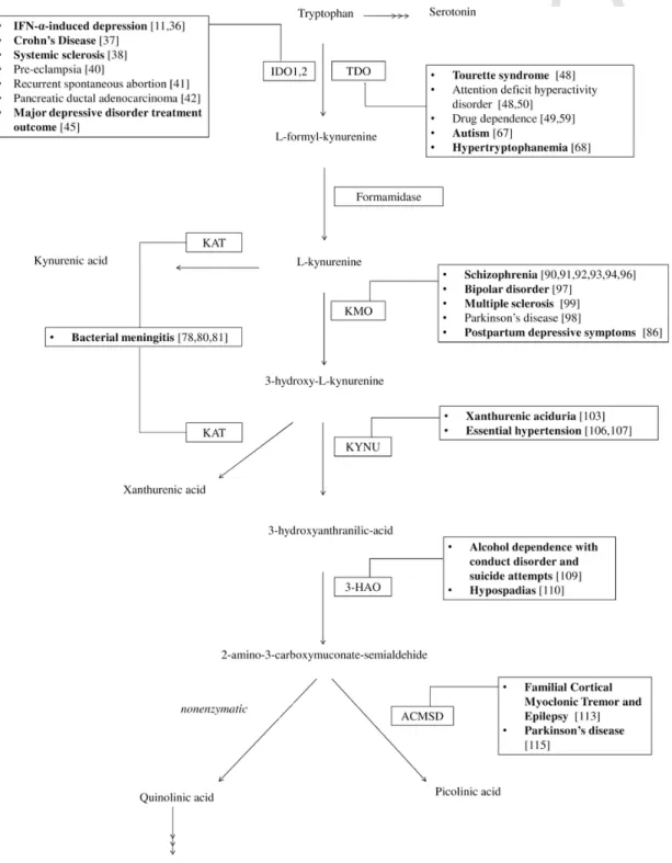
Most of the dietary tryptophan (Trp) not used for protein synthesis is metabolizedviathe kynurenine pathway (KP) [1]. The metabolites have a broad spectrum of biological actions, and have been connected
to several diseases [2–5]. The KP is one branch of Trp metabolism; the other branch provides serotonin and melatonin (Fig. 1). The first step in the KP is the conversion of Trp to N-formyl-l-kynurenine, by tryp- tophan 2,3-dioxygenase (TDO) or indoleamine 2,3-dioxygenase (IDO).
Formamidase converts N-formyl-l-kynurenine to L-kynurenine (KYN), which can be further metabolized by three distinct pathways. Kynure- nine aminotransferases (KATs) can convert KYN to kynurenic acid (KYNA), kynurenine 3-monooxygenase (KMO) can convert it to 3-hy
Fig. 1.An overview of the KP. The metabolites are indicated with full name. The names of the enzymes are given in abbreviated forms. Diseases for which association of ge- netic alteration(s) resulting in malfunction of a particular enzyme has been investigated are listed with references. (Bold: association have been found).Abbreviations: IDO1,2:
Indoleamine-2,3-dioxygenase 1,2; TDO: Tryptophan 2,3-dioxygenase; KAT: Kynurenine aminotransferase; KMO: Kynurenine 3-monooxygenase/Kynurenine 3-Hydroxylase; KYNU:
Kynureninase/L-Kynurenine Hydrolase; 3-HAO: 3-hydroxyanthranilate 3,4-dioxygenase; ACMSD: Aminocarboxymuconate semialdehyde decarboxylase/Picolinate carboxylase; NAD+:
Nicotinamide adenine dinucleotide.
UNCORRECTED
PROOF
droxykynurenine (3-HK), while kynureninase (KYNU) converts it to an- thranilic acid (AA). 3-HK is either processed by KATs to xanthurenic acid (XA), or degraded to 3-hydroxyanthranilate (3-HAA), which is metabolized by 3-hydroxyanthranilate 3,4-dioxygenase to form 2-amino-3-carboxymuconate semialdehyde (ACMS). ACMS can be processed for either synthesis of 2-aminomuconate semialdehyde by aminocarboxymuconate-semialdehyde decarboxylase (ACMSD), or can form quinolinic acid (QUIN) by non-enzymatic cyclization. QUIN can be further metabolized for nicotinamide-adenine-dinucleotide (NAD+) synthesis, while 2-aminomuconate semialdehyde can form picolinic acid or be subsequently processed to acetyl coenzyme A.
Intensive research is focused on the effects and alterations of the dif- ferent KP metabolites and enzymes, however information regarding ge- netic alterations of this pathway is scarce. Our review aims to collect the data available on mutations and genetic alterations of the enzymes of the KP and reveal possible connections with various diseases. We will follow the metabolic route of Trp (Fig. 1) and collect the data available on the enzymes directly connected to the KP.
2. Genetic alterations of the genes of kynurenine pathway enzymes and their association with diseases
As a result of genome sequencing projects all genes encoding KP en- zymes have been identified. Results of traditional human genetic analy- sis studies and targeted or non-targeted high throughput genome wide association (GWA) studies indicate that in seven genes which encode KP enzymes (IDO1/2, TDO2, KATII, KMO, KYNU, 3-HAO, ACMSD), al- terations can be identified that might be causally linked to disease states. However, it has to be emphasized that the majority of the cases summarized here only indicate an association, and not a causal relation- ship, between the genetic alteration and the respective disease. Most of these alterations are single nucleotide polymorphisms (SNP) present in variable frequencies in different population groups. In some cases, the nucleotide changes are found within coding regions of KP genes and re- sult in amino acid changes or early translational termination of the con- cerned protein product. Nucleotide alterations can also occur outside of exons. Polymorphisms affecting introns, promoters, enhancer and si- lencer regions can play a crucial role modulating gene expression both at the levels of transcription and translation. Single nucleotide changes in regulatory regions can result in elimination or formation of tran- scription factor binding sites and consequently these changes can alter the binding efficiency of DNA binding domains [6]. In a few cases nu- cleotide alterations outside of exons affect gene function by changing intronic regions and thus modifying mRNA splicing and/or translation.
It is worth noting that deletions or duplications have not been identified for any of the KP enzyme genes, which most probably reflects the essen- tial functions this pathway performs.
Through the next sections we summarize data available on genetic alterations of genes of KP enzymes following the steps of the pathway.
For each enzyme the description starts with a short introduction pro- viding the enzyme function, protein size, gene designation, location, ex- pression and/or other specifics.
2.1. Indoleamine-2,3-dioxygenases
IDO (EC 1.13.11.52) is the first enzyme of the KP. It catalyzes the conversion of Trp to N-formyl-l-kynurenine together with TDO. In hu- mans, two IDO enzymes, IDO1 and IDO2 have been identified [7,8].
They are very similar in structure and function but are expressed in dif- ferent patterns. IDO1 was first isolated from rabbit small intestine by Yamamoto and Hayaishi in 1967 [9].
It can be found ubiquitously in all tissues [10]. It is expressed in several types of immune cells, such as macrophages, monocytes, den- dritic cells and microglia [11]. It is a 45kDa protein of 403 amino
acids. The gene encoding it is on the short arm of the 8th chromosome (8p11.21). Similarly, in mice it is also coded on chromosome 8, while in rats it localizes to chromosome 16. In humans, the transcribed region extends to 26516 nucleotides and consists of 10 exons. The expression of the gene is induced by lipopolysaccharides (LPS), cytokines, tumor necrosis factors (TNFs) and interferons (IFNs), especially IFN-γ[12].
IDO1 is a heme enzyme located in the cytosol. It can convert various substrates, such as L-tryptophan, 5-hydroxy-tryptophan, serotonin and melatonin [13–15]. The crystal structure of the human enzyme was re- ported in 2006 by Sugimoto et al. [16].
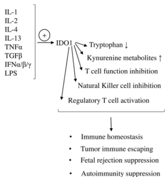
The physiologic role of the IDO1 enzyme is to conduct immune acti- vation in various states (Fig. 2). Upregulation of the enzyme leads to Trp depletion, which inhibits the proliferation of bacteria and other virulent agents. However, the mechanism of the decrease in Trp levels and the role that IDO plays in infectious states is controversial. Recently Badawy et al. argued that under specific conditions such as pregnancy Trp in fact is not depleted, rather its uptake by immune cells is increased greatly.
Therefore IDO exerts its immune-regulating effectviathe production of kynurenine metabolites [17].
Another mechanism by which IDO1 affects immune response is by inducing T cells to differentiate into T regulatory cells, thus block- ing T cell responses [18]. This might explain the diverse expression of IDO1 throughout the human body. The gene is highly expressed in tissues that are highly exposed to antigens, such as mucosal surfaces of the lung and the intestinal system, lymph nodes and spleen. Anti- gen-presenting cells such as macrophages and dendritic cells also ex- press IDO1. The anti-inflammatory effect of IDO1 activity contributes to maintain the immune homeostasis of different tissues. Thus it is con- ceivable that impairment in the activity of IDO1 can lead to the devel- opment of autoimmune diseases [19]. IDO1 could be involved in the process of immune editing, thus contributing to the immune escape of tumors [20]. Tumor derived factors have been reported to cause the differentation of dendritic cells with altered function. Immature den
Fig. 2.The role of IDO1 in immune regulation.IDO1 expression can be enhanced by cytokines (IL-1, IL-2, IL-4, IL-13, TNFα, TGFβ, IFNα/β/γ) and lipopolysaccharides (LPS).
IDO1 induction leads to a decrease in tryptophan amount and the formation of kynurenine metabolites. By activating regulatory T cells, andviathe inhibition of effector T cells and natural killer cells, IDO1 plays an important role in the maintenance of immune homeostasis, tumor immunity and in the suppression of fetal rejection and autoimmunity.
UNCORRECTED
PROOF
dritic cells in the tumor microenvironment show high expression of IDO1 [21]. The upregulation of IDO1 leads to a differentation shift to- wards regulatory T cells instead of effector T cells and natural killer cells, thus leading to immune tolerance [18]. High levels of IDO1 ex- pression in placenta indicates the importance of the enzyme in the sup- pression of fetal rejection by suppressing the reactivity of maternal T cells [22].
Until the discovery of the second IDO enzyme,’IDO’stood for what now is known as IDO1. By now a similar second enzyme has been rec- ognized that is referred to as indoleamine 2,3-dioxygenase-like protein, proto-indoleamine 2,3-dioxygenase, or most commonly indoleamine 2,3-dioxygenase-2, IDO-2 or IDO2.
IDO2 is structurally and functionally similar to IDO1, supporting the hypothesis that the two proteins are products of genes originated from duplication of a common ancestralIDOgene [23]. In all mammalian species twoIDOgenes (IDO1and2) can be found.IDO1is thought to be specific to mammals, and the majority of lower vertebrate species possess onlyIDO2. However,IDO1has also been found in several fish and two turtle species. This suggests that gene duplication occurred be- fore the divergence of vertebrates, andIDO1was later lost in several lower vertebrate lineages [24]. This notion is further supported by the fact that in mammalsIDO1andIDO2are located next to each other–in case of humans on the short arm of chromosome 8. Despite their similar structures [25],IDO2is significantly bigger as it is 81778 nucleotides long and consists of 11 exons. The regulation of the two genes is in some way coordinated, which is supported by the observation that the expres- sion ofIDO2was found to be significantly decreased inIDO1knockout mice. An explanation for this finding could be the loss ofIDO2regula- tory elements in consequence of removal of theIDO1gene region [7].
The product of theIDO2gene is a 420 amino-acid protein, with a molecular weight of over 47kDa. IDO2 and IDO1 proteins show 43%
amino acid identity both in humans and mice [7]. Those residues which are critical for catalytic functions are conserved in the two proteins.
In vitrostudies suggest that IDO2 can convert the same substrates as IDO1 (L-tryptophan, D-tryptophan, 5-hydroxy-tryptamine, tryptamine and serotonin), however, the catabolic efficiency of IDO2 is much lower than that of IDO1 [26]. Interestingly, the enzymatic parameters of in- vertebrate IDOs’are similar to those of vertebrate IDO2. This suggests that the common ancestral IDO was similar to the present IDO2s in re- spect to their low catalytic efficiency [24]. In the distal heme pocket of IDO1 a Ser residue (distal-Ser, which corresponds to Ser167 of hu- man IDO1) was found to be conserved in all IDO1 enzymes and also in a lizard (green anole,Anolis carolinensis) IDO2. In contrast with that in other IDO2 enzymes at this position a Thr (distal-Thr) residue is found [24]. Considering that IDO1 has a higher affinity to L-Trp than IDO2, and IDO2 of the green anole lizard shows similar enzymatic parameters to mammalian IDO1, this distal-Ser change seems to be crucial in the conversion of IDO1 from a low efficiency enzyme to a moderate effi- ciency one [24]. Besides the distal-Ser, another residue suspected to be
− perhaps the most − critical residue for the high affinity for L-Trp is the distal-Tyr, which corresponds to Tyr126 in the human IDO1 [27].
IDO2 is expressed in the liver, kidney, epididymis and brain in mouse [7,28], and in the liver, kidney and specific tumors in humans [29].
2.1.1. Diseases related to the genetic alterations of indoleamine 2,3- dioxygenase genes
A large number of SNPs within the transcribed regions of IDO1andIDO2genes have been described. None of these are associated unequivocally with any disease, nonetheless linkage has been suggested between several SNPs and specific disease states (Fig. 1).
As IDO plays important roles in immune response, the effects of ge- netic changes inIDOgenes were studied mostly in relation to diseases characterized by impaired immune function. It was suggested that in tu- mors elevated expression of IDO proteins can locally regulate the im- mune environment, enhancing immune tolerance and thus increasing the survival of cancer cells [18–21]. Similarly, moderation of immune functions can underlie the effects of IDO on allogenic fetal rejection [22,30–32]. Changes in IDO function have also been reported in neu- rodegenerative diseases [33].
One of the main inducers of IDO1 is IFN-α, which is used in the treat- ment of patients suffering from chronic hepatitis C. Several earlier stud- ies reported a decrease in blood Trp concentrations and increase of KYN in association with IFN‐αinduced depression, suggesting the role of IDO in the process [34,35].
In search of genetic variations that can be associated with the de- velopment of IFN‐αprovoked depressive symptoms, Galvão-de Almeida et al. investigated three SNPs of the IDO1gene in 2011. However, none of the investigated polymorphisms (rs3824259, rs10089084, and rs35099072) were found to be associated with the disease [36]. The fol- lowing year, Smith and colleagues investigated four SNPs (rs9657182, rs7820268, rs3739319 and rs6991530) of the gene with the same aim [11]. They found that one of the investigated SNPs had a significant effect on the evolution of depressive symptoms of IFN-αtreated Cau- casian patients with major depressive disorder (MDD) [11]. The SNP rs9657182 is a C/T nucleotide change in the promoter region of the gene. The nucleotide change has not been proven to alter any transcrip- tion binding site. Still, theoretically it could lead to altered gene func- tion either alone, orviathe effects of other polymorphism which may be associated with it [11].
In light of the normal physiological functions of the enzyme, the involvement of IDO1 in autoimmune diseases has been investigated.
The induction ofIDO1gene by pro-inflammatory stimuli results in Trp depletion and formation of KP metabolites, leading to the reduction of inflammation and the promotion of immune tolerance [37]. Conse- quently, impaired function of the enzyme might contribute to the devel- opment of autoimmune diseases. Guided by this notion the possible role ofIDO1have been investigated in Crohn’s disease (CD) and systemic sclerosis.
Lee et al. [37] found thatIDO1polymorphisms were rare among pa- tients with CD. Out of the six investigated polymorphisms, they de- tected four in a group of clinically phenotyped CD patients (Table 1).
Their results indicate that patients carrying specificIDO1SNPs are more prone to extraintestinal manifestations, such as uveitis and arthritis.
Their observation was that the presence of minor alleles was associ- ated with a more severe disease course. Among carriers of the SNPs, a lower KYN level and KYN/Trp ratio could be detected as compared to the control group, leading to the assumption that the detected polymor- phisms in CD patients result in impaired IDO1 function [37]. This study also included polymorphisms of the structurally and functionally similar IDO2gene. It was found that although SNPs of the gene were common among a group of patients suffering from CD, none of the investigated polymorphisms had effect on the severity of the disease [37].
IDO1polymorphism can also be related to another autoimmune disease, systemic sclerosis. Tardito et al.investigated 5 SNPs of the IDO1gene, of which one (rs7820268, a C/T change in intron 5) was found to be present at a significantly higher frequency among systemic sclerosis patients compared to the non-patient group [38]. In the carri- ers of the minor allele a decrease in the CD8+ regulatory T cell sup- pressing capacity could be detected, suggesting an association between this gene variant and regulatory T cell function [38].
The involvement of IDO in immunological mechanisms gave ground to the notion that the enzyme could be involved in the suppression of
UNCORRECTED
PROOF
Table 1
Genetic alterations affecting the enzymes of the kynurenine pathway.
Gene rs number Base change/amino
acid change Localization in the gene Change in
enzyme function Related disease Reference
IDO1 rs9657182 C > T(fwd) promoter region IFN-αinduced depressive
symptoms in patients with Hepatitis C
[11]
rs35059413 C > T(rev)/Ala4Thr diminishment in
enzyme activity Severity of Crohn's disease [37]
rs35099072 C >
T(rev)/Arg77His
C > A exon 7
9 base pair deletion exon 7
rs7820268 C > T(fwd) intron 5 Treg suppression
is diminished systemic sclerosis [38]
IDO2 rs2929115 C > T(rev) non-conserved region
between 26kb and 28kb downstream from the 3′
end
Citalopram responsiveness in major depressive disorder
[45]
rs2929116 G > A
TDO2 G > T intron 6 Tourette syndrome [49]
G > A
rs3755910 A > C (fwd) promoter region Autism [67]
c.491dup/premature
stop codon leads to a
truncated protein (premature stop codon)
Hyperserotoninemia [68]
G > C/Met108Ile accelerated
degradation of the enzyme
AADAT rs1480544 C > T (fwd) exonic splicing silencer elevated enzyme
production Immune response in
bacterial meningitis [78,81,80]
KMO rs1053230 A > G (rev);C > T
(fwd)/Arg452Cys exon 15 schizophrenia, bipolar
disorder, PDS [93,94,96,97,86]
rs2275163 C > T (fwd) intronic region schizophrenia [93,90,94]
rs1053221 A > G (rev) utr region Sclerosis multiplex [99]
rs1053183 C > T (rev)
KYNU rs606231307 A >
G(fwd)/Thr198Ala exon 7 Xanthurenic aciduria [103]
rs9013 A >
G(fwd)/Lys412Glu unknown Essential hypertension [106]
rs2304705 G >
A(fwd)/Arg188Gln enzyme activity
reduced by 50% [107]
HAAO rs375554 A > G (fwd) Alcohol dependence
accompanied with conduct disorder or suicide attempts
[109]
rs13027051 C >T(fwd) intronic region
rs2374442 A > G (fwd) rs3816184 A > G (fwd) rs3816182 A > C (fwd) rs737148 A > G (rev)
rs3816183 C > T/Ile37Val Hypospadiasis [110]
ACMSD G > A/premature
stop codon leads to a
truncated protein (premature stop codon)
Familial cortical myoclonic
tremor and epilepsy [113]
rs6710823 A > G(fwd) Parkinson's disease [115]
fwd/rev: allele reported to forward/reverse orientation to genome.
fetal rejection. Published data on this issue are however controver- sial. An earlier study by Munn and collegues reported that inhibition of tryptophan catabolismviathe implementation of the IDO–inhibitor 1-methyl-tryptophan resulted in fetal loss in a mice model. This is sup- posedly due to the lack of the T cell proliferation suppressing effect of cells catabolizing tryptophan. As 1-methyl-tryptophan is a known IDO inhibitor, it is likely that the above described fetal rejection was a consequence of maternal T cell proliferation due to the inhibition of IDO [22]. Interestingly, later Baban and colleagues found that IDO de- ficiency due to genetic manipulation did not affect pregnancy success in mice. They hypothesized that in IDO deficient mice IDO-indepen- dent mechanisms –such as the activation of another key enzyme of the KP, TDO– compensate for the lack of the enzyme. This notion was strengthened by the finding that assessment of the IDO inhibitor 1-methyl-tryptophan in IDO deficient animals did not cause changes in
pregnancy outcomes on contrary to IDO–sufficient mice pregnancies [39]. Another interesting question that still needs to be elucidated is the question of the decrease in Trp level during pregnancy. Badawy and col- leagues hypothesized that the decrease in the level of Trp is not due to the depletion of the amino acid but is the consequence of its aug- mented utilization by the cells of the immune system–similarly as in the case of infection. They claim that during pregnancy increased avail- ability of maternal Trp is needed to fulfill the increased demand for pro- tein synthesis by the fetus and the pregnant mother. Elevated maternal free plasma Trp is then utilized in the KP, ensuring the production of immunosuppressive kynurenine metabolites which support the mainte- nance of the balance of tolerance and immunity. Therefore immunosup- pression is not achieved by Trp depletion itself, but by the increased uti- lization of the amino acid [17].
UNCORRECTED
PROOF
The findings of animal studies raised the question of the role of IDO during human pregnancy. One of the most serious and life-threatening conditions during human pregnancy is pre-eclampsia. Its etiology has not been fully understood so far, but several environmental and genetic factors have been proposed to contribute to its evolvement. The findings that in the placenta of pre-eclamptic womenIDO1expression is reduced and a correlation can be observed between the severity of the illness and the activity of the enzyme [31,32] turned the attention to the KP.
Taking this into consideration, Nishizawaet al.investigated the possible association of 19 SNPs of theIDOgene with pre-eclampsia [40]. How- ever, none of the studied polymorphisms, each within the coding region of the gene, was found to be associated. The only genetic alteration that might be linked to the condition based upon this study was a four nu- cleotide deletion within the intronic region of the gene; however, it is unclear how this change affects expression of the gene, if at all [40].
The controversial findings of animal experiments raised the ques- tion of the role of the enzyme in fetal rejection in humans. However, the examination of 10 SNPs among Iranian women suffering from recur- rent spontaneous abortion revealed no linkage between the investigated polymorphisms and the disease [41].
Following the observation that IDO1 helps tumor cells to survive and proliferate by facilitating a local immunotolerant environment [20], Witkiewicz et al.investigated two polymorphisms of the functionally similarIDO2gene in patients with pancreatic ductal adenocarcinomas (PDAs) [42]. The two polymorphisms they studied show high preva- lence in the population and result in either complete or very severe decrease (90%) in the catalytic activity of IDO2 [8]. Based on a rela- tively small patient cohort in which the frequencies of these polymor- phisms were comparable to that of the control group, they did not find any link betweenIDO2genotype and PDA [42]. In other words, these SNPs do not increase the risk of PDA development. On the other hand, their results show that the majority of investigated patients had at least one wild-type or functioning allele. This observation gives support to the argument that the IDO inhibitor D-1-methyl-tryptophan might be useful in treatment of PDA due to the inhibition of the anti-inflam- matory effects of IDO [42]. However, this treatment should be pre- ceded by genetic investigation of the patients to identify those with at least one activeIDO2allele. Furthermore, selectivity of the drug should also be taken into account. The question concerning the most potent IDO2 inhibitor remains to be answered. Several studies have found that L-1-methyl-tryptophan, the levo stereosisomer of D-1-methyl-tryp- tophan has a more effective inhibitory effect of IDO2 [43] [44].
Besides the above discussed diseases, the relation of theIDOgenes and a common psychiatric disorder has also been examined. Cutleret al.
studied the potential role forIDO1andIDO2in MDD treatment outcome [45]. They found that two polymorphisms of theIDO2gene have effect on the responsiveness to citalopram treatment in patients with MDD.
The two polymorphisms (rs2929115 and rs2929116) should be consid- ered as one marker, because they were found to be in very high linkage disequilibrium [45]. Both are in a non-conserved region between 26kb and 28kb downstream from the 3′end ofIDO2. They are near to tran- scriptional binding sites and are surrounded by two histone modifica- tion sites, but the functional consequences of these polymorphisms have not been elucidated yet [46].
2.2. Tryptophan 2,3-dioxygenase
TDO enzyme (EC 1.13.11.11) is a 47kDa protein of 403 amino acids. The encoding gene,TDO2is rather large, consisting of 65 669 nu- cleotides and containing 12 exons. It is located on the long arm of chro- mosome 4 in humans, while it is located on chromosome 3 in mice and on chromosome 2 in rats.
TDO is a homotetrameric heme enzyme that catabolizes the conver- sion of L-Trp to N-formyl-l-kynurenine, which is a rate limiting step of the KP.Viathis conversion the enzyme also modulates serotonin levels by reducing the amount of Trp available for synthesis of the neurotrans- mitter. TDO is found mainly in the liver, thus modulating the available quantity of Trp throughout the body. The expression of TDO is induced by glucocorticoid hormones and the enzyme is also regulated by the availability of its substrate, L-Trp [47].
As the serotoninergic system is involved in the modulation of mood, sleep, aggression and other states,TDO2is a target of investigations in relation to psychiatric and neurological diseases characterized by changes in the circadian rhythm, behavior and the reward system.
2.2.1. Diseases related to the genetic alterations of tryptophan 2,3- dioxygenase gene
A study by Comingset al.. showed significant decrease in the Trp levels and in the serotonin/platelet ratio of patients with Tourette syn- drome (TS) [48]. A decline in the serotonin/platelet ratio was also de- tected in the parents of the patients. Similar metabolic changes were reported among patients with attention deficit hyperactivity disorder (ADHD) and their parents. The similar findings suggest the existence of a link between TS and ADHD [48]. The observed alterations of sero- tonin level and the inheritance pattern of TS indicate the possible under- lying genetic alteration: variants of theTDO2gene are possible candi- dates contributing to these disorders. Mutations causing higher expres- sion level, or leading to an enzyme that is more prone to induction can be a culprit behind the excessive N-formyl-l-kynurenine formation, thus diverting Trp from serotonin synthesis [48].
Further studies by Comingset al.showed a significant association be- tween two intronicTDO2polymorphisms (a G to T and a G to A change) and TS (Fig. 1). One of these SNPs (the G to A change) was found to be significantly associated with altered platelet serotonin levels as well [49].
In a more recent study from the same laboratory [50] 20 genes were investigated in patients with ADHD. As this disorder is likely to be multigenic, caused by additive effects of several genes [51], the 20 studied genes were grouped into three sets. Those with activities related to dopaminergic neurotransmission formed one group (6 of the investi- gated genes), those related to the noradrenergic neurotransmission an- other group (7 of the genes) and the remaining 6 genes each involved in serotoninergic neurotransmission formed the third group. One SNP was investigated for each gene in the study. ForTDO2, it was a G to A nucleotide change in the intronic region of the gene. According to the results noradrenergic genes were more likely to contribute to the ADHD phenotype than genes belonging to the other two groups [50]. Nonethe- less this finding does not exclude the possibility that genetic alterations of theTDO2gene affect the manifestation of ADHD.
The involvement of serotoninergic neurotransmission in the function of the reward system underpins the search for possible associations of specific changes inTDO2with drug abuse. Previous findings show an increase in brain serotonin levels due to nicotine exposure, and an in- crease in the level of the neurotransmitter during withdrawal [52,53].
Clinical data also showed beneficial effects of the serotonin re-uptake inhibitor Fluoxetine during the decrease of nicotine intake of smok- ers. By applying the medication, weight gain, a common consequence of increased food intake frequently accompanying cessation of smok- ing, could be decreased [54]. Based on this notion, it is hypothesized that impairment in serotoninergic neurotransmission contributes to the mood and appetite disturbances associated with nicotine withdrawal and therefore might have a role in drug dependence [49].
In another study Comingset al. [49] investigated the association of drug dependence and two intronic polymorphisms of theTDO2gene.
Both of these SNPs are in intron 6 and potentially cause alteration in a binding site of the YY-1 transcription factor [6]. One SNP is a G to T
UNCORRECTED
PROOF
change at position 666, the other is a G to A change at position 663. Nei- ther was, however, found to be associated with drug dependence [49].
Another unfortunately commonly used and abused drug in our soci- ety is alcohol. Changes in serotoninergic neurotransmission have been reported during acute ethanol administration. Shortly after alcohol in- take, an induction of serotonin biosynthesis can be observed [55,56].
After 5–8h, this is followed by TDO stimulation, which causes a de- crease in the serotonin level and simultaneously increases KYN produc- tion [56–58]. The gene activation is believed to be transcriptionally mediated by glucocorticoids (GC). The promoter region of the human TDO2gene contains four functional glucocorticoid responsive elements (GRE) [59], which can serve as target sites in this activation. Soichotet al.aimed to identify SNPs in the GREs of theTDO2promoter region that cause alterations in the transcription regulatory effects of GCs. They in- vestigated the effects of 12 SNPs of theTDO2promoter region, three of which were in putative GREs. Inin vitroexperiments under basal con- ditions (without glucocorticoid receptor (GR) over-expression, or Dex- amethasone exposure) the studied nucleotide changes did not cause sig- nificant differences in promoter activity. However, upon GR over-ex- pression without, or combined with Dexamethasone exposure, a sta- tistically significant difference in promoter activity could be observed [59]. The data suggest that theseTDO2polymorphisms exert their ef- fects only duringTDO2gene activation. Since a similar pattern of gene activation changes has been observed also in the case of a polymor- phism not located in a known GRE, this indicates the possibility of an- other, yet unexplored response element [59]. However, in contrast with results obtainedin vitro, in vivomeasurements did not reveal association between the polymorphisms and plasma KYN/Trp ratio, an indicator of TDO/IDO enzyme activity [59] (Table 2).
The findings regarding the effects of genetic alterations in the TDO2promoter region serve as good examples to highlight the impor- tance of SNPs localized outside coding regions and splicing sites. In this respect it should be emphasized that genetic alterations might have im- pact not only by changing protein structure and/or function. Polymor- phisms affecting regulatory regions such as the promoter can lead to the elimination or the formation of transcription binding sites, thus altering the binding efficiency of DNA binding domains [6].
Besides ADHD, TS and drug dependence, impairment in serotoniner- gic neurotransmission has been reported in autism as well. In patients suffering from the disease impairments in serotonin synthesis in the brain have been detected [60,61], and more than a third of patients seems to have elevated platelet serotonin levels [62]. The involvement of serotoninergic neurotransmission in the disease is also supported by studies showing that Trp depletion exacerbates symptoms in patients with autistic disorder [63,64]. Family and twin studies suggest a ge- netically determined predisposition to autism [65,66]. In light of these data, genes involved in serotonin metabolism are potential disease-caus- ing factors. Since investigation of genetic alterations of the sero
Table 2
Identified SNPs in the humanTDO2promoter region.
rs number base change
rs3755908 1605T > C
rs3775085 1532A > C
rs17033763 1471G > A
Unknown 1342C > A
rs3836580 1248_1247InsA
rs10857287 1131A > G
rs11935082 1059T > C
rs60426490 935_933delGTT
Unknown 769T > G
rs3775086 722T > A
rs3755909 455C > T
s3755910 311C > A
tonin transporter gene did not result in an unambiguous answer [49],TDO2arose as a potential candidate. Nabiet al.studied the pres- ence of fiveTDO2polymorphisms in families with members diagnosed with autism using transmission disequilibrium test [67]. Four polymor- phisms included in the study are located in the promoter region of the gene, while one is at the 3′slice region of exon 11. For one of the inves- tigated nucleotide changes affecting the promoter region (rs3755910, an A/C change) a significant difference was found in the transmission to autistic subjects. The more frequent C allele of the polymorphism was preferentially transmitted to family members with autistic disor- ders. This strongly suggests that though this SNP is not likely to be a risk factor in autism, it is in a strong linkage disequilibrium with another polymorphism associated with the disease that is so far unknown [67].
The role of TDO in Trp metabolism gives ground to the investi- gation of the enzyme not only in psychiatric disorders but in other Trp and serotonine related diseases. In a recent study Ferreiraet al. re- ported a peculiar situation, in which a single patient was diagnosed with chronic hypertryptophanemia and hyperserotoninemia [68]. Sequenc- ing of theTDO2gene revealed the patient to be compound heterozy- gous for the c.491dup and c.324G > C variant of theTDO2gene. The variant c.491dup (paternally inherited in the patient) causes a prema- ture stop codon leading to the formation of a truncated protein, which is 43% of the normal length of TDO [68]. The c.324G > C gene variant (which was maternally inherited in this patient) leads to methionine to isoleucine change at the 108. amino acid of the protein. The expression level of the c.491dup variant mRNA was found to be similar to the wild type, as the transcription of the gene was not affected by this alteration.
In vitrostudies however showed that the truncated protein changed the catabolic activity of the enzyme [68].
The c.324G > C base change affects a specific site of the enzyme.
Under normal conditions TDO has a short half-life which is regulated by the status of a non-catalytic Trp binding site. The enzyme activity and turnover depends on the occupancy of this binding site: when it is occu- pied it increases the catalytic activity, while when it is empty, it serves as a degradation signal [69]. The Met/Ile change resulting from the c.324G > C base change affects the formation of this non-catalytic Trp binding site. According toin vitrodata the activity of the enzyme is not affected [68], however, as a result of the amino acid change the binding affinity of the enzyme to its substrate is significantly decreased, and con- sequently the degradation rate of the enzyme is increased [68]. Thus, the c.324G > C genetic alteration of the gene causes the accelerated degradation of the TDO enzyme [68]. In summary, according to Fer- reiraet al., the chronic hypertryptophanemia and hyperserotoninemia observed in this patient is a consequence of the combination of altered catabolic activity and accelerated degradation of TDO, both caused by single nucleotide genetic alteration of theTDO2gene [68].
2.3. Kynurenine formamidase
Kynurenine formamidase/arylformamidase (EC 3.5.1.9) converts N-formyl-l-kynurenine to KYN. It is coded on chromosome 17, 10, 11 in humans, rats and mice, respectively (AFMIDgene). Little is known about this enzyme − it seems that it has two isoforms in humans, one a 301 AA and one a 303 AA form, and it is expressed in various tis- sues, but most abundant in liver and kidney [70]. To our knowledge, no specific alterations in theAFMIDgene were directly correlated with any human disease.
2.4. Kynurenine aminotransferases
In human brain four KATs have been identified which catalyze KYNA synthesis (EC 2.6.1.7) [71]. These are glutamine transaminase
UNCORRECTED
PROOF
K/cysteine conjugate beta-lyase 1 (KATI, EC 4.4.1.13), aminoadipate aminotransferase (KATII, EC 2.6.1.39), glutamine transaminase L/cys- teine conjugate beta-lyase 2 (KATIII, EC 4.4.1.13), and glutamic-ox- aloacetic transaminase 2/mitochondrial aspartate aminotransferase (KATIV EC 2.6.1.1) [71]. They are multifunctional enzymes with broad substrate specificity, and function as homodimers with pyridoxal 5′-phosphate as cofactor [71]. Besides the irreversible transamination of KYN to KYNA, they can catalyze the conversion of 3-HK to XA. In hu- man brain, the principal enzyme responsible for the formation of KYNA is KAT II [72,73], therefore we will focus on this enzyme below.
The KATII enzyme is a 47kDa protein consisting of 425 amino acids.
The aminoadipate aminotransferase (AADAT) gene encoding this pro- tein is located on the long arm of chromosome 4 in humans. It spans 31 478 nucleotides and contains 18 exons. The gene is located on chromo- some 8 in mice and on chromosome 19 in rats.
In vitrostudies show that KATII enzyme has very broad substrate specificity. Besides catalyzing the formation of KYNA from KYN, it cat- alyzes the transamination ofα-aminoadipate and several other amino acids [74].
Alterations in the level of KYNA have been reported in several neu- rological diseases, such as Huntington’s disease (HD), Alzheimer’s dis- ease (AD) [75] Parkinson’s disease (PD) [76] multiple sclerosis (MS) [77] and AIDS dementia. KYNA is a modulator of glutamatergic neu- rotransmissionviaitsN-methyl-d-aspartate (NMDA) receptor antagonist effect and has neuroprotective features [74–76]. According to this, im- pairment in the synthesis of KYNA could thus contribute to the devel- opment of neurodegenerative diseases. Since KATII plays a crucial role in the formation of this neuroprotective agent, the enzyme has been mainly investigated in disorders related to neuronal damage.
2.4.1. Diseases related to the genetic alterations of aminoadipate aminotransferase gene
Predicated on previous studies reporting the involvement of the KP in inflammatory diseases, de Souza et al. [78] searched for genetic alter- ations affecting KP enzymes in patients with bacterial meningitis (BM) (Fig. 1). Two polymorphisms of theAADATgene were included in the study: rs17852900, causing a G to T change, and rs1480544, a C/T change. The rs1480544 polymorphism affects a putative regulatory el- ement, an exonic splicing silencer–consequently it is thought to affect mRNA and peptide synthesis [79]. This polymorphism was found to be significantly more frequent in patients with BM considering both allelic and genotypic frequencies. In patients homozygous for the minor allele (TT), a significant decrease was detected in the levels of tumor-necro- sis factor alpha (TNF-α), interleukin 1 beta (IL1-β), macrophage inflam- matory protein 1-alpha (MIP-1αCCL3), macrophage inflammatory pro- tein-1-beta (MIP-1βCCL4) and in the number of cells counted in the col- lected cerebrospinal fluid (CSF), and an increase in immunoglobulin G (IgG) levels. These findings suggest that the rs1480544 polymorphism causes impairment in the immune response against virulent agents. The C/T change in the exonic splicing silencer region of the gene supposedly affects the expression of inflammatory markers such as TNF-α, IL1-β, MIP1αCCL3 and MIP1βCCL4, and causes impairment in the recruitment of leukocytes. The elevation of IgG levels in patients with TT geno- type suggests the influence of KYNA on immunoglobulin production, the mechanism of which, however, is so far unknown [78].
In addition to de Souza’s findings, Coutinhoet al. found elevated KYNA levels in the CSF of patients with BM carrying the T allele of the rs1480544 polymorphism [80]. A tendency for increase in KYNA level was also noted among CT genotype patients suffering from acute BM.
These results give support to the assumption that this particular SNP leads to an increase in the amount ofAADATmRNA and subsequently causes enhancement of KATII enzyme synthesis [80].
In 2015 Fontes et al. searched for possible combinations of geno- types that could have an effect on the course of BM [81]. Based on pre- vious findings of de Souzaet al., they examined the occurrence of the rs1480544C/T variant of theAADATgene in combination with poly- morphisms of genes previously reported to cause impairment in DNA repair mechanisms, such as Asn148Glu of Apurinic/Apyrimidinic Endo- deoxyribonuclease (APEX1), Ser326Cys of 8-Oxoguanine DNA Glycosy- lase (OGG1) and Val762Al of Poly(ADP-Ribose) Polymerase 1 (PARP1).
Results of gene interaction analysis showed a statistically significant combined occurrence of these genetic alterations among BM patients, suggesting the possibility of synergism existing between different path- ways involving these genes in the development and course of BM [81].
2.5. Kynurenine 3-monooxygenase
KMO (Kynurenine 3-monooxygenase, also known as Kynurenine 3-Hydroxylase) (EC 1.14.13.9) is a 55kDa molecular mass protein of more than 480 amino acids. In the KP it catalyzes the KYN to 3-HK conversion. It is a mitochondrial flavoprotein, utilizing O2and NADPH for the catalyzed reaction [82]. Downstream of KMO in the pathway, 3-HAA and QUIN are synthesized. Elevated levels of 3-HK, which is an endogenous oxidative stress generator, have been reported in sev- eral neurodegenerative disorders [77,83]. In contrast to 3-HK, 3-HAA may be neuroprotective due to its hemeoxygenase-1 (HO-1) inducing effects in astrocytes. HO-1 is an antioxidant enzyme with anti-inflam- matory and cytoprotective features [84]. In the central nervous system QUIN acts as a neurotoxin,viaactivating NMDA receptors, thus creat- ing an enormous calcium influx into astrocytes and neurons, causing cell damage [85]. QUIN has an effect on the appearance of depressive symptoms by inducing the nitric oxide − cyclic guanosine monophos- phate (NO-cGMP) pathway, promoting oxidative stress and interfering with the translation of brain derived neurotrophic factor [86].
TheKMOgene in humans is localized on the long arm of chromo- some 1. It is 63 759 nucleotides long and consist of 17 exons.Kmois coded on chromosome 13 in rat and on chromosome 1 in mice.
2.5.1. Diseases related to the genetic alterations of kynurenine 3- monooxygenase gene
Several studies have reported linkage between genetic loci on the long arm of chromosome 1 and psychiatric disorders with psychotic symptoms, such as bipolar disorder and schizophrenia (Fig. 1) [87–89].
Since theKMOgene is located at that locus, it became a candidate in the eye of researchers looking for a predisposing factor for the diseases men- tioned above. Results obtained from postmortem tissue analysis of schiz- ophrenic patients support this suspicion. A significant and correlated de- crease in the expression of theKMOgene and the activity of the KMO enzyme was found in brain tissues obtained from schizophrenia patients compared to control patients [90]. Data concerning association between genetic alterations of theKMOgene and schizophrenia are, however, in- conclusive, as no linkage could be identified between the presence of any one of 15 studied polymorphic forms of the gene in Scandinavian patients [91].
Among Japanese patients the association of the rs2275163 (aC/T change) polymorphism with schizophrenia was found to be significant both by single marker comparisons and haplotype analysis. However, among a second, independent sample the significance of haplotype as- sociation could not be reproduced [92].
Results are similar regarding the rs2275163 polymorphism among Russian patients − no significant difference was found between the fre- quency of the minor allele in patients and in healthy controls [93]. How- ever, this study revealed a significant intergroup difference concerning another SNP, rs1053230.
The polymorphism is an A/G base change in exon 15 of the KMOgene, causing an arginine to cysteine change at the 452nd amino
UNCORRECTED
PROOF
acid of the enzyme (in fact the nucleotide change in the coding sequence is C/T, however the database records the SNP in reverse orientation).
The frequency of the homozygous minor GG genotype of rs1053230 polymorphism was significantly higher among patients compared to the control group. Interestingly, though the minor allele (T) of the other studied SNP rs2275163 did not prove to be a risk factor alone, in com- bination with the GG genotype of rs1053230 it seemed to increase the risk of schizophrenia. The risk of schizophrenia among subjects with the GG genotype of the rs1053230 locus combined with at least one minor allele of the SNP rs2275163 (either in the form of a CT or TT geno- type) was shown to be twice as high as in subjects with a different allele combination [93]. Though data concerning the association of polymor- phism rs2275163 with the incidence of schizophrenia are equivocal, it is likely that is has an impact on the expression of theKMOgene. In schiz- ophrenic patients with at least one minor allele of the single nucleotide change rs2275163, a slightly higherKMOmRNA level could be detected compared to those who had CC genotype [90].
Patients homozygous for the major allele of rs2275163 (CC geno- type) were found to perform more poorly in neurocognitive tasks such as predictive pursuit and visuospatial working memory than members of the CT genotype subgroup [94,90]. However, when comparing patients with CC and TT genotypes, the difference was not significant [90]. It should be mentioned here that among healthy subjects with TT or CT genotypes inequality could also be observed in cognitive performance, though the difference was again not significant [94].
Effects upon cognitive functions were also reported for homozygous carriers of the SNP rs1053230 major allele. Similarly to rs2275163, in- dividuals of CC genotype regarding the SNP rs1053230 reached lower composite scores compared to the CT or TT genotype [94].
Based on the fact that elevated KYNA levels have been reported in the CSF of schizophrenic patients and KMO is in charge of the formation of 3-HK, thus decreasing the amount of KYN available for KYNA synthe- sis, changes in KYNA levels can serve as an indirect indicator of KMO activity [95]. Andreassenet al.found that both in control and schizo- phrenic patients the presence of the T allele of the SNP rs1053230 (in- dicated in forward orientation) was associated with a 45% increase of KYNA level in the CSF [96].
Besides schizophrenia, KMO has been investigated in bipolar disor- der and depression as well. A decrease inKMOgene expression was de- tected in the prefrontal cortex (PFC) of bipolar disorder patients with psychotic features compared to patients without psychotic features [97].
This observation is in accord with results of postmortem brain tissue analysis of schizophrenic patients. However, in contrast with results of studies carried out among schizophrenic patients, the C allele of the rs1053230 (forward) polymorphism was found to be more common among bipolar disorder patients with psychotic features. The major al- lele was also associated with higher KYNA level in the CSF of bipolar pa- tients, accompanied with a reduction in KMO activity. HapMap3 project data analysis revealed that this allele is also associated with a reduction in theKMOexpression detected in lymphoblastoid cell lines. The link between the C allele of the rs1053230 SNP andKMOgene expression can also be observed in epileptic patients [97].
The same polymorphism ofKMO(rs1053230) was also found to be significantly associated with postpartum depressive symptoms (PDS) in Chinese women. AG genotype women with PDS were found to have sig- nificantly higher serum 3-HK concentration and 3-HK/KYN ratio com- pared to those with the GG genotype. These findings suggest the possi- bility of the rs1053230 SNP causing higher KMO activity [86].
Alongside psychiatric disorders, genetic alterations of theKMOgene have also been investigated in diseases involving neurodegeneration, such as PD and MS.
Töröket al.investigated 4 polymorphisms of theKMOgene in search for a genetic link between the KP and PD. Two of the examined
SNPs (rs2275163 and rs1053230) have been associated with psychiatric disorders in other studies (see above). Both (rs2050518 and rs6661244) are localized in intronic regions of the gene; the former one of them is an A to T change (rs 2050518), the latter is a C to T. Neither of these polymorphisms was found to be associated with PD in the population studied [98].
In search for genetic alterations contributing to MS, a focused GWA study was carried out targeting chromosome 1q43. Two polymorphisms, rs1053221 (A/G) and rs1053183 (C/T) located in theKMOgene were found to be in significant association with the disease [99].
2.6. Kynureninase/L-kynurenine hydrolase
In humans, KYNU (EC 3.7.1.3) catalyses the 3-HK/3-HAA conver- sion. It is expressed in several tissues, such as bone marrow and or- gans of the immune system, kidney and urinary bladder, lung, brain, but most abundantly in the liver. It is a large protein composed of 465 amino acids, with a molecular weight over 52kDa. It functions as a ho- modimer requiring PLP as a cofactor [100]. The gene is almost 307 000 nucleotides long. It is located on the long arm of chromosome 2 at the 22.2 position and contains 21 exons in humans. In mice it localizes to chromosome 2, while in rats to chromosome 3.
2.6.1. Diseases related to the genetic alterations of kynureninase gene In 2007 Christensen et al. reported a family with xanthurenic aciduria (also known as hydroxykynureninuria). The diagnosis was based on detection of large quantities of urinary excretion of XA, 3-hy- droxykynurenine (3-OHKYN) and KYN in one child and one of his siblings (sibling 1). Slightly increased urinary excretion of 3-OHKYN of the mother, father and another sibling (sibling 4) was also de- tected, each observation suggesting the deficiency of KYNU. Analysis of theKYNUgene showed that both the proband and sibling 4 were ho- mozygous for a minor allele of the rs606231307 polymorphism of the gene (Fig. 1), whereas the parents and sibling 1 carried one minor and one major allele. The SNP is an A/G change in the 7 exon of the gene, and causes a threonine to alanine amino acid change at the 198 position.
The alteration can cause impairment in KYNU function, thus leading to the metabolic changes listed above. Impairment in the enzymatic func- tion of the KYNU enzyme has already been hypothesized to be the un- derlying cause of xanthurenic aciduria [101,102]; however, this was the first case establishing an association at molecular genetic level [103].
The involvement of the KP in blood pressure regulation has been confirmed in animal experiments. A 40 Hgmm decrease of mean arterial blood pressure could be reached in spontaneously hypertensive (SHR) rats − widely used as models for the investigation of human hyperten- sion − by injecting KYNA into the rostral ventrolateral medulla of the animals, an area with a key role in regulating arterial blood pressure [104]. Mizutaniet al. hypothesized that high blood pressure in SHR rats might be due to malfunction of one of the enzymes related to KYNA me- tabolism. Therefore, they investigated theKynugene of the SHR strain, and found an A to G change at the 1291 position in exon 16, resulting in an isoleucine to valine switch in the enzyme. Another nucleotide change –a G to A substitution–was reported in intron 11 [105]. Comparison of theKynumRNA levels in the brainstem of SHR animals and animals of a non-hypertensive strain revealed a higher expression level in rats of the SHR strain. These findings–in concert with earlier results of Itoet al.
–strongly suggest that the reported genetic alterations of theKynugene have an impact on enzyme function and participate in the development of hypertension [105].
The discovery of such drastic effects of KYNA in SHR rats, raised questions regarding the relationship between blood pressure regulation and the KP in humans. In 2005 Zhang et al.examined 16 polymor
UNCORRECTED
PROOF
phisms of theKYNUgene among patients diagnosed with essential hy- pertension. An A/G change polymorphism, causing a lysine to glutamic acid change at the 412 amino acid of the enzyme, was found to be sig- nificantly more frequent among hypertensive patients than in the con- trol group, considering both allele- and genotype distribution [106].
In a more recent experiment of Zhanget al.a further polymorphism of theKYNUgene was found to be associated with essential hyperten- sion. The SNP rs2304705 is a G to A base change, which leads to an Arg to Gln switch in the KYNU protein at the 188. amino acid position. The minor allele (GA and AA genotypes) was found to be significantly more frequent among patients than in the control group. The overwhelming majority of patients with GA genotype had hypertension in their family history (96,97%), while this ratio was remarkably smaller (50%) among the individuals of the control group. Those who carried the minor al- lele had significantly higher systolic and diastolic blood pressure, mean arterial pressure and serum creatinine levels compared to those who were homozygous to the major allele. Results of the genetic analysis of genotype-discordant sibling-pairs were in accordance with the find- ings presented above. Those who carried the minor allele had signifi- cantly higher systolic and diastolic blood pressure compared to those who were homozygous to the major allele (GG genotype).In vitrostud- ies also revealed the link between the rs2304705 polymorphism and reduced KYNU enzyme activity, with the finding that the Arg188Gln amino acid change diminished the enzyme activity by 50%. Interest- ingly this association could not be detected in the case of the Lys412Glu polymorphism despite its association with essential hypertension (see above) [107].
2.7. 3-hydroxyanthranilate 3,4-dioxygenase
The 3-hydroxyanthranilate 3,4-dioxygenase (3-HAO, EC 1.13.11.6) enzyme catalyzes the conversion of 3-HAA acid to acroleyl aminofu- marate, which converts to QUIN through non-enzymatic cyclization.
The gene of 3-HAO is also known asHAAO. It is localized at 2p21, with a length of over 26 000 bases, and it contains 11 exons.
The 286 amino acid enzyme has a molecular mass of 32kDa. It is found in the cytosol as a monomer. 3-HAO can be found in several tis- sues throughout the body, among others in liver and kidney, and it is also expressed in low amounts in the central nervous system [108].
By the conversion of 3-HAA the enzyme decreases the amount of a neuroprotective metabolite while increases the amount of a product with neurotoxic properties. Therefore, not surprisingly, alterations of 3-HAO are found in diseases which are associated with the elevated lev- els of QUIN.
2.7.1. Diseases related to the genetic alterations of 3-hydroxyanthranilate 3,4-dioxygenase gene
Collaborative Study on the Genetics of Alcoholism (COGA) data have shown a linkage between the p14-q14.3 region of chromosome 2 and alcohol dependence combined with behavioral disorder or suicide at- tempts. Several of the genes found in this region have already been as- sociated with the conditions mentioned above. One of these genes is HAAO. In a follow-up study of the COGA data, SNPs of theHAAOgene were investigated with respect to their linkage to alcohol dependence accompanied by behavioral disorder or suicide attempts. Out of the 13 investigated polymorphisms, 6 (rs375554, rs13027051, rs2374442, rs3816184, rs3816182 and rs737148) were found to be significantly as- sociated with the diseases (Fig. 1) [109].
Another association of anHAAOSNP with a disease was identified by Geller et al.By GWA studies they found a significant association between the rs3816183 SNP of the HAAOgene and the occurrence of hypospadiasis. This disease is a birth defect, the development of which is caused by the contribution of both environmental and genetic risk factors have been identified [69]. The rs3816183 polymorphism of
HAAOis a C to T change that causes an isoleucine to valine change at the 37 amino acid position of the enzyme [110]. The mechanism by which this alteration results in the development of hypospadiasis re- mains to be elucidated.
2.8. Aminocarboxymuconate semialdehyde decarboxylase
The ACMSD (EC 4.1.1.45) is a 38kDa protein composed of 336 amino acids. The encoding gene is located on the long arm of chromo- some 2. It is 63 729 nucleotides long and comprises 13 exons in humans.
In mice is coded on chromosome 1 while in rat on chromosome 13.
The enzyme can be found in the kidney, liver and brain. It plays a crucial role in the conversion of ACMS to 2-aminomuconate-semialde- hyde. In the event of impaired ACMSD function, the reaction shifts to- wards production of QUIN, and through its neurotoxic properties may contribute to the development and progression of diseases such as PD, HD, AD and epileptic seizures [13,111,112].
2.8.1. Diseases related to the genetic alterations of aminocarboxymuconate semialdehyde decarboxylase gene
Recently Martí-Massóet al.reported on a family suffering from famil- ial cortical myoclonic tremor and epilepsy (FCMTE). Symptoms of the patients, such as epileptic seizures, tremor, gait disturbances and cog- nitive impairment–the latter ones symptoms often related to neurode- generative diseases–turned the attention to theACMSDgene [113].
Whole Genome Sequencing (WGS) revealed a mutation which results in a premature stop codon (Trp26Stop) (Fig. 1). Supported by findings of Fukuokaet al. [114], it is believed that this genetic alteration causes impairment in the enzymatic function. Due to decreased activity of the ACMSD enzyme, the pathway is shifted in the direction of QUIN for- mation. The excessive amount of QUIN can explain the symptoms of the patients, as elevated brain QUIN levels can lead to the development of epileptic seizures and promote the loss of neurons. This leads to the conclusion that the Trp26Stop mutation of theACMSDgene can be a causative genetic alteration in FCMTE [113].
Findings of Martí-Massó et al., specifically the association of ACMSDwith the reported patients, support the hypothesis of the vul- nerability of the nigrostriatal dopaminergic system to the alterations of this gene. This assumption raised after the meta-analysis of GWASs car- ried out in 2011 by the International Parkinson Disease Genomics Con- sortium. The aim of the study was to reveal so far unidentified genetic risks for PD. A polymorphism in theACMSDlocus was found to have a significant impact on the risk of development of the neurodegenerative disease (Fig. 1) [115].
3. Conclusion
The KP plays a pivotal role in the metabolism of Trp. Some of the enzymes of the pathway have multiple forms in different tissues of the human body.
In recent decades large amounts of data on the human genome have been accumulated. Results of traditional genetic analyses and targeted or non-targeted high throughput GWS revealed links between disease states and several variants of seven of the KP genes (IDO1/2, TDO2, KATII, KMO, KYNU, 3-HAO, ACMSD) which result in expression of KP enzymes in somewhat altered forms. However, the majority of these findings represent associations linking genetic constitution to disease, and the alterations in the affected genes are not yet proven to be direct causes of the diseases.
Genetic alterations of the IDO enzymes were found to be associ- ated mainly with autoimmune diseases such as CD and systemic scle- rosis and disorders related to MDD. Association was found between TDO2gene polymorphisms, hypertryptophanaemia and psychiatric dis