Morpholine-Thiosemicarbazone Hybrids: Insights into the Anticancer and Antibacterial Mode of Action
Kateryna Ohui, Eleonora Afanasenko, Felix Bacher, Rachel Lim Xue Ting, Ayesha Zafar, Nuria Blanco- Cabra, Eduard Torrents, Orsolya Domotor, Nora V. May, Denisa Darvasiova, Eva A Enyedy, Ana D.
Popovi#-Bijeli#, Johannes Reynisson, Peter Rapta, Maria Babak, Giorgia Pastorin, and Vladimir B. Arion
J. Med. Chem., Just Accepted Manuscript • DOI: 10.1021/acs.jmedchem.8b01031 • Publication Date (Web): 03 Dec 2018 Downloaded from http://pubs.acs.org on December 5, 2018
Just Accepted
“Just Accepted” manuscripts have been peer-reviewed and accepted for publication. They are posted online prior to technical editing, formatting for publication and author proofing. The American Chemical Society provides “Just Accepted” as a service to the research community to expedite the dissemination of scientific material as soon as possible after acceptance. “Just Accepted” manuscripts appear in full in PDF format accompanied by an HTML abstract. “Just Accepted” manuscripts have been fully peer reviewed, but should not be considered the official version of record. They are citable by the Digital Object Identifier (DOI®). “Just Accepted” is an optional service offered to authors. Therefore, the “Just Accepted” Web site may not include all articles that will be published in the journal. After a manuscript is technically edited and formatted, it will be removed from the “Just Accepted” Web site and published as an ASAP article. Note that technical editing may introduce minor changes to the manuscript text and/or graphics which could affect content, and all legal disclaimers and ethical guidelines that apply to the journal pertain. ACS cannot be held responsible for errors or consequences arising from the use of information contained in these “Just Accepted” manuscripts.
New Water-Soluble Copper(II) Complexes with Morpholine-Thiosemicarbazone Hybrids: Insights into the Anticancer and Antibacterial Mode of Action
Kateryna Ohui,a, Eleonora Afanasenko,a, Felix Bacher,a Rachel Lim Xue Ting,b Ayesha Zafar,c Núria Blanco-Cabra,d Eduard Torrents,d Orsolya Dömötör,e Nóra V. May,f Denisa Darvasiova,g Éva A.
Enyedy,e Ana Popović-Bijelić,h Jóhannes Reynisson,c Peter Rapta,g Maria V. Babak,*,i,j Giorgia Pastorin,b Vladimir B. Arion*,a
aUniversity of Vienna, Institute of Inorganic Chemistry, Währinger Strasse 42, A-1090 Vienna, Austria,
bDepartment of Pharmacy, National University of Singapore, Singapore, 3 Science Drive 2, 117543 Singapore
cSchool of Chemical Sciences, University of Auckland, Auckland, New Zealand
dBacterial Infections: Antimicrobial Therapies, Institute for Bioengineering of Catalonia (IBEC), The Barcelona Institute of Science and Technology, Barcelona, Spain
eDepartment of Inorganic and Analytical Chemistry, University of Szeged, Dóm tér 7. H-6720 Szeged, Hungary
fResearch Centre of Natural Sciences, Hungarian Academy of Sciences, Magyar tudósok körútja 2. H- 1117, Budapest, Hungary
gInstitute of Physical Chemistry and Chemical Physics, Slovak Technical University of Technology, Radlinského 9, 81237 Bratislava, Slovak Republic
hFaculty of Physical Chemistry, University of Belgrade, 11158 Belgrade, Serbia
iDepartment of Chemistry, National University of Singapore,3 Science Drive 2, 117543 Singapore
jDrug Development Unit, National University of Singapore,28 Medical Drive, 117546 Singapore
these co-authors contributed equally
2 3 4 5 6 7 8 9 10 11 12 13 14 15 16 17 18 19 20 21 22 23 24 25 26 27 28 29 30 31 32 33 34 35 36 37 38 39 40 41 42 43 44 45 46 47 48 49 50 51 52 53 54 55 56 57 58
Keywords: Thiosemicarbazones, Cu(II)-TSC, solution equilibrium, stability constants, spectroelectrochemistry, molecular modelling, antitumor activity, antibacterial activity, mechanism of action, cellular accumulation, ribonucleotide reductase inhibition, ER stress, apoptosis, cell cycle
Abstract
Six morpholine-(iso)thiosemicarbazone hybrids HL1‒HL6 and their Cu(II) complexes with good-to- moderate solubility and stability in water were synthesized and characterized. Cu(II) complexes [Cu(L1‒6)Cl] (1‒6) formed weak dimeric associates in the solid state, which did not remain intact in solution as evidenced by ESI-MS. The lead proligands and Cu(II) complexes displayed higher antiproliferative activity in cancer cells than Triapine. In addition, complexes 2‒5 were found to specifically inhibit the growth of Gram-positive bacteria S. aureus with MIC50 values at 2 to 5 µg/mL.
Insights into the processes controlling intracellular accumulation and mechanism of action were investigated for 2 and 5, including the role of ribonucleotide reductase (RNR) inhibition, endoplasmic reticulum (ER) stress induction and regulation of other cancer signaling pathways. Their ability to moderately inhibit R2 RNR protein in the presence of DTT is likely related to Fe chelating properties of the proligands liberated upon reduction.
2 3 4 5 6 7 8 9 10 11 12 13 14 15 16 17 18 19 20 21 22 23 24 25 26 27 28 29 30 31 32 33 34 35 36 37 38 39 40 41 42 43 44 45 46 47 48 49 50 51 52 53 54 55 56 57
Introduction
Progress in modern anticancer therapy has led to significant improvements in survival rates of cancer patients. However, even though more patients achieve remission nowadays, their life-span often remains short.1 One of the major causes of cancer death is the malignant process itself, which is associated with extensive formation of metastases, but it is not commonly mentioned that a significant number of immunocompromised cancer patients die due to infections, such as pneumonia and peritonitis.2 Despite that cancer patients are very prone to develop infections during chemotherapy, their preventive antibiotic treatment is hampered by additional adverse effects.3 However, recent clinical evidence demonstrated that benefits of antibiotic prophylaxis of cancer patients outweighed its risks.4 The simultaneous suppression of pathogenic microorganisms during anticancer chemotherapy could not only interrupt or abolish tumor growth but eventually protect cancer patients from infection. Hence, the development of novel drugs which exhibit dual anticancer and antibacterial properties in comparable concentration range would affect the malignant process and simultaneously decrease the risk of patients’ death due to infection, febrile neutropenia and bacteraemia.
Both cancer and bacterial cells share similar properties, such as high rate of proliferation, rapid adjustment and quick spreading within the host and aggressive disease progression.5 In order to sustain such rapid proliferation, cancer and bacterial cells have to support DNA replication and production of RNA by increasing de novo nucleotide synthesis. Ribonucleotide reductase (RNR) is the key enzyme that catalyzes the reduction of ribonucleotides to their corresponding deoxyribonucleotides, thereby initiating DNA synthesis or repair, making it an important biomolecular target for drugs with anticancer and antibacterial properties.6,7 RNR consists of a large subunit (NrdA or R1), which contains the allosteric site that regulates and catalyzes substrate reduction, and a small subunit (NrdB or R2) with the diferric-tyrosyl radical cofactor, essential for the catalytic activity of the RNR enzyme. Small molecules,
2 3 4 5 6 7 8 9 10 11 12 13 14 15 16 17 18 19 20 21 22 23 24 25 26 27 28 29 30 31 32 33 34 35 36 37 38 39 40 41 42 43 44 45 46 47 48 49 50 51 52 53 54 55 56 57 58
that can sequester Fe(III) from the dinuclear metal center and/or scavenge the tyrosyl radical, inhibit R2 activity, thereby preventing de novo DNA synthesis in cancer cells and bacteria.
α-N-heterocyclic thiosemicarbazones (TSCs) are excellent transition metal chelators and have a broad
range of activities, including anticancer and antibacterial properties, which are believed to be at least partially due to their RNR inhibition.8 To date, several TSC compounds, namely 3-amino-2- pyridinecarboxaldehyde thiosemicarbazone (Triapine),9,10,11 di-2-pyridylketone 4-cyclohexyl-4-methyl- 3-thiosemicarbazone (DpC)12,13 and (E)-N´-(6,7-dihydroquinolin-8(5H)-ylidene)-4-(pyridine-2- yl)piperazine-1-carbothiohydrazide (COTI-2)14 are undergoing phase I and II clinical trials against various types of cancer. The main caveat in the use of TSCs is their high toxicity which is reflected in a number of side-effects in clinical trials involving Triapine.9,10,11 Many studies have shown that Cu(II)- TSC complexes often display better selectivity than their TSC proligands, which can be attributed to the induction of different intracellular signaling pathways.15
Despite advances in the design and synthesis of new TSCs and Cu(II)-TSC complexes over the years, these compounds are facing the problem of low aqueous solubility, thereby hampering their further development. Therefore, the synthesis of water-soluble and cytotoxic TSCs and Cu(II)-TSC complexes requires a thoughtful selection of the functional groups that can be attached at the TSC backbone without reducing their biological activity.16,17,18,19,20 With the aim to reach the optimal combination of aqueous solubility and high cytotoxicity, we designed new (iso)thiosemicarbazone-morpholine hybrids and their Cu(II) complexes. Notably, morpholine moiety was chosen since it confers excellent water solubility, which typically translates into an improved pharmacological effect. Additionally, morpholine derivatives possess a broad spectrum of biological activities, including anticancer and antibacterial therapeutic potential.21 For example, commercial anticancer drugs Aprepitant and Gefinitib, as well as antibacterial drugs Finafloxacin and Levofloxacin include a morpholine fragment in their structures.
2 3 4 5 6 7 8 9 10 11 12 13 14 15 16 17 18 19 20 21 22 23 24 25 26 27 28 29 30 31 32 33 34 35 36 37 38 39 40 41 42 43 44 45 46 47 48 49 50 51 52 53 54 55 56 57
N N
O
N N
H NH2 S
N N
O
N N
H N
S
Me Me
N N
O
N N
H N
S
N N
O
N N
H N
S
O
N N
O
N N
H N
H S
N N
O
N N NH2 SMe
N N
O
N N NH2 Cu S
Cl
N N
O
N N N S
Me Me Cu
Cl
N N
O
N N N Cu S
Cl
N N
O
N N N S
O Cu
Cl
N N
O
N N N H Cu S
Cl
N N
O
N N SMe Cu NH
Cl
HL1 1
HL2 2
HL3 3
HL4 4
HL5 5
HL6 6
Chart 1. Line drawings of proligands HL1‒6 and Cu(II) complexes [Cu(L1‒6)Cl] (1‒6) reported in this work. All proligands and Cu(II) complexes, but HL3 and 2, were investigated by single crystal X-ray crystallography.
Herein we report on the synthesis of water-soluble proligands HL1‒6 and Cu(II) complexes 1‒6 (Chart 1), which were characterized by analytical and various spectroscopic techniques. X-ray diffraction structures of HL1,HL2, HL3‒HL6 and 1, 3‒6 were established and solution equilibria studies for HL1 and Cu(II) complex formation with HL1 by pH-potentiometry, UV‒vis, EPR and 1H NMR spectroscopy, as well as electrochemistry were performed. Antiproliferative activity against human ovarian cancer A2780 and cisplatin resistant A2780cis cell lines, non-cancerous HEK293 cell line, as well as antibacterial activity against Gram-positive (S. aureus) compared to Gram-negative bacteria (P.
aeruginosa) were investigated and structure-activity relationships discussed. The processes controlling cellular accumulation of 2 were investigated. Preliminary insights into the mode of action of 2 and 5,
2 3 4 5 6 7 8 9 10 11 12 13 14 15 16 17 18 19 20 21 22 23 24 25 26 27 28 29 30 31 32 33 34 35 36 37 38 39 40 41 42 43 44 45 46 47 48 49 50 51 52 53 54 55 56 57 58
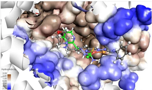
including mouse R2 RNR inhibitory potential, studied by molecular modeling and tyrosyl radical quenching monitored by EPR spectroscopy, as well as by Western Blotting and flow cytometry are also presented.
Results
Synthesis and characterization of (iso)thiosemicarbazone-morpholine hybrids and their Cu(II) complexes. The 5-methylmorpholine-pyridine-2-carboxaldehyde H was prepared in seven steps as shown in Scheme 1 and the detailed synthesis is described in the Supporting Information. First pyridine- 2,5-dicarboxylic acid A was converted into diester B that was further reduced to the diol C. The latter was selectively oxidized with SeO2 to the aldehyde D. After protection of the aldehyde group the alcohol E was converted into the chloride F that was further reacted with morpholine to give species G. Finally, the hydrolysis of the methyl ester function in acidic conditions afforded the required aldehyde H for condensation reactions with thiosemicarbazide, 4N-dimethyl-3-thiosemicarbazide, 4N-pyrrolydinyl-3- thiosemicarbazide, 4N-morpholinyl-3-thiosemicarbazide, 4N-phenyl-3-thiosemicarbazide and S- methylisothiosemicarbazide hydroiodide to give the hybrids HL1‒6 (Scheme 1), respectively. One- and two-dimensional NMR spectra were in agreement with the proposed structures for HL1‒6, enabling the assignment of all 1H and 13C resonances. The ESI mass spectra recorded in a positive ion mode showed strong peaks corresponding to [M+H]+ and [M+Na]+ ions, respectively. The proligands HL1‒6 were reacted with Cu(II) chloride dihydrate and triethylamine in 1:1:1 mole ratio in methanol to give [Cu(L1‒6)Cl] (1‒6) in 35‒91% yields. Positive ion ESI mass spectra showed strong peaks attributed to [Cu(L1‒6)]+ ions. The structures of 1, 3‒6 were also established by single crystal X-ray diffraction (vide infra).
2 3 4 5 6 7 8 9 10 11 12 13 14 15 16 17 18 19 20 21 22 23 24 25 26 27 28 29 30 31 32 33 34 35 36 37 38 39 40 41 42 43 44 45 46 47 48 49 50 51 52 53 54 55 56 57
HO N B
N
D
N O
N O
C
H N
OMe MeO
E (i)
HO N
OH
O A
O
O N
O
O
O
O
(ii) (iii)
H N
OMe MeO
F
OH OH
OH Cl
H N
OMe MeO
G
N O
H
(iv)
(vii) (vi) (v)
Scheme 1. Synthesis of 5-methylmorpholine-pyridine-2-carboxaldehyde. Reagents and conditions: (i) thionyl chloride,
methanol, 0 °C → room temperature, 12 h;22 (ii) sodium borohydride, ethanol, acetone, potassium carbonate, chloroform, 0
°C, 1 h → reflux, overnight;23 (iii) selenium dioxide, dioxane, water, 100 °C, 3 h;24 (iv) trimethyl orthoformate, methanesulfonic acid, methanol, reflux, 48 h; (v) thionyl chloride, dichloromethane, ‒80 °C → room temperature, overnight;
(vi) moprholine, triethylamine, THF/CH2Cl2 1:1, 50 °C, overnight, purification by column chromatography; (vii) HCl, water, 60 °C, overnight.
X-ray Crystallography. The results of X-ray diffraction studies of HL1, HL2 and HL4‒6 are shown in Figure S1, while those of [Cu(L1)Cl] (1), [Cu(L3)Cl] (3), [Cu(L4)Cl] (4), [Cu(L5)Cl] (5) and [Cu(L1)Cl(H2O)] (1´), [Cu(L6)Cl] (6) in Figures 1 and 2, respectively. Selected bond distances and bond angles are quoted in Tables S1 and S2. The proligands adopted different isomeric configurations in the solid state depending on substituents at the terminal nitrogen atom of the thiosemicarbazide moiety.
Complexes 1, 3, 4 and 5 form dimeric associates as shown in Figure 1 (co-crystallized solvent was omitted for clarity). Each Cu(II) ion has a distorted square-planar coordination geometry. Intermolecular contacts supporting the dimeric associates in the crystals are of different nature in 1 and 3‒5, respectively. The presence of long intermolecular contacts Cu···Cl or Cu···Cl and Cu···S (see legend to Figure 1) provides evidence of weak association of complexes in dimers, which most probably dissociate in solution with formation of monomeric species. There was no evidence from ESI mass spectra on the presence of dimeric species in solution.
2 3 4 5 6 7 8 9 10 11 12 13 14 15 16 17 18 19 20 21 22 23 24 25 26 27 28 29 30 31 32 33 34 35 36 37 38 39 40 41 42 43 44 45 46 47 48 49 50 51 52 53 54 55 56 57 58
Figure 1. ORTEP views of weak dimeric associates of (a) [Cu(L1)Cl] (1), (b), [Cu(L3)Cl] (3), (c) [Cu(L4)Cl]2 (4) and (d) [Cu(L5)Cl] (5).
Moreover, HL1 reacts with CuCl2 in aqueous solution with formation of five-coordinate complex [Cu(L1)Cl(H2O)] (1´·2H2O) (Figure 2b). The monomeric square-planar complex [Cu(L6)Cl] forms an
2 3 4 5 6 7 8 9 10 11 12 13 14 15 16 17 18 19 20 21 22 23 24 25 26 27 28 29 30 31 32 33 34 35 36 37 38 39 40 41 42 43 44 45 46 47 48 49 50 51 52 53 54 55 56 57
infinite chain via weak coordination of the morpholine oxygen atom of one complex to the Cu(II) atom of the next molecule as shown in Figure S2. The intermolecular Cu···O contact is of 2.603(3) Å.
Figure 2. ORTEP views of (a) [Cu(L1)Cl(H2O)] (1´) and (b) [Cu(L6)Cl] (6).
Solution Chemistry. In order to establish the presence of isomers of the proligands in aqueous solution and proton dissociation processes, in which these can be involved solution equilibrium studies have been performed. Likewise, the solution speciation of the copper(II) complexes, as well as their stability have been investigated to elucidate the species, which is the most stable and abundant at physiological pH.
The proligand HL1 (Chart 1) was chosen for the detailed solution equilibrium studies since it has the simplest structure and the best aqueous solubility among the proligands prepared. The presence of different isomers in solution was excluded by measurements of 1H NMR spectra in 10% D2O/90% H2O (Figure S3), which showed only one set of signals for the proligand in accordance to its low (C1) molecular symmetry. The proligand most probably adopts the E configuration found in the solid state (Figure S1). The same configuration was reported previously for the reference compound 2- formylpyridine thiosemicarbazone in polar solvents.25 Proton dissociation processes were monitored in aqueous solution by pH-potentiometric and 1H NMR titrations and three pKa values were determined by both methods (Table S3). According to the obtained pKa values HL1 is mainly neutral (97% HL, 3%
H2L+) at physiological pH (Figure S4). Inspection of 1H NMR spectra revealed stepwise deprotonation
2 3 4 5 6 7 8 9 10 11 12 13 14 15 16 17 18 19 20 21 22 23 24 25 26 27 28 29 30 31 32 33 34 35 36 37 38 39 40 41 42 43 44 45 46 47 48 49 50 51 52 53 54 55 56 57 58
of three functional groups in the following order: NpyridiniumH+ → NmorpholiniumH+ → NhydrazineH (Figure S3).
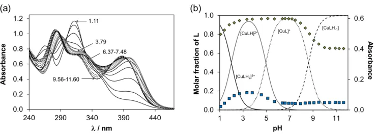
The solution speciation of the Cu(II) complexes with HL1 was characterized by the combined use of pH-potentiometry, UV−vis spectrophotometry (via charge transfer (CT) and d-d bands) and EPR spectroscopy. The spectral changes (Table S4, Figures 3a and S5) in the UV and visible regions measured at 1:1 metal-to-ligand ratio show the high-extent formation of a Cu(II) complex already at strongly acidic pH values (e.g. pH 1) and its stepwise deprotonation by increasing the pH. At pH 7.4 the dominant species is [CuL]+ (Figure 3b).
0.0 0.2 0.4 0.6
0.0 0.2 0.4 0.6 0.8 1.0
1 3 5 7 9 11
Asorbance
Molar fractionof L
pH
[CuLH2]3+
[CuLH]2+ [CuL]+ [CuLH-1]
(B)
0.0 0.2 0.4 0.6 0.8 1.0 1.2
240 290 340 390 440
Absorbance
/ nm
3.79 1.11
9.56-11.60
6.37-7.48
(A)
Absorbance
(a) (b)
Figure 3. UV–vis spectra recorded for the Cu(II)–HL1 (1:1) system at various pH values (a), and concentration distribution curves and measured absorbance values at 346 nm (♦) and 440 nm (■) for the same system (b). {cCu(II) = clig = 121 µM; T = 298 K; I = 0.10 M (KCl); l = 0.5 cm}.
Due to the high stability of the Cu(II) complexes formed, the cumulative constant (logβ) for the [CuL]+ species (Table S4) was determined via EDTA displacement studies (Figure S6). Then the pKa and the logβ values for the other type of complexes ([CuLH2]3+, [CuLH]2+, [CuLH-1]) were computed using each method and data were in a fairly good agreement (Table S4). It is worth noting that formation of a bis- ligand complex [CuL2] at ligand excess was also confirmed by the UV−vis CT and EPR titrations. The
2 3 4 5 6 7 8 9 10 11 12 13 14 15 16 17 18 19 20 21 22 23 24 25 26 27 28 29 30 31 32 33 34 35 36 37 38 39 40 41 42 43 44 45 46 47 48 49 50 51 52 53 54 55 56 57
d-d bands (Figure S5) recorded from pH 3.4 to 7.5 at 1:1 metal-to-ligand ratio revealed that the proton dissociation process of [CuLH]2+ → [CuL]+ is accompanied by rather weak spectral changes as most probably the non-coordinating morpholinium NH+ is deprotonating. In order to further characterize the coordination modes of the various complexes in solution EPR parameters (Table S5) obtained by the deconvolution of the recorded spectra (Figure S7) were analyzed. The isotropic g and A values of [CuLH]2+ and [CuL]+ are quite similar indicating the like coordination mode.
Comparison of the EPR parameters of the Cu(II) complexes of HL1 with those of 2-formylpyridine thiosemicarbazone25 and Triapine26 permitted to conclude that in the [CuLH]2+ and [CuL]+ complexes a typical coordination mode via the Npyridyl,N,S− binding site is realized in accordance with the X-ray diffraction data. [CuLH2]3+ contains the diprotonated ligand in which the protons are attributed to the non-coordinating N2H and morpholinium NH+ moieties, while [CuLH-1] is most probably a mixed hydroxido complex [CuL(OH)] formed by the deprotonation of water molecule coordinated in the fourth equatorial position. At ligand excess besides the bis-ligand complex a minor dinuclear species [Cu2L3H]2+ was detected resulting in the weak exchange coupling between neighboring Cu(II) centers smearing out the expected hyperfine structure.
Cu(II)-TSC complexes undergo quasi-reversible one-electron reduction at biologically accessible potentials. In order to assess the redox properties of 1‒6, detailed electrochemical and spectroscopic studies in various solvents were performed using cyclic voltammetry as well as EPR–
spectroelectrochemistry. Additionally, the UV‒vis spectra of 1‒5 were measured (Figure S8).
Electrochemical and spectroscopic data are summarized in Table S6. Almost reversible one-electron cathodic reduction was observed for 1‒5 in DMSO with half-wave redox potentials from –0.77 V to – 0.81 V vs Fc+/Fc (for comparison from –0.13 V to –0.17 V vs NHE) (Table S6 and Figure S9). In general, the values of redox potentials of 1‒6 for the first reduction step decreased in the following rank
2 3 4 5 6 7 8 9 10 11 12 13 14 15 16 17 18 19 20 21 22 23 24 25 26 27 28 29 30 31 32 33 34 35 36 37 38 39 40 41 42 43 44 45 46 47 48 49 50 51 52 53 54 55 56 57 58
order (E1/2(5) > E1/2(2) = E1/2(3) = E1/2(4) > E1/2(1) > E1/2(6)). The second reduction step occurred at – 1.8 V vs Fc+/Fc (–1.16 V vs NHE) and was less reversible indicating a ligand based reduction. This was confirmed by the CVs of the corresponding proligands which exhibited the first reduction step at around –1.9 V vs Fc+/Fc (not shown). In aqueous solutions the reduction potentials shifted to the less negative values and reduction was less reversible as shown for 2 in Figure S10. Complex 6 exhibited different redox behavior with the lowest electrochemical reversibility and the most negative reduction potential of –0.86 V vs Fc+/Fc (–0.26 V vs NHE) (Figure S9c). Unlike the cathodic reduction, the anodic oxidation of 1‒6 was irreversible with potential values in the region from 0.4 to 0.8 V vs Fc+/Fc (0.68 V to 1.44 V vs NHE). Similar response was observed for the corresponding proligand HL2 (Figure S11), indicating ligand based oxidation in 2.
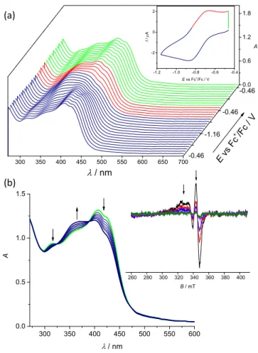
The biologically accessible reduction of Cu(II)-TSC complexes is Cu-centered. To investigate whether the biologically accessible reduction is metal-centered, the reversible one-electron reduction of 5 was further studied by in situ UV‒vis-spectroelectrochemistry (Figure 4a). Upon cathodic reduction at the first reduction peak two isosbestic points at 389 and 325 nm were detected. The spectral changes of the S→Cu(II) charge transfer bands (~425 nm) clearly confirmed the reduction of Cu(II) to Cu(I).
Additionally, upon voltammetric reverse scan, reoxidation and a nearly full recovery of the initial optical bands were observed, attesting the chemical reversibility of the cathodic reduction even at low scan rates (Figure 4). To confirm the involvement of Cu(II) in the reduction processes, in situ EPR electrochemistry of 1‒6 in nBu4NPF6/DMSO and water was performed, since metal-based reduction would result in the formation of EPR-silent Cu(I) species (Figure 4b). As can be seen for electrochemical reduction of 2 in nBu4NPF6/DMSO in the region of the first one–electron reduction step (see inset in Figure 4b), a significant decrease of EPR signal was observed in accord with the formation of diamagnetic Cu(I) d10 complex. For aqueous solutions the reversibility was significantly reduced, implying a more complex mechanism involving the release of the proligand. However, by decreasing the scan rate and going to
2 3 4 5 6 7 8 9 10 11 12 13 14 15 16 17 18 19 20 21 22 23 24 25 26 27 28 29 30 31 32 33 34 35 36 37 38 39 40 41 42 43 44 45 46 47 48 49 50 51 52 53 54 55 56 57
the more positive potentials upon reverse scan, a partial recovery of the initial optical bands was also observed in aqueous solutions as shown for 2 in Figure S12. Thus, electrochemical data indicated a likely reduction of Cu(II)-TSC complexes to Cu(I) species with the subsequent release of the proligands.
-1.2 -1.0 -0.8 -0.6 -0.4
-2 0 2
I / A
E vs Fc+/Fc / V
300 350 400 450 500 550 600 650 700
0.0 0.6 1.2 1.8
-0.46
-1.16 -0.46
/ nm
E vs Fc
+/Fc / V
A
-0.46
300 350 400 450 500 550 600
0.0 0.5 1.0 1.5
A
/ nm
260 280 300 320 340 360 380 400 B / mT
(a)
(b)
Figure 4. Spectroelectrochemistry of 2 in nBu4NPF6/DMSO in the region of the first cathodic peak. (a) Potential dependence of UV‒vis spectra with respective cyclic voltammogram (Pt-microstructured honeycomb working electrode, scan rate v = 5 mV s−1); (b) evolution of UV‒vis spectra in 2D projection in forward scan (Inset: EPR spectra measured at the first reduction peak using Pt mesh working electrode).
Lead TSCs and their Cu(II) complexes exhibited marked antiproliferative activity in a nanomolar concentration range. The in vitro anticancer activity of 1‒6 and their respective TSCs was determined in ovarian carcinoma cells (A2780 and A2780cisR) and non-cancerous human embryonic kidney cells (HEK293) by the colorimetric MTT assay with an exposure time of 72 h. The IC50 values for HL1‒HL6
2 3 4 5 6 7 8 9 10 11 12 13 14 15 16 17 18 19 20 21 22 23 24 25 26 27 28 29 30 31 32 33 34 35 36 37 38 39 40 41 42 43 44 45 46 47 48 49 50 51 52 53 54 55 56 57 58
and 1‒6 in comparison with Triapine, CuCl2, Cu-Triapine and cisplatin are listed in Tables 1 and 2, respectively, and concentration-effect curves are depicted in Figure S13.
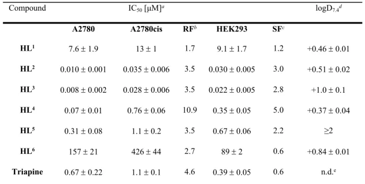
Table 1. Cytotoxicity of proligands HL1‒6 and their n-octanol/water distribution coefficients (logD7.4)
Compound IC50 [μM]a logD7.4d
A2780 A2780cis RFb HEK293 SFc
HL1 7.6 1.9 13 1 1.7 9.1 1.7 1.2 +0.46 0.01
HL2 0.010 0.001 0.035 0.006 3.5 0.030 0.005 3.0 +0.51 0.02 HL3 0.008 0.002 0.028 0.006 3.5 0.022 0.005 2.8 +1.0 0.1 HL4 0.07 0.01 0.76 0.06 10.9 0.35 0.05 5.0 +0.37 0.04
HL5 0.31 0.08 1.1 0.2 3.5 0.67 0.06 2.2 ≥2
HL6 157 21 426 44 2.7 89 2 0.6 +0.84 0.01
Triapine 0.67 0.22 1.1 0.1 4.6 0.39 0.05 0.6 n.d.e
a 50% inhibitory concentrations (IC50) in human ovarian carcinoma cell lines A2780 and A2780cisR and human embryonic kidney cell line HEK293, determined by means of the MTT assay after 72 h exposure. Values are means ± standard deviations obtained from at least three independent experiments, bResistance Factor (RF) is determined as IC50(A2780cisR)/IC50(A2780), c Selectivity Factor (SF) is determined as IC50(HEK293)/IC50(A2780), d Distribution coefficients between n-octanol and buffered aqueous solution determined at physiological pH by UV‒vis spectroscopy. Values are means ± standard deviations obtained from at least three independent experiments, e n.d. – not determined.
With the exception of HL1 and HL6, the proligands demonstrated marked antiproliferative activity in a submicromolar to nanomolar concentration range in both A2780 and A2780cisR cells. The efficacy of compounds HL2, HL3 and HL5 in cisplatin-resistant A2780cisR cells decreased by factor 3.5 in comparison to that in sensitive A2780 cells, whereas a 4.6- and 11-fold drop of activity was observed for Triapine and HL4, respectively. The activity of proligands HL1‒6 and Triapine increased in the following rank order HL6 < HL1 < Triapine < HL5 < HL4 << HL2 HL3 (Table 1).
2 3 4 5 6 7 8 9 10 11 12 13 14 15 16 17 18 19 20 21 22 23 24 25 26 27 28 29 30 31 32 33 34 35 36 37 38 39 40 41 42 43 44 45 46 47 48 49 50 51 52 53 54 55 56 57
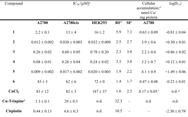
Table 2. Cytotoxicity of Cu(II)-TSC complexes 1‒6, CuCl2 and cisplatin and their n-octanol/water distribution coefficients (logD7.4)
Compound IC50 [μM]a Cellular
accumulation,d nmol Cu/
mg protein
logD7.4e
A2780 A2780cis HEK293 RFb SFc A2780
1 2.2 0.1 13 4 16 2 5.9 7.3 0.63 0.09 –0.81 0.04
2 0.012 0.002 0.030 0.003 0.032 0.008 2.5 2.7 3.9 0.6 +0.30 0.01 3 0.26 0.02 0.60 0.05 0.78 0.20 2.3 3.0 2.2 0.6 +0.86 0.02 4 0.08 0.01 0.26 0.04 0.24 0.02 3.3 3.0 3.2 0.7 +0.12 0.01 5 0.009 0.002 0.017 0.002 0.020 0.003 1.9 2.2 4.1 0.9 +1.49 0.06
6 43 3 62 6 72 0 1.4 1.7 0.47 0.08 –0.23 0.01
CuCl2 83 12 82 3 187 37 1.0 2.3 0.17 0.05 f n.d. g
Cu-Triapineh 1.3 0.1 29 0.3 n.d. 22.3 - n.d. n.d.
Cisplatin 0.44 0.13 4.6 0.3 n.d. 10.5 - - –2.30 0.79i
a 50% inhibitory concentrations (IC50) in human ovarian carcinoma cell lines A2780 and A2780cisR and human embryonic kidney cell line HEK293, determined by means of the MTT assay after exposure for 72 h. Values are means ± standard deviations obtained from at least three independent experiments, bResistance Factor (RF) is determined as IC50(A2780cisR)/IC50 (A2780), c Selectivity Factor (SF) is determined as IC50(HEK293)/IC50(A2780), d Cellular accumulation in A2780 cells, determined by ICP-MS after 24 h exposure at concentration of 1 μM. Values are means ± standard deviations obtained from at least three independent experiments, e Distribution coefficients between n-octanol and buffered aqueous solution determined at physiological pH by UV‒vis spectroscopy. Values are means ± standard deviations obtained from at least three independent experiments, f Cellular accumulation ofCuCl2 was detected at concentration of 2.5 μM, g n.d. – not determined, h the IC50
values (exposure for 72 h) were taken from the ref..27,i Log Po/w value was taken from the ref. 28.
As can be seen in Table 2, with the exception of 3, all Cu(II)-TSC complexes were equally or more cytotoxic than the respective proligands. The activity of complexes 1‒6 increased in the following order 6 < 1 < 3 < 4<< 2< 5, similar to the trend in electrochemical redox potentials for Cu-based reduction
2 3 4 5 6 7 8 9 10 11 12 13 14 15 16 17 18 19 20 21 22 23 24 25 26 27 28 29 30 31 32 33 34 35 36 37 38 39 40 41 42 43 44 45 46 47 48 49 50 51 52 53 54 55 56 57 58
step (Table S6). In order to compare the cytotoxicity of 1‒6 with that of [Cu(H2O)6]2+, A2780 cells were treated with aqueous solution of CuCl2. In agreement with the literature,29 CuCl2 revealed antiproliferative activity in the high micromolar range, in contrast to high activity of 1‒6, which should be regarded as individual entities with their own biological, pharmacokinetic and metabolic profiles.
Complexes 1‒6 were up to ~50 times more cytotoxic than cisplatin in A2780 cells and up to ~270 times more cytotoxic in A2780cisR cells. Additionally, the differences in cytotoxicity of 1‒6 in A2780 and A2780cisR cells were significantly lower than for HL1‒HL6 and cisplatin, indicating high potential of Cu(II) complexes for the treatment of cisplatin-resistant tumors. The most active TSC proligands and their respective Cu(II) complexes were more selective towards A2780 cells over HEK293 cells;
however, they did not show any selectivity towards A2780cisR cells over HEK293 cells. On the contrary, Triapine was significantly more cytotoxic towards non-cancerous HEK cells than A2780 or A2780cis cells.
Antiproliferative activity of Cu(II)-TSC complexes correlates with their cellular accumulation and lipophilicity. The ability of anticancer drugs to penetrate biological membranes, tissues and barriers underlies their biological, pharmacokinetic and metabolic properties. These properties can be predicted by calculating physicochemical characteristics of drug candidates using molecular descriptors.30 In order to estimate if the proligands HL1‒HL6 and corresponding Cu(II) complexes demonstrate drug-like properties, they were evaluated with main stream molecular descriptors, such as molecular weight (MW), number of hydrogen bond donors and acceptors (HBD/HBA), n-octanol-water partition coefficient (log P), polar surface area (PSA) and rotatable bonds (RB). The results are shown in Table S7 in the Supporting Information. Based on the calculated physicochemical parameters, we determined if novel compounds belonged to the drug-like chemical space, which is commonly defined by the well- known Lipinski rule, or to the known drug space (KDS), which includes all small compounds in medical use.31 As can be seen from Table S7, all compounds fell within the boundaries of drug-like chemical
2 3 4 5 6 7 8 9 10 11 12 13 14 15 16 17 18 19 20 21 22 23 24 25 26 27 28 29 30 31 32 33 34 35 36 37 38 39 40 41 42 43 44 45 46 47 48 49 50 51 52 53 54 55 56 57
space with the exception of HL4 which demonstrated higher HBA value, referring to KDS space. Based on these results, novel compounds are expected to be sufficiently cell permeable. Following the calculations, we determined the lipophilicity of all compounds experimentally (logD7.4, Tables 2 and 3).
It is known that cancer cells accumulate hydrophilic compounds to a smaller extent relative to hydrophobic compounds, thereby affecting their activity. Hence, the differences in the lipophilicity of compounds might be related to the observed trends in cytotoxicity of 1‒6. In general, with the exception of morpholine derivative HL4, proligands which were obtained by systematic substitution at the terminal thioamide nitrogen, demonstrated higher lipophilicity when compared to that of HL1. As expected, proligands were more lipophilic than the corresponding Cu(II) species, which are positively charged at physiological pH (Figure 3b). Among Cu(II)-TSC complexes, only the least cytotoxic complexes 1 and 6, as well as cisplatin, demonstrated negative logD7.4 values, indicating their higher hydrophilicity. The lipophilicity of 2‒5 increased in the following order 4 < 2 < 3 < 5, and correlated well with their cytotoxicity in cancer cells, with the exception of 3. Subsequently, we determined the total cellular accumulation of Cu in A2780 cells by ICP-MS upon 24 h exposure to 1‒6 in comparison with CuCl2
(Table 4). The IC50 values from MTT assays with 72 h drug exposure varied from nanomolar to high micromolar concentrations; therefore, in the cellular accumulation experiment cells were treated with 1 μM of compounds of interest for 24 h to minimize cell detachment. The cellular accumulation of all
Cu(II)-TSC complexes was significantly higher than for CuCl2, indicating the role of lipophilic TSC ligands in the delivery of the complexes into the cells. The accumulation increased in the order 6 < 1 <
3 < 4 < 2 < 5.
Cellular accumulation and efflux of Cu(II)-TSC complexes are energy- and/or temperature- dependent processes. Dependence of cytotoxicity on lipophilicity of 1‒6 indicates that their cellular accumulation at least partially occurs via passive diffusion. In order to gain additional insights into the mechanisms controlling the accumulation of Cu(II)-TSC complexes, we investigated the effects of
2 3 4 5 6 7 8 9 10 11 12 13 14 15 16 17 18 19 20 21 22 23 24 25 26 27 28 29 30 31 32 33 34 35 36 37 38 39 40 41 42 43 44 45 46 47 48 49 50 51 52 53 54 55 56 57 58
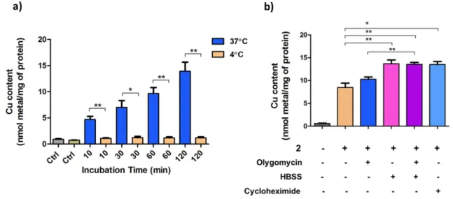
temperature and various inhibitors on total cellular accumulation of 2 with the results shown in Figure 5. A2780 cells were treated with 2 at 3 μM for 30, 60 and 120 min at 37 and 4 C and intracellular Cu content was measured by ICP-MS. Prolonged incubation at low temperatures may result in the changes in membrane fluidity, decreasing membrane permeability and restricting drug uptake;32 therefore, the shorter time point of 10 min was also included. The cellular accumulation of 2 dramatically decreased at low temperature even after 10 min treatment, indicating the involvement of active carrier-mediated transport or facilitated diffusion. Likewise, the decrease of cellular accumulation at low temperatures has been reported for both Cu(II)-TSC complexes and metal-free TSCs.33,34 To further clarify whether the uptake of 2 requires energy, the cellular ATP production was blocked by incubating cells in a saline solution (HBSS), resulting in total starvation and rapid reduction of intracellular ATP content, and/or by addition of oligomycin, leading to the inhibition of oxidative phosphorylation.
Figure 5. Effects of temperature and inhibitors on the accumulation of 2 in A2780 cells. Intracellular content was determined
by ICP-MS: a) A2780 cells were treated with 2 (3 μM) at 37 or 4 C for the indicated time periods. b) A2780 cells were pre- treated with oligomycin (5 μM), and/or pre-incubated in HBSS for 1 h, or pre-treated with cycloheximide (100 μM) for 4 h and subsequently co-treated with 2 (3 μM) for 1 h. The presence of inhibitors did not show any effects on the intracellular Cu level of untreated A2780 cells (data not shown). Statistical analysis was performed by two-tailed T-test using GraphPad Prism software (GraphPad Software Inc., CA) with p < 0.05 considered as significant (* p < 0.05, ** p < 0.01).
2 3 4 5 6 7 8 9 10 11 12 13 14 15 16 17 18 19 20 21 22 23 24 25 26 27 28 29 30 31 32 33 34 35 36 37 38 39 40 41 42 43 44 45 46 47 48 49 50 51 52 53 54 55 56 57
When A2780 cells were pre-treated with 5 μM of oligomycin for 1 or 4 h and subsequently co-treated with 3 μM of 2, the intracellular Cu levels were not affected, even though the mitochondrial respiration of the cancer cells was successfully inhibited (data not shown). However, when A2780 cells were starved in HBSS for 1 h and subsequently treated with 3 μM of 2 for 1 h, an increase in intracellular Cu content was observed. Co-treatment of 2 with 5 μM of oligomycin in HBSS did not result in a further increase of intracellular Cu levels. The increase of Cu content in HBSS-starved cells might stem from the inhibition of energy-dependent efflux processes. In order to assess the role of proteins in both cellular accumulation and efflux of 2, A2780 cells were pre-treated with cycloheximide, which is a known inhibitor of protein synthesis. Significant increase of intracellular Cu content was observed, similar to HBSS starvation, providing further evidence for the involvement of protein-dependent efflux processes.
Similar effects have been reported for related Cu(II)-TSC complexes.33 Accumulated data provide evidence that cellular accumulation of 2 occurs via carrier-facilitated diffusion, which might co-exist with passive permeation through the lipid bilayer.35 The efflux processes, however, are actively mediated by proteins and upon inhibition of protein synthesis or energy production a strong increase of intracellular Cu concentration was observed.
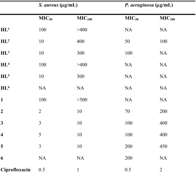
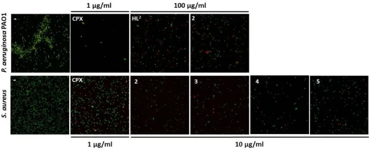
Cu(II)-TSC complexes demonstrated high antibacterial activity against S. aureus in a comparable concentration range to ciprofloxacin. The antibacterial activity of HL1‒HL6 and 1‒6 on planktonic cells of P. aeruginosa and S. aureus was investigated by determination of MIC50 and MIC100 values (Table 3). Overall, compounds showed a higher antimicrobial activity against Gram-positive (S. aureus) compared to Gram-negative bacteria (P. aeruginosa). The highest activity against S. aureus was observed for 2‒5, which specifically inhibited its growth with MIC50 values ranging between 2 and 5 µg/mL and completely abolished its growth at 10 µg/mL (MIC100). This activity is comparable but slightly lower that the antibacterial activity of a well-known benchmarked antibiotics ciprofloxacin
2 3 4 5 6 7 8 9 10 11 12 13 14 15 16 17 18 19 20 21 22 23 24 25 26 27 28 29 30 31 32 33 34 35 36 37 38 39 40 41 42 43 44 45 46 47 48 49 50 51 52 53 54 55 56 57 58
![Figure 1. ORTEP views of weak dimeric associates of (a) [Cu(L 1 )Cl] (1), (b), [Cu(L 3 )Cl] (3), (c) [Cu(L 4 )Cl] 2 (4) and (d) [Cu(L 5 )Cl] (5)](https://thumb-eu.123doks.com/thumbv2/9dokorg/1316736.106049/9.892.256.630.71.867/figure-ortep-views-weak-dimeric-associates-cu-cl.webp)
![Figure 2. ORTEP views of (a) [Cu(L 1 )Cl(H 2 O)] (1´) and (b) [Cu(L 6 )Cl] (6).](https://thumb-eu.123doks.com/thumbv2/9dokorg/1316736.106049/10.892.92.779.116.480/figure-ortep-views-cu-l-cl-cu-cl.webp)