Antibiotics
Η. Boyd Woodruff and Ian M. Miller
I. Introduction 23 II. Requirements for Optimum Production 25
III. Production in Nature 25 IV. Selectivity of Antibiotics 26
V. Biochemical Significance of Antibiotics 27 VI. Mode of Action of Antibiotics; Cellular Effects 28 VII. Mode of Action of Antibiotics; Biochemical Significance 30
A. The Bacterial Cell Wall 32 B. The Cell Membrane 37 C. Nucleic Acid Biosynthesis 41
D. Protein Synthesis 42 E . Uncoupling A g e n t s ; Respiration Inhibitors 44
F. Specific Enzyme Effects 45 G. Vitamin Interrelationships 45
H. Chelation 46 VIII. Conclusion 47
References 47
I. INTRODUCTION
1"An antibiotic is a chemical compound, derived from a microorganism, which has the capacity in dilute solution of inhibiting the growth of, and even destroying, other microorganisms." This definition, devised by
1 For the purposes of this chapter on antibiotics as metabolic inhibitors, no attempt has been made to present a complete literature survey. References have been cited for key points. Summary or review works usually have been selected in preference to the original report, as such summaries present up-to- date knowledge.
For an extension of the comments in this chapter, the reader should consult the general reviews by Verwey in Annual Review of Microbiology, Volume 13
(2); by Chain in Annual Review of Biochemistry, Volume 28 (8); and by Hahn in the Proceedings of the Fourth International Congress of Biochemis-
2 3
Waksman (1)> serves to group several thousand compounds which may be subclassified on the basis of differences in chemical or biological prop
erties. The complete chemical structure is known for only a few of the antibiotics, but these are of diverse nature. It is evident that micro
organisms have the capacity to synthesize a variety of compounds having a wide range of inhibitory activity against other microorganisms. Fre
quently, the inhibitory or toxic action extends to higher plants and animals.
The word antibiotic in the above limited sense has been in use only since 1942; however, research on antibiosis is of much earlier origin. The general area of study has been defined by Waksman as existing "between true parasitism—one organism living in or upon the body of another—
and true saprophytism—one organism merely destroying the waste prod
ucts and the dead cells of another" (10). As the science of microbiology developed in the early 1900's, emphasis was placed on the applied areas, especially soil microbiology. Because of the complex associations among the microorganisms contained in the dynamic population of soil, antago
nistic relationships became obvious to soil microbiologists and were the source of research studies and finally of review articles (11).
Antibiotics are produced with great frequency by microorganisms iso
lated from substrates such as the soil, air, and plant materials. Detailed descriptions of screening procedures have indicated that as many as 10%
of the streptomycetes isolated from soil and grown in artificial culture produce antibiotic activity (12). Many filamentous fungi also produce antibiotics. Of 65 species isolated from soil, half have been found to form antibiotics (13). The accumulation of antibiotic activity by bacteria can be shown with ease. Sterile agar media inoculated with soil suspensions at low dilutions usually show clear zones where growth of the spreading sur
face bacteria is inhibited. Frequently, these zones surround colonies of sporeforming bacteria and are due to antibiotics produced by the spore- formers (14)- However, critical studies demonstrate that antibiotic pro- try ( 4 ) . Chemotherapeutic approaches are described in "The Strategy of Chemotherapy," the Eighth Symposium of the Society for General Micro
biology ( 5 ) , and yearly progress in the field of antibiotic therapy has been summarized in "Antibiotica et Chemotherapia" (Volume 10 appeared in 1962)
(6). Nonchemotherapeutic properties of antibiotics are discussed in "Antibi
otics, Their Chemistry and Non-Medical Uses," edited by H. S. Goldberg (7).
Very complete presentations concerning antibiotics and their properties are contained in the two-volume work, "Antibiotics," by Florey and his collabo
rators at Oxford University (8) and in the "Handbook of Toxicology," Vol
ume II, distributed by the Division of Biology and Agriculture, National Academy of Sciences (9).
duction is limited to a rather small number of bacterial types and that the antibiotics they produce usually have a narrow spectrum.
II. REQUIREMENTS FOR OPTIMUM PRODUCTION
Antibiotics are formed readily under conditions which promote good growth of the producing microorganisms. High concentrations of available nutrients, such as digests of protein and fermentable carbohydrates, and availability of oxygen promote formation of antibiotics with most micro- organisms and serve as an adequate basis for screening. Each micro- organism has its own specific requirements for optimal antibiotic production. For example, streptomycin is produced in best yield if phos- phate ions are restricted in the presence of excess concentrations of other nutrients (15). For maximum benzylpenicillin yields, adequate nutrients are required for formation of metabolically active mycelium of Penicil- lium chrysogenum, but thereafter penicillin formation is promoted under a condition of restricted carbohydrate supply in the presence of sources of nitrogen and sulfur, and a phenylacetic acid derivative (16). The anti- biotic grisein contains iron, and maximum antibacterial potential can be realized only in a culture medium containing an excess of this element (17). Special conditions have been defined for optimum yield of each antibiotic culture studied in detail.
III. PRODUCTION IN NATURE
Studies on the significance of antibiotic formation in the natural habitat present an enigma. The high number of antibiotic-producing micro- organisms found in a competitive environment, such as fertile garden soil, suggests that the ability to produce antibiotics has exerted a favorable selection pressure during the course of evolution of soil microbes. Surely, if the formation of an antibiotic represented merely a wasteful drain of carbon into a nonmetabolizable, useless compound, organisms which pro- duce antibiotics would have great difficulty in competing with non- producers for the relatively restricted amount of nutrients in the soil.
Antibiotic-producing organisms have not died out; in fact, studies have shown that the ratio of antibiotic-producing microbes to the total popula- tion remains the same in a soil during the increase of several fold of the total microbial population resulting from the introduction of a nutrient into that soil (18).
It is tempting to speculate that antibiotic producers may be favored in the competitive environment by the formation of antibiotic activity, at least in the local microenvironment, to the extent that competition for nutrients would be lessened. All attempts to prove this hypothesis have failed. Generally, antibiotic formation under the conditions of restricted nutrients available in soil is very poor. In the soil, many antibiotics lose their activity through absorption on soil colloids, instability in the pres
ence of soil chemicals, or by biological attack.
A number of antibiotics have been produced in sterile soil inoculated with a specific antibiotic-producing microorganism, particularly if addi
tional nutrient supplements are added. Also, traces of the antibiotics gliotoxin, gladiolic acid, frequentin, griseofulvin, chloramphenicol, actino- mycin, and certain other antibiotics derived from Streptomyces and Bacillus species have been observed in normal and nutrient-enriched soils.
Although data supporting the advantage granted a microorganism by its ability to produce an antibiotic in the soil are limited and drawn pri
marily from studies of microbial combinations under artificial conditions, the probability is in favor of such an advantage. Brian has summarized the evidence and, on the basis of it, contends that antibiotic production is beneficial at least within microenvironments {19). Without question, the problem demands much further study and may be solved finally only by application of principles of quantitative genetics and the study of anti
biotic formation as an evolutionary factor.
IV. SELECTIVITY OF ANTIBIOTICS
The definition of an antibiotic adequately encompasses a class of com
pounds and excludes unrelated forms. It is restrictive in accepting only compounds produced by microorganisms, but several of these compounds, after the original demonstrations, have been found also in higher organ
isms or have been made by chemical synthesis. In fact, inhibitors of microbial growth may be isolated first as normal metabolic products and confirmed as true antibiotics only later, for example, griseofulvin {20).
Many of the recently synthesized derivatives of 6-aminopenicillanic acid are inhibitors of microbial growth which have the mode of action and properties of penicillin but which may never be strictly classified as anti
biotics because microbiological synthesis has not been accomplished {21, 22).
Even more characteristic than the origin of antibiotics is the property of selectivity in inhibitory activity. In fact, selectivity is inherent in the biosynthesis of antibiotics, for a microorganism which produces an anti-
biotic must have some mechanism of protecting itself against the action of the antibiotic; that is, the producer must be resistant. One could assume that other microorganisms having biochemical similarities to the producer also will be resistant.
The selectivity crosses species and strain lines as well as class distinc- tions in microorganisms. Because of the selectivity of action, antibiotics have sometimes been called broad spectrum or narrow spectrum, depend- ing on the breadth of microbial species inhibited. Broad spectrum has come to mean inhibition of many gram-positive and gram-negative micro- organisms, and possibly certain of the Rickettsia and large viruses. The term narrow spectrum is associated frequently with inhibition of gram- positive bacteria. These terms provide a false implication, however, when one realizes that many antibiotics have a "narrow spectrum" of activity limited to the filamentous fungi, others are specific inhibitors of yeasts, still others inhibit protozoa, and some inhibit a few members of each of several large groups of microorganisms without regard to their taxonomic classification.
It is the high order of selectivity of antibiotics which gives them their commercial utility. Some can be employed as therapeutic agents because they inhibit pathogenic bacteria without harming the host plant or ani- mal. The selectivity of action results from their selective biochemical activity. They may block formation of a structure such as the bacterial cell wall which is not present in higher organisms; they may form an inactive complex with an element required for growth by a microorganism;
they may block a synthetic reaction essential for rapidly multiplying cells but not essential to the human; they may fail to penetrate into the animal cell while freely entering the microbial cell. The clinical utility of antibiotics has established them firmly as a preferred field for industrial research and has given rise to intensive screening programs which have multiplied tremendously the number of antibiotics which have been de- scribed. The selective action of the antibiotics recovered is of great con- cern to the medical practitioner or the plant pathologist faced with a choice among several antibiotics for treatment of a specific disease condi- tion. A correct diagnosis is required to guide selection of an antibiotic having the appropriate spectrum. The toxicity of the antibiotic selected must be weighed against the expectation of chemotherapeutic effect.
V. BIOCHEMICAL SIGNIFICANCE OF ANTIBIOTICS
A preoccupation with the practical attributes of antibiotics has slowed appreciation of these molecules as fundamental tools in biochemistry.
Penicillin, cycloserine, bacitracin, and griseofulvin have contributed materially to our knowledge of cell wall biosynthesis; chloramphenicol, to the interrelationship of nucleic acid and protein biosynthesis; anti- mycin, to terminal electron transport. However, little use has been made of the many possibilties for expanding our knowledge of biochemical pathways through the study of the nonclinically effective antibiotics. The biochemist who desires to elucidate a biochemical pathway in a micro
organism should have little difficulty in finding an antibiotic which will influence the pathway.
Antibiotics are of prime importance to the biochemist because they consist of unusual, even revolutionary, type structures which are signifi
cant in normal metabolic pathways. A close examination of the anti
biotic structures in relation to known metabolites may give leads to the site of action of the antibiotics, but frequently this relationship is obscure.
Even after the specific point of action is known, as for example in the inhibition by D-cycloserine of formation of D-alanyl-D-alanine for intro
duction into the cell wall peptide (23), the specificity of the antibiotic structure for the specific metabolites involved is unexpected. One im
mediately becomes aware that competitive antagonism within the com
plexities of the microbial cell is more demanding of a specific structure than was recognized by the early proponents of this method of chemo- therapeutic attack upon disease microorganisms.
VI. MODE OF ACTION OF ANTIBIOTICS; CELLULAR EFFECTS
Early workers in the field of antibiotics recognized that these com
pounds acted by a variety of mechanisms. Differences were observed in their gross effects on microorganisms. Some of the more toxic antibiotics, such as patulin (24), are strongly bactericidal in their action. Death of the cells is rapid, and no amount of dilution or neutralization of the antibiotic allows the microorganisms to resume growth. A bacteriostatic action, or inhibition of bacterial growth without death at the lower anti
biotic concentrations, is a more common characteristic of the antibiotics.
Removal of the antibiotic by dilution, or loss of potency through inactiva- tion, frequently results in renewed growth.
Plotting of the growth curve on the basis of the numbers of surviving organisms as related to time of exposure yields a variety of curves char
acteristic for each antibiotic tested. This observation indicates that the antibiotics differ in their attack upon microorganisms but gives little insight into specific mechanisms of action.
Two generalized properties of antibiotics which may be observed in liquid culture point to a difference in basic mechanism of action. The degree of antibiotic activity may be influenced directly by the nutrients contained in the culture medium. For example, actithiazic acid (25) and bacillin (26) do not behave as antibiotics for cells grown in complex media containing digests of animal products, as their antibiotic action is antagonized by natural products present therein. Second, the expression of antibiotic activity may be dependent on the rate of growth of the microorganism at the time it comes under the influence of the antibiotic.
Penicillin and other inhibitors of cell wall synthesis are ineffective on nonmultiplying cells (27).
Various additional responses, which point to differing modes of action, occur with microbial colonies on agar plates exposed to a diffusing solu- tion of an antibiotic. Some antibiotics exert their effect at any stage of microbial growth and at any density of population. The edge of the zone of inhibition with such antibiotics is indistinct, as the cultures are stopped at various intensities of growth when the advancing antibiotic front reaches the inhibitory concentration. Other antibiotics are capable of inhibiting only young cultures at a low inoculum density. In these in- stances, the zone of inhibition is very sharp since, as soon as the culture initiates growth, it is able to overcome further antibiotic action. Fre- quently, evidence of enhanced growth may be seen just beyond the in- hibitory point.
Another phenomenon characteristic of antibiotics is resistance. In an agar test, resistance is evidenced by individual colonies which develop to normal size within the clear zone of antibiotic action. With most anti- biotics, resistance is infrequently encountered. Resistant colonies are seldom seen within the zone of inhibition produced by penicillin. Usually, four or five colonies will be apparent within an inhibitory zone of 20-mm diameter produced by a solution of streptomycin. With the antibiotic grisein, resistance may reach the extent that over 10% of the cells within the population are capable of making growth. Resistant colonies within the inhibitory zones with this antibiotic are so frequent that individual colonies cannot be observed, but only a lessening of the density of growth within the confines of the zone is observable. D i Marco has discussed antibiotic resistance from the biochemical standpoint (28).
A striking measure of antibiotic activity at the cellular level may be seen by microscopic examination. Long filamentous bacterial cells are produced at partially inhibitory levels with many antibiotics. Rods of Azotobacter increase as much as 100-fold in length with no detectable cross walls under the influence of actinomycin. The elongated forms of
sporeforming bacteria or exceptionally large bizarre staphylococci and streptococci which develop under the influence of penicillin have been described many times (29).
Nuclear abnormalities can be observed under the influence of anti
biotics with appropriate staining techniques. These usually result from altered concentrations of deoxyribonucleic acid. Striking demonstrations of this effect have been produced from the action of actinomycin on HeLa cells (80).
The nuclear abnormalities suggest that antibiotics may have signifi
cance as a tool in basic genetics. This has proved to be the case. Anti
biotic resistance is a convenient marker for studying the quantitative and qualitative influence of mutagenic agents on microbial cells. Furthermore, antibiotic resistance is a property associated with the transforming prin
ciple and may be carried from one cell to another during the course of transfer of deoxyribonucleic acid units (81). Certain antibiotics, which selectively kill growing cells, have been used to destroy growing proto- trophs and cause the enrichment of nongrowing antibiotic-resistant auxotrophs in a population. Such antibiotics provide a strong selection pressure for isolation of new auxotrophic mutants of bacteria (82, 38).
Lysis, or dissolution of microbial cells, is a phenomenon sometimes observed with antibiotics. Lysis may be the final response to antibiotics which inhibit cell wall formation. The lytic effect can be prevented with an adequate concentration of salts or sugar to make an isotonic prepara
tion with the cell protoplasm. Under these conditions, protoplasts may be produced (84). With certain organisms, growth in the presence of anti
biotics leads to the formation of L-forms, with a change in cell size and colony morphology (85).
Antibiotics can cause lysis as a nearly instantaneous response not dependent upon growth. A lytic response of this nature can result from a direct enzymic attack on the living cells or may arise from a combination of a proteolytic enzyme with a bactericidal antibiotic which kills the microorganism and makes it susceptible to enzymic attack.
VII. MODE OF ACTION OF ANTIBIOTICS;
BIOCHEMICAL SIGNIFICANCE
Numerous reports of the effects of antibiotics on the biochemical activi
ties of microorganisms have appeared in the scientific literature. It is very difficult to correlate the biochemical abnormalities described with
the initial or primary mode of action of the antibiotic. Because antibiotics act by inhibiting the growth of and eventually killing microorganisms, all biochemical activity in the cell eventually ceases. The responses which are observed following the addition of antibiotics are initiated at different time intervals. It has been assumed that the earlier responses may be the direct result of antibiotic action and that the secondary responses are the result of the death process in the cell. Even primary responses, how- ever, may result from loss of an unstable enzyme early in the death process.
Biochemical reactions in the microbial cell are not isolated phenomena but exist as parts of chains of reactions serving to degrade nutrient mate- rials, serving to conserve energy in the form of high energy compounds, or acting as synthetic reactions leading to the formation of complex cel- lular material. An antibiotic may disrupt any one of these biochemical reactions and in large measure alter many previous and subsequent steps in a reaction chain of cellular metabolism.
A microorganism which possesses multiple pathways can shift its meta- bolic activities with the result that life is preserved, but a completely new pattern of metabolic intermediates will accumulate, any one of which could be isolated and be claimed provisionally to be the site of action of the antibiotic. For example, an antibiotic which acts as an uncoupling agent will also be observed to prevent protein synthesis. Furthermore, it can prevent uptake of certain amino acids, as the adaptive transferase system cannot be formed or operate without transfer of energy from the respiratory pathway. Unless the facts are carefully sorted out, the claim could be made that the primary point of action of the antibiotic was on the transferase system.
Interpretations of the mechanism of antibiotic action are further com- plicated by the possibility that antibiotics may have multiple sites of action. The multiplicity of effects from a growth-inhibiting agent have been studied in great detail with the nonantibiotic bacterial inhibitor sulfanilamide, in which syntheses involving methionine, purines, serine, and thymine are blocked by the drug at successively higher inhibition ratios (36). Similar competition studies with antibiotics have not been as productive, but there have been reports of reversing agents for different antibiotics, each acting in a competitive fashion over a narrow range of inhibition (37). In assessing the primary point of action of an antibiotic, one must conduct such studies at approximately the growth-inhibiting concentration and not in presence of excess antibiotic.
Although the definition of the primary point of action of an antibiotic is often insecure, antibiotics can be important tools in defining biochemi-
cal pathways. N o attempt will be made in this chapter to present a com
prehensive listing of all of the reported activities of antibiotics. A few examples will be discussed in which the availability of antibiotics as biochemical tools has aided in our understanding of bacterial structure or bacterial metabolism.
A. The Bacterial Cell Wall
In the past 5 years, great progress has been made in elucidating the chemical nature of the bacterial cell wall. Progress has been rapid follow
ing development of techniques for separating cell wall components from the remainder of the bacterial cell. Many of the cell wall components have been identified after vigorous treatment to free them from their bound form. Some insight into the structural relationships of these components has been gained from studies of the products obtained from enzymic action. Further knowledge has been gained by metabolic inhibitors, nota
bly antibiotics such as penicillin and cycloserine, which have been used to cause accumulation of precursors of the cell wall structure.
Analyses of cell wall constituents have revealed differences between organisms; however, there are enough similarities to make the following generalizations. Both the gram-positive and gram-negative organisms contain mucopolysaccharide in their cell walls in which have been found the amino sugars glucosamine, galactosamine, and muramic acid ( 3 - 0 - lactyl-D-glucosamine); the reducing sugars glucose, galactose, mannose, arabinose, and rhamnose; and the amino acids glutamic acid, lysine or diaminopimelic acid, alanine, glycine, aspartic acid, and serine. Although the mucopolysaccharide appears to be the major wall constituent of the gram-positive organism, it amounts to only about 20% of the gram- negative cell walls, the remainder consisting mainly of lipoprotein which contains all of the normal amino acids. In addition, a group of compounds called the teichoic acids has been found in some gram-positive bacteria (88). These acids vary in composition, depending on the organism from which they were isolated, and consist of polymers of ribitol phosphate or glycerol phosphate with alanine and often glucose or ΛΓ-acetylglucosamine attached to the alcohol phosphate chain.
The cell wall imparts rigidity to the bacterial cell, so that its removal exposes the osmotically fragile cell membrane with subsequent lysis of the cell in conventional media. Removal of the cell wall without injury to the rest of the cell was accomplished by Weibull (89), who exposed cells of Bacillus megaterium to the enzyme lysozyme in a hypertonic solution with the resulting liberation of bacterial protoplasts. Later, it
was observed that penicillin also caused the liberation of cells free of walls in hypertonic solution (84, 40,41) · These observations confirmed the well- known fact that penicillin was acting only on growing cultures and indi
cated that the antibiotic was blocking cell wall synthesis rather than causing the destruction of pre-existing wall. Park and Strominger (42) quickly correlated these observations with the accumulation in penicillin- inhibited cells of the uridine nucleotides first observed by Park and Johnson (48). The structure of the principal nucleotide which has been isolated is shown in (I) (44)*
ι Ο 1
Ο OH OH
I I
HC—C Ο
Ι I
Ν Η Η 0 = C CH / \
! II
Ν CH
\ / C
I
OH
OH OH
COCH3 N H Η -C—CH20—Ρ—Ο—P—O—C—C-
I I
H
II
ο ο
Η ΗI I
- C -
I ο
OH
- C C—CH,OH
I
Η Η
I I
0 = C — C — C H3
Η
I
L-alanyl D-glutamyl 1
1
L-lysyl
I
D-alanyl
1
D-alanine (I)
The Staphylococcus aureus cell wall has the same molar proportions of iV-acetylmuramic acid, D - and L-alanine, D-glutamic acid, and L-lysine as is found in the nucleotide, suggesting that the acetylmuramic acid- peptide sequence is transferred intact into the cell wall from the uridine nucleotide. Other nucleotides have been isolated in which the peptide chain has additional amino acids, including glycine and aspartic acid
(45).
The exact mechanism of the penicillin action is still unknown. I t could be the blocking of an enzymic system concerned with the transfer of the iV-acetylmuramic acid peptide into the cell wall, or it could be the block-
ing of the synthesis of an acceptor site. Quantitative studies have shown that binding of penicillin to the cell is related to the sensitivity of the cell to the antibiotic. Over 1500 molecules must be bound per cell for bacteri
cidal action to occur; the external concentration required to prevent growth of the culture was that which would cause this amount of binding
(46). The binding was specific for the penicillin structure and did not occur with inactivated penicillin. The bound material could not be re
moved easily by detergents or caustic. Binding occurred as readily with ruptured cells and with purified cell walls as with the intact micro
organisms. Upon further degradation of the wall complex and ultra- centrifugation, the bound penicillin was found to be associated with a protein fraction which possibly originated from the cell membrane (47).
D-Cycloserine also is involved in cell wall synthesis but at a step prior to that at which penicillin is implicated. When S. aureus is exposed to D-cycloserine, the nucleotide uridine diphosphate-muramic acid-L-alanyl- D-glutamyl-L-lysine accumulates (23). This is the same nucleotide that accumulates in the presence of penicillin, except for the absence of the two terminal D-alanine molecules. Strominger et al. (48) have shown that D-cycloserine is a strong competitive inhibitor of two enzymic reactions involved in the formation of the dipeptide D-alanyl-D-alanine prior to its introduction into the peptide chain of the nucleotide. These two reactions are catalyzed by the enzymes alanine racemase and D-alanyl-D-alanine synthetase as shown in Eqs. (1) and (2).
Pyridoxal phosphate
L-Alanine ;= — D-alanine (1) ATP
D-Alanine • D-alanyl-D-alanine (2) The incorporation of the dipeptide into the peptide chain is not affected
by cycloserine. D-Alanine and D-cycloserine have structural similarities which probably account for the competitive relationship, and as would be expected, L-alanine does not compete with cycloserine in the dipeptide synthetase reaction.
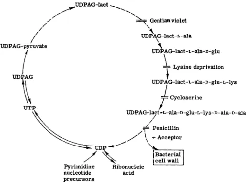
Strominger and Threnn (49) have summarized the present knowledge of the synthesis of the S. aureus wall mucopolysaccharide in Fig. 1, which has been updated to show the inhibition point of cycloserine.
Bacitracin at growth-inhibitory levels causes S. aureus to accumulate uridine phosphate iV-acetyl sugar derivatives (50). Lysis of the cells occurs at the same bacitracin concentration. Vancomycin also inhibits the synthesis of cell wall mucopeptide of S. aureus (51) and causes the accu
mulation of a material containing hexosamine, uracil, alanine, glutamic
UDPAG-lact ^
Gentian viplet
UDPAG-pyruvate
UTP
\
UDP*AG-lact-L-ala
\ Λ
UDPAG-lact-L-ala-D-glu
=y Lysine deprivation
UDPAG-lact-L-ala-D-glu-L-lys J= Cycloserine
/
UDPAG-lact-L-ala-D-glu-L-lys-D-ala-D-ala /
/
Penicillin + Acceptor
Pyrimidine nucleotide precursors
Ribonucleic acid
Bacterial cell wall
FIG. 1. Synthesis of 5 . aureus wall mucopolysaccharide. UDPAG-lact = uri
dine diphosphoacetylglucosamine lactic acid ether.
acid, and lysine (52). It will be interesting to see where these two anti
biotics fit into the scheme diagramed above.
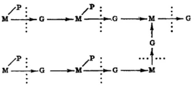
Lysozyme obtained from egg white has yielded some interesting results bearing on cell wall structure. Salton (53) found that the products obtained from the action of lysozyme on three gram-positive bacteria are fragments varying in size from disaccharides to materials with molecular weights of the order of 10,000-20,000. The disaccharide was composed probably of iV-aeetylglueosamine and acetylmuramic acid, and the larger fragments contained iV-acetylhexosamines as terminal groups. These re
sults led Salton to postulate that lysozyme was involved in breaking the amino sugar linkages. Zilliken (54) has found that among the products of lysozyme action on gram-positive bacteria are a number of amino sugars containing peptides which, on hydrolysis, yielded muramic acid, glucosamine, alanine, glutamic acid, and lysine. Ratios of muramic to glucosamine of 1:1 and 1:2 were both found. Based on work from a num
ber of laboratories, Zilliken has proposed the sequence in Fig. 2 as a possible basic structure of the bacterial cell wall.
Μ - - Μ • - Μ
Μ - - Μ
Ι
• 1 · · ·
- Μ
FIG. 2. Sequence proposed by Zilliken as a possible basic structure of the bacterial cell wall. M, muramic acid; G, iV-acetyl-D-glucosamine; P , peptide moiety; , action of lysozyme; M-»G, muraminidoglucosamine.
An early antibiotic named actinomycetin is capable of lysing killed gram-negative cells or living gram-positive cells. Actinomycetin has been shown to contain a number of enzymes capable of degrading the cell wall, including such enzymes as esterases, peptidases, and a lysozyme-like enzyme (55). Although the picture is complicated by the presence of a number of enzymes, actinomycetin has proved to be a tool in elucidating the structure of the cell wall.
The prevalence of iV-acetyl-D-glucosamine in the wall structure sug
gests that it is an important compound in cell wall synthesis. Further
more, iV-acetyl-D-glucosamine serves as a precursor of muramic acid for Lactobacillus bifidus var. pennsylvanicus. This organism has a growth requirement for ΛΓ-acetyl-D-glucosamine, preferably in the form of its β-glucosides or in the form of disaccharides such as iV-acetyllactosamine.
The Lactobacillus incorporated a,^-methyl-iV-acetyl-D-glucosaminide- 1-C1 4 directly into the muramic portion of the call wall (56). Because of the essential nature of iV-acetyl-D-glucosamine in cell wall structure, one would assume that this compound should serve as a susceptible point of antibiotic inhibition. This is the case. The antibiotic bacillin derived from Bacillus subtilis is a competitive inhibitor of N-aeetyl-D-glueosamine in greater than a tenfold range (57, 58). Even in the presence of a high con
centration of the antibiotic, normal growth of a bacterial cell occurs if the culture medium is supplemented with iV-acetylglucosamine. Separation of the antibiotic from iV-acetyl-D-glucosamine by mild chemical treat
ment resulted in restoration of the antibiotic activity.
Great progress has been made in elucidating cell wall structure through the application of the antibiotics penicillin, cycloserine, actinomycetin, and bacillin. These results promise further exciting discoveries through the detailed knowledge of the intermediates accumulating with bacitracin
and vancomycin, as well as with other y e t undiscovered antibiotics active in the cell wall biosynthesis.
B. The Cell M e m b r a n e
The bacterial membrane immediately adjacent to the cell wall is of vital importance to the cell. In large measure, the cellular enzymes are located either in or adjacent to the membrane. It is logical to assume that the membrane would be the site of action of many antibiotics.
The membrane has been studied from a crude analytical standpoint and found to consist of as much as 2 0 % phospholipid. Carbohydrates, the full complement of amino acids, and lipids have been isolated from the membrane, but no glucosamine is present. The membrane serves as an osmotic barrier and can maintain over 5 0 0 times increase in concentra- tion of glutamic acid within the cell as compared with the nutrient solu- tion external to the membrane. The internal osmotic pressure is as great as 2 0 atmospheres.
A number of antibiotics are known which have a destructive influence on the membrane. In gross effect, the action of several antibiotics on the cellular membrane is that of detergents. Tyrocidin is a cationic detergent.
It destroys the osmotic barrier property of the membrane, and amino acids leak from the cell. The amount of lysine released from the amino acid pool in the presence of 1 jug/ml tyrocidin is equivalent to that re- leased by boiling of the cells. Larger molecules, such as proteins, are not released, and study by electronmicroscopy shows that the cell wall has remained intact. Gramacidin S, another cyclic peptide, has similar activity in destroying the permeability of the membrane. Its molecular structure provides solvent-soluble groupings and water-soluble groupings which can be sterically oriented to fulfill the requirements for detergent action
(59).
A series of antibiotics isolated from Pseudomonas aeruginosa, named the pyos (60), are anionic detergents, sharing in common with chemically synthesized anionic detergents the properties of action against gram- positive microorganisms and reversibility by phospholipids and steroids.
Polymyxin also causes disruption of the cell membrane. When treated with bactericidal concentrations of polymyxin B, washed cell suspensions of sensitive bacteria release 260-m/x absorbing material in a linear relation to the percentage of cells killed. With P. aeruginosa, this loss of purines is accompanied by equimolar release of pentose and phosphate and results from breakdown of ribonucleic acid of the cells. In the presence of excess polymyxin, the enzymic degradation of ribonucleic acid is inhibited and little 260-m/i absorbing material is liberated, although amino acids,
metals, and other cytoplasmic, low molecular weight molecules may be lost.
Electron photomicrographs of cells treated with minimal concentrations of polymyxin show empty ghosts of cells that have lost intracellular material. With an excess of polymyxin, the electron-dense intracellular material remains, but a change in the surface of the cell is indicated as extraneous material agglomerates with it.
Loss of membrane permeability may be demonstrated by exposing normal and polymyxin-treated cells to A^-tolyl-a-naphthylamine-8- sulfonic acid. The dye will fluoresce in ultraviolet light when combined with the negatively charged groups of proteins. N o fluorescence is ob
served with washed cells of Pseudomonas fluorescens. Addition of poly
myxin causes immediate fluorescence as the dye penetrates into the cell.
Polymyxin does not alter the electrophoretic pattern of most cells, indicating that it is not attached to the cell surface. Calculations show that the minimum depth to which polymyxin must penetrate in order not to affect the surface change is 100 A, indicating the possibility of membrane binding.
Polymyxin will adsorb to the membrane of protoplasts made by en- zymic removal of the walls of polymyxin-sensitive gram-positive bacteria.
The point of cellular attachment may be visualized with a fluorescent complex of polymyxin. Fluorescent microscopy of 1-dimethylamino- naphthalene-5-sulfonyl chloride-polymyxin treated cells, before and after removal of the cell wall with lysozyme, shows the association of the antibiotic with the cell membrane.
The specific binding loci of polymyxin have been suggested to be the polyphosphates of the membrane. There is a close correlation between the reversal of charge of phosphate colloids by specific cations and the protection of cells against the bactericidal action of polymyxin by these same cations. This interpretation is strengthened by the observation that cephalin and lipid extracted from disrupted Pseudomonas denitrificans form strong complexes with polymyxin.
The specificity of bactericidal action of polymyxin probably depends on the composition of the bacterial surface. The cell walls of resistant bac
teria have little affinity for the antibiotic and may not allow penetration of polymyxin to the membrane. The primary difference observed in chemical composition of the wall-membrane complex of resistant gram- negative bacteria as compared to sensitive bacteria is a lower P / N ratio (61).
Streptomycin, at bactericidal concentrations, exerts a more subtle action on the cell membrane. Anand, Davis, and Armitage (62) have shown that
a rapid uptake of streptomycin occurs with Escherichia coli cells and that the antibiotic is bound to the cell by an ionic linkage. After 10 minutes, a secondary phase of uptake is initiated, and an additional three- to fourfold amount of streptomycin is bound. The secondary uptake can be prevented by chloramphenicol, but occurs very rapidly upon addition of toluene or polymyxin Β to destroy the cell membrane.
Associated with the secondary phase of binding, nucleotides and amino acids are excreted from the cell. These products arise from continued synthesis in the presence of the streptomycin, as shown by studies with isotopic carbon compounds and through quantitative measurements.
Twelve times the initial intracellular amount of adenosine monophosphate is excreted after streptomycin is added. Larger molecules, such as diphos- phopyridine nucleotide, are also excreted, but in smaller amounts. D a m age to the cell membrane is also shown by the loss of potassium ions from the cell in the presence of streptomycin (63). This effect can be detected even sooner than the loss of the other cell constituents.
Streptomycin-damaged cells are impaired in ability to concentrate valine within the cell. However, the metabolism of citrate, to which the membrane of E. coli is relatively impermeable, is increased in strepto
mycin-treated cells.
The above observations have been interpreted to indicate that the primary point of action of streptomycin is on the cell membrane and that streptomycin does not damage existing membrane, but causes defects in the new membrane synthesized by antibiotic-inhibited cells (64). The defective membrane allows the loss of intracellular components, but also permits entry of normally impermeable compounds into the cell, including streptomycin. During the entry of streptomycin through the damaged membrane, secondary binding occurs, associated with an immediate ces
sation of protein synthesis and a somewhat delayed inhibition of nucleic acid synthesis.
Further studies on those constituents of the cell membrane whose syn
thesis is blocked by streptomycin should provide identification of vital structural components of the cell. Streptomycin-resistant cells will be valuable in this research, as these cells also show the initial binding of streptomycin but do not progress to damaged membranes, indicating a change in the membrane-forming system.
A difference in the binding capacity of other microorganisms has been shown by Hancock (65) with B. megaterium, S. aureus, and B. subtilis.
With these bacteria, the initial step of membrane binding has not been found. Uptake of streptomycin parallels the increase in cell mass, and at the point of growth inhibition, over 9 5 % of the bound streptomycin is
contained in the cytoplasm in a form not easily displaced ionically. Only 1 % as much streptomycin is bound by resistant cells. Hancock's studies are in agreement with those on E. coli in the observation that actively metabolizing cells are required for binding to occur. The uptake of strep
tomycin is associated with aerobic respiratory processes.
The polyene antibiotics, which are specific in their action to the fungi, serve as interesting examples for the multiplicity of actions which may be reported in the literature before the actual primary point of attack on the cell is elucidated. These agents have been reported at low concentra
tions to stimulate endogenous respiration and glycolysis of yeasts, whereas at high concentrations respiration and glycolysis are strongly inhibited.
Further studies on isolated enzymes led to a variety of suggestions, in
cluding initiation of the destruction of the enzymes converting fructose- 1,6-diphosphate to pyruvate, release of a mechanism controlling phos
phorylation of reserve carbohydrates, or release of proteolytic enzymes which cause generalized destruction of the enzymes of the glycolytic system. Binding studies showed considerable similarity to penicillin in that sensitive organisms bound the agent, whereas resistant ones did not, and binding, once it occurred, could not be reversed by drastic solvent treatments.
Protoplasts prepared from Saccharomyces cerevisiae by enzymic re
moval of the cell wall have the same susceptibility to nystatin as do intact cells. It was observed that membranes treated with nystatin lose semipermeability to potassium. The enzymic effects of nystatin can be explained on the basis of a potassium deficiency caused by loss of potas
sium through the damaged cell membrane (66, 67). Furthermore, low levels of nystatin can be overcome by the addition of potassium ion to the fermentation medium. Potassium cannot reverse high concentrations of nystatin because leakage of other small molecules through the mem
brane occurs under these conditions.
Experiments on the mechanism of antibiotic action have not allowed elucidation of the basic structure of the microbial membrane to the same extent that studies with antibiotics have furthered our knowledge of the structure of the cell wall. These compounds do have great specificity of action, however, in their disruptive effect on the membrane. Some anti
biotics primarily attack gram-positive organisms; others destroy permea
bility of gram-negative organisms; still others destroy permeability of yeast and filamentous fungi; and antibiotics such as streptomycin cross over usual taxonomic lines. As the reasons for these specificities are un
raveled, much fundamental information should become available con
cerning the membrane structure.
C. Nucleic Acid Biosynthesis
Nucleic acid synthesis is an essential requirement for multiplication of bacterial cells, and one would assume it to be a sensitive point for attack.
This assumption is strengthened by the fact that certain abnormal purines, for example, puromycin, nucleocidin, and nebularine, can act as antibiotics. Also, purine bases will neutralize the antibiotic psicofuranine
(68).
Azaserine has a direct effect on the synthesis of purines and confirms a biosynthetic scheme derived by other methods. Azaserine blocks the incorporation of labeled carbon from formate-C1 4, glycine-l-C1 4, or β-serine-C1 4 into nucleic acid, but incorporation into only the purines is influenced, not the pyrimidines. The subsequent conversion of purine to nucleic acid is not prevented. Formation of inosinic acid is blocked, and formylglycinamide ribotide and formylglycinamide riboside accumu
late in the blocked reaction. Azaserine inhibition of bacterial growth is competitively overcome by glutamine. The reaction prevented by the antibiotic is formylglycinamide ribotide -f- glutamine + A T P -» formyl- glycinamidine ribotide + A D P -f- glutamic acid + orthophosphate.
Azaserine competes with glutamine for the enzyme site, and the combina
tion enzyme-azaserine is irreversible (69). Azaserine also inhibits the other reactions in the de novo synthesis of purines involving glutamine, but much higher concentrations of the antibiotic are required. These other sites of action are the formation of 5-phosphoribosylamine from 5-phos- phoribosyl pyrophosphate and glutamine and the formation of guanylic acid from xanthylic acid and glutamine (70).
The antibiotic hadacidin inhibits another reaction in purine biosyn
thesis, namely, the formation of adenylosuccinic acid from inosinic acid and aspartic acid. The inhibition by hadacidin is competitively reversed by L-aspartate (71).
Recently, actinomycin has assumed considerable importance as a bio
chemical tool through its ability to inhibit ribonucleic acid synthesis that is directed by deoxyribonucleic acid. Actinomycin forms a complex with deoxyribonucleic acid, which can be readily dissociated, and it is thought that this is the method by which actinomycin acts (72, 73). The addition of actinomycin to growing cells of S. aureus inhibited ribonucleic acid and protein synthesis, and this inhibition was antagonized by deoxyribonucleic acid (78). A cell-free system capable of synthesizing ribonucleic acid from the four ribonucleoside triphosphates in the presence of deoxyribonucleic acid was inhibited by actinomycin, and the addition of excess deoxyribo
nucleic acid antagonized this inhibition (74). On the other hand, Reich
et al. (75) found that deoxyribonucleic acid synthesis was not inhibited in mammalian L-cells by concentrations of actinomycin which appreciably inhibited ribonucleic acid synthesis. These authors also showed that Mengo virus, a ribonucleic acid virus, was not inhibited by 0.005 ftg actinomycin per milliliter. They concluded that they were able to dif
ferentiate ribonucleic acid-directed synthesis of new ribonucleic acid, which is resistant to actinomycin, from deoxyribonucleic acid-directed synthesis of ribonucleic acid, which is sensitive to actinomycin. Recently it has been found that actinomycin is bound to deoxyribonucleic acid through the guanine residues of the nucleic acid, and the degree of in
hibition of ribonucleic acid-polymerase reaction is a function of the guanine content of the deoxyribonucleic acid primer (76).
D. Protein Synthesis
Protein synthesis is clearly associated with nucleic acids, and lack of protein synthesis may be a secondary effect of inhibition of nucleic acid synthesis. The clearest demonstration of the action of an antibiotic on protein synthesis is the inhibition produced by chloramphenicol. This antibiotic has become a useful tool in the study of protein synthesis in bacterial, plant, and animal systems, as is shown by the rapid accumula
tion of literature on the subject. Some recent reviews dealing with protein synthesis have been written by Roberts et al. (77), Novelli (78), Jacob and Monod (79), and Simpson (80).
The available evidence suggests that there are at least three stages involved in protein synthesis. The first stage is the enzymic activation of the amino acids in the presence of adenosine triphosphate to form enzyme- adenyl-amino acid complexes. The second stage is the transfer of the activated amino acids to ribonucleic acid-amino acid complexes. These ribonucleic acids are referred to as transfer (soluble) ribonucleic acids and comprise about 1 0 - 1 5 % of the ribonucleic acid of the cell. The third stage involves the transfer of the amino acids from the transfer ribonu
cleic acids to protein. It is now thought that this transfer is directed by messenger ribonucleic acid (79) which serves as a template for the pro
tein. The messenger ribonucleic acid, which comprises less than 5 % of the cellular ribonucleic acid, has a nucleotide composition reflecting the base composition of the deoxyribonucleic acid of the cell and may have a rapid turnover rate. The instability and small amount of the messenger ribonucleic acid has made its detection extremely difficult, and it has only been revealed by some extremely elegant experiments.
The relationship of chloramphenicol to protein synthesis was indicated
by the ability of the antibiotic to block adaptive enzyme formation in E. coli (81). Growth-inhibitory levels of chloramphenicol depressed pro- tein synthesis by 90% in washed suspensions of S. aureus (82). The same levels of chloramphenicol had no effect on respiration or the accumulation of free intracellular glutamic acid, and the rate of formation of nucleic acid was increased. The effect is reversible, and protein synthesis resumes immediately upon removal of the chloramphenicol.
The first stage of protein synthesis, the activation of the amino acids, is not affected by chloramphenicol (83, 84); nor is the second stage, the binding of the amino acids to the transfer ribonucleic acids (85). How- ever, chloramphenicol does inhibit the synthesis of protein by cell-free extracts of E. coli (86) suggesting that the antibiotic interferes with the transfer of the amino acids from the transfer ribonucleic acids to protein.
Experimental evidence for this site of action has been presented by Nathans and Lipmann (87). Chloramphenicol does not inhibit protein synthesis by cell-free extracts of rat liver (88) or rabbit reticulocytes (89). Relevant to these findings is the observation that the E. coli enzyme system which transfers the amino acids from the transfer ribonucleic acids to the ribosomes is specific for E. coli ribosomes, and the transfer enzyme system from liver is specific for liver ribosomes (87).
Several workers have observed that the ribonucleic acid formed in the presence of chloramphenicol is rapidly broken down when the chloram- phenicol is removed (90). Aronson and Spiegelman (91) have indicated that the unstable ribonucleic acid formed in the presence of chloram- phenicol is a normal constituent of the uninhibited cell, and Kurland, Nomura, and Watson (92) have found that the ribosomes made during chloramphenicol inhibition contain the same ribonucleic acid types as normal ribosomes. The principal difference between the two ribosomes is the smaller amount of protein in the inhibited ribosomes. Hahn and Wolfe (98) have shown that the ribonucleic acid which accumulates in the presence of chloramphenicol has the characteristic instability and base composition of messenger ribonucleic acid and suggest that the anti- biotic interferes with the function of the messenger ribonucleic acid.
The subject of protein synthesis is under intensive investigation at the present time, and it is certain that our knowledge of the mechanisms involved and the nature of the intermediates will be rapidly expanded.
The action of chloramphenicol depends on its structural configuration.
The i,(-\-)-erythro stereoisomer of chloramphenicol has less growth- inhibitory ability than the natural D ( —) - t h r e o form and does not affect protein synthesis. However, the h(-\-)-erythro isomer does inhibit the synthesis of D-glutamic acid polypeptide by B. subtilis (94).
Several antibiotics other than chloramphenicol have been implicated in protein synthesis, although none of them has been the subject of as much work as chloramphenicol. Chlortetracycline and oxytetracycline inhibited protein synthesis in washed suspensions of S. aureus at growth-inhibitory levels (82). There was no effect on nucleic acid synthesis, glucose fermen
tation, or free intracellular glutamic acid accumulation until the concen
trations were increased to much higher levels. Chlortetracycline also inhibits cell wall synthesis in growing cells of S. aureus (50), but again at higher levels that it inhibits protein synthesis. Chloramphenicol has no effect on cell wall synthesis (95, 96).
Erythromycin inhibits protein synthesis but not nucleic acid synthesis (97). Several other antibiotics have been observed to inhibit protein synthesis. A partial list of these antibiotics includes streptomycin (98), dihydrostreptomycin (99), and puromycin (100), all of which have been shown to inhibit amino acid incorporation into protein. Cyclohexamide
(101) j gliotoxin (102), and viomycin (103) have been shown to inhibit protein and nucleic acid synthesis. Careful study of the mode of action of a number of antibiotics including those listed above should contribute materially to our knowledge of protein synthesis.
E. Uncoupling Agents; Respiration Inhibitors
Energy-generating systems are necessary to drive the reactions of pro
tein synthesis and nucleic acid synthesis in the cell. Any uncoupling agent, therefore, should block cell development. Likewise, any inhibitor of the respiratory chain will prevent release of energy for synthetic action.
The antifungal agent antimycin is an excellent example of an antibiotic that is too toxic for clinical applications but has found a place as a power
ful tool for the biochemist (104). It was first observed that antimycin in concentrations of less than 0.1 /Ag/ml inhibited oxygen uptake by yeast while carbon dioxide evolution was increased, and it was soon found that antimycin was inhibiting an essential component of the electron transport system. The exact point of attack was not clear until the discovery of the coenzyme Q system as an intermediary in the electron transport chain between succinic dehydrogenase and cytochrome c^ The reduction of coenzyme Q is not affected by antimycin but its reoxidation is inhibited by the antibiotic (105,106).
Tappel (107) has drawn some interesting activity-structure relation
ships with antimycin, showing that the presence of chelating groups and a lyophilic group are essential for the antimycin activity. Coenzyme Q is associated with lipid fractions of mammalian mitochondria (108).
Umbreit and his associates found that streptomycin inhibited the oxida- tion of pyruvate and oxalacetate by E. coli cells grown under anaerobic conditions {109). None of the known reactions involved in the oxidation of these compounds was inhibited by streptomycin. Umbreit {110) found that streptomycin blocked the uptake of radioactive phosphorus into a compound which was chromatographically similar to the 7-carbon com- pound 2-phospho-4-hydroxy-4-carboxyadipic acid and proposed that this material was an intermediate in an alternative streptomycin-sensitive pathway for pyruvate oxidation. It is doubtful that these results can be the explanation for the primary site of streptomycin action. There is a lack of correlation between the rate of killing of E. coli and inhibition of respiration by streptomycin {111), and the main oxidative pathway in E. coli proceeds through the oxidation of citrate which is insensitive to streptomycin.
F. Specific Enzyme Effects
Direct inhibition by antibiotics of specific enzymic reactions essential to the cell have been described and can be of great significance to bio- chemists. The action of D-cycloserine on alanine racemase and D-alanyl- D-alanine synthesis has been described in the section on the cell wall.
Cycloserine as produced by streptomycetes is the D-isomer. Synthetic L-cycloserine also behaves as an inhibitor of microbial growth but does not cause accumulation of the UDP-muramic acid peptide of the cell wall. I t is competitively neutralized by L-alanine rather than D-alanine and, at the enzymic level, the specific enzymic process inhibited is the transaminase between L-alanine, pyruvate, glutamate, and a-ketoglutarate {112). These findings at the enzyme level with D - and L-cycloserine point to the specificity of antibiotics for individual enzyme systems. Similar detailed knowledge of enzyme effects should be the end result of investi- gations of the mode of action of other antibiotics.
G . Vitamin Interrelationships
Vitamins are important as cofactors in many metabolic pathways, and excess synthesis of vitamin antagonists by microorganisms should result in a typical antibiotic response. There are few instances of clear-cut com- petitive relationships, however. The competitive relationship between biotin and actithiazic acid (acidomycin, mycobacidin) has led to the suggestion that the antibiotic blocks biotin synthesis {25, 113).
Foster and Pittillo have demonstrated that, at threshold concentrations of antibiotic in a chemically defined agar medium, physical isolation of each cell can be accomplished and complications due to resistance devel
opment or physiological factors are eliminated. Under this condition, many antibiotics may be reversed over a range of two to several fold by complex mixtures of nutrients. Purines, pyrimidines, and amino acids more commonly were the reversing agents, but in some instances vitamins served as neutralizers for antibiotics. Riboflavin not only neutralized the antibacterial activity of chlortetracycline over a sixteenfold range, but riboflavin synthesis was markedly depressed in cells partially inhibited by chlortetracycline (114).
H. Chelation
Numerous instances of metal effects in enzymic reactions have been reported, and it has been proposed that any organic compound capable of forming a stable organometallic complex is a potential enzymic inhibi
tor and a suitable substance for chemotherapeutic research. Streptomycin, aspergillic acid, usnic acid, and the tetracyclines all have an affinity for metallic cations and in many instances may be considered to serve as direct chelating agents. Other inhibitory compounds, especially antibiotics derived from molds, are known to have chelating properties.
Several routes whereby these chelating agents may exert their antibiotic activity have been suggested. For example, the compounds may inhibit microorganisms by depriving the cells of essential cations, or they may inhibit microorganisms in the form of the specific metal chelates. The antibiotic activity of the former compounds is suppressed by excess con
centrations of the cation. The activity of the latter toxic antibiotic-chelate complexes can be neutralized by certain metallic cations which protect susceptible protoplasmic groups from the toxic chelate (115). Tetracycline is an example of an antibiotic which is more toxic to cells in a chelated form.
Metal ions may have a more generalized effect on antibiotic activity.
The attachment of an antibiotic to the microbial cell may be suppressed by metal-binding agents and enhanced by metallic cations, for example, penicillin; or the antibiotic may be suppressed by metallic cations and enhanced by metal-binding agents, for example streptomycin and poly
myxin.
The importance of the metal-antibiotic relationship is highlighted by studies of Saz et al. (116), who have shown that cell-free nitroreductase obtained from resistant cells is not inhibited by chlortetracycline, whereas
the enzyme from sensitive cells is inhibited. It appears that the resistant cells produce altered enzymes which, in contrast to the sensitive enzymes, contain firmly bound, conjugated proflavin capable of competing success
fully with chlortetracycline for the essential metallic cations.
Grisein and related antibiotics called sideromycins are examples of antibiotics which prevent access of cell enzymes to the required metal cofactor iron. In this instance, however, the antibiotics do not exert their effect by direct competition for iron but apparently prevent iron from reaching the cytoplasm. The antibiotics are most active when chelated with iron and act competitively with iron transport complexes, variously called ferrichrome, copragen, terregens factor, or sideramines (117). It is assumed that the sideromycins are competitive with the transport factors for binding with the specific transferases in the cell membrane.
VIII. CONCLUSION
The few examples of antibiotic activity discussed in detail indicate the complexity of conducting biochemical research with antibiotics and the complications imposed by the variety of responses reported from the action of antibiotics on the intact cells. While much spadework and many independent observations are required to accumulate a series of effects of the antibiotics, one cannot minimize the thrill which the antibiotic special
ist receives from the successful dovetailing of pieces in a metabolic path
way through research on antibiotics, such as has been accomplished from studies on cell wall synthesis. With over 2000 antibiotics available as biochemical tools, we should clearly anticipate further significant ad
vances in our knowledge of biochemical pathways as the modes of action of these abnormal biological molecules become known.
REFERENCES
1. S. A. Waksman, Am. Scientist 41, 8 (1953).
2. W. F. Verwey, Ann. Rev. Microbiol. 13, 177 (1959).
3. Ε. B. Chain, Ann. Rev. Biochem. 27, 167 (1958).
4. F. E . Hahn, Proc. 4th Intern. Congr. Biochem., Vienna, 1958 5, Sym
posium 5, p. 104. Pergamon Press, N e w York, 1959.
5. S. T. Cowan and E. Rowatt, eds., "The Strategy of Chemotherapy," Sym
posium Soc. Gen. Microbiol., No. 8. Cambridge Univ. Press, London and N e w York, 1958.
6. O. Gsell, "Antibiotica et Chemotherapia," Vol. 10. Karger, Basel, Switzer
land, 1962.
7. H. S. Goldberg, ed., "Antibiotics, Their Chemistry and Non-medical Uses." Van Nostrand, Princeton, N e w Jersey, 1959.