THE
JOURNAL • RESEARCH • www.fasebj.org
Suppression of AMPK/aak-2 by NRF2/SKN-1 down- regulates autophagy during prolonged oxidative stress
Monika Kosztelnik,*,1 Anita Kurucz,†,1Diana Papp,*,2 Emily Jones,‡,§Timea Sigmond,* Janos Barna,*
Maria H. Traka,{Tamas Lorincz,kAndras Szarka,kGabor Banhegyi,†,#Tibor Vellai,* Tamas Korcsmaros,*,‡,§,3 and Orsolya Kapuy†,4
*Department of Genetics, E ¨otv ¨os Lor´and University, Budapest, Hungary;†Department of Medical Chemistry, Molecular Biology and Pathobiochemistry, and#Pathobiochemistry Research Group, Hungarian Academy of Sciences, Semmelweis University, Budapest, Hungary;
‡Gut Health and Microbes and{Food Innovation and Health, Quadram Institute, United Kingdom;§Earlham Institute, Norwich, United Kingdom; andkLaboratory of Biochemistry and Molecular Biology, Department of Applied Biotechnology and Food Science, Budapest University of Technology and Economics, Budapest, Hungary
ABSTRACT:NF-E2–related factor 2 (NRF2) transcription factor has a fundamental role in cell homeostasis mainte- nance as one of the master regulators of oxidative and electrophilic stress responses. Previous studies have shown that a regulatory connection exists between NRF2 and autophagy during reactive oxygen species–generated oxi- dative stress. The aim of the present study was to investigate how autophagy is turned off during prolonged oxidative stress, to avoid overeating and destruction of essential cellular components. AMPK is a key cellular energy sensor highly conserved in eukaryotic organisms, and it has an essential role in autophagy activation at various stress events. Here the role of human AMPK and its Caenorhabditis elegans counterpart AAK-2 was explored upon oxidative stress. We investigated the regulatory connection between NRF2 and AMPK during oxidative stress induced bytert-butyl hydroperoxide (TBHP) in HEK293T cells andC. elegans. Putative conserved NRF2/protein skinhead-1 binding sites were found inAMPK/aak-2genes byin silicoanalysis and were later confirmed experi- mentally by using EMSA. After addition of TBHP, NRF2 and AMPK showed a quick activation; AMPK was later down-regulated, however, while NRF2 level remained high. Autophagosome formation and Unc-51–like autophagy activating kinase 1 phosphorylation were initially stimulated, but they returned to basal values after 4 h of TBHP treatment. The silencing of NRF2 resulted in a constant activation of AMPK leading to hyperactivation of autophagy during oxidative stress. We observed the same effects inC. elegansdemonstrating the conservation of this self- defense mechanism to save cells from hyperactivated autophagy upon prolonged oxidative stress. We conclude that NRF2 negatively regulates autophagy through delayed down-regulation of the expression of AMPK upon pro- longed oxidative stress. This regulatory connection between NRF2 and AMPK may have an important role in understanding how autophagy is regulated in chronic human morbidities characterized by oxidative stress, such as neurodegenerative diseases, certain cancer types, and in metabolic diseases.—Kosztelnik, M., Kurucz, A., Papp, D., Jones, E., Sigmond, T., Barna, J., Traka, M. H., Lorincz, T., Szarka, A., Banhegyi, G., Vellai, T., Korcsmaros, T., Kapuy, O. Suppression of AMPK/aak-2by NRF2/SKN-1 down-regulates autophagy during prolonged oxidative stress.
FASEB J. 33, 000–000 (2019). www.fasebj.org
KEY WORDS:AMPK • prolonged stress • aging • C. elegans • network
ABBREVIATIONS:aak-2,Caenorhabditis elegansortholog of AMP-activated protein kinase; ARE, antioxidant response element;atg-11, autophagy-related protein 11; BOD, Bodipy-[11C]; DCF, dichlorofluorescein-diacetate; GAPDH, glyceraldehyde-3-phosphate dehydrogenase; GFP, green fluorescent protein; HO-1, heme oxygenase 1; KEAP1, Kelch-like ECH-associated protein 1; LC3-II, microtubule-associated protein 1A/1B-light chain 3; LGG-1, Caenorhabditis elegansortholog of mammalian LC3 protein; mTOR, mammalian target of rapamycin; NGM, nematode growth medium; NQO1, NAD(P)H:
quinone oxidoreductase 1; NRF2, NF-E2–related factor 2; qPCR, quantitative PCR; RNAi, RNA interference; ROS, reactive oxygen species; siRNA, small interfering RNA; SKN-1, protein skinhead-1; TBHP,tert-butyl hydroperoxide; ULK1, Unc-51–like autophagy activating kinase 1
1These authors contributed equally to this work.
2Current affiliation: John Innes Centre, Norwich, United Kingdom.
3Correspondence: Earlham Institute, Norwich Research Park, Norwich NR4 7UZ, United Kingdom. E-mail: tamas.korcsmaros@earlham.ac.uk
4Correspondence: Department of Medical Chemistry, Molecular Biology and Pathobiochemistry, Semmelweis University, T}uzolt ´o utca 37-47, 1094 Budapest, Hungary. E-mail: kapuy.orsolya@med.semmelweis-univ.huu
This is an Open Access article distributed under the terms of the Creative Commons Attribution 4.0 International (CC BY 4.0) (http://creativecommons.
org/licenses/by/4.0/) which permits unrestricted use, distribution, and reproduction in any medium, provided the original work is properly cited.
doi: 10.1096/fj.201800565RR
This article includes supplemental data. Please visithttp://www.fasebj.orgto obtain this information.
Oxidative stress is an inevitable and often disadvanta- geous condition affecting essentially all living organisms.
It can arise from the imbalance of the antioxidant defense mechanisms and the reactive oxygen species (ROS) such as hydroxyl•OH, superoxide O2•–, and hydrogen peroxide H2O2, and reactive nitrogen species such as nitric mon- oxide •NO (1). These highly reactive molecules can be produced endogenously (e.g., in mitochondria, peroxi- some, cytosol) or generated due to exposure to numerous exogenous agents (e.g., infrared, UV, cytokines, growth factors, drugs, toxins, high temperature) (2). By taking electrons from macromolecules, ROS can generate harm- ful chemical processes leading to alteration and damage of cell structure, or even to cell death (3). If oxidative stress–
induced defense mechanisms are not sufficient, or the stress is prolonged, it leads to many pathophysiologic conditions (atherosclerosis, cancer, diabetes, cardiovascu- lar diseases, chronic inflammation, stroke, and neurode- generative diseases such as Parkinson’s or Alzheimer’s disease) (4). If cells cannot recover by eliminating the harmful effects of oxidative stress, programmed cell death (i.e., apoptosis, necrosis, autophagic cell death) can occur to maintain the functional integrity of the affected tissue (1).
The major transcriptional regulator of the antioxidant stress response in animals is NF-E2–related factor 2 [NRF2 (NFE2L2)] (5, 6). NRF2 regulates the cellular antioxidant response mechanism by modulating the expression of hundreds of cytoprotective genes with antioxidant re- sponse elements (AREs) in their promoters, including antioxidant enzymes, phase II detoxifying enzymes, xe- nobiotic transporters, and other stress response proteins (5, 7). At physiologic conditions, NRF2 has some basal level, but it is kept inactive by binding to a stoichiometric inhibitor labeled Kelch-like ECH-associated protein 1 (KEAP1) and targeted for ubiquitination (8, 9). Various oxidative agents result in conformational changes of KEAP1, thereby blocking NRF2 degradation. InCaenorhabditis ele- gans, the NRF2 protein is represented by the 3 currently known isoforms of the protein skinhead-1 (SKN-1).
Although NRF2 works as a dimer, SKN-1 binds to the DNA in a unique monomeric form (5, 10). SKN-1 func- tions similarly to NRF2 in response to oxidative stress and is required for oxidative stress resistance and longevity in C. elegans. NRF2 also up-regulates other processes, in- cluding a cellular self-digesting pathway called auto- phagy (11). During oxidative stress, autophagy is a key element of cellular homeostasis that eliminates harmful agents from the cytosol such as protein aggregates and damaged mitochondria, and it thereby limits ROS pro- duction and restores oxidative balance. It also has an important role in guaranteeing the proper functions of the cell by self-digesting the dispensable elements of the cell during starvation. The focus of the present study was on the regulation of the most well-known type of autophagy, namely (macro)autophagy (12).
The conjugated form of the microtubule-associated protein 1A/1B-light chain 3 (LC3-II) is recruited to auto- phagosomal membranes, and the LC3-II level thus directly estimates the abundance of autophagosomes before lyso- somal digestion. Because it reflects the autophagic activity, detecting LC3-II levels is a reliable and well-characterized
method for monitoring autophagy flux and autophagy- related processes (13). The p62 protein binds both LC3 and unfavorable proteins; therefore, p62-targeted proteins are selectively transferred to the autophagosome and can be removed by autophagy (14, 15). In the subsequent degra- dation, both p62 and its substrate are destroyed; conse- quently, the p62 level quickly drops during intensive autophagy (16, 17). Blocking of autophagy results in an increase in cellular p62 level, and this feature is therefore used to detect dysfunctional autophagy (18, 19).
The induction of autophagosome formation is con- trolled by the Unc-51–like autophagy activating kinase 1 (ULK1) complex, containing ULK1, ATG13, and FIP200 (20). ULK1 activity is tightly regulated by the nutrient and energy sensors of cellular homeostasis, mammalian target of rapamycin (mTOR), and AMPK, respectively (21). Be- cause mTOR is fully active at physiologic conditions to maintain general protein synthesis in the cell, mTOR is a key negative regulator of the initial steps of autophagy (22). However, AMPK becomes activated when the ATP:
ADP ratio is dropping, and it induces autophagy. AMPK activation in turn inhibits mTOR, and AMPK can also in- duce ULK1 activity directly (through phosphorylation of Ser317, Ser555, and Ser777) (21, 23, 24).
The most relevant connection of autophagy and oxi- dative stress response mechanism is achievedviaNRF2.
Namely, activation of NRF2 occurs by the NRF2- dependent transcription of the ubiquitin-binding and autophagosome cargo protein p62. p62 quickly binds the NRF2 inhibitor (KEAP1) upon oxidative stress, resulting in the fast activation of the transcription factor (25). Recently, active NRF2 was found to bind to the ARE sequence of various autophagy regulators (e.g., Sqstm1/p62, ATG4, ATG5, ATG7, Gabarapl1), suggesting that NRF2 has a positive effect on autophagy (26). In addition, AMPK can also stimulate NRF2 signaling upon oxidative stress (21, 27). AMPK directly phosphorylates NRF2 at Ser550, pro- moting nuclear accumulation of NRF2 for ARE-mediated gene transcription (28). AMPK also induces the anti- oxidative heme oxygenase (HO-1) gene expression in hu- man vascular cells and rat arteries via the NRF2/ARE pathway (29). InC. elegans, metformin activates AAK-2 (the worm ortholog of AMPK) to promote SKN-1/NRF2 nuclear translocation (30). Although several papers have shown the importance of autophagy activation upon oxidative stress, oftenviaNRF2 (reviewed in refs. 31–33), how autophagy is down-regulated when oxidative stress is already balanced to avoid overeating and destruction of further and essential cellular components has not yet been explored.
Previously, genetic studies found that autophagy down-regulation could indeed be beneficial upon pro- longed oxidative stress, and, interestingly, this survival mechanism involves NRF2. Anthocyanins from Chinese bayberry extract, while activating NRF2 inbcells, were found to negatively regulate oxidative stress–induced autophagy, and this outcome provided a protective effect after the transplantation of rat b cells under the renal capsules of the recipient mice (34). In an- other study, NRF2 was induced by an anticancer redox agent, mitoquinone, and the direct involvement of NRF2 in autophagy down-regulation was confirmed
with genetic studies (35). Similarly, in mice,NRF2de- ficiency contributed to autophagy deregulation upon age-related retinopathy, implying the importance of NRF2 regulation of autophagy upon prolonged oxida- tive stress (36). In cardiomyocytes exposed to pro- longed oxidative stress, AMPK levels were found to be decreased, resulting in autophagy inhibition (37).
Down-regulation of autophagy in these studies was not detected when NRF2 activity was compromised, in- dicating a direct role for NRF2 in autophagy inhibition.
Importantly, autophagy down-regulation in these studies was not observed when oxidative stress was short term, explaining why most of the previous oxidative stress–
related experiments focusing on NRF2 and autophagy could not find this negative regulatory effect. To our knowledge, none of the published works provided a molecular mechanism for how NRF2 could down- regulate autophagy. Identifying which genes and how they are involved in the adaptation to prolonged oxida- tive stress will contribute to our understanding of anti- oxidant stress response and the pathogenesis of related diseases, including cardiovascular disease and cancer, as well as aging.
Prompted by these questions, in the present study we investigated the relationship between NRF2 and auto- phagy. Using computational regulatory network analysis, we identified a conserved NRF2 binding site in the pro- moter region of AMPK. We validated the functional role of this binding site, and also the critical effect of NRF2 in autophagy down-regulation, both in human cell lines and in the model organismC. elegans. Our results uncovered the mechanism of how NRF2 inhibits autophagy hyper- activation upon prolonged oxidative stress.
MATERIALS AND METHODS Cell culture and oxidative treatment
Human embryonic kidney (HEK293T) and human colonic (Caco- 2) cells were cultured in DMEM (41965039; Thermo Fisher Sci- entific, Waltham, MA, USA) supplemented with 10% fetal bovine serum (10500064; Thermo Fisher Scientific) and 1% antibiotics/
antimycotics (15240062; Thermo Fisher Scientific). This cell line was maintained in adherent culture in a humidified incubator at 37°C in 95% air and 5% CO2. To simulate oxidative stress, LuperosTBH70x, tert-butyl hydroperoxide (TBHP) solution (458139; MilliporeSigma, Burlington, MA, USA) was used for different lengths of time (i.e., 0.5, 1, 2, and 3 h).
Immunofluorescence
HEK293T cells (73104) were seeded on glass coverslips (ECN 631–1577; VWR International, Radnor, PA, USA). Cells were washed in PBS, fixed with ice-cold 100% methanol for 10 min, and washed again. Coverslips were blocked for 30 min with PBS buffer with 0.1% Tween-20 containing 3% bovine serum albumin (A 9647; MilliporeSigma) and then incubated overnight at 4°C with primary antibody (LC3 A/B, 4108S; Cell Signaling Tech- nology, Danvers, MA, USA) at 1:200 dilution in 1% bovine serum albumin containing PBS buffer with 0.1% Tween-20. Samples were washed with PBS and then incubated for 1 h at room tem- perature with Alexa Fluor 488 (4412S; Cell Signaling Technology)
secondary antibody at 1:600 dilution. Cells were washed with PBS and treated with DAPI at 1:1000 dilution in PBS for 10 min, followed by wash with PBS. Coverslips were mounted by using FluorSave Reagent (345789; MilliporeSigma) and visualized by using a Zeiss LSM 710 laser confocal microscope (Carl Zeiss Microscopy GmbH, Jena, Germany). Three images per given conditions were taken. All data are expressed as means6SEM. Comparisons between groups were made by using ANOVA with Tukey’s multiple comparisonpost hoctest. Statistical sig- nificance was evaluated, and values ofP,0.05 were considered significant.
Quantification of mRNA
Total RNA was isolated from HEK293T cells at 3 d of cultivation by using Trizol reagent (Thermo Fisher Scientific). RNA was re- verse transcribed by using the SuperScript II First-Strand Syn- thesis System (Thermo Fisher Scientific). Quantification ofNRF2 andAMPKmRNA was performed by using the GoTaq Quanti- tative PCR (qPCR) Master Mix (A6001; Promega, Madison, WI, USA). Forward and reverse primer sequences used for qPCR were as follows: NRF2: 59-TCCAGTCAGAAACCAGTGGAT- 39and 59-GAATGTCTGCGCCAAAAGCTG-39; AMPK: 59- ACTGTACCAGGTCATCAGTACACC-39and 59-TCCAGGTA- CATCAGATTTCCTTC-39; andGAPDH: 59-TGCACCACCAA- CTGCTTAGC-39and 59-GGCATGGACTGTGGTCATGAG-39.
The PCR thermal program included the following: 10 min at 95°C (1 cycle), and 30 s at 95°C, 45 s at 58°C, 30 s at 72°C (40 cycles), 5 min at 95°C, 1 min at 55°C, 30 s at 97°C (1 cycle).
Measures were performed on an Mx3005P qPCR System (Stratagene, San Diego, CA, USA). Melting curve analysis was performed to confirm correct PCR product size and absence of nonspecific bands. Relative mRNA levels were determined by normalizing the PCR threshold cycle number of NRF2and AMPKwith that of theGAPDHreference gene. Normalized data were analyzed by using independent 2-sample Student’s t tests with Bonferroni correction (SPSS v.20 software; IBM, Armonk, NY, USA).
RNA interference in HEK293T cells
In the case of human cells, 1 d before the transfection, 2.53105 HEK293T cells were plated in 2.5 ml growth medium without antibiotics. Duplex RNA to targeting human NRF2 [sense 59-GGUUGAGACUACCAUGGUU(dTT)-39antisense 59-AAC- CAUGGUAGUCUCAACC(dTT)]-39was transfected into cells at 50–60% confluency. Small interfering RNA (siRNA) and Lipo- fectamine RNAiMax were diluted into Opti-MEM I (GlutaMax-I) in accordance with the manufacturer’s protocol (Thermo Fisher Scientific). siRNA duplex-Lipofectamine RNAiMax complexes were added to each well. Cells were assessed at 24 h after transfection by using real-time PCR. For negative control scramble, siRNA was used.
Western blot analysis
Treated HEK293T or Caco-2 cells were collected and lysed by using RIPA buffer (20 mM Tris, 135 mM NaCl, 10% glycerol, 1%
NP40, pH 6.8). Protein concentrations were measured by using Pierce BCA Protein Assay (Thermo Fisher Scientific, 23225).
Equal amounts of protein were separated on a 10–15% SDS- PAGE gel and transferred to PVDF membrane (0.45; Milli- poreSigma). Membranes were blocked with 5% nonfat dry milk or 1% bovine serum albumin in Tris-buffered saline, 0.1% Tween for 1 h followed by overnight incubation at 4°C with primary antibodies in 1% milk or 1% bovine serum albumin. The antibody
sources are as follows: phospho-AMPKa Thr172 (2531S; Cell Signaling Technology), AMPKa(23A3; Cell Signaling Technol- ogy), NAD(P)H:quinone oxidoreductase 1 (NQO1) (A180; Cell Signaling Technology), anti-LC3b (sc-16755; Santa Cruz Bio- technology, Dallas, TX, USA), 4EBP1P (9459S; Cell Signaling Technology), phospho-ULK1 Ser555 (5869S; Cell Signaling Technology), ULK1 (8054S; Cell Signaling Technology), anti–glyceraldehyde-3-phosphate dehydrogenase (GAPDH) (sc-32233; Santa Cruz Biotechnology), HO-1 (D60G11; Cell Signaling Technology), NRF2 (C15410242; Diagenode, Den- ville, NJ, USA), and horseradish peroxidase conjugated sec- ondary antibodies (sc-2020, sc-2005, Santa Cruz Biotechnology;
7074S, 7076S, Cell Signaling Technology). To detect both the phosphorylated and total forms of the same proteins, equal amounts of samples were blotted onto different PVDF mem- branes. Cross-reactivity and protein removal problems were avoided by using this approach. Each experiment was repeated 3 times. The quantification of Western blot bands was con- ducted on ImageQuant 5.2 software (GE Healthcare Life Sciences, Little Chalfont, United Kingdom). Relative band densities were normalized to appropriate total protein or GAPDH used as ref- erence protein. The average of 3 independent measurements was calculated. All data are expressed as means6SEM. Comparisons between groups were made by using ANOVA with Tukey’s multiple comparison post hoc test. Statistical significance was evaluated, and values ofP,0.05 were considered significant.
Measurement of oxidative stress by flow cytometry
HEK cells were seeded on 6-well plates and treated as indi- cated. After treatment, cells were trypsinized, washed once with PBS, and 0.5 3106 cells/ml were labeled with 10mM dichlorofluorescein-diacetate (DCF) or 2mM Bodipy-[11C] (BOD) for 30 min in HBSS at 37°C. After labeling, cells were washed once with PBS, resuspended in PBS, and analyzed with a BD FACS- Calibur flow cytometer (BD Biosciences, Franklin Lakes, NJ, USA). The fluorescence of DCF and BOD from excitation with a 488 nm laser was recorded in FL1 (530 nm). Cell population was gated through the SSC-FSC scatter plot by using unlabeled cells, and histograms normalized to the samples’ mode were con- structed by using FlowJo software (FlowJo, Ashland, OR, USA). A minimum of 10,000 cells were analyzed per condition.
Nuclear extracts and 2-color fluorescence EMSA
Nuclear extracts were prepared from Caco-2 cells treated with 100mM TBHP for 4 h, using the Nuclear Extraction Kit (Abcam, Cambridge, MA, USA) according to the manufacturer’s instruc- tions. Single-stranded oligonucleotide probes containing either AMPK query Nrf2 binding site (59-AAACCATGACTCTGCA- TAAAA-39 and 59-GATCTTTTATGCNNNGTCAATG-39) or AMPK NRF2 consensus binding site (59-GATCTTTTATGC- TGTGTCATGG-39 and 59-AAACCATGACACAGCATAAAA- 39) were manufactured by Integrated DNA Technologies (Coralville, IA, USA) and annealed at 95°C for 5 min, then cooled slowly to room temperature for several hours. Gel shift analysis was conducted by using the EMSA kit with SYBR Green and SYPRO Ruby (Thermo Fisher Scientific) according to the manufacturer’s instructions. Briefly, binding reactions were performed in a total volume of 10ml containing 5mg of nuclear extract, 1 ng of DNA probe, and 5 times binding buffer (750 mM KCl, 0.5 mM DTT, 0.5 mM EDTA, 50 mM Tris, pH 7.4) for 20 min at room temperature. In supershift experiments, nuclear extracts were preincubated with 2mg of anti-Nrf2 antibody (sc365949-X;
Santa Cruz Biotechnology) for 30 min before the addition of the oligonucleotide probe. At the end of the incubation period,
63gel-loading solution was added to the binding reaction and protein–probe complexes separated by electrophoresis on a nondenaturing Novex 4–20% Tris-Glycine Mini Gel (Thermo Fisher Scientific) at 100 V for 2 h at 4°C. To visualize nucleic acids, the gel was stained with SYBR Green EMSA gel stain in TBE buffer (89 mM Tris base, 89 mM boric acid, 1 mM EDTA, pH
;8.0), washed in dH2O, and imaged by using the AlphaImager system (ProteinSimple, San Jose, CA, USA). Proteins were sub- sequently visualized on the same gel by using SYPRO Ruby EMSA gel stain. After washing in dH2O, proteins were imaged by using the AlphaImager system. Gel bands were quantified by using the ImageJ/Fiji software package (National Institutes of Health, Bethesda, MD, USA).
C. elegansstrains and maintenance
Unless otherwise indicated, nematodes were maintained and propagated at 20°C on nematode growth medium (NGM)- containing plates and fed with Escherichia coli OP50 bacteria.
Experiments were performed at 20°C on young adult her- maphrodites. The followingC. elegansstrains were used in this study: Bristol (N2) as wild-type, BU071 green flurescent pro- tein (GFP)::LGG-1, TG38aak-2(gt33),atg-11(tm2508), EU31skn- 1(zu135) IV/nT1 [unc-?(n754) let-?] (IV;V) [zu135 allele is a nonsense mutation that prevents DNA binding by all 3 SKN-1 isoforms (38)], skn-1(lax120), TTV453 aak-2::gfp;skn-1(zu135), GFP::LGG-1;aak-2(gt33). The translational fusionaak-2::gfpre- porter strain was a kind gift of Hyeon-Sook Koo (39).
RNA interference treatment inC. elegans
skn-1was silenced by feedingC. eleganswithE. coliHT115 bac- teria that expressedskn-1double-stranded RNA from the vector pL4440. Theskn-1RNA interference (RNAi) bacteria were a kind gift of T. Keith Blackwell (40). Overnight culture ofskn-1RNAi bacteria were seeded onto NGM plates containing 50 mg/ml ampicillin, 6.25 mg/ml tetracycline, and 0.4 mM isopropyl b-D-thiogalactoside (R0393; Thermo Fisher Scientific) in final concentration. Worms were grown on RNAi bacteria from hatching until the L4/young adult stage. Empty vector contain- ing HT115 bacteria was used as a control in the assays.
Oxidative stress (paralysis) assay inC. elegans
Twenty to thirty age-synchronized young adult worms were transferred onto NGM or RNAi plates, respectively, containing 10 mM TBHP (A13926; Alfa Aesar, Ward Hill, MA, USA). All TBHP plates were prepared 1 d before the assays. Mobile and paralyzed animals were counted in every hour. Animals were counted as paralyzed if they failed to move upon prodding with a worm pick. At least 15 worms were assayed in all conditions, and the experiments were repeated 3 times. All experiments were conducted at 20°C. Statistical significance was determined by using independent 2-sample Student’sttests.
Fluorescent microscopy
For fluorescent microscopy, young adult worms were immobi- lized by using 0.1 M levamisole or 10 mM NaN3in M9 buffer.
Images for quantitative analysis were taken by using an Olym- pus BX51 microscope (Shinjuku, Tokyo, Japan), Leica DMI6000B microscope (Leica Camera AG, Wetzlar, Germany), or Zeiss AxioImager Z1. To observe AAK-2::GFP expression upon oxi- dative stress, age-synchronized L4 larvae were transferred to TBHP assay plates containing 1, 2, or 4 mM TBHP; NGM or RNAi
plates without TBHP were used as control. Images of whole animals were taken after 1, 5, or 24 h incubation, and fluorescence intensity was measured by using ImageJ software. At least 15 worms were analyzed in all conditions, and the experiments were repeated at least 3 times. To study GFP::LGG-1 puncta upon oxidative stress, young adult age-synchronized worms were in- cubated on TBHP assay plates (1 mM TBHP) for 3 h. Epifluor- escence images of the whole animals and the first 2 intestinal cells behind the pharynx were taken and analyzed by using ImageJ software; the GFP::LGG-1 positive area were measured relative to the area of the cell on each image. Up to 36 worms were ana- lyzed in all conditions, and the experiments were repeated 2 times. All experiments were conducted at 20°C. Statistical sig- nificance was determined by using independent 2-sample Stu- dent’sttests.
RESULTS
Putative conserved NRF2/SKN-1 binding sites were found in AMPK/aak-2genes
Previously, we developed an online resource, NRF2ome (http://nrf2.elte.hu/), which provides an integrated and systems-level database for NRF2 (6, 41). NRF2ome con- tains experimentally verified and predicted interactions of NRF2, and it lists known and potential NRF2 target genes, NRF2-regulating transcriptional factors, and microRNAs (41). We also developed the Autophagy Regulatory Network database (http://arn.elte.hu/) that contains autophagy components (proteins involved in the mechanisms of autophagy), their regulators, and their transcription factors (42). Comparing NRF2 target genes from NRF2ome with autophagy genes from the Autophagy Regulatory Network database, we found sev- eral autophagy-related genes that contain NRF2/ARE–
binding sites (GCNNNGTCA) (43) in their promoter or coding region. We focused on the AMPK gene, because He et al. (37) had showed earlier that prolonged oxidative
stress can attenuate adaptive AMPK signaling and in- hibit autophagy. We uncovered consensus NRF2 bind- ing sites in both the promoter region and the first intron of the human AMPK gene. One putative binding site is located 100 bp upstream and the other 2.2 kb downstream of the ATG translational initiation site. We hypothesized that during oxidative stress, possible NRF2-dependent suppression of autophagy by down-regulating the AMPK signaling pathway may be a strategy to control cell survival.
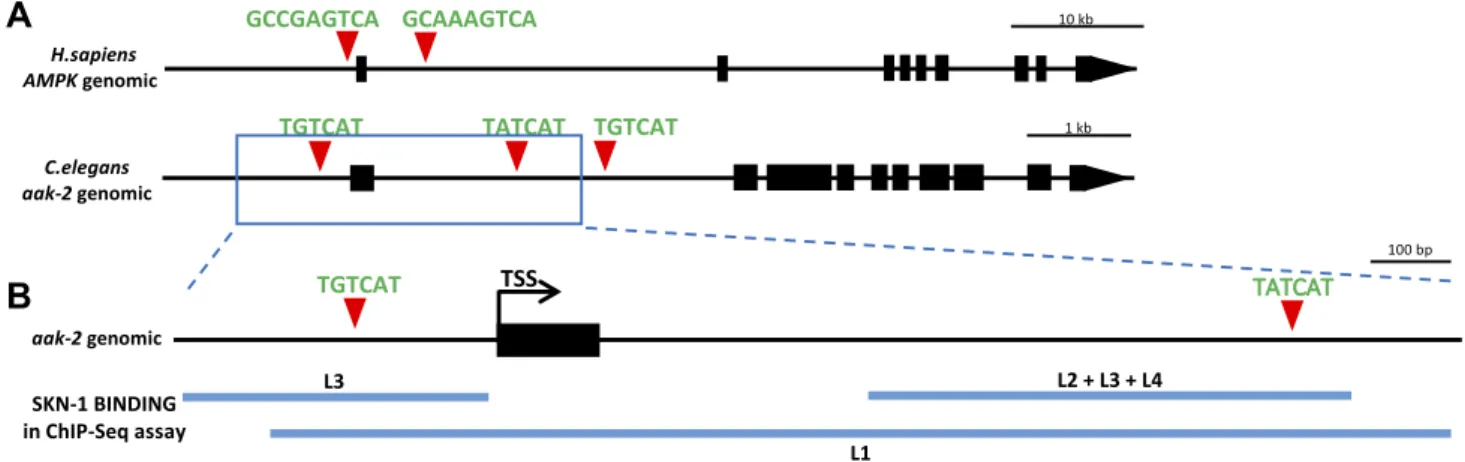
The canonical NRF2/ARE binding sites are present in the proximity ofAMPKgenes of other vertebrate and in- vertebrate species, including C. elegans(Fig. 1A, B). Our genome-wide sequence analysis identified 3 canonical SKN-1 (NRF2 ortholog) binding sites (WWTRTCAT, where R = A or G) in the regulatory and coding regions of theC. elegans aak-2gene (worm ortholog ofAMPK), 100 bp upstream and 1073 and 1573 bp downstream of the ATG translational initiation site, respectively. These sites are also highly conserved in the aak-2 genomic region of closely related Caenorhabditisspecies (Fig. 1A). Next, we compared these in silico predicted SKN-1 binding sites with the experimentally identified regulatory elements found in the relevant analysis of the model organism Encyclopedia of DNA Elements (modENCODE) project (44). According to a chromatin immunoprecipitation and multiplex sequencing analysis (Brdliket al.)(44), the second binding site of SKN-1 seems most likely to be functional, as SKN-1 protein was found to bind to this region of theaak-2 gene in every larval stage tested (L1–L4) (Fig. 1C). SKN-1 was also found to bind to the promoter region ofaak-2but not at every larval stage. These results suggest that these ARE sites might be functional and that the second poten- tial binding site is most likely to be an actual ARE. These data, together with the presence of conserved NRF2/
SKN-1 binding sites in the AMPK/aak-2 locus, raise the possibility that putative molecular interactions between
Figure 1.Putative conserved NRF2/SKN-1 binding sites were found inaak-2/AMPKgenes. A) Location and sequences of the conserved putative NRF2/SKN-1 transcription factor binding sites, relative to the transcription start site (+1) in different species.
(N denotes arbitrary nucleotides, R indicates adenine or guanine, and S indicates cytosine or guanine.)B) The genomic region of AMPK andaak-2gene with predicted NRF2 and SKN-1 binding sites. Black boxes correspond to exonic sequences; connecting lines represent introns. Red triangles indicate conserved NRF2/SKN-1 binding sites.C) The genomic region of theaak-2gene with 2 possible SKN-1 binding sites. The blue lines indicate the genomic regions where Brdliket al.(44) identified binding sites for SKN-1 in their chromatin immunoprecipitation and multiplex sequencing (ChIP-Seq)assay in any of the 4 larval stages ofC.
elegans. Black boxes correspond to exons; connecting lines represent introns. Red triangles indicate conserved SKN-1 binding sites. TSS indicates the transcription start site. L1, 2, 3, and 4 are the 4 larval stages ofC. elegansbefore reaching adulthood.
NRF2/SKN-1 and AMPK/aak-2 are evolutionarily con- served from worms to humans.
NRF2 repressesAMPKexpression
To analyze the regulatory effect of NRF2 on AMPK expression during oxidative stress, relative levels of both AMPK and NRF2 mRNA were measured with qPCR under oxidative stress–induced circumstances.
We established a protocol to induce oxidative stress in HEK293T cells by using the organic peroxide TBHP.
TBHP is a widely used agent that results in oxidation of reduced glutathione, peroxidation of cellular lipids, and altered Ca2+homeostasis, which leads to cell death (45).
The oxidizing effect of TBHP was verified by using flow cytometry (Supplemental Fig. S1). TBHP induced ROS production and lipid peroxidation (see the table and his- togram red line: 300mM TBHP on Supplemental Fig. S1) as opposed to untreated cells (table and histogram black line with gray background on Supplemental Fig. S1) as measured by increased DCF and BOD fluorescence, re- spectively. Silencing of NRF2 through attenuated both TBHP-induced ROS production and lipid peroxidation (table and histogram green line: 300mM TBHP + NFkB siRNA on Supplemental Fig. S1), but an oxidative envi- ronment was observed.
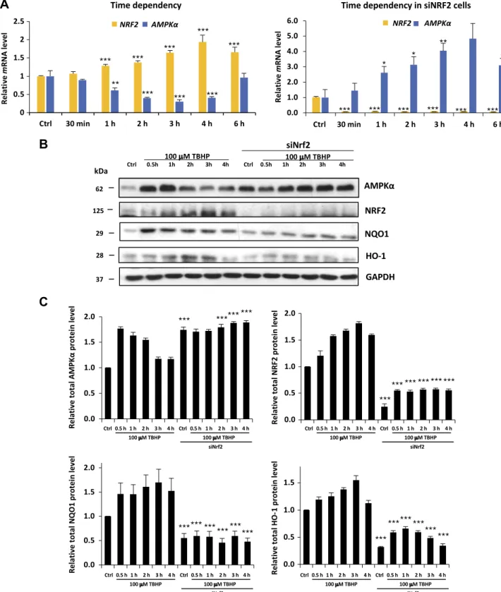
Generating oxidative stress by adding various concen- trations of TBHP clarified that 1.5 h of treatment with 100 mM TBHP was able to increase the level of NRF2 mRNA, whereas addition of 300mM TBHP resulted in a slight decrease in theNRF2 mRNA level in control cells (Fig. 2and Supplemental Fig. S2). Due to the fact that NRF2 is mainly controlled at the posttranscriptional level, we also analyzed NRF2 activity on the protein level. In addition to immunoblotting NRF2, two well-known NRF2 targets, NQO1 (46) and HO-1 (47), were used to follow the time- dependent activation of NRF2 on a protein level. Therefore, we chose 100 mM TBHP treatment to focus on the time dependency of oxidative stress in HEK293T.
We found that the effect of TBHP treatment onAMPK and NRF2 mRNA levels occurs in a time-dependent manner (Fig. 2A). The longer the cells were treated with TBHP, the moreNRF2and lessAMPKmRNA were present in cells. In cells growing for 1, 2, 3, or 4 h in 100mM final concentration, TBHP medium was significantly lessAMPK (at 4 h, only 41% mRNA relative to control cells) and more NRF2mRNA (at 4 h 194% mRNA relative to control cells).
These qPCR analysis results suggest that oxidative stress lowers the transcription ofAMPKwith a certain time de- lay while quickly enhancing the transcription of NRF2.
Interestingly, theAMPKmRNA level did not show any decrease when TBHP treatment was preceded by using siRNA to silence NRF2. The fact that lowering ofAMPK mRNA levels requires NRF2 suggests a regulatory in- teraction between theAMPKgene and NRF2 protein.
To verify the relationship betweenAMPKand oxidative stress, the expression level of total AMPK proteins was analyzed by using Western blot in HEK293T cells. Time dependency was clearly shown in AMPK levels because they were elevated (from 4 to 9 times) in each sample treated with TBHP (1–4 h) relative to the control samples
(Fig. 2B, C and Supplemental Table S1). Both AMPK phosphorylation and its total protein level showed a transient peak during TBHP treatment, and this activation profile is correlated to NRF2, NQO1, and HO-1 level supposing that NRF2 might have some regulatory role in AMPK. The densitometry data of AMPK protein level normalized for GAPDH directly show us that the AMPK protein level was also decreased after 3 h of TBHP treat- ment. It is well known that the half-life of AMPK is rela- tively long; therefore, the AMPK protein level could remain high much longer compared with the AMPK mRNA level during permanent oxidative stress. To further analyze the effect of NRF2 on AMPK during oxidative stress, Western blot experiments were also performed when NRF2 was silenced by using siRNA. NRF2 silencing was detected by depletion of NRF2, NQO1, and HO-1 protein levels. Relative to control cells, AMPK levels were significantly increased in cells treated with siNRF2. In these cells, AMPK protein levels were not suppressed;
however, a 7 times higher basic AMPK level was present in control and treated cells relative to the unsilenced samples.
These data suppose that oxidative stress–induced NRF2 is able to suppress human AMPK levels in both a time- and dose-dependent manner.
NRF2 directly binds to the ARE binding site at AMPK
To assess whether NRF2 is able to bind to its potential ARE sequence in the promoter region of AMPK (query se- quence), an EMSA study was performed with nuclear extracts isolated from Caco-2 cells treated with TBHP for 4 h. We used the well-known, consensus ARE sequence as a positive control. Assembly of the NRF2 protein–probe complex was visualized by using a 2-color fluorescence staining technique to observe both the nucleic acid and the protein components of the assay (48). As shown inFig. 3A, the gel shift assay identified protein–probe complex for- mation in the presence of either AMPK query (lane 5) or consensus (lane 4) oligonucleotides, compared with no complex formation in control lanes containing oligonu- cleotide probe (lanes 1–2) or nuclear extract only (lane 3).
Supershift assays using a specific anti-NRF2 antibody showed a 11 and 10.5% decrease in the protein–probe complex formation for both the AMPK query and the consensus oligonucleotide probes, respectively (Fig. 3B), indicating the presence of NRF2 in the protein–probe complex. Although no retarded NRF2 complex was identified, it has been shown that binding of the antibody to NRF2 protein can reduce its oligonucleotide probe-binding capacity (49). These results indicate NRF2 binds to both AMPK query and consensus oligonucleotide sequences.
NRF2 negatively regulates prolonged autophagyviaAMPK
To address the impact of oxidative stress and NRF2 on autophagy, we also analyzed the time dependency of certain key autophagy markers in TBHP treatment by immunoblotting in HEK293T cells (Fig. 4). To detect
Figure 2.NRF2 down-regulates both mRNA and protein levels of AMPK in the HEK293T cell line after long exposure to oxidative stress.A) Time dependency of bothNRF2andAMPKmRNA levels in oxidative stress with/without silencing ofNRF2by siRNA.
HEK293T cells were treated with 100mM TBHP. The relative level of mRNA was measured by quantitative real-time PCR.Nrf2 gene expression was depleted by Nrf2 siRNA. TBHP-treated samples were compared with control (Ctrl) samples. B) Time dependency of total AMPK protein levels (AMPK-T), NRF2, NQO1, and HO-1 during oxidative stress. Cells were treated with 100 mM TBHP, while the AMPK level was detected by immunoblotting. Nrf2 gene expression was depleted by Nrf2 siRNA.
GAPDH was used as a loading control. C) Quantification and statistical analysis of Western blot assays. Densitometry data represent the intensity of AMPK-T, NRF2, NQO1, and HO-1 normalized for GAPDH. Samples were compared with their partner with siNRF2 background. Error bars represent 6SEM. *P,0.05, **P, 0.01, ***P,0.005 (independent 2-sample Student’s ttests).
whether AMPK is activated upon oxidative stress, the level of active, phosphorylated AMPK was measured.
Because not only does the level of p-AMPKAalpha change upon oxidative stress but AMPKAalpha is also up-regulated, on Fig. 4Bwe show the time profile of p- AMPKAalpha/GAPDH during oxidative stress. We also analyzed the phosphorylation status of its direct target, ULK1. Phosphorylated Ser555 on ULK1 assumes an activated autophagy process induced by phospho-
AMPK. Interestingly, we found a transient activation for AMPK upon prolonged oxidative stress. Both AMPK and ULK555 were fully phosphorylated after 1 h of TBHP treatment, suggesting an essential role of AMPK in autophagy induction. However, AMPK activity later quickly dropped (as indicated by its dephosphorylation after 3 h of TBHP treatment), suggesting a delayed negative feedback on AMPK ac- tivity during oxidative stress. Some key autophagy markers (e.g., LC3, p62) also indicated that auto- phagy exhibits an activation peak during oxidative stress, but long treatment with an oxidative agent sup- presses autophagy. To confirm that this time profile of autophagy activation occurs in an NRF2-dependent manner, the experiment was repeated in NRF2-silenced HEK293T cells. Remarkably, both AMPK-P and ULK555P were increased in NRF2-silenced control cells. Further- more, levels of each autophagy markers we used (AMPK- P, ULK555P, and LC3-II) remained high, whereas the autophagy substrate p62 was robustly reduced after 4 h of TBHP treatment in cells pretreated with NRF2 siRNA.
These results confirm that NRF2 negatively regulates autophagyviaAMPK.
Densitometry data of autophagy markers (i.e., LC3-II/
LC3I, p62, ULK555) show that autophagy has a transient activity change during oxidative stress (Fig. 4B). To verify the delayed negative effect of NRF2 on autophagy with respect to permanent oxidative stress, autophagy was analyzed by using immunofluorescence microscopy (LC3 ICC staining) with and without NRF2 silencing (Fig. 5). To generate oxidative stress, TBHP was added to HEK293T cells for several hours prior to with/without NRF2siRNA treatment. Cells were fixed and immuno- labeled for endogenous LC3-I/II (green). Thus, LC3-I shows diffused cytoplasmic staining, and LC3-II can be observed in discrete foci. As positive controls, bafilo- mycin A1 and rapamycin were used. Although bafilo- mycin A1 is a well-known inhibitor of the late phase of autophagy resulting in the latent accumulation of auto- phagosomes, rapamycin hyperactivates autophagy via mTOR inhibition (50–53). We detected a 5-fold increase in the relative amount of autophagosomes in control cells treated with TBHP; however, after 4 h of treatment, the amount of autophagosomes was similar to the basal level. In contrast, the relative amount of autophago- somes remained significantly high both in the absence of NRF2 under control conditions and during excessive levels of TBHP. These results fit to the Western blot analysis and confirm at the cellular level that transient autophagy is down-regulated by NRF2 upon prolonged oxidative stress (Supplemental Fig. S3).
In vivoevidence for the relationship of aak-2 and SKN-1
As a model organism,C. elegansprovides several charac- teristics that can confirm ourin vitro/in silicoand cellular models. The conservation of disease/signaling pathways betweenC. elegansand higher organisms makes the worm an effectivein vivomodel. We used anaak-2::gfpreporter strain (39) and tested how its expression is changed in Figure 3. NRF2 binds to AMPK query and consensus
oligonucleotide sequences. Nuclear extracts (NE) of Caco-2 treated with TBHP were incubated with query (Q) (lane 4) or consensus (C) (lane 5) oligonucleotide probes and compared with nuclear extract only control (lane 1). Image of the EMSA gel stained with SYBR Green EMSA stain identified nucleic acids (green), and subsequent SYPRO Ruby EMSA stain identified protein (red) components. NRF2 protein–probe complexes (yellow) formed only in lanes containing nuclear extracts + probe. B) Caco-2 nuclear extracts were incubated with 2mg of anti-NRF2 antibody (Ab.) before the addition of oligonucleotide probe (lanes 3 and 5) and analyzed for complex retardation.
response to oxidative stress. The translational fusion re- porter construct contains the whole genomic region ofaak- 2gene and the 2 kb upstream promoter region (Fig. 6A).
Broad GFP expression was observed in the pharynx, gut,
and nervous and reproduction systems of the young adult animals. The reporter was up-regulated inskn-1(2) null mutants (P, 0.005) (Fig. 6B), as well as in skn-1RNAi- treated animals (P,0.0001) (Supplemental Fig. S4). The Figure 4. NRF2 down-regulates autophagy proteins after long exposure to oxidative stress in the HEK293T cell line.A) Time dependency of autophagy down- regulation by NRF2 in oxidative stress. Western blot results of HEK293T cells in 100mM TBHP- containing media. The Western blot shows the time course levels of autophagy-related proteins.
Cells were cultured with TBHP for 0.5, 1, 2, 3, and 4 h, while NRF2 gene expression was de- pleted byNRF2siRNA. GAPDH was used as a loading control.
B) Quantification and statistical analysis of the Western blot assays.
Densitometry data represent the intensity of p62 normalized for GAPDH, LC3-II normalized for LC3-I, ULK555-P normalized for total level of ULK, and AMPK-P normalized for GAPDH.
Samples were compared with their partner with siNRF2 background.
Error bars represent6SEM. *P, 0.05, **P , 0.01, ***P , 0.005 (independent 2-sample Student’s ttests).
most apparent/striking differences regarding the intensity of fluorescence were observed in the head and pharynx of the animals. In acute oxidative stress (5 h), theaak-2::gfp expression was decreased (P,0.0001), but this reduction in fluorescence disappeared when theskn-1gene was si- lenced (Fig. 6C). The inhibition of aak-2::gfp expression by oxidative stress was dose dependent: we observed the maximal inhibition at 2 mM TBHP concentration (Supplemental Fig. S5 and Supplemental Table S2).
However, this decrease of aak-2::gfp expression was suppressed when worms were maintained on TBHP- containing plates for 24 h (Fig. 6D). Altogether, these results providein vivoevidence for the time- and dose-
dependent relationship of aak-2expression, oxidative stress, and SKN-1.
To test the role and the position ofaak-2andskn-1in oxidative stress response, we measured the oxidative re- sistance of skn-1 RNAi-treated and/or aak-2 and skn-1 mutant worms. In response to higher level of oxidative stress (10 mM TBHP),C. elegansdisplays loss of motility and progressive paralysis. Without SKN-1, this paralysis is more definite after 7 h (76% of paralyzed animals in wild type vs. 85% in SKN-1 depleted animals) (Fig. 6E and Supplemental Table S3). Eleven percent of theaak-2(gt33) deletion mutant animals were paralyzed after 7 h, whereas 76% of the N2 animals lost their motility. Without SKN-1, Figure 5. NRF2 silencing positively regulates autophagy. The connection of NRF2-dependent oxidative stress and autophagy induction was checked by immunofluorescence microscopy. LC3 was stained by green fluorescence dye (Alexa Fluor 488);
therefore, the green dots in the cells represent functional autophagosomes.A) Time dependency of TBHP-induced (100mM) autophagy regulation by NRF2. Rapamycin (Rap, 100 nM, 2 h) and bafilomycin (Baf, 10mM, 2 h) were used as positive controls.
B) Quantification and statistical analysis of immunofluorescence microscopy data. Error bars represent6SEM. **P,0.01, ***P,0.005 (independent 2-sample Student’sttests).
this difference was smaller (N2vs. aak-2(gt33): 85vs.23%).
Most of theskn-1(lax120)gain-of-function mutant animals remained mobile even after 7 h, but this resistance to oxi- dative stress was suppressed in skn-1 silenced animals.
skn-1gain-of-function mutants displayed TBHP resistance reminiscent ofaak-2loss-of-function mutants. Thus, SKN-1 is likely to promote TBHP resistance by negatively regu- latingaak-2activity.
InC. elegans, tagging LGG-1 (worm ortholog of Atg8/
LC3) with a fluorophore has become a widely accepted method to visualize autophagosomes (54, 55). The quan- titative assessment of the fluorescence signal of cells with numerous LC3-positive puncta can be assessed accurately at a single-cell level (56). GFP::LGG-1 is expressed throughout development in multiple tissues and dis- plays a diffuse expression pattern, whereas under con- ditions that induce autophagy, GFP::LGG-1 labels positive punctate structures, and its overall expression increases in multiple tissues, including intestinal cells. The amount of GFP::LGG-1–positive vesicles correlates with the forma- tion of autophagosomes, making GFP::LGG-1 an accept- able marker for detecting autophagosomes. Thus, we could test whether AAK-2 or SKN-1 is required for
autophagy by monitoring changes in the number of GFP-positive structures or the ratio of GFP-positive area (autophagosomes) in the cells of transgenic ani- mals (57, 58).
To further analyze the possible relationship between oxidative stress, SKN-1, AAK-2, and autophagy, we also measured relative autophagy activity in aak-2 mutant worms. We incubated the young adult animals on plates containing 1 mM TBHP for 3 h with or withoutskn-1RNAi treatment, using an integrated GFP-tagged LGG-1 re- porter strain our group generated earlier (59). There was no significant autophagy activity difference in the control RNAi N2 andaak-2mutant animals without TBHP treat- ment. Upon oxidative stress, the area of GFP::LGG-1–
positive loci decreased drastically by 80% (Fig. 7A, Band Supplemental Table S4) in wild-type animals (P,0.0001).
Inaak-2(gt33)mutant worms, however, this reduction was not as definite [only 40% (P,0.0001)]. This finding means that in animals lacking functional AAK-2 protein, the effect of oxidative stress on autophagy is suppressed. If skn-1 gene expression is depleted, autophagy decreased by 33%
(P,0.0001) in the cells of the wild-type animals. However, in TBHP-treatedaak-2mutants, the ratio of GFP-positive Figure 6.SKN-1/NRF2 down-regulatesaak-2/AMPKexpression upon oxidative stress inC. elegans. A) The genomic region ofaak-2 with predicted SKN-1 binding sites, and the structure of the translational aak-2::gfp reporter gene, is shown. Black boxes correspond to exonic sequences, connecting lines represent introns. Red triangles indicate conserved SKN-1 binding sites.B) Expression ofaak-2::gfptransgene was increased inskn-1(-)mutant background.C) In acute oxidative conditions (2 mM TBHP, 5 h),aak-2::gfpexpression was decreased in animals with wild-type background. The absence ofskn-1gene products eliminated this change of gene expression.D) In chronic oxidative conditions (2 mM TBHP, 24 h),aak-2::gfpgene expression was not inhibited.
E) Paralysis assay of worms held on 10 mM final concentration TBHP plates. Data are expressed as the ratio of paralyzed/
nonparalyzed worms. The number of animals examined: n = 15–100. Error bars represent 6 SEM. All experiments were independently reproduced 3 times. *P,0.05, ***P,0.005 (independent 2-sample Student’sttests).
area of autophagosomes did not decrease as much as in wild-type animals (76vs.58% reduction in TBHP treated N2 vs. aak-2(gt33)worms, respectively) (Fig. 7Band Sup- plemental Table S4). According to this result, in the pres- ence of the aak-2 mutation, autophagy is more active during oxidative stress. Oxidative stress lowers the level of autophagy both in wild-type and inaak-2mutant animals, but this effect is smaller if theskn-1gene is silenced.
We also examined the resistance/tolerance of auto- phagy deficiency upon oxidative stress by using atg-11 mutant nematodes [tm2508allele containing a 548 bp de- letion (atg-11has a role in the initiation of autophagosome formation)]. After 7 h of 10 mM TBHP treatment, 76% of the wild-type animals became paralyzed (Fig. 7C and Supplemental Table S5), whereas only 16% of autophagy mutantatg-11 animals exhibited a paralyzed phenotype.
Thus, the stress resistance/tolerance of the autophagy- deficient mutant animals is better than the wild-type’s during oxidative stress, indicating that autophagy could not be beneficial upon long-term oxidative stress. The presence or absence ofskn-1gene products changed this
ratio only in theatg-11mutant animals. With thesein vivo experiments, we confirmed that the effect of SKN-1 on autophagy occurs through the inhibition ofaak-2expres- sion, showing that this down-regulatory mechanism is well conserved between worms and humans.
DISCUSSION
NRF2, as a key antioxidant response regulator, and auto- phagy, as an important cellular mechanism to eliminate ROS and reactive nitrogen species, act together to promote cell survival upon oxidative stress (3, 32, 60). ROS and oxidative stress/damage alter autophagic flux rates, and excessive autophagy can promote cell death through the degradation of important components within the cell (32, 61). However, if oxidative stress is prolonged or already contained, we showed that it is also NRF2 that acts as a master regulator to turn off autophagy to avoid the ex- cessive destruction of cellular components, and possibly to save the cell from autophagic cell death (32). Our results Figure 7.Oxidative stress decreases the level of autophagy.A) Fluorescent images showing GFP-tagged LGG-1 accumulation in control animalsvs.in animals treated with 1 mM TBHP for 3 h. In one half of the animals, theskn-1gene expression was depleted by skn-1 RNAi. GFP::LGG-1 accumulates in punctuate areas that are supposed to indicate autophagosomal structures. B) Quantification and statistical analysis offluorescent images of GFP::LGG-1 foci in individual cells. GFP::LGG-1–positive dots that are believed to correspond to autophagosomal structures were quantified. C) Paralysis assay of worms held on 10 mM final concentration TBHP plates. Data are expressed as ratio of paralyzed/nonparalyzed worms.skn-1gene expression was depleted by skn-1 RNAi. The number of animals examined:n = 40–70. Error bars represent 6SEM. All experiments were independently reproduced 3 times. Detailed statistics for each assay can be found in the Supplemental Data. Ns, not significant. *P,0.05, ***P, 0.005 (independent 2-samplettests).
show that NRF2 binds to both AMPK query and consen- sus oligonucleotide sequences, suggesting that AMPK can be directly regulated by NRF2. We predicted and con- firmed an evolutionary conserved mechanism by which NRF2 regulates the expression of the autophagy inducer AMPK, and, by this action, decreases the activation of autophagy to physiologic levels. We also presented that the NRF2-mediated autophagy down-regulation can only be observed upon prolonged stress, highlighting why this exciting mechanism was not yet discovered. Because both the function of NRF2 and the regulation of autophagy are highly complex and context dependent (6, 41, 62), we cannot exclude other important factors in relation to NRF2, AMPK, and autophagy. Here, we highlighted a seamless but effective molecular mechanism for how the antioxi- dant master regulator NRF2 turns off autophagy when autophagy has performed its function.
Exploring the regulatory network of NRF2, we pro- posed a model of a direct transcriptional regulation be- tween NRF2 and the autophagy regulator AMPK. Many experimental data have shown that AMPK can stimulate NRF2 signaling during oxidative agents (e.g., hydrogen peroxide) (30, 63). The detailed mechanism of this regu- latory connection, however, is yet unknown. NRF2 pro- motes autophagy through p62. BecauseNRF2knockout reduces cell viability, we can assume that NRF2 has a crucial role in promoting autophagy-dependent survival during oxidative stress. In the present study, we confirmed that NRF2 was able to bind the ARE binding site atAMPK (Fig. 3), suggesting that NRF2 directly regulates the ex- pression of AMPK. We proved this binding by EMSA;
however, this connection needs further confirmation to show more direct proof for NRF2 binding to ARE elements (e.g., by using chromatin immunoprecipitation analysis).
We also described that NRF2 is able to down-regulate AMPK, and this negative effect was manifested at the transcriptional level during prolonged cellular stress in both the human cell line andC. elegans(Figs. 2A, Band 6B, C). By following in time the level of main components with respect to TBHP-induced oxidative stress, it turned out that the timing has an important role in this network (Figs.
2A, B and 6C, D). Namely, AMPK quickly responds to oxidative exposure. On the one hand, AMPK becomes active via phosphorylation; on the other hand, the AMPKAalpha protein amount is also increased. The active AMPK can activate NRF2. This regulatory loop implies that at the very beginning of oxidative stress, AMPKAalpha protein abundance is independent of NRF2 effects. In addition, this fast response leads to autophagy induction. The autophagy markers (i.e., LC3-II/LC3-I and p62) illustrate (Fig. 4B) that autophagy has a transient ac- tivity change during oxidative stress. LC3-II level was quickly induced, whereas the p62 level was decreased when TBHP was added to HEK293T cells. The LC3-II/LC3- I level started to decrease, while the p62 level increased back after 3 h of oxidative stress. The key autophagy activator ULK1 has also shown a transient activatory phosphoryla- tion on its Ser555, further supposing a transient activation of autophagy. This result seems to fit to microscopy data when the LC3-II–positive foci were detected upon oxida- tive stress (Fig. 5). Both data confirm that autophagy has a
delayed down-regulation during permanent TBHP treat- ment. However, after a certain time delay, NRF2 is able to down-regulateAMPK. Down-regulation ofAMPKresults in the inactivation of its targets (including NRF2). The si- lencedNRF2gene results in the continuous up-regulation of AMPK during extended oxidative stress because the delayed negative feedback has been diminished from the control network.
TheC. elegansexperiments performed here have further uncovered this important regulatory mechanism of auto- phagy and confirmed the in vitroresults. In the present study, we found that in response to oxidative stress, SKN-1 down-regulates aak-2 in C. elegans as well. Relative to control animals, aak-2::gfpexpression levels were signifi- cantly increased in animals treated withskn-1RNAi. SKN- 1 was required to keepaak-2expression at low levels upon acute oxidative stress, whereas this effect was not seen under chronic oxidative stress circumstances. We noted that SKN-1 down-regulatesaak-2(Fig. 6C, D) not only as part of the antioxidant response but under normal cir- cumstances as well. Similarly,.60 genes were previously described as being down-regulated under basal conditions by SKN-1 (64, 65). In the paralysis assay, theaak-2mutant animals tolerated oxidative stress better than the wild-type animals. However, this increased stress tolerance seemed SKN-1 dependent (Fig. 6E). The decreased stress tolerance of theskn-1 RNAi-treatedskn-1gain-of-function mutant animals (relative to the control skn-1 mutant animals) proves that the gene silencing with RNAi worked, and it was effective. Animals carrying thegain-of-functionmuta- tion display high stress resistance among. The reason for the elevated but not significant increase (4 vs. 30%
paralyzed animals) of stress tolerance of theskn-1gain- of-function mutant animals (compared with skn-1 RNAi animals) may be a function of the not completely skn-1gene-silenced neuronal cells. In these cells, some SKN-1 protein is constitutively active and can influence motility despite the RNAi.
We believe that this delayed negative feedback between AMPK and its targets plays a significant role in the fine- tuning of autophagy induction. To further explore the role of NRF2 in the delayed down-regulation of AMPK during oxidative stress in human cell line, we propose a theoret- ical mathematical model to illustrate and interpret the important connections and feedback of the NRF2/AMPK regulatory network (Fig. 8A). According to our experi- mental data, AMPK has a quick transient activation fol- lowed by autophagy induction with respect to oxidative stress (Fig. 8B). After 4 h of treatment with an oxidative stressor, NRF2-dependent down-regulation ofAMPKoc- curred, controlled by a delayed negative feedback be- tween AMPK and NRF2. This connection immediately induces the inactivation of autophagy during prolonged oxidative stress. A quick activation of autophagy is es- sential with respect to oxidative stress because the bulk of damaged components have to be reduced during oxida- tive stress. Obviously, the extended autophagic event is not beneficial for the cellular system; therefore, the cell tries to reduce autophagy to avoid a harmful self-digestion. We propose that this delayed negative feedback between AMPK and NRF2 has a main role in guaranteeing that
autophagy does not become hyperactivated even at high levels or prolongation of cellular stress. In the absence of NRF2, the delayed negative feedback becomes disrupted; therefore, the AMPK levels remain high even after 4 h of treatment with an oxidative stressor (Fig. 8C). The high level of AMPK is able to keep the autophagy processes active. Our results sug- gest that for the proper cellular stress-dependent acti- vation of autophagy, both AMPK and NRF2 are required. At an early stage of the autophagy process, AMPK is required for autophagy activation, while in the later phase of the stress event, NRF2 is essential to down-regulate the processvia inhibiting its own acti- vator. In this way, with certain delay, NRF2 start down- regulating AMPK, and therefore, the level of active autophagy is also dropping. However, in our model, we did not rule out that NRF2 has a positive effect on autophagy induction in later phases of oxidative stress.
We believe that NRF2 alone is not sufficient to maintain autophagic response when AMPK is diminished.
In healthy cells, stress induces autophagy; however, above a certain level, autophagy could result in cell death or may lead to the onset of harmful processes (1, 32, 66). Thus, we propose that SKN-1 is supposed to prevent these undesired cellular events.
Elevated levels of autophagy were detected (in anaak- 2–dependent manner) in animals exposed to oxidative stress when SKN-1 protein was depleted (Fig. 7A, B).
This is not the first known regulatory mechanism through which SKN-1 controls autophagy. SKN-1 regulates autophagy through binding to the ARE
element in the promoter ofp62gene as well, and p62 in turn delivers polyubiquitinated cargoes to auto- phagy via the LIR/Atg8 family–interacting domain (17, 25).
In summary, we established a new role for NRF2 in regulating the dynamics of autophagy. Upon prolonged oxidative stress, NRF2 decreases the activation of auto- phagy through the down-regulation of AMPK to prevent the harmful hyperactivation of autophagy, and with that, promote cellular survival. Oxidative stress has been rec- ognized as a major contributing factor in aging and in various forms of pathophysiology, including neuro- degeneration, diabetes, liver diseases, and cancer (67–75).
In the last decade, the role of autophagy in oxidative stress–related diseases has been reported in detail, show- ing that normally functioning autophagy can promote cellular survival, whereas malfunction in autophagy reg- ulation can lead to cell death and disease phenotypes.
However, the method by which activated autophagy is turned off when oxidative stress is prolonged or already contained has not been elaborated, despite several phe- notypic reports on the existence of such a crosstalk. In the present study, combining regulatory network analysis with experimental validation in a model organism,C. ele- gans, and in a human cell line, we reported the critical role of NRF2 in inhibiting autophagy hyperactivation upon prolonged oxidative stress. By unraveling how NRF2/
SKN-1 could regulate autophagy upon prolonged oxida- tive stress, we highlighted an evolutionary conserved mechanism through which cellular mechanisms interact to promote survival.
Figure 8. Proposed computa- tional model of NRF2-dependent AMPK down-regulation during oxidative stress. A) The core network of AMPK and NRF2 regulation with respect to oxida- tive stress. The dotted line de- notes how the components can influence each other.B) Numer- ical simulations of relative pro- tein levels and activities of the HEK293T cell line with a math- ematical model during oxidative stress. Both AMPK and NRF2 have a transient activation profile resulting in the down-regulation of autophagy after 4 h of oxida- tive stress.C) Numerical simula- tions of relative protein levels and activities of the HEK293T + siNRF2 cell line with a mathe- matical model during oxidative stress. The active AMPK is able to maintain the autophagic process high in the absence of NRF2.