https://doi.org/10.1007/s11033-021-06754-7 ORIGINAL ARTICLE
Anti‑neuropathic pain activity of a cationic palladium (II) dithiocarbamate by suppressing the inflammatory mediators in paclitaxel‑induced neuropathic pain model
Muhammad Naveed1,2 · Rahim Ullah3 · Adnan Khan2 · Bushra Shal2 · Ashraf Ullah Khan2 · Shahan Zeb Khan4,5 · Zia ur Rehman4 · Salman Khan2
Received: 15 April 2021 / Accepted: 10 September 2021
© The Author(s), under exclusive licence to Springer Nature B.V. 2021
Abstract
Background Neuropathic pain is a chronic pain state that negatively impacts the quality of life. Currently, available therapies for the treatment of neuropathic pain often lack efficacy and tolerability. Therefore, the search for novel drugs is crucial to obtain treatments that effectively suppress neuropathic pain.
Objectives The present study was undertaken to investigate the antinociceptive properties of (1,4-bis-(diphenylphosphino) butane) palladium (II) chloride monohydrate (Compound 1) in a paclitaxel (PTX)-induced neuropathic pain model.
Methods Initially, behavioral tests such as mechanical and cold allodynia as well as thermal and tail immersion hyperalgesia were performed to investigate the antinociceptive potential of Compound 1 (5 and 10 mg/kg, b.w). RT-PCR was performed to determine the effect of Compound 1 on the mRNA expression level of inducible nitric oxide synthase (iNOS), cyclooxy- genase-2 (COX-2), and proinflammatory cytokines such as tumor necrosis factor-alpha (TNF)-α, interleukin (IL)-1β, and IL-6. In addition, antioxidant protein, nitric oxide (NO), and malondialdehyde (MDA) levels were also determined.
Results The results demonstrated that once-daily dosing of Compound 1 significantly suppressed the PTX-induced behav- ioral pain responses dose-dependently. The mRNA gene expressions of iNOS, COX-2, and inflammatory cytokines were markedly reduced by Compound 1. Furthermore, it enhanced the level of antioxidant enzymes and lowered the level of MDA and NO production.
Conclusion These findings suggest that the antinociceptive potential of Compound 1 in the PTX-induced neuropathic pain model is via suppression of oxidative stress and inflammation. Thus, Compound 1 might be a potential candidate for the therapeutic management of PTX induced neuropathic pain.
Keywords Allodynia · Hyperalgesia · iNOS · COX-2 · Inflammatory mediators · Oxidative stress
Introduction
Neuropathic pain occurs due to the damage of peripheral or central nerves [1, 2]. It is generally characterized by extreme sensitivity to non-noxious stimuli (allodynia) and noxious stimuli (hyperalgesia). Neuropathic pain arises due to trauma, disease, and chemotherapeutic drugs [2, 3]. Macrophages are a vital component of the immune system and play pivotal roles in the regulation of inflammatory responses [4]. Macrophages are implicated in the pathogenesis of neuropathic pain [5]. Follow- ing nerve injury, resident macrophages initiate inflammatory responses and elicit long-lasting neuroinflammation through the recruitment of circulating leukocytes to the site of injury [4]. At the periphery, activated macrophages secrete cytokines/
chemokines such as TNF-α (tumor necrosis factor-α) and
* Zia ur Rehman zrehman@qau.edu.pk
* Salman Khan skhan@qau.edu.pk
1 Department of Pharmacology and Pharmacotherapy, Faculty of Medicine, University of Szeged, Szeged, Hungary
2 Pharmacological Sciences Research Lab, Department of Pharmacy, Faculty of Biological Sciences, Quaid-I-Azam University, Islamabad, Pakistan
3 Department of Pharmacy, University of Peshawar, Peshawar, Pakistan
4 Department of Chemistry, Quaid-I-Azam University, Islamabad 45320, Pakistan
5 Department of Chemistry, University of Science and Technology, KPK, Bannu 28100, Pakistan
interleukins (IL-1β, IL-6). Cytokines and chemokines secreted by macrophages are potential mediators of pain hypersensitiv- ity in neuropathic pain [5]. Paclitaxel (PTX) is a well-known chemotherapeutic agent that is widely used for the treatment of breast, ovarian, lung, and bladder cancers [6]. However, treatment with PTX may result in peripheral neuropathy that exists for several months even after discontinuation of therapy [7]. Peripheral neuropathy is a major reason for reduction of dose or even cessation of chemotherapy and therefore, results in a great impact on the survival of cancer patients [8].
Numerous studies have revealed that inflammatory responses in the spinal cord result in the development of chemotherapy-induced peripheral neuropathy [9, 10]. It is well recognized that pro-inflammatory mediators are released by activated glial cells in the spinal cord which intensifies the sensitivity and excitability of the neuron [11].
Several lines of evidence have indicated that COX-2 (an inducible enzyme) and iNOS are associated with the induc- tion and progression of neuropathic pain [12]. Peripheral nerve injury elevates the expression of COX-2 in the spinal cord which facilitates inflammation and pain hypersensitiv- ity [13]. In addition, oxidative stress plays a crucial role in the pathogenesis of neuropathic pain [14].
Currently, treatment for neuropathic pain includes NSAIDs, morphine, anti-convulsant, and anti-depressants drug. However, these treatments have been associated with a wide spectrum of adverse effects [2]. The compounds containing structural palladium thiocarbamate have been reported for various anti-inflammatory and analgesic prop- erties [15]. Pronounced pharmacological effects with a better safety profile have been considered by designing and syn- thesizing palladium-containing complexes [16]. The dithi- ocarbamates have also been reported for promising activi- ties against oxidative stress and inflammatory cytokine [17].
It has been noted that dithiocarbamates structural analogs exhibit antiarthritic activities via inhibition of NF‐κB and COX-2 signaling [18]. The characterization of Compound 1 has recognized it to be an efficient drug moiety. Earlier results have indicated that Compound 1 has suppressed the complete Freund's adjuvant (CFA)-, carrageenan-, hista- mine-, and serotonin-induced nociception [19]. Thus, bear- ing in mind the above-mentioned results, we decided to fur- ther explore the neuroprotective property of Compound 1 against PTX-induced neuropathic pain model.
Materials and methods
Chemicals and reagentsThe DNA Extraction Kit (Novel Genomic DNA Mini Kit), TRI-Reagent (Bioshop, Canada), cDNA Synthesis Kit (ABM, Canada), PCR Primers (Macrogen Korea), PCR
Master Mix (Thermo Fisher Scientific US). PTX, Compound 1, gabapentin (GBP), Griess Reagent, 1-chloro-2,4-dini- trobenzene (CDNB), dithio-nitrobenzoic acid (DTNB), hydrogen peroxide (H2O2), and all the other chemicals used were obtained from Sigma (USA). Drugs were dissolved in dimethyl sulfoxide (DMSO 2%) and diluted with normal saline up to the final volume.
Synthesis of compound 1
Compound 1 (1,4-bis-(diphenylphosphino) butane) pal- ladium (II)chloride monohydrate) was synthesized according to the literature procedure [19]. Briefly, it was prepared by simultaneous and dropwise addition of 1,4-bis- (diphenylphosphino) butane (0.426 g, 1 mmol) in acetone (40 mL) and sodium 4-benzylpiperidine-1-carbodithioate (0.273 g, 1 mmol) in methanol (25 mL) to a methanolic suspension of palladium (II) chloride (0.177 g, 1 mmol). The reaction mixture was refluxed for 6 h with constant stirring.
It was then filtered, and the filtrate was rotary evaporated.
The product was soluble in acetone, chloroform, methanol, and dimethylsulfoxide.
Animals and experimental design
Sprague–Dawley female rats (200–250 g) were used in the present study. The animals were obtained from the NIH Islamabad, Pakistan. All the rats were accommodated in standard environmental conditions (22 ± 1 ºC, 52 ± 3%
humidity) and a light/dark period of 12-h with unrestricted access to food and water.
Induction of neuropathic pain and dosing
PTX (4 mg/kg) was injected intraperitoneally (i.p.) in vol- umes of 1 ml per kg, 4 times a week (on days 1, 3, 5, and 7) [20]. PTX dose selection was selected based on a previ- ous report [20]. Compound 1 (5 and 10 mg) was admin- istered once daily for 8 days. The doses of Compound 1 were selected based on its previously reported therapeutic activities in animal models of arthritis [19]. Previously, in the first set of experiments, different doses of Compound 1 were screened to select an optimum and effective dose.
Compound 1 was tested at a dose level of 0.1, 1, and 10 mg/
kg. It exhibited optimum anti-inflammatory and analgesic effects at the doses of 5 and 10 mg/kg of body weight in mice [19]. The schedule of behavioral experimentations was on day 0 i.e., before first drug administration and then on every other day of PTX administration i.e., on days 2, 4, 6, and 8 after 1 h of treatment. Samples were collected after all the rats were sacrificed from each group on the final day of the experiment (Fig. 1).
Animal’s models
Compound 1 was tested in a PTX-induced neuropathic pain model. All the animals were allotted in normal, nega- tive (PTX-induced), positive (GBP) and treatment groups (5 and 10 mg/kg of Compound 1) with 6 animals in each group. Behavioral testing was carried out by an experimenter blinded to the treatment groups.
PTX‑induced model
Rats were distributed into five groups with six rats per group:
Group 1: Normal control.
Group 2: PTX-induced (4 mg/kg, i.p).
Group 3: GBP 75 mg/kg (once daily for 8 days, i.p).
Group 4: Compound 1 (5 mg/kg, once daily for 8 days, i.p).
Group 5: Compound 1 (10 mg/kg, once daily for 8 days, i.p).
Behavioral experiments
Evaluation of static mechanical allodynia in PTX‑induced rats
The anti-allodynic effect of Compound 1 was measured at various intervals (i.e. on days 0, 2, 4, 6, and 8) using Von Frey (VF) filaments as described previously [21–23].
Evaluation of cold allodynia (acetone test)
Compound 1 (5 and 10 mg/kg) was investigated against PTX-induced cold pain (allodynia) by performing an ace- tone test as described previously [24, 25]. In short, rats were habituated for 15 min in a transparent plastic box with a wire mesh floor. After that, a drop (0.05 mL) of acetone was put on the rat’s plantar skin surface using a syringe. The behav- iors of rats (i.e. time taken by a rat while flinching, licking, withdrawing, and biting of hind paw) after the acetone spray
was examined within 15 s and measured as paw withdrawal duration (PWD). A comparative increase in PWD (s) is con- sidered to be a sign of neuropathic pain. Acetone was ejected three times on each hind paw and their average PWD was calculated for each rat.
Evaluation of thermal hyperalgesia
To evaluate further the analgesic effect of Compound 1 in PTX-induced rats, a hot plate test was performed according to the previously reported protocols [26–28]. The once-daily dosing of Compound 1 was examined for antihyperalgesic effect on days 0, 2, 4, 6, and 8.
Evaluation of tail‑flick latency (hot)
A hot tail immersion assay was performed to evaluate the central anti-nociceptive activity of Compound 1 as described previously [29, 30]. Briefly, the rat’s tail was immersed into 52 °C water for 15 s (maximum), and the latency when the rat withdrew the tail was noted. The time taken by a rat to flick its tail from hot water was recorded as tail withdrawal latency (TWL). Each rat was exposed to three trials with a break of 5 min in between. The average of 3 trials was cal- culated as latency for each rat.
Evaluation of tail‑flick latency (cold)
The cold-water tail-flick assay was performed to evaluate the cold hyperalgesia and antihyperalgesic effect of Compound 1 by dipping the lower half of the rat’s tail into a beaker filled with cold water (0–4 °C) [29, 31]. After dipping the tail, the TWL was measured with a 15 s cut-off time. The test was studied 3 times at 5 min intervals to avoid tissue damage and the average was calculated. The antihyperal- gesic effect was represented by the average of each latency (i.e. the comparatively longer latency was considered the antihyperalgesic effect) [32].
Fig. 1 A schematic representation of overall study plan
Evaluation of motor performances
Assessment of Compound 1 treatment on motor perfor- mances of rats was evaluated using a rotarod apparatus, which was rotating at a speed up to 40 rpm for 5 min.
Before starting the experiment, all the rats were trained for 4 days by placing them on a rotating drum with a maxi- mum speed of 40 rpm as described previously [33, 34].
The rats which did not endure walking on the rotarod for more than 2 min were excluded. After that baseline trials were performed before any drug administration to start the experiment. Each rat underwent 3 trials. The latency of time to fall and the falling frequency over a 5-min period were measured. The same trials were repeated on days 2, 4, 6, and 8, 1 h after treatment with Compound 1 or GBP.
Biochemical assays RNA extraction and RT‑PCR
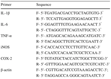
Total RNA was extracted from the L4-6 spinal cord seg- ment of rats according to the manufacturer’s protocol using the TRI-reagent and the purity of the total RNA was determined using a UV spectrophotometer. Total RNA was transcribed to cDNA using a cDNA synthesis kit. The expressions of targeted genes like TNF-α, IL-1β, IL-6, iNOS, and COX-2 were determined (Table. 1). β-actin was incorporated as the housekeeping gene. Amplified prod- ucts were isolated by the use of 1.5% agarose gel electro- phoresis and visualized through a UV trans-illuminator.
The expression level (A.U) was calculated [11, 35–38].
Nitric oxide (NO) determination
The production of NO in the spinal tissue was quantified using the Griess reagent assay according to the previously reported protocols [39–41].
Determination of antioxidant enzymes
The levels of antioxidants such as GSH, GST, and cata- lase were measured according to the previously reported method [42, 43].
Lipid peroxidation assay (LPO)
LPO was estimated by measuring the MDA concentra- tion in spinal tissue according to the previously reported
method [44–46]. A microplate reader was used to measure the absorbance at 535 nm.
Pharmacokinetics and toxicokinetic analysis
An in-silico analysis was performed to determine the phar- macokinetic behavior of Compound 1. The various phar- macokinetics factors that were assessed include absorption, distribution, metabolism, excretion, the volume of distri- bution, and plasma protein binding. The pharmacokinetic parameters were analyzed using Swiss target prediction and pK-CSM online server as reported previously [47]. Simi- larly, the toxicokinetic analysis was performed to assess the harmful effect of Compound 1 against animals, tissue, and micro-organisms, and the maximum tolerated dose was established using online computational tools [47].
Statistical analysis
Data expressed as the mean ± SD. Two-way ANOVA was applied followed by Bonferroni’s post hoc test for the assess- ment of statistical significance amongst various treated groups. Statistical analysis of the data was performed using Sigma-plot version 12.5. p” value < 0.05 was considered to be statistically significant.
Results
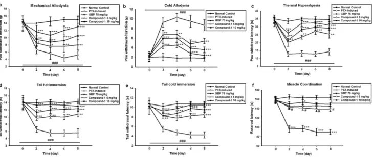
Effect of Compound 1 on static mechanical allodynia Allodynia produced by administration of PTX was consid- erably (p < 0.001) inhibited by treatment with Compound 1 and GBP in a dose-dependent manner. The antiallodynic effect was indicated by increased paw withdrawal threshold (PWT) by treatment with Compound 1 when evaluated on
Table 1 The sequences of PCR primers
Primer Sequence
IL-1β F- 5'-TGA TGA CGA CCT GCT AGT GTG-3° R- 5'- TCC ATT GAG GTG GAG AGC TT-3° IL-6 F- 5'-GGA GTT TGT GAA GAA CAA CT 3° R- 5'- CTA GGG TTT CAG TAT TGC TC-3° TNF-α F- 5'- ATG AGC ACA GAA AGC ATG ATC-3'
R- 5'-TAC AGG CTT GTC ACT CGA ATT-3' iNOS F- 5'-CAC CAC CCT CCT TGT TCA AC-3'
R; 5'-CAA TCC ACA ACT CGC TCC AA-3' COX-2 F- 5′-TGT ATG CTA CCA TCT GGC TTCGG-3’
R- 5’-GTT TGG AAC AGT CGC TCG TCATC-3’
β-actin F- 5′- CGT TGA CAT CCG TAA AGA CCTC-3';
R- 5'-TAG GAG CCA GGG CAG TAA TCT -3'
the 2nd, 4th, 6th, and 8th day. The PWT (g) was remarkably high in the treatment groups as compared to the PTX group (Fig. 2a).
Effect of Compound 1 on cold allodynia
Cold allodynia in PTX-treated rats was significantly (p < 0.001) high as compared to the normal control groups as evident from the comparative rise in PWD (s). However, this increase in PWD was reduced by treatment with Com- pound 1 and GBP (Fig. 2b).
Effect of Compound 1 on thermal hyperalgesia Treatment with Compound 1 markedly (p < 0.001) lowered the hyperalgesic responses (paw licking and jumping) dose- dependently in PTX-treated rats as denoted by increased paw withdrawal latency (PWL) in treatment groups (Fig. 2c).
Effect of Compound 1 on tail hot hyperalgesia The hot tail immersion nociception is indicative of central nociception. Rats treated with PTX exhibited shortened TWL (s) as compared to the normal control. However, Compound 1 treatment markedly (p < 0.001) increased TWL (Fig. 2d).
Effect of Compound 1 on tail cold hyperalgesia In PTX-treated rats, TWL was significantly reduced than normal animals indicating cold hyperalgesia. Compound 1 daily treatment remarkably (p < 0.001) elevated this reduc- tion in TWL showing its anti-neuropathic pain potential (Fig. 2e).
Effect of Compound 1 on motor activity
Motor coordination was assessed through a rotarod test. No abnormal effect was observed on motor activity after the treatment with Compound 1. However, GBP exhibited a significant motor deficit indicated from low rotarod latency time (sec) (Fig. 2f).
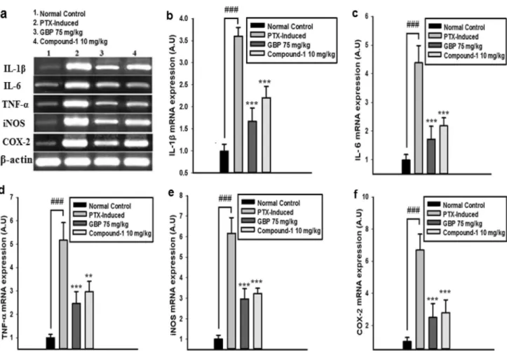
Effect of Compound 1 on inflammatory cytokines The mRNA level of inflammatory cytokines in spinal cord tissues was analyzed using RT-PCR. A significant (p < 0.001) induction in the expression of these cytokines was observed in the PTX-treated group. However, treat- ment with Compound 1 significantly (p < 0.001) lowered the expression of these cytokines as compared to the negative control group (Fig. 3b-d).
Fig. 2 Dose-dependent effect of Compound 1 in doses of 5 and 10 mg/kg in PTX-induced rats. Compound 1 pretreatment inhibited PTX-induced (a) mechanical allodynia (b) cold allodynia (c) thermal hyperalgesia (d) tail thermal hyperalgesia (e) tail cold hyperalgesia and (f) motor activity. The data is displayed as the mean (n = 6) ± SD.
ANOVA followed by a post hoc Bonferroni test was applied for com- paring statistical differences between groups. *p ≤ 0.05, **p ≤ 0.01, and ***p ≤ 0.001 represent a statistically significant difference from the PTX-induced group. (###) indicates comparison to the normal control group
Effect of Compound 1 on iNOS and COX‑2 mRNA expression levels
To further determine the inhibitory effect of Compound 1 on inflammatory mediators, we evaluated the mRNA expression level of iNOS and COX-2 in the spinal cord.
There was an up-regulation of iNOS and COX-2 mRNA expression after neuropathic pain induction which was significantly (p < 0.001) inhibited by treatment with Com- pound 1 (Fig. 3e, f).
Effect of Compound 1 on antioxidants
The levels of antioxidants were significantly (p < 0.001) reduced in the PTX-treated group. However, Compound 1 increased the level of these antioxidant proteins as shown in (Table. 2).
Effect of Compound 1 on nitric oxide (NO) production
The NO production was significantly (p < 0.001) increased in the PTX-treated group. Treatment with Compound 1 remarkably (p < 0.001) inhibited the PTX-induced NO pro- duction (Table. 2).
Effect of Compound 1 on LPO
PTX administration significantly (p < 0.001) elevated the MDA level. Compound 1 treatment showed a marked (p < 0.001) decrease in MDA level as compared PTX-treated group (Table. 2).
Pharmacokinetics and toxicokinetic analysis
The pharmacokinetic analysis of the compounds showed variable pharmacokinetic properties using online pKCSM software (http:// biosig. unime lb. edu. au/ pkcsm/ predi ction).
Fig. 3 a Effect of Compound 1 treatment on mRNA expression level of (b) IL-1β (c) IL-6 (d) TNF-α (e) iNOS and (f) COX-2. The results are shown in a relatively arbitrary unit (A.U). The data is displayed as the mean (n = 6) ± SD. ANOVA followed by a post hoc Bonferroni
test was applied for comparing statistical differences between groups.
*p ≤ 0.05, **p ≤ 0.01, and ***p ≤ 0.001 represent a statistically sig- nificant difference from the PTX-induced group. (###) indicates com- parison to the normal control group
The various properties that were predicted include drug- like properties, physicochemical properties, lipophilicity, water solubility, and pharmacokinetic properties. Simi- larly, the toxicokinetic analysis of the Compound 1 showed a good safety profile as shown in Table 3.
Discussion
PTX is an effective chemotherapeutic drug indicated in the treatment of breast, ovarian, lung, and bladder can- cers. However, PTX-induced peripheral neuropathy is a
Table 2 Effect of Compound 1 on antioxidants, LPO and NO production in PTX-induced rat’s spinal tissue
The data is presented as the mean (n = 6) ± SD (###) denotes comparison to normal control group
***p denotes comparison to PTX-induced group
Parameters Normal control PTX GBP (75 mg/kg) Compound 1 (10 mg/kg) GSH concentration (µM) 14.47 ± 0.39 3.45 ± 0.28### 11.42 ± 0.39*** 10.90 ± 0.36***
GST concentration (µM) 38.92 ± 1.27 12.43 ± 1.38### 36.70 ± 1.14*** 36.25 ± 1.66***
Catalase activity (unit/µL) 11.6 ± 0.17 3.88 ± 0.09### 11.16 ± 0.09*** 11.03 ± 0.14***
MDA level (%) 10.68 ± 0.30 100 ± 2.86### 16.89 ± 1.68*** 18.01 ± 0.68***
NO production (µM) 7.61 ± 0.51 56.06 ± 6.56### 11.29 ± 1.90*** 11.64 ± 1.34***
Fig. 4 Graphical abstract representing anti-neuropathic pain activity of Compound 1 in PTX-induced neuropathic pain model
major adverse effect and causes premature termination of cancer therapy. This adverse effect may persist up to several months even after discontinuation of therapy [7]. Current therapies have been associated with a wide spectrum of adverse effects that limit their satisfactory clinical use. It is important to identify novel drugs that effectively suppress neuropathic pain without attenuating their anticancer effects [2]. Numerous studies have shown the role of inflammatory cytokines, NO, COX-2, and oxi- dative stress in the initiation and maintenance of neuro- pathic pain [48–50]. Therefore, the drug that inhibits the release of inflammatory mediators might be the potential candidate to treat neuropathic pain. Previous findings have demonstrated that Compound 1 exhibited remarkable anti- inflammatory and analgesic activities by suppressing the inflammatory mediator’s production. Therefore, keeping in view the previous promising therapeutic activities, Com- pound 1 was further evaluated as an anti-neuropathic pain agent in the present study.
PTX significantly induced allodynia and hyperalgesia by increasing the rat’s plantar sensitivity to VF filaments, hot, and cold stimuli. The neuropathic pain produced by PTX administration was measured in the form of various pain parameters such as decreasing PWT, PWL, TWL, and increase in PWD [29]. However, these parameters of nociception were significantly improved by daily treatment with Compound 1 and GBP. Results from all the behavioral responses demonstrated the significant antiallodynic and antihyperalgesic activities of Compound 1. Furthermore, Compound 1 did not show any deteriorating effect on motor activity.
Inflammatory cytokines play a crucial role in neuropathic pain by increased hypersensitivity [1] and lead to excessive nociceptive transmission in the spinal cord [51]. The chemical mediators secreted all over the inflammatory process sensitizes the nociceptors that produce pain hypersensitivity resulting in hyperalgesia and allodynia [19]. PTX has been revealed to induce the expression of proinflammatory cytokines, such as IL-1 and TNF-α, in the rat spinal cord [52]. In the present study, PTX treatment considerably increased the expression of inflammatory cytokines in spinal tissues. These inflam- matory cytokines were remarkably reduced by Compound 1.
Similarly, the iNOS and COX-2 expression levels were also measured. The present study showed that Compound 1sig- nificantly suppressed iNOS and COX-2 expressions as well as NO production.
It is well known that oxidative stress plays a vital role in neuropathic pain [2]. In the present study, PTX administra- tion significantly lowered the level of endogenous antioxidants i.e., GSH, GST, and catalase, respectively. The results dem- onstrated that Compound 1 exhibited a protective role against the PTX-induced oxidative stress by enhancing the level of antioxidants in spinal tissues while decreasing the MDA level.
Additionally, the pharmacokinetic analysis using pK-CSM software showed that Compound 1 exhibits high GIT absorp- tion, BBB permeability, and interaction with the cytochrome p450 system, and partial risk of toxicity.
Table 3 Pharmacokinetic and toxicokinetic analysis of the Compound 1
Name Absorption Distribution Metabolism Excretion
Water solu- bility (log mol/l)
Caco2 Cell Intestinal
absorption P-gp Substrate
Cat-egorical (Yes/
No)
BBBpermeability (logBB)
Fraction Unbound Numeric (Fu)
CNS VDss Numeric (log PS) log L/kg
CYP2D6 3A4 1A2 2C19 2C9 2D6 Substrate 3A4
Inhibitor Categorical (Yes/No)
Numeric (log ml/min/
kg)
Compound 1 − 5.34 1.009 94.37 Yes 1.445 0.300 − 0.056
− 0.753 No Yes No No No No No
− 1.109
Molecular
properties Molecular
weight Logp Rotatable
bonds Acceptors Donors Surface
area
Compound 1 785.328 8.918 6 2 0 290.311
Toxicity Ames toxic-
ity Max dose hERG I
inhibitor hERG II
inhibitor Oral toxic- ity acute (LD50)
Oral chronic
toxicity Hepatotoxic-
ity Skin sensi- tivity T.
Pyriformis toxicity
Minnow toxicity
Compound 1 No 0.45 No Yes 3.263 − 0.367 No No 0.285 3.039
Conclusion
In summary, the present study revealed that the compound possesses significant antiallodynic and antihyperalgesic activities in the well-known PTX-induced neuropathic pain model. Compound 1 markedly suppressed the levels of proinflammatory cytokines, iNOS, COX-2, and oxidative stress (Fig. 4). The pharmacokinetic analysis showed that Compound 1 exhibits good ADME properties. We suggest from the present study that Compound 1 might be a useful candidate for the treatment of neuropathic pain. Additional research is needed to confirm these results.
Authors’ contributions MN, RU designed and performed research including behavioral and biochemical assays. SZK and ZR synthe- sized the compound. AK helped in behavioral and biochemical assays.
AK and MN write the original manuscript. MN, AK, BS, AUK, and SK analyzed the data. SK supervised the project. All authors read and approved the final manuscript.
Data Availability The datasets generated during and/or analyzed dur- ing the current study are available from the corresponding author on reasonable request.
Declarations
Conflict of interest The authors declare they have no conflict of inter- est.
Ethical approval “QAU guidelines for animal’s care” Islamabad were followed for the overall experiments involving animals. QAU, Islama- bad Bioethical Committee (Approval No: BEC-FBS-QAU 2017–59) approved the study. Maximum care was assured to minimize harm to animals.
Consent for publication N/A
References
1. Khan S et al (2016) Attenuation of neuropathic pain and neuroin- flammatory responses by a pyranocoumarin derivative, anomalin in animal and cellular models. Eur J Pharmacol 774:95–104 2. Khan A et al (2021) Suppression of TRPV1/TRPM8/P2Y noci-
ceptors by withametelin via downregulating MAPK signaling in mouse model of vincristine-induced neuropathic pain. IJMS 22(11):6084
3. Khan A et al (2021) 7β-(3-Ethyl-cis-crotonoyloxy)-1α-(2- methylbutyryloxy)-3, 14-dehydro-Z notonipetranone attenuates neuropathic pain by suppressing oxidative stress. Inflam Pro- Apoptotic Protein Expressions 26(1):181
4. Kiguchi N et al (2017) Pharmacological regulation of neuropathic pain driven by inflammatory macrophages 18(11):2296
5. Ristoiu VJL (2013) Contribution of macrophages to peripheral neuropathic pain pathogenesis. Life Sci 93(23):870–881 6. Manjavachi MN et al (2019) Spinal blockage of CXCL1 and its
receptor CXCR2 inhibits paclitaxel-induced peripheral neuropa- thy in mice. Neuropharmacology 151:136–143
7. Seretny M et al (2014) Incidence, prevalence, and predictors of chemotherapy-induced peripheral neuropathy: a systematic review and meta-analysis. Pain 155(12):2461–2470
8. Starobova H, Vetter I (2017) Pathophysiology of chemotherapy- induced peripheral neuropathy. Front Mol Neurosci 10:174 9. Janes K et al (2015) Spinal neuroimmune activation is independ-
ent of T-cell infiltration and attenuated by A3 adenosine receptor agonists in a model of oxaliplatin-induced peripheral neuropathy.
Brain Behav Immun 44:91–99
10. Makker PG et al (2017) Characterisation of immune and neu- roinflammatory changes associated with chemotherapy-induced peripheral neuropathy. PLoS ONE 12(1):e170814
11. Kanda H et al (2017) Microglial TNF alpha induces COX2 and PGI2 synthase expression in spinal endothelial cells during neuro- pathic pain. ENeuro. https:// doi. org/ 10. 1523/ ENEURO. 0064- 17.
12. Liang F et al (2017) Dexmedetomidine attenuates neuropathic 2017 pain in chronic constriction injury by suppressing NR2B, NF-κB, and iNOS activation. Saudi Pharmaceu J 25(4):649–654 13. Takeda K et al (2005) Role for cyclooxygenase 2 in the develop-
ment and maintenance of neuropathic pain and spinal glial activa- tion. Anesthesiology 103(4):837–844
14. Shim HS et al (2019) Peripheral and central oxidative stress in chemotherapy-induced neuropathic pain. Mol Pain 15:1744806919840098
15. Shaheen F et al (2010) In vitro assessment of cytotoxicity, anti- inflammatory, antifungal properties and crystal structures of metallacyclic palladium (II) complexes. J Organomet Chem 695(3):315–322
16. Shi C-Y et al (2010) Synthesis, crystal structure, DNA-bind- ing and cytotoxicity in vitro of novel cis-Pt (II) and trans-Pd (II) pyridine carboxamide complexes. Bioorg Med Chem Lett 20(24):7250–7254
17. El-Aarag BY et al (2014) In vitro anti-proliferative and anti- angiogenic activities of thalidomide dithiocarbamate analogs. Int Immunopharmacol 21(2):283–292
18. Cuzzocrea S et al (2002) Pyrrolidine dithiocarbamate attenuates the development of acute and chronic inflammation. Br J Pharma- col 135(2):496–510
19. Naveed M et al (2019) A new cationic palladium (II) dithiocar- bamate exhibits anti-inflammatory, analgesic, and antipyretic activities through inhibition of inflammatory mediators in in vivo models. Naunyn Schmiedebergs Arch Pharmacol 392(8):961–977 20. Tsutsumi K et al (2016) Polaprezinc reduces paclitaxel-induced peripheral neuropathy in rats without affecting anti-tumor activity.
J Pharmacol Sci 131(2):146–149
21. Khan S et al (2013) Mechanism underlying anti-hyperalgesic and anti-allodynic properties of anomalin in both acute and chronic inflammatory pain models in mice through inhibition of NF-κB, MAPKs and CREB signaling cascades. Eur J Pharmacol 718(1–3):448–458
22. Khan S et al (2014) Anti-hyperalgesic and anti-allodynic activities of capillarisin via suppression of inflammatory signaling in animal model. J Ethnopharmacol 152(3):478–486
23. Khan AU et al (2021) Inhibition of NF-κB signaling and HSP70/
HSP90 proteins by newly synthesized hydrazide derivatives in arthritis model. Naunyn-Schmiedeberg’s Arch Pharmacol 394:1–23
24. Khan A et al (2019) Anomalin attenuates LPS-induced acute lungs injury through inhibition of AP-1 signaling. Int Immunopharma- col 73:451–460
25. Ullah MZ et al (2018) Attenuation of inflammatory pain by puera- rin in animal model of inflammation through inhibition of pro- inflammatory mediators. Int Immunopharmacol 61:306–316 26. Zeeshan S et al (2019) N-Pyrazoloyl and N-thiopheneacetyl
hydrazone of isatin exhibited potent anti-inflammatory and
anti-nociceptive properties through suppression of NF-κB, MAPK and oxidative stress signaling in animal models of inflammation.
Inflamm Res 68(7):613–632
27. Khalid S et al (2019) Suppression of TRPV1 and P2Y nocicep- tors by honokiol isolated from Magnolia officinalis in 3rd degree burn mice by inhibiting inflammatory mediators. Biomed Pharm 114:108777
28. Ur Rehman F et al (2021) Surface modified multifaceted nano- carriers for oral non-conventional cancer therapy; synthesis and evaluation. Mater Sci Eng C 123:111940
29. Khan J et al (2019) Attenuation of vincristine-induced neuropa- thy by synthetic cyclohexenone-functionalized derivative in mice model. Neurol Sci 40(9):1799–1811
30. Dong J et al (2019) Berberine ameliorates diabetic neuropathic pain in a rat model: involvement of oxidative stress, inflammation, and μ-opioid receptors. Naunyn Schmiedebergs Arch Pharmacol 392(9):1141–1149
31. Yemitan OK, Adeyemi OO (2017) Mechanistic assessment of the analgesic, anti-inflammatory and antipyretic actions of Dalbergia saxatilis in animal models. Pharm Biol 55(1):898–905
32. Jung Y et al (2017) Anti-allodynic effect of Buja in a rat model of oxaliplatin-induced peripheral neuropathy via spinal astrocytes and pro-inflammatory cytokines suppression. BMC Complement Altern Med 17(1):48
33. Ahmad N et al (2017) A novel pregabalin functionalized salicyla- ldehyde derivative afforded prospective pain, inflammation, and pyrexia alleviating propensities. Arch Pharm 350(6):e201600365 34. Lee B-H et al (2016) Gintonin enhances performance of mice in
rotarod test: involvement of lysophosphatidic acid receptors and catecholamine release. Neurosci Lett 612:256–260
35. Khalid S et al (2018) Antihyperalgesic properties of honokiol in inflammatory pain models by targeting of NF-κB and Nrf2 signal- ing. Front Pharmacol 9:140
36. Khan S et al (2014) Anti-inflammatory properties of samidin from Seseli resinosum through suppression of NF-κB and AP-1-medi- ated-genes in LPS-stimulated RAW 2647 cells. Arch Pharm Res 37(11):1496–1503
37. Khan S et al (2013) Molecular mechanism of capillarisin- mediated inhibition of MyD88/TIRAP inflammatory signaling in in vitro and in vivo experimental models. J Ethnopharmacol 145(2):626–637
38. Khan A et al (2019) Antinociceptive properties of 25-methoxy his- pidol A, a triterpinoid isolated from Poncirus trifoliata (Rutaceae) through inhibition of NF-κB signalling in mice. Phytotherapy Res 33(2):327–341
39. Khan A et al (2019) Matrine ameliorates anxiety and depression- like behaviour by targeting hyperammonemia-induced neuroin- flammation and oxidative stress in CCl4 model of liver injury.
Neurotoxicology 72:38–50
40. Khan AM et al (2020) Continentalic acid exhibited nephropro- tective activity against the LPS and E coli-induced kidney injury through inhibition of the oxidative stress and inflammation. Int Immunopharm 80:106209
41. Khan S et al (2011) Suppression of LPS-induced inflammatory and NF-κB responses by anomalin in RAW 2647 macrophages. J Cell Biochem 112(8):2179–2188
42. Shal B et al (2019) Effect of 25-methoxy hispidol A isolated from Poncirus trifoliate against bacteria-induced anxiety and depression by targeting neuroinflammation, oxidative stress and apoptosis in mice. Biomed Pharmacother 111:209–223
43. Khan AU et al (2020) The newly synthesized compounds (NCHDH and NTHDH) attenuates LPS-induced septicemia and multi-organ failure via Nrf2/HO1 and HSP/TRVP1 signaling in mice. Chemico-Biol Interact 329:109220
44. Atiq A et al (2019) Diadzein ameliorates 5-fluorouracil-induced intestinal mucositis by suppressing oxidative stress and inflamma- tory mediators in rodents. Eur J Pharmacol 843:292–306 45. Kazmi Z et al (2020) Anti-epileptic activity of daidzin in PTZ-
induced mice model by targeting oxidative stress and BDNF/
VEGF signaling. NeuroToxicol 79:150–163
46. Shal B et al (2020) Neuroprotective effect of 25-Methoxyhispidol A against CCl4-induced behavioral alterations by targeting VEGF/
BDNF and caspase-3 in mice. Life Sci 253:117684
47. Ali H et al (2020) Attenuation of LPS-induced acute lung injury by continentalic acid in rodents through inhibition of inflamma- tory mediators correlates with increased Nrf2 protein expression.
BMC Pharmacol Toxicol 21(1):1–14
48. Chun J et al (2012) Alantolactone suppresses inducible nitric oxide synthase and cyclooxygenase-2 expression by down-reg- ulating NF-κB, MAPK and AP-1 via the MyD88 signaling path- way in LPS-activated RAW 264.7 cells. Int Immunopharmacol 14(4):375–383
49. Hughes JP et al (2012) Understanding chronic inflammatory and neuropathic pain. Ann N Y Acad Sci 1255(1):30–44
50. Moalem G, Tracey DJ (2006) Immune and inflammatory mecha- nisms in neuropathic pain. Brain Res Rev 51(2):240–264 51. Marchand F, Perretti M, McMahon SB (2005) Role of the immune
system in chronic pain. Nat Rev Neurosci 6(7):521–532 52. Ledeboer A et al (2007) Intrathecal interleukin-10 gene therapy
attenuates paclitaxel-induced mechanical allodynia and proinflam- matory cytokine expression in dorsal root ganglia in rats. Brain Behav Immun 21(5):686–698
Publisher's Note Springer Nature remains neutral with regard to jurisdictional claims in published maps and institutional affiliations.