RELEASE OF HYDROGEN PEROXIDE
Carl F. Nathan
INTRODUCTION
The capacity to release hydrogen peroxide is a useful bio- chemical correlate of macrophage activation, if the latter term is defined as enhanced ability to destroy intracellular microbes and extracellular tumor cells (1, 2) . Thus, acti- vated mouse peritoneal macrophages are capable of secreting substantial amounts of H2O2 (2-5), while resident peritoneal cells, or those elicited with thioglycollate broth, proteose- peptone, or fetal bovine serum, secrete much smaller amounts (2,3,5). Enhanced capacity to release H202 into the medium marks macrophages activated either in vivo (such as by injec- tion of BCG, C. parvum, T. cruzi, or Γ. gondii (2-5))/ or in vitro, by incubation in mediators from antigen- or mitogen- stimulated lymphocytes (2,5). H202 or closely related sub- stances have been directly implicated as both microbicidal
(4-8) and tumoricidal (9-12) factors in mononuclear phagocyte effector function.
METHODS FOR STUDYING Copyright © 1981 by Academic Press, Inc.
MONONUCLEAR PHAGOCYTES 4 9 9 All rights of reproduction in any form reserved.
ISBN 0-12-044220-5
Several methods are available for measuring the concentra- tion of H2O2 in biological systems (13). The following dis- cussion concerns the assay based on the loss of fluorescence of scopoletin (6-methoxy-7-hydroxy-l, 2-benzopyrone, a cou- marin) when the 7-hydroxyl group is oxidized to a ketone by H2O2 in concert with horseradish peroxidase. One mole of
H2°2 oxidizes 1 mole of scopoletin. The method was introduced in 1955 by Andreae (14), improved by Perschke and Broda in 1961 (15), first applied to the measurement of H2O2 release from leukocytes by Root et al. in 1975 (16), and extended to the study of mononuclear phagocytes by Nathan and Root in 1977 (3). The method has been used with blood monocytes and tissue macrophages of man, mouse, rat, guinea pig, and rabbit
(17).
There are two requirements in order to detect substantial H2O2 release from mononuclear phagocytes using the scopoletin method (3). First, if mouse peritoneal macrophages are em- ployed, they must be activated. H2O2 release from nonacti- vated cells occurs but is small. However, blood monocytes can secrete copious H2O2 without prior exposure to activating
(differentiative) stimuli (18,19). Second, the mononuclear phagocytes must be exposed to a triggering agent, that is, any of a variety of membrane-active stimuli, such as tumor pro- moters, antigen-antibody complexes, or phagocytic particles
(4), which elicit a rapid response. In the absence of a trig- gering agent, extremely little if any H2O2 release is detected in the extracellular medium.
II. REAGENTS
Ά. Buffer
A variety of protein-free balanced salt solutions are suitable. Hank's balanced salt solution without phenol red is used frequently with monocytes. For mouse peritoneal
macrophages, Krebs-Ringer phosphate buffer with glucose (KRPG) has been most extensively used. It is best prepared from double-distilled, glass-distilled water. The formulation is NaCl, 120 mtf; KC1, 4.8 mM; CaCl2, 0.54 mM; MgS04, 1.2 mW;
sodium phosphate, 15.6 mM; glucose, 5.5 mAf; pH, 7.30 to 7.40;
tonicity, 300 mOsm.
B. Scopoletin
The fluorochrome is obtainable from Sigma Chemical Co., St. Louis, Missouri. A ImM stock solution is prepared in buffer without glucose. Complete dissolution of the scopo-
letin usually requires 24-36 hr at 37°C. The concentration is checked by assay against known amounts of ethyl hydrogen peroxide (Polysciences, Inc., Warrenton, Pennsylvania). The stock solution is stable for many months at 4°C.
C. Horseradish Peroxidase (HRP)
This may be obtained from Worthington Biochemical Corp., Freehold, New Jersey, or from Sigma (Type II). A solution of 200 purpurogallin U/ml is prepared in buffer without glucose and stored in aliquots at -20°C/ where it is stable for many months. The optimal amount of each preparation should be determined under typical assay conditions. Some preparations are suppressive if used in amounts optimal for other prepara- tions.
D. Phorbol Myristate Acetate (PMA)
This triggering agent (12-0-tetradecanoyl-phorbol-13-ace- tate) is obtainable from Consolidated Midland, Inc., Brewster, New York, or from Sigma. A stock solution of 0.3 mg/ml is prepared in dimethylsulfoxide and stored at -80°C in the dark in tightly capped glass tubes in aliquots of about 2 ml.
Periodically, one tube is used to prepare aliquots of about 50 yl. For convenience, one 50-yl aliquot may be stored at -20°C for up to a week, after which the unused portion is discarded. Under these conditions, PMA usually retains full potency for about 1 yr. Failure of the solution to freeze at -20°C is often a sign of deterioration. A dose-response curve for the amount of PMA needed to trigger maximal H2O2 release from macrophages or granulocytes should be performed with each new lot of PMA, and as needed during the later period of use of a given lot. The optimal concentration should initially be about 10 ng/ml, and may shift with time to about 100 ng/ml.
If higher doses are required, a new lot should probably be prepared.
PMA is an extremely potent tumor promoter. It must not come into contact with the skin. Gloves should be worn throughout the assay and mouth pipetting should be strictly forbidden. Stock solutions should be tightly capped and thawed as briefly as possible. PMA-containing solutions should be disposed of according to institutional guidelines for hazardous chemicals. The PMA waste bottle in the labora- tory should be nonbreakable and should not be used for the concomitant storage of carcinogens.
III. PROCEDURES
A. Cells in Suspension, Under Continuous or Intermittent Observation
The advantages of this approach are the following: The number of mononuclear phagocytes in the cuvette can be deter- mined by counting in a hemocytometer; the cells are ready to use immediately after preparation; and the kinetics and magni- tude of the response may be observed as it is evolving, per- mitting adjustments in assay conditions. The disadvantages are the need for a thermostatted compartment in the fluoro- meter, the inability to employ surface adherence and in vitro incubation to enrich for mononuclear phagocytes, limitation of the number of samples that may be assayed at one time, ac- cording to the number of cuvettes which the fluorometer may accommodate, usually 4, and difficulty in using high doses of phagocytic particles as triggering agents.
Cell suspensions should be freed of contaminating erythro- cytes, if necessary, by hypotonie lysis. The cell pellet is resuspended in 2 to 10 ml of 0.2% NaCl (the amount depending on the size of the pellet), and promptly mixed with an equal volume of 1.6% NaCl, followed by three volumes of buffer. A 5-sec exposure to hypotonie saline is sufficient to lyse ery- throcytes in mouse peritoneal cells. The cells are then washed several times by centrifugation to remove exogenous protein. The cell number is determined with a hemocytometer, and slides are made for differential counting.
The choice of cell number, amount of scopoletin, and amount of HPO for the assay depends on the activity of the cells under study. For observations of rate of H2O2 release, typical amounts are 3 x 10^ cells, 10 to 20 nmoles of scopo- letin initially and 1 purpurogallin unit (about 6 yg of Sigma Type II) HPO in a total volume of 3 ml of buffer. With 2 x 10^ cells, the initial amount of scopoletin could be reduced to 1 nmole. With monocytes, higher values may be obtained by adding 1 mAf sodium azide, in order to inhibit the catabolism of H2O2 by reactions dependent upon catalase and myeloperoxi- dase. The cells are allowed to equilibrate for 5 min at 37°C in the thermostatted compartment of the fluorometer, such as the Perkin-Elmer MPF 44A (Perkin-Elmer Co., Norwalk, Connecti- cut) connected to a circulating water bath. The recorder is brought to the desired scale by selecting the slit widths
(usually between 2 and 10 nm) and the gain, and the spontaneous activity is recorded for several minutes with the excitation wavelength at 350 nm and the emission at 460 nm. (The appar- ent peak excitation wavelength of scopoletin may vary with assay conditions. In KRPG, it is 380 nm. Sensitivity is re-
duced 25% by exciting at the conventional setting of 350 nm rather than 380 nm. However, the precision of the determina- tion of H2O2 concentration is unaffected.) The reaction is started by adding the triggering agent in a small volume.
When the scopoletin has been 60% oxidized, additional scopole- tin is added, taking care not to exceed full scale. The re- sponse may be observed for a predetermined time, or followed to its completion, which is usually between 1.5 and 3.5 hr later. If observation lasts more than 5 min, the contents of the cuvettes should be stirred automatically, or mixed manu- ally at intervals before each reading. An automatic indexer and stirrer may be obtained from C. N. Wood Manufacturing Co., Newtown, Pennsylvania.
To confirm that the decrease in fluorescence is due to
H2°2' c o n t r° ls consist of omitting HPO, or adding catalase.
The cuvettes may be glass or quartz. Type 3H quartz cuvettes (Precision Cells, Inc., Hicksville, New York) are a reasonable choice, since they will serve also for most other fluorometric purposes. Scrupulous care of the cuvettes is advisable. Residue in cuvettes may trigger peroxide release from the cells, which may be misconstrued as spontaneous acti- vity. Higher amounts of residue may kill the cells. The following method of cleaning is satisfactory. The cuvettes are rinsed three times with water. A very dilute solution of dishwashing detergent is prepared; for example, one drop of Palmolive liquid is mixed with 50 ml of water in a beaker, the contents discarded, and the beaker refilled. Using a cotton Q-tip, the inner and outer surfaces of the cuvettes are gently washed. The cuvettes are rinsed in five changes of warm water, then filled briefly with Chromerge (potassium dichromate in con- centrated sulfuric acid). The Chromerge is decanted, and the cuvettes rinsed manually 20 times with distilled water, or rinsed on a cuvetter washer (Precision Cells) with 100 ml of distilled water each. They are drained on absorbent paper, and the outsides dried with lint-free tissues. The cuvettes are handled with disposable plastic gloves from which the starch powder has been rinsed.
B. Cells Adherent, under Continuous or Intermittent Observa- tion
The advantages of this method are as follows: The cells may be enriched for mononuclear phagocytes by discarding non- adherent cells and permitting granulocytes to die over time;
lysis of erythrocytes is unnecessary; cells may be assayed after various times in culture; phagocytic particles may be centrifuged onto the cells before starting the assay; and the kinetics of the response may be observed as it is evolving.
Disadvantages are the need for a thermostatted fluorometer;
the restriction in the number of samples that can be assayed at one time; and the need to determine the adherent cell num- ber by some means other than counting in a hemocytometer.
Cells are allowed to adhere to coverslips. The most use- ful coverslips are rectangular, with a width equal to the diagonal of the cuvette. This prevents shifts in position of the coverslip in the cuvette, which can cause artifacts in the reading. Coverslips measuring 13 x 27 x 0.1 mm (Bellco Glass, Vineland, New Jersey) are suitable with cuvettes having a 1-cm light path. The coverslips are cleaned and sterilized by soaking in 70% ethanol, dipping in absolute ethanol, touch- ing off the excess, and passing through a flame to ignite the alcohol. After cells have adhered, the coverslip is picked up with jeweller's forceps and rinsed thoroughly in at least
four beakers, each containing 100 ml of saline or KRPG, in order to remove serum proteins and nonadherent cells.
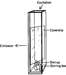
The coverslip is then placed directly in the cuvette, and the assay performed as above. Thorough mixing is important during the assay. For automatic stirring, the coverslip may be mounted above the stirring bar on a glass stirrup (see Fig. 1).
Excitation
Emission ^
Coverslip
Stirrup Stirring bar
Fig. 1. A 13 x 27 x 0.1 mm glass coverslip with adherent mononuclear phagocytes is placed diagonally in the cuvette
(1-cm light path) on a glass stirrup to permit the use of a magnetic stirring bar.
To use a phagocytic stimulus, the coverslips are placed in 2 ml of KRPG in a 35-mm dish on ice. The phagocytic par- ticles, such as opsonized zymosan, are added, and the dish is centrifuged for 2 min in the cold. The slip is gently im- mersed in cold buffer, then transferred directly to the pre- warmed reagents in the cuvette in the fluorometer. Nearly instantaneous onset of H2O2 release may be anticipated.
Matched coverslips should be rinsed in the same manner as above, and used to measure cellular protein or DNA. It is generally not satisfactory to use the same coverslip for both H2O2 release and measurement of cell number, as there is a variable loss of cells by the end of the assay.
C. Cells Adherent with Observation at a Single Time Point This method combines the advantages of using adherent, cultured cells with the ability to process large numbers of samples. A temperature-controlled fluorometer is not neces- sary. The cells may be cultured on coverslips of almost any size or shape. The disadvantage is that only one observation is made of each sample. If the amount of scopoletin added proves to be insufficient to react with all of the H2O2 secreted, the remainder of the H2O2 will be undetected. If excessive scopoletin is added, small amounts of H2O2 release will be difficult to measure accurately. The lag time before onset of H2O2 release cannot be measured, and the initial rate of secretion can only be estimated by interpolation from mul- tiple samples.
One variant of this method will be described. Cells are cultured on 13-mm round glass coverslips (Clay-Adams, New York) in 16-mm wells in plastic multiwell plates (Costar Data Pack- aging, Cambridge, Massachusetts). After incubation for the desired length of time, the coverslips are rinsed in four beakers of saline or KRPG, drained briefly on absorbent paper, and transferred to a new plate, whose wells contain 1.5 ml of KRPG and the desired amounts of scopoletin, HPO, and PMA.
The assay plate is then floated in a 37°C water bath for 2.5 hr. The medium in each well is transferred to a 10 x 75 mm glass test tube and allowed to come to room temperature. The tubes are then read in the fluorometer, either directly using a test-tube holder adaptor in the sample compartment, or by decanting into cuvettes. The readings are stable overnight.
Coverslips to which no cells are added are included as controls. The amount of rinsed fluid that is carried into the assay medium is thereby taken into account to avoid misinter- preting dilution of scopoletin as oxidation. In addition, these controls correct for concentration of scopoletin due to evaporation. Additional cell-bearing coverslips are saved for protein or DNA measurements.
IV. CALCULATION OF DATA
The two most useful ways of reporting the results are in terms of the maximal rate of H2O2 release (nanomoles per 10^
cells per initial 5 min) or as the total amount secreted (nanomoles per 10^ cells, or nanomoles per microgram cell protein).
V. CRITICAL COMMENTS
A. Purity of Cell Populations
Granulocytes are potent secretors of H202 in response to the same stimuli that trigger mononuclear phagocytes. Most preparations of freshly isolated mononuclear phagocytes con- tain at least a few granulocytes. With some regimens for activating peritoneal macrophages, the proportion of granulo- cytes may exceed 20%. It is necessary to determine the per- centage of granulocytes in each preparation and to avoid attributing their responses to macrophages. Granulocyte con- tamination can usually be eliminated by 4-18 hr incubation in vitro. However, macrophage activation is reversible (2), so that with prolonged incubation, values for H2O2 release from activated macrophages will often decline.
-B. Measurement of Net Extracellular Release
The method detects only that fraction of H2O2 which es- capes from the cell into the medium and which is not expended there in the oxidation of substances other than scopoletin.
The method does not indicate the actual rate or amount of H202 produced by the cells; it provides a lower limit for those values. Even in a cell-free system, in which H2O2 is generated enzymatically, only about 70% of the H2O2 is detect- ed by the scopoletin method (20). Cells with active catabolic pathways for ^ 0 2 , such as alveolar macrophages, may appear to perform poorly in the scopoletin assay. The method reveals
little regarding the rate of H202 accumulation in phagosomes, or in the zones of contact between the macrophage plasma mem- brane and closely adherent surfaces, such as those of tumor cells or parasites.
C. Interference by Alternate Hydrogen Donors
Many substances may be oxidized by H2O2 in competition with scopoletin because of the wide range of substrates for HPO (20). Thus, as little as 0.03% serum interferes with the assay. Interference is seen with antibody-coated red cells and tumor cells, serum-coated latex particles and starch granules, and reducing agents such as glutathione, ascorbate, NADPH, and NADH. The method is not well suited to the study of cell lysates. As a related problem, H2O2 may be consumed by catalase or glutathione peroxidase within the mononuclear phagocytes themselves, or within other cells present in the system.
D. Influence of Triggering Agents
The difference in rates of release of H2O2 from activated and nonactivated macrophages, which is elicited by exposure to PMA, is sometimes less marked when using phagocytic particles as triggering agents. This shift in ratio is difficult to in- terpret. Activated mouse peritoneal macrophages are often less phagocytic than nonactivated macrophages (21), and may have less readily detectable Fc receptors (22). Phagocytic particles themselves may interfere with the detection of H202 release (see above). Such interference may depend in part on the "leakiness" of the phagosome during its formation, that is, how freely the nascent vacuole may communicate with the extracellular space.
E. Sensitivity to Conditions of Prior Incubation
Activated mouse peritoneal macrophages tend to secrete less H2O2 after temporary exposure to alkaline media (pH 7.8 or above). The defect may persist for some time after the pH is corrected. During transfer of coverslips, or other opera- tions with bicarbonate-based media performed outside the incu- bator, it is important to control the pH.
F. Choice of Assay
At least four methods are capable of recording changes in H202 concentration of about 10~8 M. Three of these employ fluorescent indicators: scopoletin, leukodiacetyldichloro- fluorescein (LDADCF) (23-24), and homovanillic acid (25).
Assays using the latter two compounds have the advantage of detecting an increase, rather than a decrease, in the inten-
sity of fluorescence in proportion to the concentration of H2O2. However, LDADCF is reacted with the cell supernatant, not with the cells themselves, which makes the assay somewhat less versatile than the scopoletin method. Homovanillic acid may oxidize spontaneously during long-term assays. All three methods share the disadvantages of dependence on HPO (see above).
The fourth assay is spectrophotometric. It is based on the shift in absorption undergone by yeast cytochrome peroxi- dase when it forms an enzyme-substrate complex with H2O2 (26).
The assay is as sensitive as the fluoremetric methods when a dual-wavelength spectrophotometer is employed. The cytochrome peroxidase method is probably more accurate than the scopole- tin method; it is less susceptible to interference by other hydrogen donors (20). Its disadvantages are the need for specialized equipment and the lack of commercial availability of yeast cytochrome peroxidase, which is consumed in' stöichio- metric amounts with the H2O2 detected.
The cytochrome peroxidase method will probably occupy an increasingly important role in specialized applications. The scopoletin method is likely to remain in wide use for assays with intact cells, in view of its simplicity, versatility, and the low cost of the reagents.
Acknowledgments
Some of the work reported here was supported by PHS grant CA-22090. C.N. is a Scholar of the Leukemia Society of
America, and a Research Career Scientist of the Irma T. Hirschl Trust.
REFERENCES
1. Z. A. Cohn. The activation of mononuclear phagocytes:
Fact, fancy, and future. J. Immunol. 121:813-816, 1978.
2. C. Nathan, N. Nogueira, C. Juangbhanich, J. Ellis, and Z. A. Cohn. Activation of macrophages in vivo and in vitro: Correlation between hydrogen peroxide release and killing of Trypanosoma cruzi. J. Exp. Med. 149:1056-1068, 1979.
3. C. F. Nathan and R. K. Root. Hydrogen peroxide release from mouse peritoneal macrophages: Dependence on sequen- tial activation and triggering. J. Exp. Med. 146:1648- 1662, 1977.
4. R. B. Johnston, Jr. Oxygen metabolism and the micro- bicidal activity of macrophages. Fed. Proc. 37:2759- 2764, 1978.
5. H. W. Murray and Z. A. Cohn. Macrophage oxygen-depen- dent antimicrobial activity, III. Enhanced oxidative metabolism as an expression of macrophage activation.
J. Exp. Med. 152:1596-1609, 1980.
6. H. W. Murray, C. Juangbhanich, C. F. Nathan, andZ, A.
Cohn. Macrophage oxygen-dependent antimicrobial acti- vity. II. The role of oxygen intermediates. J. Exp.
Med. 150:950-964, 1979.
7. C. B. Wilson, V. Tsai, and J. S. Remington. Failure to trigger the oxidative metabolic burst by normal macro- phages. Possible mechanism for survival of intracellular pathogens. J. Exp. Med. 151:328-346, 1980.
8. M. Sesada and R. B. Johnston, Jr. Macrophage micro- bicidal activity. Correlation between phagocytosis- associated oxidative metabolism and the killing of Candida by macrophages. J. Exp. Med. 152:85-98, 1980.
9. C. F. Nathan, L. H. Brukner, S. C. Silverstein, and Z. A. Cohn. Lysis of tumor cells by activated macro- phages and granuloeytes. I. Pharmacologie triggering of the effectors and the release of hydrogen peroxide.
J. Exp. Med. 149:84-99, 1979.
10. C. F. Nathan, S. C. Silverstein, L. H. Brukner, and Z. A. Cohn. Extracellular cytolysis by activated macro- phages and granuloeytes. II. Hydrogen peroxide as a mediator of cytotoxicity. J. Exp. Med. 149:100-113,
1979.
11. C. F. Nathan, L. H. Brukner, G. Kaplan, J. C. Unkeless, and Z. A. Cohn. The role of activated macrophages in antibody-dependent lysis of tumor cells. J. Exp. Med.
152:183-197, 1980.
12. C. F. Nathan, and Z. A. Cohn. The role of oxygen-depen- dent mechanisms in antibody-induced lysis of tumor cells by activated macrophages. J. Exp. Med. 152:198-208.
13. B. Chance, H. Sies, and A. Boveris. Hydroperoxide metabolism in mammalian organs. Physiol. Rev. 59:
527-605, 1979.
14. W. A. Andreae. A sensitive method for the estimation of hydrogen peroxide in biological materials. Nature 175:
859-860, 1955.
15. H. Perschke, and E. Broda. Determination of very small amounts of hydrogen peroxide. Nature 190:257-258, 1961.
16. R. K. Root, J. Metealf, N. Oshino, and B. Chance.
H2^2 r ele a s e from human granuloeytes during phagocytosis.
I. Documentation, quantitation, and some regulating factors. J. Clin. Invest. 55:945-955, 1975.
C. F. Nathan. The release of hydrogen peroxide from mononuclear phagocytes and its role in extracellular cytosis. In "Mononuclear Phagocytes: Functional Aspects" (R. van Furth, ed.). Martinus Nijhoff, The Hague, 1980.
M. Reiss, and D. Roos. Differences in oxygen metabolism of phagocytosing monocytes and neutrophils. J. Clin.
Invest. 61:480-488, 1978.
L. M. Adler, J. B. L. Gee, and R. K. Root. H2O2 for- mation and utilization by human monocytes (MONOs).
Clin. Res. 26.-522A, 1978.
A. Boveris, E. Martino, and A. O. M. Stoppani.
Evaluation of the horseradish peroxidase-scopoletin method for the measurement of hydrogen peroxide formation in biological systems. Anal. Biochem. 00:145-158, 1977.
C. F. Nathan, and W. D. Terry. Decreased phagocytosis by macrophages from BCG-treated mice: Induction of the phagocytic defect in normal macrophages with BCG in vitro.
Cell. Immunol. 29:295-311, 1977.
C. F. Nathan, R. Asofsky, and W. D. Terry. Characteriza- tion of the nonphagocytic adherent cell from the perito- neal cavity of normal and BCG-treated mice. J. Immunol.
118:1612-1621, 1977.
A. S. Keston, and R. Brandt. The fluorometric analysis of ultramicro quantities of hydrogen peroxide. Anal.
Biochem. 11:1-5, 1965.
J. W. T. Homan-Muller, R. S. Weening, and D. Roos.
Production of hydrogen peroxide by phagocytizing human granulocytes. J. Lab. Clin. Med. 85:198-201, 1975.
F. Rossi, B. Bellazite, A. Dobryna, T. Dri, and G.
Zabucchi. Oxidative metabolism of mononuclear phago- cytes. In "Mononuclear Phagocytes: Functional Aspects"
(R. van Furth, ed.). Martinus Nijhoff, The Hague, 1980.
A. Boveris, N. Oshino, and B. Chance. The cellular pro- duction of hydrogen peroxide. Biochem. J. 128:611-630, 1972. .