SEPARATION OF HUMAN MONOCYTES AND GUINEA PIG MACROPHAGES BY DENSITY GRADIENTS OF METRIZAMIDE
Barbara A. Sherry Mathew A. Vadas
John R. David
INTRODUCTION
The use of slightly hypertonic discontinuous metrizamide gradients as an effective, reproducible method for the purifi- cation of normal human peripheral blood eosinophils and neutro- phils was developed in our laboratory by Vadas et al. (1).
High yields and purity were obtained without sacrificing the viability or functional integrity of the fractionated cells, This method employs density steps ranging between 1.10 gm/cm^
and 1.13 gm/cm^ [on a percentage basis steps are 18, 20, 22, 23, 24, and 25% (w/v) metrizamide]. In the course of experi- mentation, it was observed that a large percentage of human mononuclear cells did not enter the gradients at all, but rather formed a band above the 18% metrizamide step. It was
Present address: Walter and Eliza Hall Institute, Royal Melbourne Hospital, Parkville, 3050 VIC, Australia.
METHODS FOR STUDYING Copyright © 1981 by Academic Press, Inc.
MONONUCLEAR PHAGOCYTES 1 8 7 All rights of reproduction in any form reserved.
ISBN 0-12-044220-5
hypothesized that by modifying the method of Vadas et al. to incorporate a range of density steps between 1.088 and
1.108 gm/cm3 [on a percentage basis steps would be 16, 17, 18, 19, 20, and 21% (w/v) metrizamide], a fraction enriched for human peripheral blood monocytes might be recovered.
Using these modified conditions, certain fractions were found to be considerably enriched in human monocytes. Ten ex- periments were performed using peripheral blood white cells from six separate donor individuals (see Table I ) . With a mean starting monocyte concentration in the buffy coat of 8.9%
(individual values ranging between 4.0 and 15.4%), enriched fractions were recovered with a mean purity of 76.0% (values ranging between 61.0 and 88.2%). In these ten experiments, the mean percent recovery of total monocytes in the enriched fraction was 46.8% (values ranging between 19.0 and 85.7%).
Cells from a single donor individual were fractionated on five separate occasions to test for reproducibility of the technique
(see Table II). Starting monocyte concentrations in the buffy coat varied between 10.0 and 20.0% (mean 15.4%) depending upon the experiment. After purification of these cells on metriza- mide gradients, enriched fractions were recovered that ranged between 83.0 and 9 3.0% monocytes (mean purity 88.2%). The percentage recovery of total monocytes in enriched fractions ranged between 21.0 and 51.0% (mean recovery 34.3%). Cells in experiments 1 - 3 were tested for viability by trypan blue ex- clusion both before and after fractionation. They were found in all cases to be greater than 94% viable. No cell damage was observed by morphologic criteria, and when cells were tested for function, as assessed by uptake of latex particles or phagocytosis of sensitized sheep red blood cells, they ap- peared to be functionally intact.
This modified method was expanded to the fractionation of guinea pig macrophages from casein-induced peritoneal exudate cells, and as is shown in Table III, purities ranging from 94.8and 97.9% were achieved, with a percent recovery of macro- phages in enriched fractions ranging between 13.2 and 93.8%.
Data from all fractionation experiments can be reviewed in Tables I - III, along with results of functional assays and peroxidase staining.
II. REAGENTS
MEM complete, minimal essential medium (Microbiological Associates, Inc., Bethesda, Maryland) supplemented with 10%
heat-inactivated fetal calf serum, 100 U/ml penicillin G, 100 yg/ml streptomycin, 5 itiM L-glutamine, 25 mM HEPES buffer
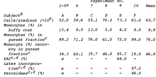
TABLE I. Purification of Human Monocytes Experiment No.
1-53 6 7 8 9 10 Mean Subject** Ά B C D E F
Cells/gradient (*106) 52.0 58.6 55.1 76.2 73.3 61.0 62.7 Monocytes (%) in
buffy coat 15.4 8.0 13.0 5.0 4.0 8.0 8.9 Monocytes (%) in
purest fraction0 88.2 71.2 76.0 61.0 72.0 88.0 76.0 Monocyte (%) recov-
ery in purest
fraction0 34.3 60.1 35.7 46.0 85.7 19.0 46.8
EACp'd (%) e 68.0
Latex incorpora-
tion0^ (%) e 87.0
Peroxidase°'d (%) e - - - 46.0
aValues in column 1 refer to mean of five separate experi- ments utilizing peripheral blood from donor A. Remaining columns (donors B - F) reflect data obtained from single ex-
periments utilizing peripheral blood from different individuals.
"Code letters specify individual blood donors.
cPurest fraction refers to the 16% metrizamide interface in experiments 1,8, and 10, to a pool of the 16 and 17% interfaces in experiments 6 and 7, and to the 17% interface for experi-
ment 9.
"Techniques used for functional tests are described in Section II.
eSee Table II.
(all from Microbiological Associates, Inc., Bethesda, Maryland), and DNase, 30 mg/liter (Worthington Biochemical Corp., Freehold, New Jersey, 2045 U/mg), pH adjusted to 7.3.
Hank's balanced salt solution, HBSS (Microbiological Asso- ciates, Inc., Bethesda, Maryland).
5x Tyrode's stock solution, each liter containing 5 gm dex- trose, 5 gm NaHC03, 1 gm KC1, 40 gm, NaCl, and 0.25 gm Na2HP04
(anhydrous).
Tyrode's gel DNase, appropriate dilution of 5* Tyrode's stock solution supplemented with 0.1% gelatin and DNase 30 mg/liter (Worthington Biochemical Corp., Freehold, New Jer- sey) , pH adjusted to 7.2.
Thirty percent working metrizamide solution. 30 gm metriza- mide (centrifugation grade, Nyegaard and Co., A/S, Oslo) diluted to 100 ml with Tyrode's gel DNase, prepared fresh before each fractionation.
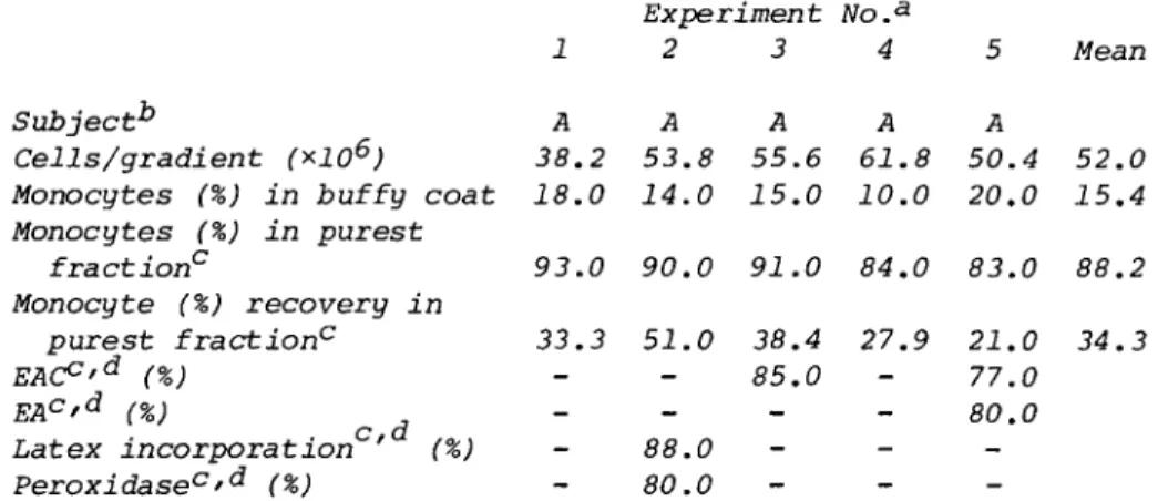
TABLE II. Purification of Human Monocytes
1 A 38.2 18.0
Experiment 2 3 A A 53.8 55.6 14.0 15.0
No.a 4 A 61.8 10.0
5 A 50.4 20.0
Mean
52.0 15.4 Subject1*
Cells/gradient (*106) Monocytes (%) in buffy coat Monocytes (%) in purest
fraction0 93.0 90.0 91.0 84.0 83.0 88.2 Monocyte (%) recovery in
purest fraction0 33.3 51.0 38.4 27.9 21.0 34.3
EAC°'d (%) - - 85.0 - 77.0
EAc'd (%) - 80.0
Latex incorporation ' (%) - 88.0 -
Peroxidasec'd (%) 80.0
aIn experiments 1 - 3 cells from all fractions were found to be 94% viable by trypan blue exclusion. Percent viability not determined in experiments 4 and 5.
Code letters specify individual blood donors. Same donor in experiments 1 - 5 (A).
cPurest fraction refers to the 16% metrizamide interface in experiments 1 - 4 , and a pool of the 16 and 17% interfaces in experiment 5.
^Techniques used for functional tests are described in Section II.
Dextran (4.5%), 4.5 gm dextran T500 (Pharmacia Fine Chemi- cals, Inc., Piscataway, New Jersey, MW 500,000) diluted to 100 ml with PBS; Heparin (Sigma Chemical Co., St. Louis, Mis- souri) diluted to 1000 U/ml in saline.
III. PROCEDURES
A. Cell Isolation
Collection of human peripheral blood white cells was ac- complished by drawing 50 ml of blood from normal individuals into a syringe containing 10 ml of 4.5% dextran and 0.5 ml heparin [final dextran concentration equals 0.9% (w/v), final heparin concentration equals 10 U/ml]. Blood was allowed to sediment at 37°C for approximately 30 min with the syringe se- cured in a vertical position. When sedimentation was complete,
TABLE III. Purification of Guinea Pig Macrophagesa Experiment No.a
11 12 13 14 Mean Cells/gradient (*106) 61.8 50.0 25.0 55.9 48.2 Macrophages (%) in unfrac-
tionated PE cells 67.0 72.0 77.0 78.0 73.5 Macrophages (%) in purest
fractions1* 94.8 97.2 96.3 97.9 96.6 Recovery of macrophages in
purest fractions1* (%) 13.2 58.9 93.8 47.2 53.5 experiments 11 -14 were done using guinea pig peritoneal exudate cells induced by injection of casein 4 days prior to harvest. Each experiment is based on the fractionation of cells from a single Hartley guinea pig.
bIn all four experiments, relatively pure macrophages were found in two successive fractions. These fractions were pooled accordingly, and chart values reflect differentials and counts on pooled fractions. In experiment 11 17% and 18%
metrizamide interfaces were pooled, in experiments 12 and 13 16% and 17% interfaces were pooled, and in experiment 14 15 and 16% interfaces were pooled.
the buffy coat material was expressed through an 18-gauge needle into a 50-ml conical centrifuge tube containing an equal volume of MEM complete. Cells were washed three times in MEM complete by centrifugation at 1000 rpm for 10 min. After the final wash, cells were counted and resuspended to between 38.0 x 10⁶ and 72.0 x 10⁶ cells/ml. One milliliter of result- ing cell suspension was layered onto each metrizamide gradient.
Casein-induced peritoneal exudate cells were collected from normal Hartley guinea pigs according to the method of David and David (2). Cells were washed three times in HBSS by centrifugation at 1000 rpm for 5 min. After the final wash, cells were counted and resuspended to between 25.0 x 10" and 61.0 x 10 cells/ml. One milliliter of resulting cell suspen- sion was layered onto each metrizamide gradient.
B. Purification of Cells on Metrizamide Gradients
Cells were fractionated on slightly hypertonic discontinu- ous metrizamide gradients. A 30% working metrizamide solution was freshly prepared, and dilutions, in Tyrode's gel DNase, of this stock solution were made on a percentage basis ranging
between 16 and 21% metrizamide (densities ranging between 1.088 and 1.108 gm/cm ). To prepare individual gradients, 2 ml vol- umes of decreasing metrizamide densities, 1% difference each, were layered into 15-ml conical centrifuge tubes (Falcon No.
2095), each tube ultimately containing six gradient steps. A 1-ml suspension of human buffy coat cells or guinea pig peri- toneal exudate cells (optimal cell concentrations ranging be- tween 40.0 x 10⁶ and 60.0 x 10⁶ cells/ml) was layered onto each gradient. Gradients were spun for 45 min at 1200 g in a centrifuge with a nonflexible shaft. Temperature was maintained at 20°C throughout centrifugation. Cells were collected from each interface using Pasteur pipettes, and washed three times in MEM complete by centrifugation at 1000 rpm for 10 min. Total
cell yield per fraction was determined, and cytocentrifuge smear preparations of each fraction were made. Smears were fixed for
5 min in methanol, allowed to air dry, and stained for 5 min in an 8% giemsa solution. Differentials were read under lOOx oil immersion. In certain experiments, cells were then taken from each fraction and tested for functional integrity.
Cytocentrifuge smears of each fraction were stained for peroxidase activity according to the method of Kaplow (3). The ability of the cells to ingest latex particles (4) and to phago- cytose antibody-coated red cells (5) were also determined.
IV. CRITICAL COMMENTS
Although absolute purity cannot be achieved by this modified metrizamide gradient method, fractions were recovered in which monocyte purity was found to be enriched at least eightfold from the buffy coat values (as is shown in Table I). This enrichment is substantial in view of the fact that it is achieved by a routine, single-step fractionation technique that is independent of the adherent capacity of human monocytes. Because the abili- ty to adhere to surfaces is not the determining factor in this purification method, the necessary and tedious process of re- covering cells from monolayers with EDTA, rubber policemen, or lidocaine treatment, all of which could possibly have an adverse effect on cell viability as well as on functional integrity, is not required. Therefore, under certain conditions this density gradient technique might be preferable to plating techniques.
In all cases tested, the percent phagocytosis of either latex particles or sensitized sheep red blood cells correlated direct- ly with the percent monocytes in fraction being analyzed (ex- periment 10, 88% monocyte/87% phagocytosis; experiment 9, 72%
monocyte/68% phagocytosis; experiment 2, 90% monocyte/88% pha- gocytosis; experiment 3, 91% monocyte/85% phagocytosis; experi-
ment 5, 83% monocyte/77 and 80% phagocytosis). This is evi- dence that differential counts were accurate and that the mo- nocytes present were viable and functionally competent. Purity of the monocyte-enriched fraction increases as percent mono- cytes in buffy coat increases. It must be noted, however, that percent recovery of total monocytes in the enriched fraction seems to decrease as purity of that fraction increases. The fact that this method also allows for a mean macrophage purity of 96.6% in the fractionation of guinea pig peritoneal exudate cells could be very useful in cases in which one requires pure guinea pig macrophages devoid of neutrophil contamination. A preliminary experiment showed that this method might also be useful in purifying mouse peritoneal exudate macrophages.
Acknowl e dgmen t
Supported by NIH grant No. AI 07685 and a grant from the Rockefeller Foundation.
REFERENCES
M. A. Vadas, J. R. David, A. Butterworth, N. T. Pisani, and T. A. Siongok. A new method for the purification of human eosinophils and neutrophils, and a comparison of the ability of these cells to damage schistosomula of
Schistosoma mansoni. J. Immunol. 122: 1228, 1979.
J. R. David and R. A. David. In "In Vitro Methods in Cell Mediated Immunity" (B. R. Bloom and P. R. Glade, eds.), p. 249. Academic Press, New York, 1970.
L. S. Kaplow. Simplified myeloperoxidase stain using ben- zidine hydrochloride. Blood 26: 215, 1965.
J. Michl, D. J. Ohlbaum, and S. C. Silverstein. 2-Deoxy- glucose selectivity inhibits Fc and complement receptor- mediated phagocytosis in mouse peritoneal macrophages.
J. Exp. Med. 144: 1465, 1976.
D. T. Fearon, K. F. Austen, and S. Ruddy. Formation of a hemolytically active intermediate by the interaction be- tween properdin factors B±D and the activated third compo- nent of complement. J. Exp. Med. 138: 1305, 1973.