Inflammation and Immunity
ROGER ROBINEAUX
Hôpital Saint-Antoine, Paris, France
Cells encountered in the phenomena of inflammation and immunity are not very good material for the study of cellular movement from a fundamental point of view. Many facts can, however, be gathered from observing them.
For several years, we have devoted a good deal of work to the study of the movement of cells involved in inflammation. This work has been resumed recently (Robineaux and Pinet, 1960). More recently still, we have studied in tissue culture the movement of cells involved in im- munity.
The technique of observation used in all these cases has been time- lapse microcinematography in phase or interference contrast, subse- quently analyzed frame-by-frame. We shall present here the results obtained in a study of cells of the adult guinea pig spleen. T h e cells were cultured under a dialysis membrane.
Material and Methods
The spleen cultures came from adult guinea pigs. Before splenec- tomy, the animals were bled by cardiac puncture and the spleens removed by lobotomy and placed in a sterile petri dish containing 10 ml of Hanks' solution. After removal of the capsule, the spleen was cut into fragments of less than 1 mm3 in culture medium.
NUTRIENT MEDIUM
The medium consisted of 7 0 % Parker 199, adjusted to pH 7.2 with sodium bicarbonate; 5 % chicken embryo extract; 2 5 % guinea pig serum, either autologous serum or a homologous serum from a pool of normal guinea pigs stocked at — 20°C; solution of antibiotics and antifungal agents—penicillin (200 units/ml), streptomycin (50 ^ig/ml), mycostatin (100 units/ml).
CULTURE TECHNIQUE
We have chosen to use the cellophane strip technique described by Rose et al. (1958). We used a standard size (2-ml) multipurpose chamber;
cover slips less than 0.15 mm in thickness were required for use with high- power and oil-immersion objectives.
351
352 ROGER ROBINEAUX
Four tissue fragments were placed on a cover slip in a small drop of nutrient medium. The cover slip was then completely covered with a strip 4 cm wide of Visking dialysis membrane previously sterilized in alcohol and washed in Hanks' solution. The silicone rubber gasket which forms the walls of the culture chamber was then put in place, followed by the second cover slip which closed the chamber. T h e whole chamber was maintained between two metal retaining plates by four screws. Two needles were placed in the gasket; one permitted the filling of the chamber above the cellophane strip with about 2 ml of nutrient medium, the other served as an air vent; both could be removed after filling.
Results
GENERAL ASPECTS
Beginning as early as the first hours of cultivation and continuing up to the third day, many cells could be observed migrating out from the expiant toward the periphery in the space delimited by the expiant, the cover slip, and the dialysis membrane. T h e most peripheral cells became flat and stationary when peripheral movement was limited by the dialysis membrane. In this zone are found neutrophilic and eosino- philic granulocytes, lymphoid cells of all ages, and many undifferentiated cells, with and without macrophagic activity. Cells with macrophagic activity typically clean the intermediary zone rather rapidly.
Between the third and the ninth days, the intermediary zone be- comes progressively organized: at about the tenth day, when many cell divisions appear, it assumes its most interesting aspect. T h e evolution of this zone will continue for 1 or 2 weeks.
It is easy to remove the entire culture on the cellophane strip for fixation and staining. The study of a fixed and stained preparation on about the fifteenth day shows all the intermediate types between reticular cells and mature plasmacytes. This method of culture then seems to represent a good system for obtaining in vitro all the stages of plasma- cyte differentiation. We shall now outline what can be found in the study of cells in this series.
C E L L MOVEMENTS
Two kinds of movement can be seen in this culture:
1. The movements of single cells: reticular cells, free or in syncytium, middle-size and small lymphocytes, plasmablasts (hemocytoblasts of the immunologists), proplasmacytes and plasmacytes, histioblasts and mono-
cytes, and polymorphonuclear leucocytes ("polymorphs") in the third day of cultivation. One can also study movements during mitosis in these different types of cells.
2. Cell interactions, of which the following aspects are the most characteristic: (a) Movements of cells in reticulolymphoplasmacytic islets;
(b) emperipolesis observable between small and middle-size lymphocytes and reticular cells or certain cells in mitosis.
Movement of Single Cells
1. Free Reticular Cells. T h e two types of cells encountered in the cultures can be distinguished clearly by the activities of their membranes.
In cells functioning as macrophages, these membranes are of the undu- lating type, pinocytosis is intense, and the cytoplasm is filled with phago- cytized material. In cells without macrophagic activity, there is no undulating membrane, no pinocytosis, and movement is restricted to
"microbubbling." These two types of cells are not polarized, and their locomotion is generally weak.
2. Syncytia. Numerous reticular cells organize themselves into syn- cytia containing numerous nuclei with nucleoli and cytoplasm containing large amounts of phagocytized material. T h e movements of the mem- brane are of the undulating type. T h e pinocytotic vacuoles which form at the periphery of the cell enter the cytoplasm in waves forming lines parallel to a tangent to the cell surface.
3. Middle-Size and Small Lymphocytes. These movements conform to the classic descriptions. In media free of plasma, they advance highly polarized, rapidly emitting many short hyaloplasmic veils at the anterior pole; these are plainly visible in phase contrast. In the small lymphocyte, these veils are smaller than in the middle-size lymphocyte, but they are just as active as the middle-size ones; the speed of migration is the same for the two types of cells. In the small, spherical lymphocytes, these veils can resemble by their length and fineness the undulating membranes of histiocytes.
In the course of their locomotion, they may have the so-called "hand- mirror" shape, but many remain oval or somewhat rectangular. T h e posterior part of the cell, dense and contracted, adheres very little to the substratum. We have never observed a lymphocyte change its polarity; it always moves straight forward, striking other cells, going around them, or engaging in emperipolesis. It can stop, and while stationary can lose its polarity, sending out only temporary ameboid pseudopodia. At this time, none of the characteristic production of hyaloplasmic veils occurs, as in the moving cell.
354 ROGER ROBIN EAU Χ
4. Plasmacytic Series. The type of culture utilized has permitted us to follow the dynamic behavior of plasmacytes of different ages.
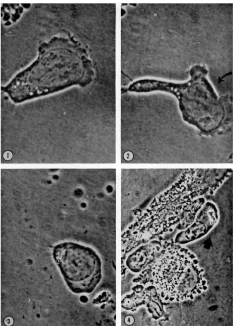
a. Plasmablast. This cell possesses much cytoplasm and an anteriorly located nucleus; it forms hyaloplasmic veils quite comparable to those of polymorphs; in addition, one can observe in the front of the cell during locomotion a moderate pinocytic activity. The hand-mirror form de- scribed for lymphocytes is clearly visible and a "tail" region of the cell appears then with clarity. T h e cell leaves behind, on the substratum, some trailing cytoplasmic fibrils (Fig. 1). These cells can change their polarity and one can then observe between the new anterior pole and the remains of the old one, the formation of a cortical gel (Fig. 2).
b. Proplasmacyte. At this stage the cell has reduced its volume and no longer shows hyaloplasmic veils, but only temporary cytoplasmic pro- trusions which then become the front of the cell in locomotion. The nucleus is generally situated in a median position, with the centrosome behind it still visible; the "tail" of the proplasmacyte hardly adheres at all to the substratum (Fig. 3).
c. Plasmacyte. These cells, which are very easily identified in our cultures, can locomote easily but slowly; while the speed of migration of polymorphs and of lymphocytes is comparable (20-35 μ/min), the plas- mocytes move much more slowly (20-25% as rapidly); as for the lympho- cytes, the locomotion is polarized, and the cells do not change their polarization. When the cell is moving, it takes on a trapezoidal form, the longest side located at the front of the cell. The nucleus is median in location; the cell then resembles a cylindrical epithelial cell, with the mitochondria occupying principally the anterior part of the cytoplasm.
The movement occurs without intervention of the hyaloplasmic veils by a mass cytoplasmic streaming relative to the substratum. It is clear that that maturation is accompanied by a radical modification of dynamic behavior in speed as well as morphology (Fig. 4).
5. Histiocytes, a. Histioblasts. This type of cell, quite rarely en- countered in our cultures, moves very little; when it does move, it re- sembles all histiocytic cells: large undulating membranes and intense pinocytic activity. One can recognize in this type a bipolarity on the same axis.
b. Monocytes. These are histiocytic elements with feeble locomotion and hyaloplasmic veils of the undulating membrane type. This cell can also change polarity. In Figs. 5 to 7 it is to be noted that the change in orientation of the cell is accompanied by the formation of a new mem- brane. The gelated zone easily observable behind the hyaloplasmic veils at the anterior pole disappears before the veils themselves disappear, as the poles change. T h e gel zone then reappears after the hyaloplasmic veils
FIG. 1. Plasmablast exhibiting numerous hyaloplasmic veils at the anterior end.
Note the pinocytic activity. Magnification: χ 1368.
FIG. 2. T h e same plasmablast showing a gelated zone between the old and the new veil formation. Magnification: χ 1368.
FIG. 3. Proplasmacyte. No hyaloplasmic veils are visible at the anterior pole and the cell has an ameboid movement. Magnification: χ 1368.
FIG. 4. Mature plasmacyte resembling a cylindrical epithelial cell. No hyaloplas- mic veils are visible. T h e cell flows in mass on the substratum. Magnification: χ 900.
356 ROGER ROBINEAUX
appear at the front of the cell. The change of polarity is thus accompanied by a parallel change in the distribution of gel. It should be added that the tail of the monocyte adheres to the substratum and leaves behind easily visible cytoplasmic fibrils.
6. Mitoses. Of all the cells encountered in our cultures three types can be distinguished: reticular cells coming directly from the reticulum,
FIGS. 5, 6, 7. Monocyte. T h e polarity of the cell changes and at the same time the gelated zone is reorganized behind the veils of the anterior pole (10 sec between each picture). Magnification: χ 1254.
plasmablasts derived from elements of the reticulum, and middle-size lymphocytes which we think issue from germinative centers of the ex- plant. A point of some interest was the fact that movements of the cells disappeared during division, only to return after division and separation into daughter cells was completed. It is, for example, very striking to see the middle-size lymphocytes mobilize themselves rapidly after their divi- sion and recoup their original mode of interphase movement.
7. Clasmatosis. This is the segregation of one part of the cytoplasm
lacking a nucleus; it is easily seen in the cells of the plasmacytic series.
Clasmatosis can occur also in reticular cells. This segregation occurs after the production of a large ameboid process, which becomes somehow separated, apparently by "strangulation." During this process, cytoplasmic streaming is clearly visible, especially in dividing plasmablasts.
Cellular Interactions
I. Emperipolesis. Rapid cellular contacts between small and middle- size lymphocytes, on the one hand, and reticular cells, on the other (with
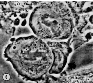
FIG. 8. Mitosis of a plasmablast. Emperipolesis by a lymphocyte inhibits bubbling of the two daughter cells. Magnification: χ 1900.
or without macrophagic function) are very frequent. They are similar to those which one can observe between lymphocytes and cancer cells in culture. This emperipolesis represents, probably, a surface phenomenon;
it must be distinguished from certain situations where lymphocytes, plas- macytes, and monocytes are included and moving into the syncytium.
A particular type of emperipolesis is to be noted in middle-size lym- phocytes and plasmacytes in mitosis. Cell contact is generally established at the moment of cytokinesis, and on the cytoplasmic bridge; it is to be noted that at the moment of contact, the bubbling which is so violent at this stage of mitosis stops in both of the daughter cells (Fig. 8). T h e movement continues once the lymphocyte has broken off contact. We have
358 ROGER ROBINEAUX
observed this phenomenon with plasmablasts, but not with the middle- size lymphocytes or with the reticular cells.
2. Lymphoplasmacytic Reticular Islets. We were able to observe such an islet in culture for 6 days. T h e center is formed by reticular cells be- coming progressively flat. The reticular cell exhibits weak membrane activity on the part of the microvilli, but without any microscopically visible pinocytosis. The population which surrounds this cell changes with time. At the beginning one can observe essentially lymphocytes;
48 hr later, plasma cells are in the majority; after 72 hr, large mononu- cleate cells appear; then by the fifth to sixth day, the plasmacytes are present once again. The movements of the cells which surround the reticular cell are the same as those described earlier for single cells.
In addition, there are two other types of cell movements to be men- tioned:
a. Emperipolesis of lymphoid cells bordering on the reticular cell, or crossing on top of, or beneath the reticular cell.
b. Close relations between reticular cells, on the one hand, and plasmacytes and mononucleate cells (proplasmacytes?), on the other. On accelerated projection of a film, it is almost impossible to distinguish the closely associated cytoplasms of the two cells.
It should be added that during this entire period of observation of 6 days, the reticular cells were not observed to divide.
Discussion and Conclusions
We can thus review the observed facts seen in our cultures concerning cell movements:
1. All the migrating cells, except mature plasmacytes, exhibit hyalo- plasmic veils at their anterior pole. These veils take on, in the monocytes, the typical character of the undulating membrane. In general, hyalo- plasmic veils vary from one type of cell to another in number, size, and speed. It seems to be true that one can observe some intermediate types of veils between those described in the polymorph and those in cells with histiocytic function.
2. Stationary histiocytic cells, syncytia, macrophagic reticular cells, and histioblasts produce undulating membranes which are large and numerous. These cells are not polarized, except for the histioblasts.
3. Nonmacrophagic reticular cells produce only microvilli.
4. The tail of locomoting cells is clearly visible at the opposite end of the cell from the hyaloplasmic veils, except in the mature plasmacytes.
In the tail one can observe contractions, particularly in histiocytes.
5. The histioblasts which are rich in undulating membranes, and
polymorphs which are rich in hyaloplasmic veils, can have a double polarity and extend in two opposite directions. If one of the two poles disappears, the hyaloplasmic expansions disappear first, the adjacent gel layer persists, becomes denser, and organizes itself as the tail for the cell during movement.
6. If the cell has two "anterior" poles, then these are separated by a cortical gel.
7. Cytoplasmic streaming is visible only in polymorphs; streaming is not continuous even though its direction is the same as the direction of locomotion. It is doubtful whether the streaming plays a role in the locomotion of the cell.
8. T h e emperipolesis of the lymphocytes characterized by fast con- tacts is observed in nonmacrophagic reticular cells and plasmablasts at the time of cytokinesis.
9. Some prolonged contacts can be established, on the one hand be- tween middle-size lymphocytes or plasmacytes and a macrophagic reticu- lar cell rich in phagocytized material, or, on the other hand, between young or old plasmacytes and nonmacrophagic reticular cells. These relations have completely different aspects in the two types of reticular cells.
How should we interpret all these observations?
C E L L MOVEMENTS
Lewis (1931, 1939, 1942) and De Bruyn (1944, 1945, 1946, 1947) be- lieve that the movements of the ameba and of leucocytes are essentially of the same nature. Their conclusions are based on: (1) the similarity of form of these cells during movement; and (2) the behavior of lateral protuberances and of granules, showing that in the leucocytes as in the ameba, the lateral parts of the cytoplasm are stationary. There exists at the surface a superficial gel layer where processes of contraction take place. These contractions would explain a forward displacement of the plasmasol. A solation must occur in the body, since plasmasol is con- stantly present at the anterior pole.
In regard to the observations, it seems that insufficient attention has been paid to the production of hyaloplasmic veils in the leucocyte (with the exception of mature plasmacytes).
These numerous and active veils are quite visible with phase and in- terference contrast microscopy. Their existence suggests a mechanism of migration analogous to that described by Ambrose (1961) for moving fibroblasts on a solid substratum. The migration was postulated to be due to the undulations of the cell membrane apposed to the substrate. These undulations would themselves be produced by contractile fibrils embedded
360 ROGER ROBINEAUX
in the cytoplasm, parallel and close to the contact surface. Such a mecha- nism evidently could be invoked to explain movement in leucocytes.
It is, however, evident that some contractile phenomena are observable in leucocytes in motion, in particular at the posterior pole. We have pre- viously presented a series of interference contrast pictures of this (Robin- eaux and Pinet, 1960). This progressive and continued contraction was marked by the migration of two vacuoles toward the Golgi zone, and the anteriolateral production of a hyaloplasmic expansion.
It is, besides, easy to observe the stationary gelated lateral zones of a cell in movement, just as they were described by De Bruyn (1947).
The formation itself of undulating membrane in a stationary cell shows the production of gelated zones which are linear and radial, and between which form pinocytosis vacuoles; their orientation indicates the direction of the retraction of the membrane. The coexistence within the leucocytes of gel formations and of movement is certain; the variation of movement probably bears some strict relationship with the variation of gel structure.
If one compares these observations with the observations of Gold- acre (Goldacre and Lorch, 1950a,b; Goldacre, 1961) on the ameba, one finds agreement concerning the formation de novo of membrane in the front of leucocytes (at least during pinocytosis) and concerning tail contraction.
But, the existence of gelated zones in other parts of the cell in the anterior pole of a leucocyte in movement or in the undulating membranes of stationary cells of macrophagic function would support the presence of local zones of contraction wherever the movement is produced.
A possible relationship between the zone of contraction and cytoplas- mic streaming, such as has been suggested by Allen (1961a,b; Allen et ah,
1960) in the frontal contraction theory, is at present impossible to deter- mine in the case of leucocytes.
In the particular case of movement in plasmacytes, we can affirm that movement is in strict agreement with the production of hyaloplasmic veils. Numerous in plasmablasts, these structures are correlated with a high velocity of migration. Less numerous in proplasmacytes, they are correlated with a less rapid locomotion. There are no veils in mature plasmacytes where the movements have become slower and where mass cytoplasmic streaming is observable. The progressive disappearance of veils in the course of plasmacyte maturation is perhaps in agreement with the development of endoplasmic reticulum in these cells. The extreme development of this reticulum might confer on the mature plasmacyte some aspects of its characteristic movement.
Thus, besides the phenomena of contraction, the movements of the
hyaloplasm appear to play a role of great importance in the movement of leucocytes. The movement of the leucocyte would then seem to resem- ble, in part, both the ameba and the fibroblast.
The production of these veils raises the question of the formation of new membranes, or perhaps of the reutilization of material from resorbed membranes. As far as the ameba is concerned, the formation of new membranes during locomotion is contested by Griffin and Allen (1960) and more recently by Wolpert (1963). This appears true for the ameba, but would not seem applicable to leucocytes, because of the importance of providing a new supply of membrane for the formation of pinocytotic vesicles. The relation between movement and the production of new surface as described by Bell (1961) appears to us to be especially applica- ble to leucocytes, with the exception of mature plasmacytes.
CELLULAR INTERACTIONS
We have discussed this question elsewhere (Robineaux et al., 1962a,b) in detail, but will now summarize some of the principal cellular inter- actions.
1. Emperipolesis. We have observed the same phenomena in tissue cultures of spleen under dialysis membrane as described by Humble et al.
(1956) in agar cultures, between lymphocytes and neoplastic cells; be- tween lymphocytes and megakaryocytes; and between lymphocytes and megaloblasts. In our experiments, these surface relations become estab- lished between lymphocytes and reticular cells, and between lymphocytes and plasmablasts in mitosis.
No interpretation can, as yet, be given to this phenomenon. T h e sug- gestion of Humble et al. (1956), according to which the lymphocytes are the mobile sources of enzymes or of metabolites necessary to the cell for growth or mitosis still remains only a hypothesis. It is possible that the speed of their movement, the passage of one reticular cell to another, can suggest a transport function, but one cannot say more. However, a recent study by Loutit (1962) marshals many arguments in favor of a trophic role for lymphocytes.1
2. Lymphoplasmacytic Reticular Islets. These islets have been des- cribed by Undritz (1950) in fixed and stained lymph node smears. They were found again by Thiéry (1960) in fresh preparations of dissociated lymph node, and in thin sections studied by electron microscopy. We have just described them in differentiated cultures of spleen.
These islets have both an anatomical and functional significance: T h e
l Two varieties of lymphocytes exist. One type incorporates tritiated thymidine very slowly; it plays a role in immune phenomena. T h e other variety with a short tritiated thymidine incorporation time has no known function (Everett et al., 1963).
362 ROGER ROBINEAUX
close relation between proplasmacytes and reticular cells suggests a trans- fer system.
It is important to recall at this time that the experiments conducted by Sharp and Bur well (1960) and by Fishman (1961) have advanced the notion of interrelationships between macrophages and antibody-forming cells in sensitized systems in tissue culture.
Finally the passage, by micropinocytosis, of macromolecules from a reticular cell into a plasmacyte (Thiéry, 1962) is in support of some kind of messenger system.
A possible immunological significance of strict relationships between reticular cells and plasmacytes might be suggested: some information (in the form of ribonucleic acid?) might be transmitted from the reticular cell reservoir containing antigenic structure to the plasmacyte which forms specific protein antibodies.
Summary
The culture of adult guinea pig spleen under dialysis membrane has made it possible to obtain a population of growing, differentiating cells.
The cells encountered are involved in the phenomena of inflammation and immunity. Their movements, their divisions, and their interactions with other cell types have been studied. Particular attention has been given to the plasmacytic series, of which all stages of development of the various movement patterns could be found. The anatomical and physiolo- gical relations between plasmacytes and reticular cells have been studied from a dynamic point of view.
REFERENCES
Allen, R. D. (1961a). In "The Cell" (J. Brächet and Α. Ε. Mirsky, eds.), pp. 135-216.
Academic Press, New York.
Allen, R. D. (1961b). Exptl. Cell Res. Suppl. 8, 17-31.
Allen, R. D., Cooledge, J . W., and Hall, P. J . (1960). Nature 187, 896-899.
Ambrose, E. J . (1961). Exptl. Cell Res. Suppl. 8, 54-73.
Bell, L. G. E. (1961). / . Theoret. Biol. 1, 104-105.
De Bruyn, P. P. H. (1944). Anat. Record 89, 43-63.
De Bruyn, P. P. H. (1945). Anat. Record 93, 295-315.
De Bruyn, P. P. H. (1946). Anat. Record 95, 177-192.
De Bruyn, P. P. H. (1947). Quart. Rev. Biol. 22, 1-24.
Everett, N. B., Caftrey, R. W., and Rieke, W. O. (1963). In "Leukopoiesis in Health and Disease," N.Y. Acad. Sei. In preparation.
Fishman, M. (1961). / . Exptl. Med. 114, 837-856.
Goldacre, R. J . (1961). Exptl. Cell Res. Suppl. 8, 1-16.
Goldacre, R. J . , and Lorch, I. J . (1950a). Nature 166, 497-500.
Goldacre, R. J . , and Lorch, I. J . (1950b). Intern. Rev. Cytol. 1, 135-164.
Griffin, J . L., and Allen, R. D. (1960). Exptl. Cell Res. 20, 619-622.
Humble, J . G., Jayne, W. H. W., and Pulvertaft, R. J . V. (1956). Brit. J. Haematol.
2, 283.
Lewis, W. H. (1931). Bull. Johns Hopkins Hosp. 4 9 , 29-36.
Lewis, W. H. (1939). Arch. Exptl. Zellforsch. Gewebezücht. 2 3 , 1-7.
Lewis, W. H. (1942). In "Structure of Protoplasm" (W. Seifritz, ed.). Iowa State College Press, Ames, Iowa.
Loutit, J . F. (1962). Lancet 2 4 , 1106-1108.
Robineaux, R., and Pinet, J . (1960). Ciba Found. Symp. Cellular Aspects Immunity, pp. 5-40.
Robineaux, R., Pinet, J . , and Kourilsky, R. (1962a). Compt. Rend. Soc. Biol. 1 5 6 , 1025.
Robineaux, R., Pinet, J . , and Kourilsky, R. (1962b). Nouvelle Rev. Franc. Hematol.
2 , 797-811.
Rose, G. G., Pomerat, C. M., Shindler, T . O., and Trunnel, J . B. (1958). ./. Biophys.
Biochem. Cytol. 4 , 761-764.
Sharp, J . S., and Burwell, R. G. (1960). Nature 1 8 8 , 474.
Thiéry, J . P. (1960). Ciba Found. Symp. Cellular Aspects Immunity, pp. 59-91.
Thiéry, J . P. (1962). / . Microscopie 1, 275-286.
Undritz, E. (1950). Folia Haematol. 7 0 , 32-42.
Wolpert, L. (1963). Personal communication.
DISCUSSION
DR. RHEA: DO you have any evidence of active movement on the part of the nucleus, or was it all passive?
DR. ROBINEAUX: This is an important problem. In the cells of these cultures the nuclei are very deformable, especially in polymorphs and macrophages, and to a lesser degree in lymphoid cells. However, these are probably passive movements.
DR. REBHUN: I noticed two very interesting things in some of your films. In one, surface bubbling appeared only at telophase, while the cell was smooth during the rest of mitosis.
In one of your other films, an elongated cell with pseudopods at the polar regions divided, but the mitosis occurred perpendicularly to the axis of the cell. T h e furrow cut in, in a direction parallel to the long axis. Do these two things occur often in these cells?
DR. ROBINEAUX: Concerning the mitosis of reticular cells, it is usually seen that:
(1) the cells are bipolar, and (2) the spindle is perpendicular to the polar axis. I am not sure whether this is always true.
The bubbling is ordinarily seen at anaphase and telophase, but may also be seen earlier, for example, in metaphase. In the case that you referred to, two lymphocytes established cell contact with the reticular cell (emperipolesis); the bubbling appeared after the departure of the lymphocytes in telophase.
DR. D E BRUYN: I would like to comment on the nomenclature of these cells, even though it was stated in your film that the problem of nomenclature is not as yet settled. All of the cells that you called "plasmablasts," I would call "medium-sized or large lymphocytes." Similarly, I believe that your plasmacytes are cells in the eryth- rocytic series.
If I understood you correctly, cells (lymphocytes, or whatever they were) were believed to penetrate the macrophage or reticular cell. I have seen things like this, too, but I always assumed that these cells moved between the cover glass and the larger cell. If really true, I think penetration of one cell by another would be an ex- tremely interesting phenomenon.
364 ROGER ROBINEAUX
DR. ROBINEAUX: T O answer your first question, I do not agree that our plasmacytes are erythroblasts. T h e size, shape, and color characteristics of these cells are those of plasmacytes. In fact, there are no erythroblasts in our cultures. Certainly an ultrastruc- tural study could settle the matter.
About the relations between lymphocytes, plasmacytes, monocytes, and reticular cells in syncytia. First, lymphocytes in emperipolesis: I think this is a surface phenomenon.
I asked Pulvertaft what he thought, but he was not certain whether the phenomenon was intracytoplasmic or at the surface. Second, lymphocytes, plasmacytes, monocytes inside syncytia: this phenomenon is similar to what is seen in cancer cells and mega- karyocytes. These cells are included in an empty round surface in the cytoplasm, and the cell membranes are quite distinct. It could not be a superposition, because the depth of field is very thin and the details are just as distinct in the syncytia as in the reticular cells.
DR. BISHOP: Have you studied antibody production or release?
DR. ROBINEAUX: I think this kind of culture will permit many studies of this kind to be carried out, but I have had no experience in this field. T h e animals from which the cultures came were not sensitized, but the media were heterologous, containing chicken embryo extract.