Acta Physiologica. 2020;228:e13345.
|
1 of 16https://doi.org/10.1111/apha.13345 wileyonlinelibrary.com/journal/apha
1 | INTRODUCTION
The abundance and convenience of energy‐rich foods with high hedonic value poses a homeostatic challenge for modern
humans and predisposes them to obesity and associated car- diometabolic and psychiatric diseases.1 While the maladap- tive peripheral and central responses to chronic overeating have been extensively explored,2-4 present knowledge of the R E G U L A R PA P E R
Blunted leptin sensitivity during hedonic overeating can be
reinstated by activating galanin 2 receptors (Gal2R) in the lateral hypothalamus
Este Leidmaa
1,2,3| Mary Gazea
1| Alexandre V. Patchev
1| Anna Pissioti
1|
Nils Christian Gassen
1| Mayumi Kimura
1| Zsolt Liposits
4| Imre Kallo
4|
Osborne F. X. Almeida
1This is an open access article under the terms of the Creative Commons Attribution License, which permits use, distribution and reproduction in any medium, provided the original work is properly cited.
© 2019 The Authors. Acta Physiologica published by John Wiley & Sons Ltd on behalf of Scandinavian Physiological Society 1Max Planck Institute of Psychiatry,
Munich, Germany
2Graduate School of Systems
Neuroscience, Munich University, Planegg‐
Martinsried, Germany
3Institute of Molecular Psychiatry, Bonn, Germany
4Institute of Experimental
Medicine, Hungarian Academy of Sciences, Budapest, Hungary
Correspondence
Osborne F. X. Almeida, Max Planck Institute of Psychiatry – NeuroAdaptations, Munich, Germany
Email: osa@psych.mpg.de Funding information
European Union (FP7 Health), Grant/
Award Number: 259772 to OFXA; National Science Foundation of Hugary (Országos Tudományos Kutatási Alapprogramok), Grant/Award Number: K101326 to IK
Abstract
Aim: Since foods with high hedonic value are often consumed in excess of energetic needs, this study was designed to identify the mechanisms that may counter anorexi- genic signalling in the presence of hedonic foods in lean animals.
Methods: Mice, in different states of satiety (fed/fasted, or fed/fasted and treated with ghrelin or leptin, respectively), were allowed to choose between high‐fat/high‐
sucrose and standard foods. Intake of each food type and the activity of hypothalamic neuropetidergic neurons that regulate appetite were monitored. In some cases, food choice was monitored in leptin‐injected fasted mice that received microinjections of galanin receptor agonists into the lateral hypothalamus.
Results: Appetite‐stimulating orexin neurons in the lateral hypothalamus are rap- idly activated when lean, satiated mice consume a highly palatable food (PF); such activation (upregulated c‐Fos expression) occurred even after administration of the anorexigenic hormone leptin and despite intact leptin signalling in the hypothala- mus. The ability of leptin to restrain PF eating is restored when a galanin receptor 2 (Gal2R) agonist is injected into the lateral hypothalamus.
Conclusion: Hedonically‐loaded foods interrupt the inhibitory actions of leptin on orexin neurons and interfere with the homeostatic control of feeding. Overeating of palatable foods can be curtailed in lean animals by activating Gal2R in the lateral hypothalamus.
K E Y W O R D S
galanin, ghrelin, lateral hypothalamus, leptin, orexin, overeating
proximal neurobiological mechanisms that initiate overeating is poor. In light of the “hedonia hypothesis of obesity” that posits that foods imbued with pleasurable signals override (and/or reset) homeostatic set‐points,5 we here aimed at iden- tifying mechanisms that may counter anorexigenic signalling when lean mice encounter hedonic foods.
The orexigenic peptide ghrelin stimulates appetite; it is the only “hunger hormone” known to date.6,7 Ghrelin levels are downregulated in the obese state, making its direct involve- ment in the pathophysiology of obesity unlikely.8,9 However, the early role of ghrelin in triggering obesity should not be underestimated since the start and termination of the feeding cycle depends on a fine balance between the orexigenic ef- fects of ghrelin and those of anorexigenic hormones such as leptin. Leptin, released into the blood as energy reserves are replenished, reduces appetite and stimulates energy expen- diture10; thus, leptin serves as a major satiety signal. While the ability of leptin to act as a physiological brake on eating is compromised in the obese state because of the develop- ment of leptin resistance,11,12 it seems plausible that transient leptin resistance may explain why metabolically healthy indi- viduals indulge in foods of high hedonic value. The hedonic properties of palatable foods (PF) can be mainly ascribed to their high content of energy‐dense fats and sugars that stimu- late the senses of taste, smell and texture.
Ghrelin and leptin receptors are prominently expressed in hypothalamic nuclei (arcuate nucleus [Arc] and lateral hy- pothalamic area [LHA]) that are critical for the regulation of energy balance13-15 where these orexigenic (ghrelin) and an- orexigenic (leptin) hormones exert opposing effects. Ghrelin stimulates orexigenic neuropeptide Y/agouti‐related peptide (NPY) neurons in the Arc16,17 and orexin (OX/hypocretin) neurons that are expressed exclusively in the LHA.7,18-20 Leptin activates anorexigenic pro‐opiomelanocortin (POMC) neurons and inhibits orexigenic NPY neurons 3,21 which ulti- mately innervate OX neurons in the LHA. Besides increasing appetite, OX is also implicated in reward‐related behaviour and as a driver of hedonic feeding.22,23 It is important to note that OX neurons that are subject to inhibition by leptin24,25 do not express LepRb14,26 but in addition to their POMC inputs, receive dense inputs from neighbouring LepRb‐positive neu- rons. The latter co‐express galanin (Gal), neurotensin (Nts) and gamma‐aminobutyric acid (GABA).14,24,26-28 There is evidence that Gal mediates the inhibitory actions of leptin on OX neurons from studies using acute hypothalamic slices and knockout mouse models.24,25
In this study, using lean, adult male mice, we tested the hypothesis that overeating of PF results from disruption of the mechanisms through which leptin inhibits OX neurons.
To begin dissecting how different elements of this com- plex neurocircuitry contribute to hedonic overeating, we first tested whether acute PF affects the efficacy of leptin or ghrelin on food intake. First, we compared food consumption
in mice that received peripheral injections of either saline, ghrelin or leptin before being given access to standard chow (SC, here considered to be hedonically neutral) for 1 h vs sa- line‐, ghrelin‐ or leptin‐injected mice exposed to a so‐called
“choice paradigm” in which mice could choose between SC and PF (hedonically loaded) or SC over 1 h. Brain areas possibly contributing to hedonic eating were mapped using immediate early gene c‐Fos, and c‐Fos was monitored in first‐ and second‐order neurons (POMC, OX); in addition, we examined the efficacy of leptin signalling (induction of pSTAT3)11,12 when leptin‐treated mice were confronted with hedonically‐loaded PF. Lastly, we examined whether intra‐
LHA injections of Gal can restore the ability of leptin to re- duce hedonic eating.
Briefly, the present experiments demonstrated that expo- sure of mice to PF triggers hyperphagia, a phenomenon that can be attributed to an escape of OX neurons from the inhibi- tory actions of leptin. Furthermore, we show that the anorexi- genic effects of leptin can be pharmacologically reinstated by stimulation of the Gal receptor 2 (Gal2R) in the LHA. These observations suggest that descending hedonic information impinges on Gal neurons which, in turn, gate the access of peripheral signals of satiety to OX neurons.
2 | RESULTS
2.1 | Palatable food challenges homeostatic control mechanisms in LHA
In order to study the efficacy of homeostatic signals dur- ing initial acute exposure to hedonic foods, non‐obese mice were injected with either ghrelin to induce hunger or leptin to mimic satiety, before testing their intake of standard chow (SC) in a no‐choice paradigm (SC/SC) vs palatable food (PF) in a choice paradigm (SC/PF), as shown schematically in Figure S1A,B. These tests were conducted at ZT3 in the light‐phase (resting period) to increase the level of conflict between the homeostatic state of the animal and the hedonic motivation to eat.
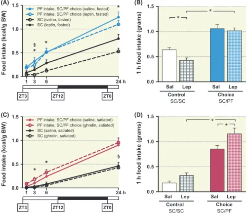
In lean, fasted mice, independent experiments using within‐subject (Figure 1A) and between‐subject (Figure 1B) designs showed that systemic leptin injections are more ef- fective at suppressing SC ingestion than PF within the first hour of food presentation (for normalized data, see Figure S1C). Pre‐treatment with leptin before exposure to SC/SC suppressed eating, as expected. In contrast, leptin‐pretreated mice that were subsequently given access to SC/PF choice escaped from the anorexigenic effects of the peptide (Figure 1B). As shown in Figure S1D, the amount of SC ingested in the SC/PF choice paradigm was negligible in both saline‐ and leptin‐treated mice. The same experiment was repeated in the dark phase (ZT16) when similar results were obtained (data not shown).
A converse set of experiments showed that ghrelin (Ghr) administration to mice that had been previously satiated on SC stimulated only PF and not SC consumption (compare Control SC/SC vs Choice SC/PF) (Figure 1C within‐subject and Figure 1D between‐subject experiment, normalized data in Figure S1F).
We next sought to identify the initial neural correlates of hedonic eating (ZT16, 4 h into the dark phase) when mice would have satiated their homeostatic energy needs. In this experiment, SC‐satiated mice that were allowed to choose between a palatable milk solution and SC, preferentially consumed milk (0.52 ± 0.1 kcal/g BW) as compared to SC (0.02 ± 0.02 kcal/g BW) during the first hour. Neuronal ac- tivity in brain areas likely involved in the physiological and behavioural regulation of hedonic eating was monitored by measuring c‐Fos (mRNA and protein) expression.29,30 After 1 h of exposure to milk, increased activity was observed in brain regions implicated in the processing of reward/hedonic information (Figure 2A), eg piriform cortex (olfaction), insular cortex (taste), ventral pallidum (hedonic, reward)
orbitofrontal cortex, lateral septum (association) and amyg- dala (emotional learning and reward valuation). Hedonic eat- ing was also accompanied by strong activation of two satiety centres, the ventromedial and dorsomedial hypothalamic nu- clei, as well of the lateral hypothalamic area (LHA). Orexin (OX) neurons located in the LHA19,31 are normally activated during hunger to stimulate food intake. In the present study, evaluation of pre‐pro‐OX mRNA transcript levels in the LHA of SC‐satiated mice revealed that OX mRNA levels are up- regulated within 1 h of consuming milk and return to baseline levels after 24 h. Interestingly, OX mRNA was again upreg- ulated when mice, previously satiated on milk for 24 h, were offered a different hedonic stimulus (solid PF comprised of high‐fat/high‐sucrose), as shown in Figure 2B. These data imply that, in addition to hunger per se, hedonic foods in- crease the activity of OX neurons in the LHA. Under both conditions (presentation of milk for 1 h and presentation of solid PF for 1 h to mice previously satiated with milk), the in- creases in OX mRNA levels were accompanied by elevations in serum corticosterone (CORT) levels (Figure 2C).
FIGURE 1 Overeating of highly palatable food (PF) is enhanced by ghrelin but is not suppressed by leptin. A, Leptin (3 mg/kg ip, after 24 h fast) suppresses consumption of SC (within‐subject design (n = 18); F1,34 = 50.2, P < .0001; time × drug interaction: F3,34 = 33.1, P < .0001,
*Sidak's post hoc P < .05) but only partially reduces PF intake (F1,34 = 7.1, P < .05; time × drug interaction: F3,34 = 24.9, P < .0001, §P < .05). B, Leptin initially (during 1 h) suppresses SC but not PF intake (between‐subject design; n = 20‐22/group; leptin effect: F1,81 = 4.564, P = .036; food type × leptin interaction: F1,81 = 2.00, P = .16, *Tukey's post hoc P < .05). C, In an independent set of satiated mice exposed to the SC/PF choice paradigm, ghrelin (Ghr) injections (0.3 mg/kg) stimulated the cumulative intake of palatable food (PF) over 24 h (F1,34 = 12.6, *Sidak's post‐hoc P < .01); Ghr did not increase SC consumption (F1,34 = 6.6, P < .05, §post‐hoc P < .05), within‐subject design (n = 18). (D) Ghr stimulates PF ingestion and not that of SC (between‐subject design, n = 10/group; measurements 1 h‐post treatment; drug effect: F1,35 = 10.2, P = .003, *Tukey's post‐hoc P < .05)
1 3 6 24 h
0.0 0.5 1.0 1.5
Food intake (kcal/g BW)
SC (saline, satiated) SC (ghrelin, satiated)
PF intake, SC/PF choice (saline, satiated) PF intake, SC/PF choice (ghrelin, satiated)
* *
ZT3 ZT12 ZT0
1 3 6 24 h
0.0 0.5 1.0 1.5
Food intake (kcal/g BW)
SC (leptin, fasted) SC (saline, fasted)
PF intake, SC/PF choice (saline, fasted) PF intake, SC/PF choice (leptin, fasted)
ZT3 ZT12 ZT0
* *
*
* *
0.0 0.5 1.0 1.5
1 h food intake (grams)
Sal Lep Control
SC/SC Choice
SC/PF Sal Lep
0.0 0.5 1.0 1.5
1 h food intake (grams)
Sal Lep Control
SC/SC Choice
SC/PF Sal Lep
* *
(A) (B)
(C) (D)
(C) (D)
(A) (B)
Milk Milk Milk
2.2 | Orexin neurons in LHA escape inhibition by leptin during eating
Since PF intake is not decreased by leptin (Figure 1A,B) and upregulates OX expression (Figure 2B), we next measured the effects of leptin on orexigenic peptide expression in the LHA. As observed in SC‐satiated mice (Figure 2B), PF up- regulated OX expression (Figure 2D). Unexpectedly,4,32 ad- ministration of exogenous leptin to previously fasted mice did not influence OX mRNA expression in the LHA (Figure 2D). Notably, neither PF nor leptin altered mRNA expression of another LHA orexigenic peptide, melanin‐concentrating hormone (MCH) (Figure 2D). Nevertheless, leptin reduced the number of c‐FOS/OX co‐expressing LHA neurons in mice offered SC/SC. Consistent with the results of previous
electrophysiological studies,19 our observations indicate that leptin suppresses OX‐ergic activity when mice are exposed to control food (SC). Interestingly, however, leptin did not affect OX neuron activity in mice given the SC/PF choice (Figure 3A,B); this result matches the observation that leptin does not curtail the eating of PF (Figure 1A,B).
2.3 | Initial contact with PF does not interrupt leptin signalling in LHA
Effective dosage of leptin was confirmed by assaying serum levels of leptin (Figure 4A) and of insulin and Ghr (Figure S2). Moreover, measurements of pSTAT3 (Tyr705), which mediates the actions of leptin,2,12 revealed that acute PF in- gestion does not disrupt leptin signalling in the brain. Briefly, FIGURE 2 Hedonic eating upregulates c‐Fos and orexin expression in the LHA. A, Qualitative relative scores of c‐Fos mRNA expression in various relevant brain areas of mice after they had consumed either a hedonic solution (milk) or water for 1 h; the mice were already satiated on standard chow (SC) (liquids co‐presented with SC). Mice consumed significantly greater amounts of milk vs water (0.52 ± 0.1 and 0.02 ± 0.02 kcal/g BW respectively). B, Acute access (1 h) to hedonic foods (either milk or a solid high‐fat‐containing food [PF]) during a state of satiety (ZT 16) also upregulates pre‐pro‐OX mRNA expression in the LHA (n = 2‐3/group). C, Serum corticosterone (CORT) levels were increased after PF presentation for 1 h. Serum was collected at ZT 16 from control (SC‐exposed) mice (n = 10), mice that had ingested milk for 1 h (n = 10) or 24 h (n = 6), and mice that were satiated (24 h) on milk before being allowed to eat a novel palatable food (PF) for 1 h (n = 4). D, Ingestion of PF (high‐fat/high sucrose food) by fasted mice increases mRNA transcript levels of orexin (OX) (F1,20 = 16.92, P = .0005), but not pro‐MCH.
Importantly, exogenous leptin (3 mg/kg ip) does not influence OX expression in either SC/SC‐ or SC/PF‐exposed animals. Numerical data are mean ± SEM (n = 6‐7/group). *indicates P < .05
FIGURE 3 Leptin fails to suppress OX neuron activity in the LHA during hedonic eating. A, Double immune‐labelling of c‐FOS (black) and orexin (brown) neurons in the LHA. The high‐power inset shows neurons that express either c‐FOS (arrowheads) or, orexin (open arrow) and neurons that display colocalized c‐FOS and orexin (arrows) immunoreactivity. B, Leptin treatment significantly decreased the number c‐FOS/OX co‐expressing neurons in the LHA of mice on the SC/SC diet. In contrast, the number of c‐FOS/OX double‐labelled neurons was not reduced when leptin was administered to mice given the SC/PF choice (drug effect: F1,21 = 27.4, P < .0001; food type effect: F1,21 = 13.9, P = .0012; drug × food type interaction: F1,21 = 12.2, P = .02). Scale bar represents 50 µm. Semi‐quantitative analysis; mean ± SEM; n = 6‐7/group; *Tukey's post‐hoc P < .05)
Leptin + SC/SC
Leptin + SC/PF Saline + SC/SC
Saline SC/PF
* *
0.0 0.2 0.4 0.6 0.8 1.0
% C-FOS/OX cells
Sal Lep Control
SC/SC Choice
SC/PF Sal Lep
* *
(A) (B)
exogenous leptin induced pSTAT3 levels in the hypothala- mus (Figure 4B,C), including the Arc (Figure 4E) and LHA (Figure 4D,F).
Leptin is known to stimulate anorexigenic POMC‐ex- pressing neurons in the Arc which, in turn, inhibit orexi- genic NPY neurons in that nucleus.3,33,34 We here found that FIGURE 4 Hedonic PF eating does not interrupt proximal leptin signalling in hypothalamus. Leptin was injected (ip 3 mg/kg at ZT 3) 30 min before access to SC/SC and PF/SC choice PF over 1 h. A, Serum levels of leptin 1.5 h post‐injection (n = 8‐10/group). B, Representative results for total and pSTAT3 in hypothalamic lysates, as measured by capillary electrophoresis. C, Semi‐quantitative analysis, based on capillary electrophoretic detection, of phosphorylated STAT3 (pSTAT3, Tyr705) in the hypothalamus after leptin injection. Leptin induced pSTAT3 expression to a similar extent in SC/SC‐ and SC/PF‐exposed groups (main leptin effect: F1,20 = 12.38, P = .0022, food type effect: F1,20 = 0.51, P = .86). D, Number of pSTAT3‐positive nuclei in the LHA (main leptin and food type effects: F1,21 = 25.3, P < .0001 and F1,21 = 0.0149, P = .90 respectively). E and F, Immunostaining of pSTAT3 in the Arc (Bregma −1.46) and LHA (Bregma −1.82) respectively. Scale bar: 50 µm. *P < .05
0 10 20 30 40 50
Leptin levels in serum (ng/ml)
Sal Lep Control
SC/SC Palatable SC/PF Sal Lep
* *
0.0 2.0 4.0 6.0
pSTAT3/actin chemiluminecence
Sal Lep Control
SC/SC Palatable SC/PF Sal Lep
* *
0 50 100 150
Nr. of pSTAT3 + nuclei
Sal Lep Control
SC/SC Palatable SC/PF Sal Lep
*
(A) (B)
(C)
(E) (F)
(D)
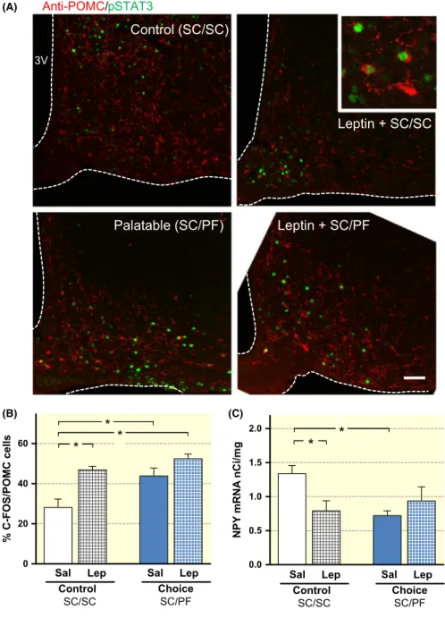
exogenous leptin concomitantly activates POMC neurons (increased c‐FOS expression; Figure 5A,B), and downreg- ulates NPY mRNA expression (Figure 5C) in mice given SC/SC. Moreover, saline‐ and leptin‐treated mice showed similar anorexigenic responses when presented with SC/
PF (Figures 4C,D and 5B,C; also see food intake data in Figure 1B), indicating that PF does not immediately disrupt Arc components of the neurocircuitry that regulates feed- ing behaviour.35-37 It deserves pointing out that, since PF is known to induce the release of endogenous leptin from fat tissue,38 it was not surprising to find that exogenous leptin did not potentiate the POMC cell‐activating effects of PF per se (Figure 5A,B). The apparent discrepancy between these findings and those showing that PF abrogates the inhibitory effects of leptin on OX neurons (Figure 3B) led us to con- sider that additional regulatory pathways and mechanisms
may be engaged when hedonic foods are encountered during a state of satiety.
2.4 | Galanin (Gal) restores leptin efficacy during hedonia‐driven overeating
Given the unlikelihood that POMC and NPY neurons in the Arc are the loci at which hedonic foods act to override lep- tin actions (Figure 5B,C), we hypothesized that PF perturbs signal reception and/or transmission in the LHA. This was based on previous in vitro work (acute hypothalamic slices) that demonstrated that Gal mediates leptin‐induced hyperpo- larization of OX neurons25 and the data depicted in Figure 6A that show galanin (Gal) neurons terminating in the vicinity of OX neurons in the LHA, as also described by previous authors.14,26
FIGURE 5 Neurons in the hypothalamic arcuate nucleus (Arc) remain responsive to leptin during hedonic eating. A, Immunoflourescent images of the Arc of leptin‐treated mice that could choose between either SC/SC or SC/
PF show co‐expression of POMC (red) and c‐FOS (green); the upregulation of c‐FOS expression in a subset of POMC‐
positive neurons is shown at high power in the inset. Scale bar: 20 µm. B, Semi‐
quantitative analysis of c‐FOS and POMC double‐immunostained Arc neurons. Data (mean ± SEM; n = 6‐7/group; *Tukey's post hoc P < .05) shows increased activation (c‐FOS) of POMC neurons after either leptin or SC/PF exposure (main leptin and food type effects: F1,20 = 19.9, P = .0002 and F1,20 = 12.1, P = .002, respectively;
leptin × food type interactions: F1,20 = 2.66, P = .12). C, Levels of NPY mRNA expression are reduced in mice exposed to either leptin and SC/SC or SC/PF (leptin × food type interaction: F1,25 = 7.5, P = .01). Numerical data depicted are mean ± SEM (n = 6‐7/group); *Tukey's
post hoc P < .05 *
*
0.0 0.5 1.0 1.5 2.0
NPY mRNA nCi/mg
*
Sal Lep Control
SC/SC Choice
SC/PF Sal Lep
*
0 20 40 60
% C-FOS/POMC cells
* *
Sal Lep Control
SC/SC Choice
SC/PF Sal Lep
(B) (C)
(A)
To examine the role of Gal in the escape from leptin‐in- duced suppression of PF consumption, leptin‐treated mice were given intra‐LHA microinjections (Figure 6B) of M617
and M1145 (agonists of the galanin receptor 1 [Gal1R] and galanin receptor 2 [Gal2R], respectively)39 immediately be- fore being presented with PF. The ability of leptin to suppress FIGURE 6 The ability of leptin to suppress hedonic eating is reinstated by intra‐LHA injections of a galanin receptor 2 (Gal2R) agonist. A, Galanin‐immunoreactive fibres (blue) terminating in the vicinity of activated (c‐FOS‐immunoreactive, green) orexin A‐immunoreactive neurons (red) neurons in the LHA (arrowheads, sites of contact). Tissues analysed were derived from experimental mice used in Figures 1B and 3-5. B, Verification of sites of galanin agonist microinjections; correctly targeted injections of saline (black rectangles), Gal1R (purple triangles) and Gal2R (blue circles) are shown on coronal maps (from Franklin and Paxinos, 2008); LHA outlined (dotted lines) on cresyl violet‐stained coronal sections are shown in the lowest panel. C and D, Intra‐LHA Gal2R agonist M1145 restores the suppressive effects of ip leptin (3 mg/kg) on PF ingestion. Compared to saline‐treated (control) mice, PF intake was significantly reduced in M1145‐treated mice after 1 h (in C, *P < .05; in D, drug effect: F1,21 = 7.2, P = .014; time effect F3,63 = 142.6, P < .0001; n = 10‐12/group). E, Intake of PF over 1 h in 24 h‐fasted mice. Data in columns 1 and 2 are from experiment shown in Figure 1B (n = 23/ group); columns 3‐5 show data obtained in mice co‐treated with leptin and either intra‐LHA saline, GalR1 (M617; 0.1 nmol/L) or GalR2 (M1145; 0.1 nmol/L) 10 min before food presentation leptin (F4,74 = 2.96, P = .025;
n = 10‐12/group). Numerical values represent means ± SEM
0.0 0.5 1.0 1.5
Food intake (grams)
60 min PF
* Saline
Leptin
Leptin + Sal (LHA) Leptin + M617 (LHA) Leptin + M1145 (LHA)
*
60 min PF
0.0 0.5 1.0 1.5
* 20 min PF
0.0 0.5 1.0 1.5
Food intake (grams)
*
i.p.
intra-LH
Leptin
Saline M1145 Saline M1145
Cumulative PF intake (0-3 h)
0.0 0.5 1.0 1.5 2.0
20 40 60 180 min
Food intake (grams)
lep + intra-LH Gal2R lep + intra-LH sal
(A) (C)
(B)
(D)
(E)
(PF) eating was restored after application of the Gal2R ag- onist M1145 (Figure 6C,D), whereas PF overeating per- sisted in mice receiving leptin and the Gal1R agonist M617 (Figures 6Eand S3A). On the other hand, a low (0.1 nmol/L) (Figure 6C,D), but not high (1 nmol/L) (Figure S3A), dose of the Gal2R agonist “permitted” leptin to exert its inhibitory effects on PF eating (Figure S3A). These observations indi- cate that blockade of the inhibitory actions of Gal initiate PF overeating, as evidenced by the PF feeding data over 24 h, shown in Figure S3B.
3 | DISCUSSION
Despite considerable advances in the understanding of the neurobiological mechanisms underlying overeating and obesity, the proximal mechanisms that initiate overeating are largely unknown; greater knowledge of the pathways and mechanisms involved can serve to develop preventa- tive strategies against obesity and its associated pathologies.
Accordingly, we here examined the hypothesis that disrup- tion of the homeostatic processes that normally signal satiety and terminate eating are disrupted upon initial encounters with palatable foods that have high hedonic value, therefore resulting in their overconsumption.5 The present study fo- cused on leptin, a potent satiety factor38 whose role in the regulation of hedonic eating has not been investigated to date. To this end, we developed a paradigm in which fasted, lean mice were injected with leptin before being exposed to either only standard food (SC/SC) or to a choice consisting of a highly palatable (high fat‐high sucrose) food (PF) and standard chow (SC/PF).
Two independent experiments showed that systemic leptin injections are more potent at suppressing the consumption of SC in the control condition than PF (SC/PF choice). While chronic over‐eating of energy‐dense foods is causally linked to leptin resistance,2,3,12 these data are the first to demonstrate that a single, brief exposure to hedonic food can override physiological signals of satiety.
Consistent with reports that ghrelin stimulates hedonic eating40 and that ghrelin increases the reward salience of high‐fat foods in an OX‐dependent manner,41,42 we observed that ghrelin selectively increases appetite for hedonic food.
The fact that leptin, like ghrelin, caused unrestrained con- sumption of PF indicates the validity of the choice paradigm devised for these studies.
The lateral hypothalamic area (LHA), a brain area that plays a key role in the integration of hedonic and homeostatic information, is responsive to both, ghrelin and leptin.43 The fact that the LHA innervates the VTA, an important driver of motivated behaviour,44-46 also suggests that this hypothalamic nucleus plays a key role in regulating the intake of hedonic foods. In support of the latter view, we found that access to
hedonic food increases neuronal activity in the LHA, as well as other areas responsible for processing reward/hedonic and/
or homeostatic signals. Thus, the LHA might indeed repre- sent a nexus at which PF challenges the homeostatic mech- anisms that govern food intake. Consistent with reports that hunger19,31 and hedonic foods47-50 activate OX‐expressing neurons in the LHA, we also observed a rapid induction of pre‐pro‐OX mRNA transcript levels in the LHA of mice given access to palatable milk after they were already satiated with SC. Our experiments also showed that OX neurons receive inputs that signal the hedonic properties of food: thus, OX expression was further upregulated in mice that were satiated on milk before being presented with a qualitatively different (taste, odour, texture) hedonic stimulus (solid PF comprised of high‐fat/high‐sucrose). It also deserves mentioning that PF eating was not only accompanied by increased OX neuron activity but also by elevated levels of serum corticosterone (CORT). This catabolic hormone, together with its ability to modulate cognitive and motivational behaviour,51,52 was previously shown to be stimulated by OX53 and is likely to amplify the seeking of palatable, energy‐dense foods.
The finding that PF defies leptin‐mediated signals of sa- tiety and upregulates OX expression (in both, satiated and fasted mice) prompted a detailed examination of the regula- tion of orexigenic peptide expression in the LHA by leptin.
Moreover, while leptin (predictably, see ref. 23) reduced the number of activated OX neurons in mice offered SC/SC, it failed to inhibit OX neuron activity in mice that preferentially ate PF in the SC/PF choice paradigm. The incongruence be- tween the results of the present behavioural observations, the observed insensitivity of OX neurons to leptin in the hedonic eating paradigm used here and reports that leptin inhibits OX neuron activity,19,25 prompted us to further investigate the mechanistic basis of PF‐triggered non‐responsiveness to leptin.
It is known that the hypothalamic actions of leptin depend on the phosphorylation of STAT3, with a parallel increase in the activity of POMC neurons and downregulation of NPY expression.2,3,33,34 We found that PF elicits anorexigenic re- sponses in POMC and NPY neurons and, importantly, that PF does not impair the induction of pSTAT3 by leptin per se.
Our demonstration that PF ingestion does not impair prox- imal signalling of leptin, required formulation of an alter- native hypothesis to explain how OX neurons might escape from the inhibitory control of leptin during consumption of PF. Accordingly, we chose to examine whether PF perturbs signal reception and/or transmission in the LHA by interfer- ing with the inhibitory control of OX neurons. While it is known that OX neurons do not express leptin receptors, it is worth noting that leptin receptor‐expressing galanin (Gal) neurons (some of which co‐express neurotensin/GABA) ter- minate in the vicinity of OX neurons14,25,26 (see Figure 6A).
Moreover, Gal (but neither neurotensin nor GABA) was
previously demonstrated to mediate leptin‐induced hyper- polarization of OX neurons in acute hypothalamic slices.25 Against this background, we considered the possibility that galanin neurons are the locus where PF acts to interfere with the relay of leptin signals to OX neurons, thereby negating physiological restraints on eating. Using pharmacological activation of Gal receptors in the LHA, we found that mi- croinjections of M1145, a specific Gal2R agonist, into the LHA could restore the ability of leptin to suppress PF eating.
Thus, the initiation of PF overeating would appear to result from blockade of the inhibitory actions of Gal on neighbour- ing OX neurons. While this interpretation awaits testing in future studies, our observations provide a basis for under- standing the mechanisms that initiate the excessive intake calories and, therefore, the onset of obesity. Notably, the finding that only low doses of M1145 could inhibit PF eating hints at the potential importance of the prevailing Gal‐ergic tone. Furthermore, while our results may at first appear to be at odds with previous reports that Gal induces feeding54 and food‐seeking behaviour,55 other work using mice in which leptin receptor (LepRb) expression was constitutively deleted in Gal neurons confirms that the inhibitory actions of leptin are mediated by GalR.24 The precise mechanisms underly- ing the actions of Gal are unknown. However, previous work suggested that Gal2R are coupled to either excitatory or in- hibitory G (Gi) proteins (Webling et al, 2012). Coupling of Gal2R to Gi could result in either hyperpolarization of OX neurons or inhibition of OX neurons through modulation of their presynaptic inputs; the latter mechanism would be con- sistent with the results of previously‐reported experiments.25 When considered in the light of previous publications,24,25
the present results may be interpreted as follows: deficits in the release of Gal from LepRb‐expressing galanin neurons in the LHA underpin the disinhibition of OX neurons during he- donic feeding. Unfortunately, technical limitations precluded demonstration that the Gal2 agonist specifically activated OX neurons (following Gal agonist treatments, precision of microinjection sites was verified using methylene blue – an essential step in light of the small size and difficult‐to‐access location of the LHA). Future studies will be needed to define the origin of the Gal‐ergic neurons that innervate the OX neu- rons that are involved in hedonic overeating.
Recent work shows that leptin reduces the strength of presynaptic excitatory inputs to OX neurons that project to the VTA, an effect blunted in diet‐induced obesity.4 Given that the LHA is innervated by forebrain structures such as the shell of the nucleus accumbens (NAc) and ventral pall- idum (VP)43,56 which are activated by PF (Figure 2A), it is plausible that the aforementioned areas increase LHA neuron excitability and dampen competing inhibitory Gal‐ergic in- puts (from a presently unknown brain area) to OX neurons.
At the same time, because OX neurons are known to inte- grate complex and even counteracting metabolic cues,19,43 a role for other competing influences on OX neurons cannot be excluded.
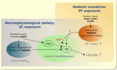
To sum up, our work demonstrates that hedonic eating results from the development of insensitivity to leptin in OX neurons, an effect which can be (at least partly) attributed to a loss of Gal‐mediated inhibitory control over OX‐ergic neurons in the LHA. This study reveals a potentially new pathway and mechanism that complements existing knowl- edge of the neural underpinnings of hedonic eating. Clearly,
FIGURE 7 Working model of the circuitry upon which leptin acts to signal energy balance and satiety (Arc nucleus is major component of the blue sphere) during SC feeding. Inhibition of OX neurons in the lateral hypothalamic area (LHA), involving leptin stimulation of Gal secretion from LepRb‐bearing Gal neurons in the vicinity of OX neurons (see Figure 5A), is considered a key mechanism in the suppression of feeding. The model postulates that besides positive emotional, cognitive and hedonic drivers, PF overeating results from a disruption of the ability of leptin to influence communication between Gal and OX neurons since activation of Gal2R in the LHA reduces PF consumption (see Figure 5C‐E)
feeding behaviour, which is essential for survival, depends on in‐built fail‐safe mechanisms, rather than simple homeo- static reflexes.57 The multiplicity of simultaneously or se- quentially‐acting regulatory factors makes dissection of the role of individual signalling molecules challenging. Thus, the hypophagic response to just a single hormone (eg leptin) may be small and probabilistic.57 In this study, robustness of the data was ascertained by repeating all behavioural ex- periments on at least two occasions. The model shown in Figure 7 is necessarily simple ‐ it does not include all of the proposed molecular players and switches through which PF may modulate leptin signalling and actions in the hy- pothalamus.58-60 Rather, it highlights input‐output relation- ships between the LHA and periphery as well as brain areas that receive and process sensory information4,43,61; among these, the central amygdala and VTA, both innervated by OX‐ergic neurons in the LHA, contribute to flavour and re- ward learning.4,62-66
4 | MATERIALS AND METHODS 4.1 | Animals
Three‐month old male C57Bl/6N mice (Charles River, Sulzfeld, Germany) were single‐housed on arrival and ac- climated (2 weeks) to local conditions before use (lights on at ZT0, lights off at ZT12), under standard conditions recommended for housing laboratory mice (temperature 21 ± 2°C, relative humidity 50 + 10%), with ad libitum ac- cess to standard chow (SC) and water, unless otherwise spec- ified. Experiments were approved by the local ethics board (Government of Upper Bavaria) and complied with Directive 2010/63/EU.
4.2 | Diet composition and sources
Standard chow (SC) was Diet 1320 from Altromin Spezialfutter GmbH & Co. KG (Lage, Germany); it contained 2.84 kcal/g (11% fat, 65% carbohydrate, of which 4.9% was sucrose). Palatable food (PF), purchased from ResearchDiets Inc (Cat. D12451; New Brunswick, NJ, USA), had a calorific content of 4.73 kcal/g (45% kcal from fat, 35% from carbo- hydrate, of which 17% was sucrose]. Low‐fat food (LF; Cat.
D12450B from Research Diets Inc), contained 4.06 kcal/g (10% energy from fat, 70% from carbohydrate, of which 35% was sucrose]; ResearchDiets Inc). The protein contents of the SC, HF and LF were similar (20%, 20% and 24% re- spectively), but the diets varied in sweetness (LF > HF>SC).
Brain activation during the hedonic experience, was assessed in mice with given access to SC and milk (60 mL) of dairy cooking crème [Molkerei Weihenstephan, Germany] diluted in water to provide 1.64 kcal/g and a final composition of 5%
fat, 1.3% carbohydrate, 1% protein).
4.3 | Food choice paradigm
Standard chow (SC) was available throughout, unless oth- erwise specified. With the exception of one test when mice were presented with milk (1.64 kcal/g; 5% fat) and SC, ani- mals could choose between SC, high‐fat/high‐sucrose (45%
fat) and/or low‐fat/high‐sucrose (LF, 10% fat) foods that var- ied in sweetness (see above). The LF option was eventually dropped since mice neglected SC and LF in favour of high‐
fat food (hereinafter, “palatable food”, PF) and comparisons of PF intake in the choice condition (SC/PF) were compared to SC intake when only SC (SC/SC) was provided.
4.4 | Drugs
Ghrelin was injected immediately before, and leptin 30 min before, presentation of test foods. Octanylated rat ghrelin (Tocris, Bristol, UK) was given at a dose of 0.3 mg/kg, ip, as described in previous studies.67-69 Leptin (LEP‐6; Protein Laboratories Rehovot Ltd., Rehovot, Israel) was injected ip at a dose of 3mg/kg.62,70,71 The galanin receptor 1 and 2 (Gal1R and Gal2R) agonists (M617 and M1145, respectively, both from Tocris) were injected (intra‐LHA) at 0.1 or 1 nmol/L in a volume of 0.5 µL, as described under “Restoration of leptin responsiveness by intra‐LHA galanin”. Saline (0.9%) served as vehicle and diluent for all drugs injections.
4.5 | Brain activation by acute hedonic food experience
Mice (n = 24) were habituated to the presence of two water‐
containing bottles and had ad libitum access to SC through- out the experiment. At ZT16, 4 groups of mice (n = 6) could choose between bottles containing either water/water or water/milk (see above) for 1‐24 h; one group received PF for 1 h (after 24 h access to milk. At sacrifice (1 or 24 h), trunk blood was collected for measuring corticosterone by radioim- munoassay (RIA‐1364 kit from DRG, Marburg, Germany) and brains were processed to detect c‐fos mRNA and c‐FOS protein (see below). Pilot studies demonstrated greater con- sumption of milk (high hedonic value ascribable to its high fat and lactose content) vs standard chow (SC); to avoid the confound from differing calorific contents of the food op- tions, the milk was diluted in water to contain 5% fat.
4.6 | Food preference as a function of natural and simulated satiety states
Using a non‐randomized treatment/within‐subject design (Figure S1A), mice were repeatedly exposed to the choice paradigm (HF, LF and SC over 24 h) or control diet (SC/
SC), with a 2 d “wash‐out” between exposures starting at ZT3, the resting phase for mice, in order to create a conflict
between homeostatic state and hedonic motivation of the animal. Animals were tested in either satiated or fasted (24 h) states (5 d recovery between fasts). Ghrelin (0.3 mg/
kg) or leptin (3 mg/kg) were injected ip immediately (Ghr) or 30 min (leptin) before presentation of SC or SC/PF at ZT3. Ghrelin‐injected mice had continuous access to SC and were naturally satiated at the time of injection; leptin was injected to mimic a state of satiety in mice that had been 24 h‐fasted (Figure S1A). Food intake was monitored after 1, 3, 6 and 24 h.
Results of the first experiment were subsequently ver- ified by applying a between‐subject factorial design to 8 groups of fasted or satiated mice with experiments started at ZT3 (n = 10; BW similar between groups) that were exposed to one of the following drug‐food combinations:
vehicle (saline)‐SC, ghrelin (Ghr)‐SC or leptin‐SC, vehicle‐
PF, and Ghr‐PF or leptin‐PF (Figure S1B). As before, test foods were presented to mice after saline or Ghr (0.3 mg/
kg) injections (groups previously satiated on SC) or after saline or leptin (3 mg/kg ip) injections (24 h‐fasted group).
Food consumption was monitored after 1 h, after which the mice were sacrificed (see the next paragraph). Mice were exposed to PF (24 h, 5 day before testing) to avoid novelty confounds. The between‐subject experiments with leptin were also repeated with starting point at ZT16 (dark‐phase);
the results were not different from those obtained at ZT3, (data not shown).
4.7 | Subjugation of leptin activity by hedonia‐driven eating:
neuroanatomical correlates
Mice from the between‐subject design (n = 56, see Figure S1B) were sacrificed 1 h after eating either SC or PF. Trunk blood was collected for determination of leptin, insulin and Ghr using multiplex assays (described later). Brains (n = 7) were excised and prepared for in situ hybridization histochem- istry and/or immunohistochemical analysis of c‐fos, orexin, melanin‐concentrating hormone (MCH) and neuropeptide Y (NPY) mRNA expression and c‐FOS protein expression (for detailed methods, see later). Brains from a subset of mice (n = 6) were perfused with 4% P‐formaldehyde and immu- nostained for c‐FOS and pSTAT3, orexin‐A and orexin‐B, c‐FOS and orexin‐B, c‐FOS and POMC, or c‐FOS, orexin‐
B and galanin. Free‐floating sections from the hypothala- mus (every 6th section from Bregma −1.06 to −2.18) were analysed.
4.8 | Restoration of leptin responsiveness by intra‐LHA galanin
Mice (n = 39) were outfitted with bilateral intra‐LHA in- jection cannulae, as described below. After 2 weeks, mice
were fasted (24 h) and given leptin (3 mg/kg ip) at ZT 3, followed by an intra‐LHA microinjection (0.5 µL) of either 0.9% NaCl (vehicle) or Gal receptor (Gal1R or Gal2R) ago- nists (0.1 nmol/L each) 20 min later. Following a 10 min re- covery period, mice were given the SC/PF choice (intake of each food type was measured for up to 24 h). After a 6 days rest, all procedures were repeated except that mice received intra‐LHA saline or a higher dose (1 nmol/L) of the Gal1R or Gal2R agonists.
In preparation for the intra‐LHA microinjections of the galanin receptor agonists, mice were anaesthetized with isoflurane, placed in a stereotaxic frame (Stoelting Co.) and subcutaneously injected with analgesics (Metacam, 0.5 mg/kg and atropine sulphate, 0.5 mg/kg; both from Braun Melsungen AG). Bilateral intra‐LHA guide can- nulas with 2‐mm stylets (Plastics One) were implanted using the coordinates A/P: −1.5, M/L ± 1 from Bregma (Franklin and Paxinos, 2013). After replacement of the stylets with removable dummy injectors (Plastics One), the guide cannulas were fixed to the skull with dental acrylic resin (Paladur; Heraeus Kulzer, Hanau, Germany).
Animals were allowed to recover for 2 weeks before ex- perimental manipulations. On the day of experiment, ve- hicle or drugs (GalR1 and GalR2 agonists) were infused bilaterally via infusion cannulas that targeted the LH at
−5.2 mm from Bregma, over 3 min, using a micro‐infu- sion pump. Infusion cannulas were held in place for 3 min after microinjection, removed and then replaced with dummy injectors. After completion of behavioural tests, 0.5 µL methylene blue was infused via re‐inserted injec- tor cannulas. The mice were then anaesthetized and their brains snap‐frozen, sectioned on a cryostat (20 µm) and counterstained with cresyl violet to verify correct place- ment of the cannulae and site of microinjection by refer- ence to a stereotaxic atlas of the mouse brain.72 Cannulae were misplaced in two mice; these animals were excluded from further analysis. The doses of Gal1R (M61773,74) or a Gal2R (M114575,76) were chosen based on the previous studies.
4.9 | mRNA detection by in situ hybridization histochemistry (ISHH)
After removal from the skull, brains were flash‐frozen in isopentane/dry ice and stored at ‐ 80°C until sagittal cryo‐
sectioning (10 µm thick; lateral coordinates: 3.25, 2.52, 1.56, 0.96, 0.48, 0.24 mm) or coronal sectioning to de- tect c‐fos mRNA (10 µm thick; 2.34, 1.70, 0.74, −0.70,
−1.43, −3.16 mm from Bregma). Sections (between Bregma −1.46 and −1.70; 10 µm) were used to detect prepro‐orexin, pro‐MCH mRNA and NPY mRNA. The ISHH protocol followed has been previously described.77 Briefly, following fixation, acetylation, dehydration and
delipidation, air‐dried sections were hybridized with cus- tom‐synthesized oligoprobes (Sigma‐Aldrich Chemie GmbH, Steinheim, Germany):
c‐fos: 5’‐GTTGACAGGAGAGCCCATGCTGGAG
AAGGAGTCGGCTGGGGAATG‐3’
prepro‐
orexin: 5’‐AGCAGCGTCACGGCGGCCCAGGGAACCT
TTGTAG‐3’
pro‐MCH: 5’‐CAACATGGTCGGTAGACTCTTCCCAGCATA CACCTGAGCATGTCAA‐3’
NPY: 5’‐GTCCTCTGCTGGCGCGTCCTCGCCCGGA
TTGTCCGGCTTGGAGGGGTA‐3’
Oligoprobes were 3′ end‐labelled with 35S d‐ATP (Perkin Elmer, Waltham, MA, USA). Specificity was controlled by hybridizing alternate sections with a corre- sponding sense sequence probe. Following an overnight incubation (37°C) and stringent washing, dehydrated sec- tions were exposed to X‐ray film (Carestream Biomax MR Film) for 24 h (OX and MCH), 3 days (NPY) or 10 days (c‐fos), before developing. Images were captured and the optical densities of radioactive signals were either quali- tatively evaluated or quantified, relative to a 14C standard (range: 0‐35 nCi/mg; American Radiolabelled Chemicals Inc), using Image J software (http://imagej.nih.gov/ij/
downl oad/zips/-j149.zip).
4.10 | Immunohistochemistry (IHC)
Brains were flash‐frozen in isopentane/dry ice immediately after sacrifice and stored at −80°C until cryosectioning (10 µm). A freezing microtome was used to cut 25 µm sec- tions from 4% P‐formaldehyde (PFA)‐fixed brains. In the latter case, brains were initially fixed in situ by intra‐cardiac perfusion of pentobarbital‐anaesthetized mice with 0.9%
saline, followed by PFA; excised brains were subsequently post‐fixed in, cryo‐preserved (30% sucrose/PBS) and stored at −20°C until sectioned. Unless otherwise specified, phos- phate‐buffered saline (PBS; 0.01 mol/L, pH 7.4) served as a general diluent and wash solution. Endogenous peroxidases were quenched 0.5% hydrogen peroxide (15 min), 0.2%
Triton X‐100 (15 min) was used for permeabilization, and non‐specific antibody binding was blocked using 2% normal horse serum (20 min). Antigens detected by immunofluores- cence were marked with direct‐conjugated fluorescent an- tibodies or biotinylated secondary antibodies from Jackson ImmunoResearch (West Grove, PA). Details of primary an- tisera used, dilutions and incubation times are given in the Table 1.
Transmitted light and fluorescent microscopic images were captured using an AxioCam MRc 5 digital camera mounted on a Zeiss AxioImager M1 microscope, outfitted with AxioVision 4.6 software (Carl Zeiss). Confocal images were captured with TABLE 1 Antisera, incubation conditions and detection systems used in immunohistochemistry
Antibodies Source/Dilution Incubation/detection
Single targets
Anti‐c‐Fos, rb 1:5000; 18 h; Millipore (PC38) BIOT‐dk‐anti‐rb IgG (1:500; 2h; Jackson (711‐65‐152), ABCa, Ni/DAB (grey‐black)
Anti‐Orexin
A, rb 1:5000;18h; Phoenix Pharmaceuticals (H‐003‐30) FITC‐dk‐anti‐rb IgG (1:500; 2h; Jackson (711‐095‐152) (green)
Anti‐Orexin
B, gt 1:2000; 18 h; Santa Cruz Biotechnology (sc‐8071) CY3‐dk‐anti‐gt IgG (1:500; 2h; Jackson (715‐165‐150) (red) Anti‐pSTAT3,
rb 1:500; 48 h, Cell Signaling Technologies (#9131) BIOT‐dk‐anti‐rb IgG, ABC, biotin‐tyramide (BT) amplifica- tionb, streptavidin (STA) 488 (green)
Multi‐targets
Anti‐c‐Fos, rb 1:6000; 48 h; Millipore (PC38) BIOT‐dk‐anti‐rb IgG, ABC, Ni/DAB, silver‐gold intensifica- tionc (black)
Anti‐Orexin
B, gt 1:3000; 48 h; Santa Cruz Biotechnology (sc‐8071) BIOT‐dk‐anti‐gt IgG (1:500; 2h, RT; Jackson 705‐065‐147), ABC, DAB (brown)
Anti‐c‐Fos, gt 1:10 000; 48h; Santa Cruz Biotechnology (sc‐52) BIOT‐dk‐anti‐gt IgG + ABC +BT amplification, STA 488 (green)
Anti‐POMC, rb 1:5000; 48 h, Phoenix Pharmaceuticals (H029‐30) CY3‐dk‐anti‐rb IgG (1:500; 2h; Jackson 715‐165‐152) (red) Anti‐galanin,
sheep 1:50 000; 48 h, kindly gifted by Dr I. Merchenthaler,
University of Maryland, College Park, MD AMCA dk‐anti‐sheep IgG (1:500; 2h; Jackson 713‐155‐003) (blue)
aavidin‐biotin complex (ABC; Vector Laboratories, Burlingame, CA); 1:1000, 2 h, RT;
bBiotin‐tyramide (BT) amplification (Hopman et al, 1998); secondary streptavidin fluorescent antibody (Alexa 488‐streptavidin from Molecular Probes, S‐11223 Thermo Fisher; 1:1000, 12 h, RT.
cLiposits, Setalo and Flerko B (1984).78
dAnti‐orexin‐Texas Red®‐X conjugate prepared using Zenon Tricolor Rabbit IgG Labeling Kit #2, Thermo‐Fischer Scientific (Z‐ 25 370, Zenon®).
an inverted Nikon Eclipse Ti‐E microscope, equipped with an A1R confocal system (Nikon, Vienna, Austria). Digital images were processed with Adobe Photoshop CS software (Adobe Systems) and equally adjusted for brightness and contrast. A summary of the antibodies, incubation conditions and detection systems used is provided in Table 1.
4.11 | Capillary electrophoretic detection of STAT3 immunoreactivity
Hypothalami were homogenized in 100 mmol/L Tris‐HCl, pH 8 (with 1 mmol/L EDTA, 250 mmol/L NaCl, 25 mmol/L MgCl2, 10% glycerol, 1% Nonidet and Roche cOmplete Protease Inhibitor Cocktail and phosphatase inhibitor cock- tails 2 and 3). After determining protein contents, sam- ples were assayed for pSTAT3, total STAT3 and actin by semi‐automatic capillary electrophoresis (2 µg protein/µL per capillary) using a Wes‐Mouse Masterkit (PS‐MK17, ProteinSimple); electrophoretic separation and quantification of concentrations of proteins of interest was performed with a Wes™ (ProteinSimple) apparatus. The following primary antibodies were used: anti‐pSTAT3, Tyr705 (1:25, #9145, Cell Signaling Technologies), anti‐STAT3 (1:100, #9139, Cell Signaling Technologies) and actin (1:3000 for pSTAT3 and 1:1000 for STAT3, #4967, Cell Signaling Technologies), using anti‐rabbit IgG (042‐206, ProteinSimple) as secondary antibody. Incubations were carried out at room temperature for 2 h. Total STAT3 and pSTAT3 signals were normalized against actin signals.
4.12 | Multiplex assay of serum leptin, insulin and ghrelin
Serum concentrations of leptin, insulin and ghrelin were measured using Luminex®‐based technology Milliplex MAP kit (#MMHMAG‐44K‐07.Mouse, Merck Chemicals, Am Kronberger, Schwalbach, Germany) and the Bio‐Plex system for signal detection (Bio‐Rad Laboratories GmbH, Munich, Bayern, Germany). The assay sensitivities for leptin, insulin and ghrelin were 40, 30 and 3 pg/mL, re- spectively; values below these limits were excluded from the analysis. Assays were performed at the Else Kröner‐
Fresenius Center for Nutritional Medicine, Technical University Munich, courtesy of Professor Hans Hauner and Manuela Hubersberger.
4.13 | Statistics
Statistical differences were tested using GraphPad Prism 6 software. Student's t‐tests were used to compare data from pairs of groups. Following 2‐way ANOVA, post hoc multiple comparisons were made using Tukey's or Sidak's (repeated measures) tests (significance level: P < .05).
ACKNOWLEDGEMENTS
We thank Barna László (Budapest), Manuela Hubersberger (Weihenstephan) and Cornelia Flackskamm and Kathrin Hafner (Munich) for excellent technical assistance. This work was partly supported by the EU FP7 Consortium SwitchBox (Contract 259772 to OFXA) and by the National Science Foundation of Hungary (OTKA K101326 to IK).
CONFLICT OF INTEREST
The authors declare that they have no conflict of interest.
AUTHORS’ CONTRIBUTION
EL, AVP and OFXA conceived the studies; EL, MG, AVP and IK planned the experiments; EL, MG, AVP, AP, NCG, MK and OFX performed experiments; EL, AVP, ZL and IK carried out histological analyses; EL, AVP, IK and OFXA analysed and interpreted the data; EL, IK and OFX wrote the manuscript; all authors read and approved the manuscript.
ORCID
Este Leidmaa https://orcid.org/0000-0001-9409-2134 Mary Gazea https://orcid.org/0000-0003-1468-8419 Imre Kallo https://orcid.org/0000-0003-3412-4100 Osborne F. X. Almeida https://orcid.
org/0000-0001-7331-6928
REFERENCES
1. Swinburn BA, Sacks G, Hall KD, et al. The global obesity pan- demic: shaped by global drivers and local environments. Lancet (London, England). 2011;378(9793):804‐814.
2. Myers MG, Leibel RL, Seeley RJ, Schwartz MW. Obesity and leptin resistance: distinguishing cause from effect. Trends Endocrinol Metab. 2010;21(11):643‐651.
3. Morton GJ, Meek TH, Schwartz MW. Neurobiology of food intake in health and disease. Nat Rev Neurosci. 2014;15(6):367‐378.
4. Liu J‐J, Bello NT, Pang ZP. Presynaptic regulation of leptin in a defined lateral hypothalamus‐ventral tegmental area neurocircuitry depends on energy state. J Neurosci. 2017;37(49):11854‐11866.
5. Berridge KC, Kringelbach ML. Neuroscience of affect: brain mechanisms of pleasure and displeasure. Curr Opin Neurobiol.
2013;23(3):294‐303.
6. Kojima M, Hosoda H, Date Y, Nakazato M, Matsuo H, Kangawa K. Ghrelin is a growth‐hormone‐releasing acylated peptide from stomach. Nature. 1999;402(6762):656‐660.
7. Kojima M, Kangawa K. Ghrelin: structure and function. Physiol Rev. 2005;85(2):495‐522.
8. Tschöp M, Weyer C, Tataranni PA, Devanarayan V, Ravussin E, Heiman ML. Circulating ghrelin levels are decreased in human obesity. Diabetes. 2001;50(4):707‐709.