ORIGINAL INVESTIGATION
Acute escitalopram treatment inhibits REM sleep rebound and activation of MCH-expressing neurons in the lateral hypothalamus after long term selective REM sleep
deprivation
Zita Kátai&Csaba Ádori&Tamás Kitka&Szilvia Vas&
Lajos Kalmár&Diána Kostyalik&László Tóthfalusi&
Miklós Palkovits&György Bagdy
Received: 5 July 2012 / Accepted: 25 February 2013 / Published online: 21 March 2013
#Springer-Verlag Berlin Heidelberg 2013
Abstract
RationaleSelective rapid eye movement sleep (REMS) deprivation using the platform-on-water (“flower pot”) method causes sleep rebound with increased REMS, de- creased REMS latency, and activation of the melanin- concentrating hormone (MCH) expressing neurons in the hypothalamus. MCH is implicated in the pathomechanism of depression regarding its influence on mood, feeding behavior, and REMS.
Objectives We investigated the effects of the most selective serotonin reuptake inhibitor escitalopram on sleep rebound following REMS deprivation and, in parallel, on the activa- tion of MCH-containing neurons.
Methods Escitalopram or vehicle (10 mg/kg, intraperitone- ally) was administered to REMS-deprived (72 h) or home cage male Wistar rats. During the 3-h-long“rebound sleep”, electroencephalography was recorded, followed by an MCH/Fos double immunohistochemistry.
Results During REMS rebound, the time spent in REMS and the number of MCH/Fos double-labeled neurons in the lateral hypothalamus increased markedly, and REMS latency showed a significant decrease. All these effects of REMS deprivation were significantly attenuated by escitalopram treatment.
Besides the REMS-suppressing effects, escitalopram caused an increase in amount of and decrease in latency of slow wave sleep during the rebound.
Conclusions These results show that despite the high REMS pressure caused by REMS deprivation procedure, escitalopram has the ability to suppress REMS rebound, as well as to diminish the activation of MCH-containing neu- rons, in parallel. Escitalopram caused a shift from REMS to slow wave sleep during the rebound. Furthermore, these data point to the potential connection between the seroto- nergic system and MCH in sleep regulation, which can be relevant in depression and in other mood disorders.
Keywords EEG . Sleep deprivation . Rebound sleep . Melanin-concentrating hormone . Escitalopram . Depression . Antidepressant . Serotonin . REM sleep . Rat
Introduction
The crucial role of the serotonergic system in the regulation of both sleep–wake states and mood is well documented (Dugovic2001; Monti2011; Ursin2002). Sleep disturbances Z. Kátai
:
C. Ádori:
T. Kitka:
S. Vas:
D. Kostyalik:
L. Tóthfalusi
:
G. Bagdy (*)Department of Pharmacodynamics, Semmelweis University, 1089 Nagyvárad tér 4., Budapest, Hungary
e-mail: bag13638@mail.iif.hu L. Kalmár
Institute of Enzymology, Research Centre for Natural Sciences, Hungarian Academy of Sciences, 1113 Karolina út 29., Budapest, Hungary
M. Palkovits
Neuromorphological Research Laboratory of the Semmelweis University and the Hungarian Academy of Sciences, 1094 Tűzoltó utca 58., Budapest, Hungary
G. Bagdy
Group of Neurochemistry, Hungarian Academy of Sciences, 1083 Budapest, Hungary
G. Bagdy
Group of Neuropsychopharmacology, Hungarian Academy of Sciences, 1083 Budapest, Hungary
belong to the core symptoms of depression, such as increase in the amount of rapid eye movement sleep (REMS) and a decrease in REMS latency (Steiger and Kimura2010).
Selective serotonin reuptake inhibitors (SSRIs) are com- monly used as effective antidepressants with a strong REMS decreasing and REMS latency increasing effects (Steiger and Kimura2010; Wilson and Argyropoulos2005). All these ef- fects were also demonstrated in rodents (Monaca et al.2003;
Vas et al.2013). The main mechanisms existing in the back- ground of REMS suppression of SSRIs imply the increase in serotonin (5-HT) concentration via the inhibition of its reuptake and the concomitant activation of postsynaptic 5-HT1Aand 5- HT1Breceptors (El Mansari et al.2005; Maudhuit et al.1996).
The involvement of the lateral hypothalamus (LH) in sleep regulation has been well established earlier (Gerashchenko and Shiromani2004). The melanin-concentrating hormone (MCH) -containing cells in the LH and zona/subzona incerta (ZI/Sub ZI) have been implicated first in the regulation of feeding and metabolism (Bittencourt et al.1992; Pissios 2009; Qu et al.
1996; Torterolo et al.2011). Later, a strong connection between MCH and REMS has been revealed (Kitka et al. 2011;
Torterolo et al.2011). Local or intracerebroventricular admin- istration of MCH to rodents caused a marked increase in REMS; moreover, an elevated activation (increased Fos immu- noreactivity) of MCH-expressing neurons has been reported during REMS rebound after a long-term sleep deprivation (Hanriot et al. 2007; Kitka et al. 2011; Modirrousta et al.
2005; Peyron et al. 2009; Torterolo et al. 2011; Verret et al.
2003). The increase in the time spent in REMS and the decrease in the REMS latency during the rebound following sleep dep- rivation are both changes similar to those described in depres- sion. Furthermore, the antidepressant-like profile of MCH-R1 antagonists suggest the role of MCH in the regulation of mood (Antal-Zimanyi and Khawaja2009; Borowsky et al. 2002;
Chung et al.2011; Lagos et al.2011a; Millan et al.2008).
Nevertheless, the effects of antidepressants on both the activation of MCHergic neurons and REMS rebound after sleep deprivation have not been studied yet. Here we demon- strate that the most selective, relatively new, and clinically effective (Ceglia et al. 2004; Leonard and Taylor 2010;
Montgomery et al.2001; Wade et al.2002) SSRI compound escitalopram reduces Fos expression in MCH-immunoreactive neurons in the LH. Moreover, escitalopram also diminishes the time spent in REMS during REMS rebound following the 72-h REMS deprivation.
Materials and methods
Animals
All animal experiments and housing conditions were carried out in accordance with the European Community Council
Directive of 24 November 1986 (86/609/EEC) and the National Institutes of Health “Principles of Laboratory Animal Care” (NIH Publications No. 85–23, revised 1985) as well as specific national laws (the Hungarian Governmental Regulation on animal studies, 31 December Psychopharmacology 1998 Act). Permission was obtained from local ethical committees.
Male Wistar rats (n=36) were purchased from Animal Facility (Semmelweis University, Budapest, Hungary). Rats, weighing 346±6.14 g (mean ± SEM) at surgery, were kept under controlled environmental conditions (temperature at 21±1 °C and a 12-h light–dark cycle starting at 10:00A.M.).
Food and water were available ad libitum during the whole experiment.
Surgery
Rats were chronically equipped with electroencephalographic (EEG) and electromyographic (EMG) electrodes, under halo- thane (2 %) anesthesia (Fluotec 3), as described earlier (Kantor et al. 2004). Briefly, stainless steel screw electrodes were implanted epidurally over the left frontal cortex (L=2.0 mm and A=2.0 mm to bregma) and left parietal cortex (L=2.0 mm and A=2.0 mm to lambda) for frontoparietal EEG recordings.
The ground electrode was placed over the cerebellum. EMG electrodes (stainless steel spring electrodes embedded in sili- con rubber; Plastics One Inc., Roanoke, VA, USA) were placed into muscles of the neck. Rats were kept in single cages in the recording chamber, and after a 7-day recovery period in order to habituate the animals to the recording conditions, rats were attached to the polygraph by a flexible recording cable and an electric swivel, fixed above the cages, permitting free movement of the animals (Filakovszky et al.1999; Graf et al.
2004). To assess motor activity, electromagnetic transducers were used in which potentials were generated by movements of the recording cable as described earlier (Kantor et al.2004).
Habituation period was 7 days long.
Groups
Animals were randomly divided into four groups as follows:
vehicle-treated home cage (HC-veh, n= 7) group, escitalopram-treated home cage (HC-SSRI, n= 7) group, vehicle-treated REMS-deprived group (RD-veh, n=9), and escitalopram-treated REMS-deprived (RD-SSRI,n=9) group.
REMS deprivation procedure
After the habituation period, rats were detached from the electric cable and a 72-h-long REMS deprivation was performed as described earlier (Kitka et al. 2009). Briefly, the HC animals were placed into single cages, and each RD animal was placed on a round platform (diameter =
6.5 cm, surface was 0.5 cm above the water level) situated in the middle of a round water tank (diameter=
41 cm). The REMS deprivation was started at lights on and finished 72 h later. All animals were kept undisturbed, and food and water were available ad libitum during the REMS deprivation period. The “flower pot” sleep deprivation paradigm was established to be stressful for the animals; we addressed this question extensively in our previous papers (Kitka et al. 2009, 2011). The conclusion of our studies was that REM rebound and the activation of MCH neurons were strong- ly associated with each other, but not with stress.
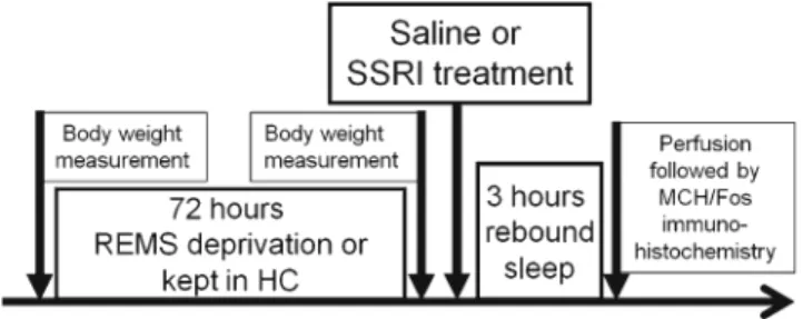
Furthermore, rats spent a considerable amount of time swimming during our pilot experiments (unpublished da- ta), suggesting that immobilization-induced stress is not a major factor regarding this method. The schema of the detailed experimental design is shown in Fig. 1.
Body weight and food intake measurement
Body weights of both HC and RD animals were measured before and after the 72-h sleep deprivation period. For further evaluations, the percent change in body weight was used.
Food intake of each animal was measured during both REMS deprivation period and sleep rebound period. Rat chow (150 g) was placed into the feeding tube of each container for deprived animals and also 150 g for the HC animals into their cages to allow ad libitum feeding for all animals. The feeding tube was prepared in a way that the loss was a strict measure of direct voluntary feeding.
Drug administration
After removing the animals from the platforms or home cages and finishing body weight measurements, vehicle (saline) or escitalopram oxalate (10 mg/kg, dissolved in saline, provided by EGIS Plc., Hungary) were injected in a
volume of 1 ml/kgi.p. The injections were administered at the beginning of sleep rebound to permit an undisturbed sleep for both REMS-deprived and HC animals during the 3-h EEG recording (Sogaard et al.2005; Vas et al.2013).
EEG recording and evaluation
Immediately after the injections, all animals were re- attached to recording cables in single cages. EEG, EMG, and motility were recorded during a 3-h-long period (sleep rebound). Rats were undisturbed throughout the recordings and had free access to standard rodent chow and tap water.
The vigilance states were classified by SleepSign for Animal sleep analysis software (Kissei Comtec America, Inc., USA) for 4-s periods, as described earlier (Kantor et al. 2004). In active wakefulness (AW), the EEG is charac- terized by low-amplitude activity at beta (14–30 Hz) and alpha (8–13 Hz) frequencies accompanied by high EMG and intense motor activity. In passive wakefulness (PW), also used as quiet wakefulness in the literature, the EEG is characterized by low amplitude activity at beta (14–30 Hz) and alpha (8–13 Hz) frequencies accompanied by high EMG activity but minimal or no motor activity. In light slow wave sleep (SWS1), high-voltage slow cortical waves (0.5–4 Hz) were interrupted by spindles (6–15 Hz) accom- panied by reduced EMG and no motor activity. In deep slow wave sleep (SWS2), there were continuous high-amplitude slow cortical waves (0.5–4 Hz) with reduced EMG and no motor activity. In REMS, low-amplitude and high- frequency EEG activity with regular theta waves (5–9 Hz) were accompanied by silent EMG and motor activity with occasional twitching.
The following parameters were calculated: duration of time spent in each sleep stage per hour, the interval between sleep onset and the occurrence of the first REMS and SWS2 period (REMS latency and SWS2 latency, respectively), the number and the average duration of REMS episodes per hour, and the length of the first REMS episode. In order to exclude short REMS attempts, a REMS episode was defined as a period of REMS lasting for≥16 s and not interrupted by≥16 s of other vigilance states (Gandolfo et al.1996; Kitka et al.2009,2011;
Vyazovskiy et al.2007).
MCH/Fos double immunohistochemistry
After the 3-h-long sleep rebound period, animals of both HC and RD groups were anesthetized with sodium pentobarbital (Nembutal, 35 mg/kg, i.p.; CEVA-Phylaxia) and transcardially perfused using 4 % paraformaldehyde in 0.1 M phosphate buffer, pH=7.4 (PB). The brains were removed, postfixed at 4 °C overnight, and cryoprotected in 20 % sucrose in PB overnight. Then, 50-μm-thick coronal sections throughout the hypothalamus were cut using a frigomobile (Frigomobile;
Fig. 1 Schematic illustration of the experimental design. Saline or SSRI (escitalopram, 10 mg/kg, i.p.) treatments were applied following the REMS deprivation (flower pot), or home cage stay, just before the beginning of a 3-h sleep rebound (at lights on). Body weight changes during the 72-h REMS deprivation and sleep parameters during the sleep rebound were recorded. Immediately after the sleep rebound, all groups were sacrificed and MCH/Fos double immunohistochemistry staining was done. For more details, see“Materials and methods”section
Reichert-Jung, Vienna, Austria). For immunostaining, solu- tions were dissolved in PB and the primary antibodies were applied for 2 days at 4 °C; all the other incubations were performed at room temperature. The sections were washed extensively for 3×10 min in PB between the incubation steps.
The sections were first permeabilized with 0.5 % Triton X-100 for 1 h. The endogenous peroxidase activity and the non- specific antigen binding sites were blocked with the incubation of the sections in 3 % hydrogen-peroxide solution and in 10 % normal goat serum for 15 min and for 1 h, respectively. After these steps, the sections were incubated in rabbit anti-Fos (1:30,000, in PB; Santa Cruz Biotechnology, Inc., Heidelberg, Germany) primary antibody, and then in biotinylated goat anti-rabbit IgG (1:1,000) and in avidin–bio- tin–peroxidase complex (ABC, 1:500) for 1 h in both solutions (both from Vector Laboratories, Burlingame, CA, USA). The immunostaining was visualized by nickel-enhanced diaminobenzidine (NiDAB) chromogen resulting in a dark blue reaction product. Then, MHC immunostainings were performed in the same way as the first one except using rabbit anti-MCH as primary antibody (1:10,000 in 3 % BSA/0.5 % Triton X-100 from Phoenix Europe GmbH, Karlsruhe, Germany) and DAB, with chromogen resulting a brown reaction product. Finally, the sections were col- lected on gelatin-coated slides, dehydrated, and mounted with DPX Mountant (Sigma-Aldrich, Budapest, Hungary) mounting medium.
Morphometry analysis of MCH/Fos double immunolabeling The amounts of MCH-immunoreactive (MCH-IR)/Fos-im- munoreactive (Fos-IR) double-labeled and MCH-IR/non- Fos-IR cells were determined in the lateral hypothalamus (LH) bilaterally, in at least five 50-μm-thick coronal sections between bregma −2.5 and −3.5 using a Visopan microscope (No. 361977; Reichert, Austria) (Paxinos and Watson 2007). Briefly, four randomly se- lected non-overlapping areas were selected in each sec- tion (0.64 mm2 altogether) under 40× objective, and the quantification was made by the same observer in all cases. Finally, the number of cells was calculated to cells per square millimeter values.
For further analysis, activated portion of MCH- containing neurons (MCH/Fos), namely the percent ratio of MCH-IR/Fos-IR double-labeled cells per total number of MCH-IR cells, was used.
Statistical analysis
Sleep parameters were evaluated by repeated measures ANOVA with three main factors: rebound (as a result of sleep deprivation; non-repeated; HC or RD), treatment (non- repeated; vehicle or SSRI), and time (repeated; first, second,
and third hour of rebound) or by two-way ANOVA with two main factors: rebound (as a result of sleep deprivation; HC or RD) and treatment (vehicle or SSRI). Post hoc analysis (Tukey HSD test) was based on this latter two-way ANOVA and indicated on figures. Immunohistochemical data was also analyzed by two-way ANOVA with two main fac- tors: rebound and treatment (see above). For post hoc analysis, Tukey HSD test was used. For statistical anal- ysis, STATISTICA 7.0 (StatSoft Inc., Tulsa, OK, USA) software was used. Data in all figures are expressed as mean ± SEM.
Results
Sleep parameters during the sleep rebound and the activated portion of MCH-containing neurons in the LH were deter- mined in the saline- (HC-veh and RD-veh) and escitalopram- treated groups (HC-SSRI and RD-SSRI). Food intake and body weight changes of all animals were measured during the procedure.
Effects of REMS deprivation on sleep rebound Time spent in different vigilance stages
The “time effect” was significant (p< 0.005) in each vigilance stage (AW, PW, SWS1, SWS2, and REMS) in the investigated time period (from the first to the third hour). In general, in the RD groups, the time spent in AW, PW, and SWS2 decreased, while REMS increased during sleep rebound, compared to HC groups (Fig. 2a, b, d and Fig. 3a). The effects of rebound, as a result of sleep deprivation, were significant in AW and REMS (F1;17= 5.42, p= 0.0326 andF1;17= 213.98, p< 0.0001, re- spectively), although in passive wake, only a strong trend was observed (F1;17= 4.45, p= 0.0501). The time × rebound interactions were significant both in SWS2 and REMS (F2;34= 10.43, p< 0.001 and F2;34= 21.45, p<
0.0001, respectively).
Specific REMS parameters
Comparing the HC and RD groups, the REMS latency decreased, while the number and the average duration of REMS episodes as well as the length of the first REMS episode showed an increase during sleep rebound (significant rebound effects—F1;17= 419.70, p< 0.0001;
F1;17= 64.36, p= 0.0001; F1;17= 122.41, p= 0.0001; and F1;17= 104.48, p< 0.0001; REMS latency, number of REMS episodes, average duration of REMS episodes, and length of the first REMS episode, respectively;
Fig. 3b–d).
SWS2 latency
We found significant rebound effect as a results of depriva- tion, namely a decrease in the SWS2 latency in RD groups compared to HC groups (F1;17= 16.22, p= 0.000875;
Fig.3e).
Activated portion of MCH-containing neurons in the LH The number of MCH-expressing neurons was not affected by the REMS rebound (F1;17=0.0342,p=0.855).
The number of activated (Fos-positive) MCH- containing neurons increased dramatically during sleep
rebound following REMS deprivation (RD vs. HC re- bound effect—F1;17= 517.95, p< 0.0001; Figs. 4 and 5).
Effects of SSRI treatment in home cage and REMS-deprived animals
Time spent in different vigilance stages
SSRI treatment decreased the time spent in REMS in the home cage group (HC-SSRI) and even during sleep rebound after deprivation (RD-SSRI; Fig3a).
In case of REMS during the rebound, the following signif- icant effects were found: a treatment effect (F1;17=29.04,p<
Fig. 2 The time spent in active wake (AW,a), passive wake (PW,b), light slow wave sleep (SWS1,c), and deep slow wave sleep (SWS2,d) in each hour of the 3-h-long sleep rebound. Saline or SSRI treatment (escitalopram, 10 mg/kg, i.p.) was applied following the 72-h REMS deprivation just before the beginning of sleep rebound. Note that REMS deprivation has a decreasing effect on the time spent in AW, PW, and SWS2 and escitalopram increases SWS2 in REMS deprived
rats. Data are represented as mean ± SEM. For significant ANOVA results seeResultssection. *Significant post hoc effect of SSRI com- pared to the appropriate saline control in the same hour (p<0.05).
Groups: vehicle-treated home cage (HC-veh), escitalopram-treated home cage (HC-SSRI), vehicle-treated REMS-deprived group (RD- veh), and escitalopram-treated REMS-deprived (RD-SSRI) groups
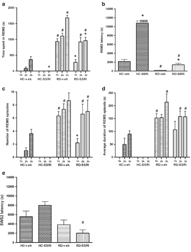
Fig. 3 Changes in time spent in rapid eye movement sleep (REMS;a), REMS latency (b), the number of REMS episodes (c), the average duration of the REMS episodes (d), and SWS2 latency (e) in the 3-h- long sleep rebound. Saline or SSRI treatment (escitalopram, 10 mg/kg, i.p.) was applied following the 72-h REMS deprivation before the beginning of sleep rebound. Data are presented as mean ± SEM. For significant ANOVA results seeResultssection. *Significant post hoc
effect of SSRI compared to the appropriate saline control (p<0.05).
#Significant post hoc effect of REMS deprivation compared to the appropriate home cage group in the same hour (p<0.05). Groups:
vehicle-treated home cage (HC-veh), escitalopram-treated home cage (HC-SSRI), vehicle-treated REMS-deprived group (RD-veh), and escitalopram-treated REMS-deprived (RD-SSRI) groups
0.0001), time × treatment, rebound × treatment, and time × rebound × treatment interactions (in all cases,p<0.01).
Summarizing data of the 3-h-long sleep rebound, a marked, almost three times increase in SWS2 was observed in RD-SSRI group compared to RD-veh group (rebound × treatment interaction—F1;17= 4.20, p= 0.0562), and a 10-fold increase in the first hour of SWS2 in RD-SSRI group compared to HC-SSRI animals was also observable (Fig. 2d).
Specific REMS parameters
In HC animals (HC-SSRI group), SSRI treatment almost completely abolished REMS.
As a result of escitalopram, we found an increase in REMS latency as well as a decrease in both the number and the average duration of REMS episodes during sleep rebound in RD-SSRI group, compared to RD-veh animals (treatment effect—F1;17= 315.97, p< 0.0001; F1;17= 7.09, p= 0.0164;
andF1;17=10.71, p=0.0045, respectively; Fig.3b–d). A re- bound × treatment interaction in REMS latency and a time × rebound × treatment interaction in the number of REMS episodes were also significant (F1;17= 176.63, p< 0.0001;
andF2;34=5.58,p=0.0080, respectively).
SWS2 latency
In SWS2 latency, a significant rebound ×treatment interac- tion was found (F1;17=4.94, p=0.040167). Namely, SSRI has an SWS2 latency-elevating effect when administered to HC animals, but reduced SWS2 latency in REMS-deprived animals during the sleep rebound (see Fig.3e).
Activated portion of MCH-containing neurons in the LH SSRI treatment significantly decreased the activated portion of MCH-containing neurons during sleep rebound in RD- SSRI group compared to RD-veh animals (Figs. 4 and 5).
Interestingly, a significant rebound × treatment interaction was also found (F1;17=8.87, p=0.0084). In vehicle-treated ani- mals, REMS rebound following REMS deprivation caused a 40-fold increase (RD-veh vs. HC-veh) in the activated (Fos positive) portion of MCH-containing neurons. In contrast, in SSRI-treated animals (RD-SSRI vs. HC-SSRI), there was only a 10-fold increase in this parameter (see Fig.4).
Correlations between REMS parameters and the activated MCH-containing neurons
A general positive correlation could be seen between REMS parameters and Fos% within the MCH-immunopositive Fig. 4 Activated (Fos-positive) portion of MCH-containing neurons,
namely the percent ratio of MCH-IR/Fos-IR double-labeled cells per total number of MCH-IR cells in the LH 3 h after the saline or SSRI treatment (escitalopram, 10 mg/kg, i.p.) and the cessation of REMS deprivation or beginning of sleep rebound. The drug treatment was applied immediately after the REMS deprivation before the beginning of sleep rebound. Data are presented as mean ± SEM. *Significant post hoc effect of SSRI compared to the appropriate saline control (p<0.05).
#Significant post hoc effect of REM sleep deprivation compared to the appropriate home cage group in the same hour (p<0.05). Groups: vehi- cle-treated home cage (HC-veh), escitalopram-treated home cage (HC- SSRI), vehicle-treated REMS-deprived group (RD-veh), and escitalopram-treated REMS-deprived (RD-SSRI) groups
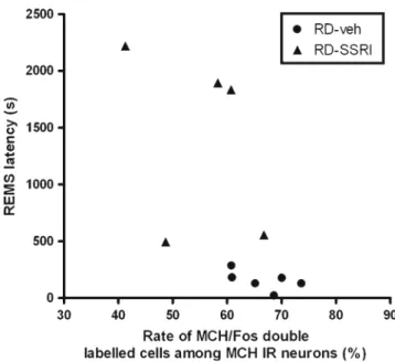
Fig. 5 Representation of individual REMS latency and activated Fos- positive portion parameters in REMS-deprived vehicle (filled circles) and REMS-deprived SSRI-treated (filled triangles) groups. REMS latency during the 3-h REMS rebound and activated (Fos-positive) portion of MCH-containing neurons, namely the percent ratio of MCH-IR/Fos-IR double-labeled cells per total number of MCH-IR (MCH-IR and MCH-IR/Fos-IR) cells in the LH 3 h after the saline or SSRI treatment (escitalopram, 10 mg/kg, i.p.). Saline or SSRI treat- ment was applied following the 72-h REMS deprivation just before the beginning of sleep rebound. For correlation values seeResultssection
cells when both rebound groups (RD-veh and RD-SSRI) are plotted together, but the difference between the two groups was not significant in any case, possibly due to the small size of the sample. In the case of REM latency, where escitalopram had a very strong effect, there were no overlapping REMS latency data in the RD-veh and RD- SSRI group; different slopes for the negative correlation can be seen and calculated, but again, the correlation was significant only if both groups were included in the same analysis (r2= 0.309, y=−9.298x+ 771.575, p=0.252, n= 6;
r2= 0.119, y=−27.715x+ 2,928.939, p= 0.570, n= 5; and r2= 0.426, y=−57.2832x+ 4,235.36, p= 0.0296, n= 11 for RD-veh, RD-SSRI, and combined RD groups calculated together, respectively; Fig.6).
Changes in food intake and body weight during REMS deprivation
Significant effects of REMS deprivation were found in both parameters, namely an increase in food intake and a de- crease in weight gain compared to home cage group (per- cent body weight change;F1;18=13.70,p<0.01 andF1;16= 15.16,p<0.001, respectively). These effects were relatively small in magnitude; the food intake during 72 h was 91.6±
4.4 g and 119.6±6.2 g in HC and REMS-deprived groups, respectively. The percent body weight change during the 72 h of REMS deprivation was 108.4±1.2 % and 100.31±
1.7 % in HC and RD groups, respectively. Food intake and percent body weight changes during the rebound period did not differ significantly from zero (data not shown).
Discussion
Our study provides the first evidence that acute SSRI treat- ment attenuates the activation of MCH-expressing neurons in the lateral hypothalamus during the sleep rebound after 72-h-long selective REMS deprivation in rats. Furthermore, our findings show that during the rebound sleep, escitalopram is still able to reduce REMS despite the strong REMS pressure. However, instead of increasing wake, which is the case in home cage animals, it increases deep slow wave sleep during the rebound sleep of the previously REMS-deprived rats.
Sleep deprivation in different fashion was widely inves- tigated as part of clinical therapy since it could reverse depressive symptoms in approximately 50–60 % of de- pressed patients (Gillin1983; Hemmeter et al.2010; Kuhs and Tolle1991; Landsness et al.2011; Wirz-Justice and Van den Hoofdakker 1999; Wu and Bunney 1990), showing rapid effect on mood (Selvi et al. 2007). However, these are acute effects; relapses usually come with the first epi- sode of sleep, which is associated with a high REMS
pressure (Adrien2002). Changes in vigilance stages during rebound sleep of rodents after sleep deprivation are similar to symptoms observable in depressed patients (Riemann et al. 2001). The SSRI antidepressant escitalopram inhibits REMS in depressed patients and, according to our results, even during REMS rebound in rats. The usual REMS- decreasing effect of escitalopram is observable in both HC and RD animals in our study, although considering the activation of the MCH-containing neurons, escitalopram had a significant effect only in RD but not in HC animals.
The latter is probably due to a floor effect, as in basal condition these neurons show a minimal, 1–2 % Fos posi- tivity. It is in accordance with our earlier results where a similarly small amount of MCH-containing neurons was activated in control HC animals (Kitka et al. 2011). In addition, the MCH-containing neurons occasionally fire during transition from wake to SWS, and they reach their maximal firing activity during REMS (Hassani et al.2009), which is probably lower than it could be during REMS rebound (Peyron et al.2009).
The acutely administered escitalopram, increasing extra- cellular serotonin concentration, interferes with REMS and MCH/Fos activity. Our previous paper (Kitka et al. 2011) has demonstrated pronounced individual correlation be- tween the number and the time spent in REMS versus Fos activation of the MCH neurons during “rebound”. One explanation could be the inhibition of the LDT/PPT cholin- ergic neurons; the structure possesses a pivotal role in the promotion of REMS. This inhibition is probably mediated via 5HT1A receptors (Monaca et al. 2003). As serotonin causes hyperpolarization of the hypothalamic MCH- containing neurons as it has been shown in rat hypothalamic slices (van den Pol et al. 2004), we cannot exclude that a local mechanism causes hyperpolarization of MCH neurons, too. In addition, MCH neurons have projections, among others, toward the LDT/PPT nuclei and the dorsal raphe nucleus (Torterolo et al. 2011), raising the possibility of a further interaction among the two systems. Anyway, our finding about MCH/Fos reducing the effect of escitalopram raises several intriguing new questions regarding the clinical effects of escitalopram and other SSRI antidepressants. One of these could be that the effect of serotonin on MCH- containing neurons (van den Pol et al. 2004) may be a pivotal component in the antidepressant effect of SSRIs, regarding that selective MCH1-R antagonist agents show antidepressant-like effects (Borowsky et al.2002; Gehlert et al. 2009) and MCH administered into the dorsal raphe evokes a depressive-like behavior (Lagos et al. 2011b;
Torterolo et al.2011). In contrast, the role of REM reduction may be questioned by the fact that there are other effective antidepressants, such as bupropion, with its main effect on the noradrenergic and dopaminergic system without affect- ing the serotonergic system and not suppressing REMS
either in human controls or depressed patients (Wilson and Argyropoulos 2005). Besides, the antidepressant effect of therapies modulating circadian rhythm (e.g., sleep depriva- tion, phototherapy) may be based on changes in diurnal variations of patients and are independent from REMS attenuation (Bodizs et al.2010; Selvi et al.2007).
In a recent study, chronically administered fluoxetine (an- other SSRI) also failed to cause any change in the activation of MCH-containing neurons after the beginning of active phase either in control rats or in animals exposed to chronic mild stress (Nollet et al. 2011). However, the latter study was performed after the beginning of the active phase, when MCH neurons are mostly inactive, and they failed to examine the activation of MCH immunoreactive neurons during REMS or after a sleep deprivation experimental paradigm.
The high rate (452 %) activation of MCH neurons may be specific to rebound sleep only and not observable in physio- logical conditions (such as in HC rats). However, a higher activation of MCH-containing neurons is conceivable also in the sleep of depressed patients. Here, we demonstrate that
despite the high REMS pressure caused by REMS deprivation procedure, escitalopram still has the ability to suppress REMS during the rebound and, moreover, to inhibit the activation of MCH-containing neurons, in parallel. We must note, of course, that despite certain similarities between the alteration of sleep parameters during sleep rebound in our experimental design and the sleep of depressed people, the results of animal studies are scarcely generalizable to the sleep conditions of depressed patients.
Inconsistent data can be found in the literature regarding the effects of SSRIs on wake and NREM sleep, namely that most SSRIs decreased or did not change NREM sleep in depressed patients (Murck et al. 2003; Winokur et al. 2001, 2003).
Besides, animal studies showed that elevated MCH level not just antagonizes wake but has an increasing effect on SWS even if in a minor extent only (Lagos et al.2011a; Verret et al.
2003). Indeed, direct injection of MCH into the ventrolateral preoptic region (VLPO) increased NREM sleep (Benedetto et al.2013). Our data show that during the sleep rebound after long-term sleep deprivation, escitalopram is still able to reduce Fig. 6 Illustrations of DAB/NiDAB MCH/Fos immunostaining of the
melanin-concentrating hormone (MCH)-expressing neurons in the lat- eral hypothalamus (LH) on 50-μm-thick coronal sections of four experimental groups:avehicle-treated home cage (HC-veh),bvehi- cle-treated REMS-deprived group (RD-veh), cescitalopram-treated home cage (HC-SSRI), andd escitalopram-treated REMS-deprived (RD-SSRI) group. The black reaction product visualizes Fos early gene product in cell nuclei; brown depicts the MCH mostly in the
cytoplasm. Note the high rate of MCH/Fos double-labeled cells in both RD groups (b,d), while in the HC groups, MCH/Fos double-labeled cells among MCH-immunoreactive neurons are almost absent. Com- paring RD groups (b, d), the decrease in Fos labeling of MCH- containing cells in the SSRI-treated group (d) is apparent compared to the vehicle-treated group (b). Between HC groups (a,c), no striking difference in MCH/Fos double staining can be seen
REMS, but instead of increasing wake, it increases deep slow wave sleep. This is suggested also by the opposite effects of escitalopram on SWS2 latency in home cage and deprived animals. During the sleep rebound, caused by the mostly selective REMS deprivation, the REMS pressure predominates sleep, leading to a decrease in the amount of deep slow wave sleep and total slow wave sleep (Kitka et al.2009). Given the fact that REMS deprivation by the“flower pot”method also causes some slow wave sleep deprivation (Verret et al.2003) being much smaller in magnitude compared to REMS pres- sure, the existence of a slow wave sleep pressure is also feasible and suggested by rebound sleep data (Kitka et al.
2009). As the SSRI treatment interferes with the high REMS pressure, we may conclude that the increased amount of deep slow wave sleep can be the consequence of the slight SWS deprivation. This is in line with the increased activation of MCH neurons, even if decreased to some extent by the SSRI treatment, as MCH has a potent role in the promotion of sleep, and both SWS and REMS are facilitated by MCH, although REMS is considered to be more sensitive to MCH modulation.
This might explain the opposite effects of escitalopram in home cage and deprived animals. Changes in slow wave sleep may be relevant considering the beneficial cognitive effects of SSRI antidepressants, but further studies are needed to eluci- date mechanisms behind the common effects of antidepressant and sleep deprivation (Bodizs2009; Goder et al.2011).
In conclusion, our main result shows that the SSRI anti- depressant escitalopram reduces the activation of MCH- containing neurons as well as the time spent in REMS during rebound. Changes in the activation of MCH- containing neurons by escitalopram may be relevant for the clinical effects of SSRI antidepressants, although further studies are needed.
Acknowledgments This study was supported by TAMOP-4.2.1.
B-09/1/KMR-2010-0001, the 6th Framework Program of the European Community LSHM-CT-2004-503474, and the Hungarian Science Research Foundation, OTKA A-08-1-2009-0050.
References
Adrien J (2002) Neurobiological bases for the relation between sleep and depression. Sleep Med Rev 6:341–351
Antal-Zimanyi I, Khawaja X (2009) The role of melanin-concentrating hormone in energy homeostasis and mood disorders. J Mol Neurosci 39:86–98
Benedetto L, Rodriguez-Servetti Z, Lagos P, D'Almeida V, Monti JM, Torterolo P (2013) Microinjection of melanin concentrating hor- mone into the lateral preoptic area promotes non-REM sleep in the rat. Peptides 39:11–15
Bittencourt JC, Presse F, Arias C, Peto C, Vaughan J, Nahon JL, Vale W, Sawchenko PE (1992) The melanin-concentrating hormone system of the rat brain: an immuno- and hybridization histochem- ical characterization. J Comp Neurol 319:218–245
Bodizs R (2009) In waves' parlance: serotonin and sleep oscillations.
Neuropsychopharmacol Hung 11:191–199
Bodizs R, Purebl G, Rihmer Z (2010) Mood, mood fluctuations and depression: role of the circadian rhythms. Neuropsychopharmacol Hung 12:277–287
Borowsky B, Durkin MM, Ogozalek K, Marzabadi MR, DeLeon J, Lagu B, Heurich R, Lichtblau H, Shaposhnik Z, Daniewska I, Blackburn TP, Branchek TA, Gerald C, Vaysse PJ, Forray C (2002) Antidepressant, anxiolytic and anorectic effects of a melanin-concentrating hormone-1 receptor antagonist. Nat Med 8:825–830
Ceglia I, Acconcia S, Fracasso C, Colovic M, Caccia S, Invernizzi RW (2004) Effects of chronic treatment with escitalopram or citalopram on extracellular 5-HT in the prefrontal cortex of rats: role of 5-HT1A receptors. Br J Pharmacol 142:469–478
Chung S, Parks GS, Lee C, Civelli O (2011) Recent updates on the melanin-concentrating hormone (MCH) and its receptor system:
lessons from MCH1R antagonists. J Mol Neurosci 43:115–121 Dugovic C (2001) Role of serotonin in sleep mechanisms. Rev Neurol
(Paris) 157:S16–S19
El Mansari M, Sanchez C, Chouvet G, Renaud B, Haddjeri N (2005) Effects of acute and long-term administration of escitalopram and citalopram on serotonin neurotransmission: an in vivo electro- physiological study in rat brain. Neuropsychopharmacology 30:1269–1277
Filakovszky J, Gerber K, Bagdy G (1999) A serotonin-1A receptor agonist and an N-methyl-D-aspartate receptor antagonist oppose each others effects in a genetic rat epilepsy model. Neurosci Lett 261:89–92
Gandolfo G, Gauthier P, Arnaud C, Gottesmann C (1996) Influence of paradoxical sleep deprivation on the intermediate stage of sleep in the rat. Neurosci Res 25:123–127
Gehlert DR, Rasmussen K, Shaw J, Li X, Ardayfio P, Craft L, Coskun T, Zhang HY, Chen Y, Witkin JM (2009) Preclinical evaluation of melanin-concentrating hormone receptor 1 antagonism for the treatment of obesity and depression. J Pharmacol Exp Ther 329:429–438
Gerashchenko D, Shiromani PJ (2004) Different neuronal phenotypes in the lateral hypothalamus and their role in sleep and wakeful- ness. Mol Neurobiol 29:41–59
Gillin JC (1983) The sleep therapies of depression. Prog Neuropsychopharmacol Biol Psychiatry 7:351–364
Goder R, Seeck-Hirschner M, Stingele K, Huchzermeier C, Kropp C, Palaschewski M, Aldenhoff J, Koch J (2011) Sleep and cognition at baseline and the effects of REM sleep diminution after 1 week of antidepressive treatment in patients with depression. J Sleep Res 20:544–551
Graf M, Jakus R, Kantor S, Levay G, Bagdy G (2004) Selective 5- HT1A and 5-HT7 antagonists decrease epileptic activity in the WAG/Rij rat model of absence epilepsy. Neurosci Lett 359:45–48 Hanriot L, Camargo N, Courau AC, Leger L, Luppi PH, Peyron C (2007) Characterization of the melanin-concentrating hormone neurons activated during paradoxical sleep hypersomnia in rats.
J Comp Neurol 505:147–157
Hassani OK, Lee MG, Jones BE (2009) Melanin-concentrating hor- mone neurons discharge in a reciprocal manner to orexin neurons across the sleep–wake cycle. Proc Natl Acad Sci USA 106:2418– 2422
Hemmeter UM, Hemmeter-Spernal J, Krieg JC (2010) Sleep depriva- tion in depression. Expert Rev Neurother 10:1101–1115 Kantor S, Jakus R, Balogh B, Benko A, Bagdy G (2004) Increased
wakefulness, motor activity and decreased theta activity after blockade of the 5-HT2B receptor by the subtype-selective antag- onist SB-215505. Br J Pharmacol 142:1332–1342
Kitka T, Adori C, Katai Z, Vas S, Molnar E, Papp RS, Toth ZE, Bagdy G (2011) Association between the activation of MCH and orexin
immunoreactive neurons and REM sleep architecture during REM rebound after a three day long REM deprivation.
Neurochem Int 59:686–694
Kitka T, Katai Z, Pap D, Molnar E, Adori C, Bagdy G (2009) Small platform sleep deprivation selectively increases the average dura- tion of rapid eye movement sleep episodes during sleep rebound.
Behav Brain Res 205:482–487
Kuhs H, Tolle R (1991) Sleep deprivation therapy. Biol Psychiatry 29:1129–1148
Lagos P, Torterolo P, Jantos H, Monti JM (2011a) Immunoneutralization of melanin-concentrating hormone (MCH) in the dorsal raphe nu- cleus: effects on sleep and wakefulness. Brain Res 1369:112– 118
Lagos P, Urbanavicius J, Scorza MC, Miraballes R, Torterolo P (2011b) Depressive-like profile induced by MCH microinjections into the dorsal raphe nucleus evaluated in the forced swim test.
Behav Brain Res 218:259–266
Landsness EC, Goldstein MR, Peterson MJ, Tononi G, Benca RM (2011) Antidepressant effects of selective slow wave sleep depri- vation in major depression: a high-density EEG investigation. J Psychiatr Res 45:1019–1026
Leonard B, Taylor D (2010) Escitalopram—translating molecular properties into clinical benefit: reviewing the evidence in major depression. J Psychopharmacol 24:1143–1152
Maudhuit C, Jolas T, Chastanet M, Hamon M, Adrien J (1996) Re- duced inhibitory potency of serotonin reuptake blockers on cen- tral serotoninergic neurons in rats selectively deprived of rapid eye movement sleep. Biol Psychiatry 40:1000–1007
Millan MJ, Gobert A, Panayi F, Rivet JM, Dekeyne A, Brocco M, Ortuno JC, Di Cara B (2008) The melanin-concentrating hor- mone1 receptor antagonists, SNAP-7941 and GW3430, enhance social recognition and dialysate levels of acetylcholine in the frontal cortex of rats. Int J Neuropsychopharmacol 11:1105–1122 Modirrousta M, Mainville L, Jones BE (2005) Orexin and MCH neurons express c-Fos differently after sleep deprivation vs. re- covery and bear different adrenergic receptors. Eur J Neurosci 21:2807–2816
Monaca C, Boutrel B, Hen R, Hamon M, Adrien J (2003) 5-HT 1A/1B receptor-mediated effects of the selective serotonin reuptake in- hibitor, citalopram, on sleep: studies in 5-HT 1A and 5-HT 1B knockout mice. Neuropsychopharmacology 28:850–856 Montgomery SA, Loft H, Sanchez C, Reines EH, Papp M (2001)
Escitalopram (S-enantiomer of citalopram): clinical efficacy and onset of action predicted from a rat model. Pharmacol Toxicol 88:282–286
Monti JM (2011) Serotonin control of sleep–wake behavior. Sleep Med Rev 15:269–281
Murck H, Nickel T, Kunzel H, Antonijevic IA, Schill J, Zobel A, Steiger A, Sonntag A, Holsboer F (2003) State markers of depression in sleep EEG: dependency on drug and gender in patients treated with tianeptine or paroxetine. Neuropsychopharmacology 28:348–358 Nollet M, Gaillard P, Minier F, Tanti A, Belzung C, Leman S (2011)
Activation of orexin neurons in dorsomedial/perifornical hypo- thalamus and antidepressant reversal in a rodent model of depres- sion. Neuropharmacology 61:336–346
Paxinos G, Watson C (2007) The rat brain in stereotaxic coordinates.
Academic, London
Peyron C, Sapin E, Leger L, Luppi PH, Fort P (2009) Role of the melanin-concentrating hormone neuropeptide in sleep regulation.
Peptides 30:2052–2059
Pissios P (2009) Animals models of MCH function and what they can tell us about its role in energy balance. Peptides 30:2040–2044 Qu D, Ludwig DS, Gammeltoft S, Piper M, Pelleymounter MA, Cullen
MJ, Mathes WF, Przypek R, Kanarek R, Maratos-Flier E (1996) A role for melanin-concentrating hormone in the central regula- tion of feeding behaviour. Nature 380:243–247
Riemann D, Berger M, Voderholzer U (2001) Sleep and depression— results from psychobiological studies: an overview. Biol Psychol 57:67–103
Selvi Y, Gulec M, Agargun MY, Besiroglu L (2007) Mood changes after sleep deprivation in morningness–eveningness chronotypes in healthy individuals. J Sleep Res 16:241–244
Sogaard B, Mengel H, Rao N, Larsen F (2005) The pharmacokinetics of escitalopram after oral and intravenous administration of single and multiple doses to healthy subjects. J Clin Pharmacol 45:1400–1406
Steiger A, Kimura M (2010) Wake and sleep EEG provide biomarkers in depression. J Psychiatr Res 44:242–252
Torterolo P, Lagos P, Monti JM (2011) Melanin-concentrating hor- mone: a new sleep factor? Front Neurol 2:14
Ursin R (2002) Serotonin and sleep. Sleep Med Rev 6:55–69 van den Pol AN, Acuna-Goycolea C, Clark KR, Ghosh PK (2004)
Physiological properties of hypothalamic MCH neurons identified with selective expression of reporter gene after recombinant virus infection. Neuron 42:635–652
Vas S, Katai Z, Kostyalik D, Pap D, Molnar E, Petschner P, Kalmar L, Bagdy G (2013) Differential adaptation of REM sleep latency, intermediate stage and theta power effects of escitalopram after chronic treatment. J Neural Transm 120:169–176
Verret L, Goutagny R, Fort P, Cagnon L, Salvert D, Leger L, Boissard R, Salin P, Peyron C, Luppi PH (2003) A role of melanin- concentrating hormone producing neurons in the central regula- tion of paradoxical sleep. BMC Neurosci 4:19
Vyazovskiy VV, Achermann P, Tobler I (2007) Sleep homeostasis in the rat in the light and dark period. Brain Res Bull 74:37–44 Wade A, Michael Lemming O, Bang Hedegaard K (2002)
Escitalopram 10 mg/day is effective and well tolerated in a placebo-controlled study in depression in primary care. Int Clin Psychopharmacol 17:95–102
Wilson S, Argyropoulos S (2005) Antidepressants and sleep: a quali- tative review of the literature. Drugs 65:927–947
Winokur A, DeMartinis NA 3rd, McNally DP, Gary EM, Cormier JL, Gary KA (2003) Comparative effects of mirtazapine and fluoxe- tine on sleep physiology measures in patients with major depres- sion and insomnia. J Clin Psychiatry 64:1224–1229
Winokur A, Gary KA, Rodner S, Rae-Red C, Fernando AT, Szuba MP (2001) Depression, sleep physiology, and antidepressant drugs.
Depress Anxiety 14:19–28
Wirz-Justice A, Van den Hoofdakker RH (1999) Sleep deprivation in depression: what do we know, where do we go? Biol Psychiatry 46:445–453
Wu JC, Bunney WE (1990) The biological basis of an antidepressant response to sleep deprivation and relapse: review and hypothesis.
Am J Psychiatry 147:14–21