Article
A Glial-Neuronal Circuit in the Median Eminence Regulates Thyrotropin-Releasing Hormone-
Release via the Endocannabinoid System
Erzse´bet Farkas, Edina Varga, Bala´zs Kova´cs, ..., Jean-Louis Charli, Patricia Joseph- Bravo, Csaba Fekete
fekete.csaba@koki.mta.hu
HIGHLIGHTS
Tanycytes tonically inhibit the activity of TRH axons via endocannabinoid release
Glutamate depolarizes the tanycytes and regulates their 2-AG synthesis
Glutamate released from the hypophysiotropic TRH axons influences tanycytes
A microcircuit utilizing glutamate and endocannabinoids regulates TRH release
Farkas et al., iScience23, 100921
March 27, 2020ª2020 The Author(s).
https://doi.org/10.1016/
j.isci.2020.100921
Article
A Glial-Neuronal Circuit in the Median Eminence Regulates Thyrotropin-Releasing Hormone-
Release via the Endocannabinoid System
Erzse´bet Farkas,1,15Edina Varga,1,15Bala´zs Kova´cs,1,15Anett Szilva´sy-Szabo´,1,15Antonieta Cote-Ve´lez,2 Zolta´n Pe´terfi,1Magdalini Matziari,3Mo´nika To´th,1Do´ra Zelena,4,5Zsolt Mezriczky,6Andrea Ka´da´r,1 Do´ra K}ova´ri,1Masahiko Watanabe,7Masanobu Kano,8Ken Mackie,9Bala´zs Ro´zsa,10Yvette Ruska,1 Blanka To´th,11Zolta´n Ma´te´,12Ferenc Erde´lyi,12Ga´bor Szabo´,12Bala´zs Gereben,1Ronald M. Lechan,13,14 Jean-Louis Charli,2Patricia Joseph-Bravo,2and Csaba Fekete1,14,16,*
SUMMARY
Based on the type-I cannabinoid receptor (CB1) content of hypophysiotropic axons and the involve- ment of tanycytes in the regulation of the hypothalamic-pituitary-thyroid (HPT) axis, we hypothesized that endocannabinoids are involved in the tanycyte-induced regulation of TRH release in the median eminence (ME). We demonstrated that CB1-immunoreactive TRH axons were associated to DAGLa-immunoreactive tanycyte processes in the external zone of ME and showed that endocanna- binoids tonically inhibit the TRH release in this tissue. We showed that glutamate depolarizes the ta- nycytes, increases their intracellular Ca2+level and the 2-AG level of the ME via AMPA and kainite re- ceptors and glutamate transport. Using optogenetics, we demonstrated that glutamate released from TRH neurons influences the tanycytes in the ME.
In summary, tanycytes regulate TRH secretion in the ME via endocannabinoid release, whereas TRH axons regulate tanycytes by glutamate, suggesting the existence of a reciprocal microcircuit between tanycytes and TRH terminals that controls TRH release.
INTRODUCTION
Tanycytes are specialized glial cells lining the floor and lateral walls of the third ventricle behind the optic chiasm (Rodriguez et al., 2005; Prevot et al., 2018). The small cell bodies of these cells form the ventricular wall, whereas their long basal processes project into the median eminence (ME) or into the neuropil of the arcuate, ventromedial, and dorsomedial hypothalamic nuclei (Rodriguez et al., 2005; Prevot et al., 2018).
In the external zone of the ME, tanycyte processes terminate around the fenestrated capillaries of the hypo- physial portal circulation and intermingle with the terminals of hypophysiotropic axons, suggesting that the tanycytes (b2 tanycytes) that project to the ME may be involved in the regulation of the neurohypophysial systems (Rodriguez et al., 2005; Prevot et al., 2018). Indeed, a major role of tanycytes has been demon- strated in the regulation of the hypothalamic-pituitary-thyroid (HPT) axis by multiple mechanisms (Lechan and Fekete, 2007; Rodriguez-Rodriguez et al., 2019). As tanycytes are the primary hypothalamic cell types that express the thyroid hormone activating enzyme, type 2 deiodinase (D2), these cells can regulate the activity of hypophysiotropic thyrotropin-releasing hormone (TRH) synthesizing neurons by controlling the hypothalamic availability of T3, the active form of thyroid hormones that can effectively bind to the nuclear receptors and suppresses TRH release (Fekete and Lechan, 2014). For example, endotoxin-induced increase of tanycyte D2 activity has a critical role in the development of central hypothyroidism (nonthyr- oidal illness syndrome) (Fekete et al., 2005; Freitas et al., 2010). Tanycytes also express the TRH-degrading ectoenzyme (TRH-DE) (Sanchez et al., 2009), providing a mechanism by which they can control the amount of TRH reaching the pituitary by degrading the tripeptide in the extracellular space of the external zone of the ME.
We recently observed that large number of hypophysiotropic axon terminals in the external zone of the ME contain type 1 cannabinoid receptor (CB1) (Wittmann et al., 2007), raising the possibility that endocanna- binoids may also regulate the release of hypophysiotropic hormones in the ME. In most cases, endocanna- binoids are released from postsynaptic neurons and act on CB1 receptors located on presynaptic axon
1Department of Endocrine Neurobiology, Institute of Experimental Medicine, Szigony u. 43, Budapest 1083, Hungary
2Departamento de Gene´tica del Desarrollo y Fisiologı´a Molecular, Instituto de Biotecnologı´a, Universidad Nacional Auto´noma de Me´xico (UNAM), Cuernavaca 62210, Me´xico
3Department of Chemistry, Xi’an Jiaotong-Liverpool University, Suzhou, Jiangsu 215123, China
4Department of Behavioral Neurobiology, Institute of Experimental Medicine, Budapest, Hungary
5Centre for Neuroscience, Szenta´gothai Research Centre, Institute of Physiology, Medical School, University of Pe´cs, Pe´cs 7624, Hungary
6Faculty of Information Technology and Bionics, Pa´zma´ny Pe´ter Catholic University, Budapest 1088, Hungary
7Department of Anatomy, Hokkaido University School of Medicine, Sapporo 060-8638, Japan
8Department of
Neurophysiology, Graduate School of Medicine, The University of Tokyo, Tokyo 113-0033, Japan
9Gill Center for Biomolecular Science, Department of Psychological and Brain Sciences, Indiana University, Bloomington 474052, IN, USA
10Laboratory of 3D Functional Network and Dendritic Imaging, Institute of
Continued
terminals (Piomelli, 2003; Kano et al., 2009). The external zone of the ME houses very few neuronal peri- karya, and endocannabinoids can travel only very short distances in the brain (Regehr et al., 2009) support- ing a glial origin of endocannabinoids in this brain region. Therefore, we hypothesized that tanycytes, the most abundant glial cell type of the ME, may control the release of hormones, including TRH, from the hypophysiotropic axon terminals of the ME via the endocannabinoid system. In addition to TRH, hypophy- siotropic TRH axons also release glutamate (Hrabovszky et al., 2005). Since tanycytes express glutamate receptors (Eyigor and Jennes, 1998; Kawakami, 2000), and glutamate is known to regulate endocannabi- noid synthesis and release (Katona et al., 2006), we further hypothesized that glutamate release from hypo- physiotropic axon terminals signal toward the tanycytes, establishing a local regulatory microcircuit between TRH axons and tanycytes in the external zone of the ME.
To test these hypotheses, we determined whether the tanycytes can control the release of TRH from hypo- physiotropic terminals in the ME by utilizing the endocannabinoid system and whether glutamate released from axon terminals of hypophysiotropic TRH neurons regulatesb-tanycytes of the ME.
RESULTS
Expression of CB1 in Hypophysiotropic TRH Neurons of Mice
To determine whether the hypophysiotropic TRH neurons express the CB1 receptor, double-labelingin situhybridization was performed. CB1 mRNA was observed in the majority of neurons in the hypothalamic paraventricular nucleus (PVN) where the perikaryon of the hypophysiotropic TRH neurons reside; however, the intensity of the hybridization signal was much lower than that observed in cortical or hippocampal areas. Analyses of the double-labeled sections showed that silver grains denoting CB1 mRNA were observed above 73.4G1.5% of the TRH neurons in the PVN (Figure 1A). In addition, double-labeling immu- nofluorescence showed that punctuate CB1-immunoreactive signal was present in the majority of TRH-IR axon varicosities in the external zone of the ME suggesting that the hypophysiotropic TRH axons are sensitive to endocannabinoid signaling (Figure 1B).
DAGLa-Immunoreactivity Is Present in Tanycytes in the ME and Associated with CB1-IR TRH Axons in Mice
To determine the cell type that releases endocannabinoids in the ME, the localization of diacylglycerol lipasea(DAGLa), the synthesizing enzyme of one of the main endocannabinoids, 2-arachinodonoylgly- cerol (2-AG), was studied. At light microscopic level, DAGLa-immunoreactivity was observed in tanycyte cell bodies lining the floor (b2 tanycytes) and the lateral evagination (b1 tanycytes) of the third ventricle (Fig- ure 1C). In addition, DAGLa-immunoreactivity was also observed in processes running toward the capillary plexus of the external zone of the ME (Figure 1D), reminiscent of the distribution of theb-tanycyte basal processes. Colocalization of DAGLa-immunoreactivity and the ZsGreen fluorescence in Rax/CreERT2//
Gt(ROSA)26Sor_CAG/LSL_ZsGreen1 mice where the green fluorescence labels the tanycytes (Pak et al., 2014) demonstrate that DAGLais indeed present in tanycytes (Figure 1E).
Immunoelectron microscopy also demonstrated DAGLaimmunoreactivity to be present in tanycyte cell bodies and processes in the ME. In the external zone of the ME, DAGLa-IR endfeet processes ofb2-tany- cytes were closely associated with axon terminals of hypophysiotropic neurons (Figures 1F and 1G). Triple- labeling immunofluorescence demonstrated that axon terminals containing both TRH immunoreactivity and punctate immunofluorescence labeling the CB1 immunoreactivity are closely associated to DAGLa-IR tanycyte processes (Figure 1H).
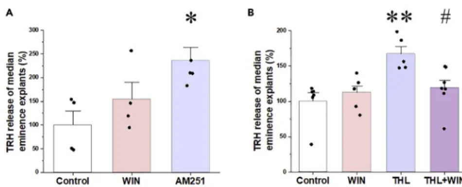
Endocannabinoids Tonically Inhibit TRH Release from the ME in Rats
To determine whether endocannabinoids affect basal TRH release, ME explants were treated with the CB1 antagonist, AM251, or CB1 agonist, WIN55,212-2, in the presence of a TRH-degrading enzyme (TRH-DE) inhibitor. AM251 stimulated TRH release causing an approximately 2-fold increase in the TRH concentra- tion of the ME explant supernatant (p < 0.05; Figure 2A). The CB1 agonist, however, had no effect (p = 0.48;Figure 2A). To test the hypothesis that the exogenous CB1 agonist was ineffective because of saturation of CB1 by its endogenous ligand, the effect of WIN55,212-2 was tested in the presence of the DAGLa inhibitor, tetrahydrolipstatin (THL). Treatment of the explants with THL caused an increase in TRH release (p < 0.01), whereas the CB1 agonist decreased THL-induced TRH release (p < 0.05;Figure 2B).
Experimental Medicine, Budapest 1083, Hungary
11Department of Inorganic and Analytical Chemistry, Budapest University of Technology and Economics, Szent Gellert ter 4, Budapest 1111, Hungary
12Medical Gene Technology Unit, Institute of Experimental Medicine, Budapest 1083, Hungary
13Department of Medicine, Division of Endocrinology, Diabetes and Metabolism, Tupper Research Institute, Tufts Medical Center, Boston 02111, MA, USA
14Department of Neuroscience, Tufts University School of Medicine, Boston 02111, MA, USA
15These authors contributed equally
16Lead contact
*Correspondence:
fekete.csaba@koki.mta.hu https://doi.org/10.1016/j.isci.
2020.100921
These data demonstrate that endocannabinoids inhibit TRH release in the ME and there is a tonic, endocannabinoid-induced inhibition of TRH release in the ME explants.
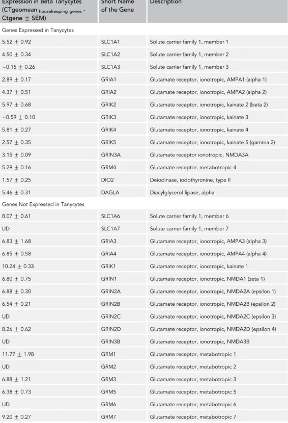
b2-Tanycytes Express Glutamate Receptors and Glutamate Transporters in Mice
To determine whether glutamate and TRH released from the hypophysiotropic TRH axons influenceb2-ta- nycytes, the expression of the glutamate receptor subunits, glutamate transporters, and TRH receptors was studied inb2-tanycyte cell bodies isolated by laser capture microdissection (Table 1). A high level of SLC1A3 (EAAC1/EAAT3) glutamate transporter mRNA and low level of SLC1A1 (GLAST/EAAT1) and SLC1A2 (GLT-1/EAAT2) mRNA was detected in b-tanycytes, whereas SLC1A6 (EAAT4) and SLC1A7 (EAAT5) glutamate transporters were not expressed.
Among the AMPA receptor subunits, mRNA of GRIA1 and GRIA2 was observed. GRIK3 was the kainite subunit with the highest expression, followed by GRIK2,4,5 that, although low, was still detectable. GRIN3A was the only NMDA receptor subunit and GRM4 the only metabotropic glutamate receptor detected in Figure 1. Elements of the Endocannabinoid System Are Present in the Hypophysiotropic TRH Neurons and in the Tanycytes in the External Zone of the ME
(A) Double-labelingin situhybridization demonstrates that the majority of TRH neurons (red) in the PVN express CB1 mRNA labeled by the presence of silver grains. Arrowheads point to double-labeled neurons expressing both TRH and CB1 mRNAs. Inset illustrates double-labeled neurons at higher magnification (arrowheads).
(B) Double-labeling immunocytochemistry demonstrates the presence of CB1 immunoreactivity (red dots) in TRH-IR hypophysiotropic axon varicosities (green) in the external zone of the ME. The CB1 immunoreactivity within the TRH axons appear yellow owing to the color mixing.
(C) High level of DAGLaimmunoreactivity is present in tanycyte cell bodies lining the floor of the third ventricle and the wall of the lateral evaginations. Dense DAGLa-IR fiber network is also present in the median eminence.
(D) Higher-magnification image illustrates that the DAGLa-IR fibers run perpendicular to the surface of the median eminence.
(E) DAGLaimmunofluorescence (red) in tanycyte processes where the green fluorescent protein labels the tanycytes. The spotted DAGLaimmunofluorescence can be observed along the tanycyte processes.
(F and G) Ultrastructural images demonstrate that the DAGLaimmunoreactivity (labeled by silver grains) is present in tanycyte endfeet processes terminating around portal capillaries.
(H) The CB1-containing (red) TRH-IR (green) axon varicosities are closely associated to DAGLa-IR (blue) tanycyte processes.
Scale bars, 100mm in (A) and (C), 10mm in inset and (B), 50mm in (D), 10mm in (E), 0.5mm in (G) that corresponds to (F) and (G), and 5mm in (H). Abbreviations: III, third ventricle; a, axon varicosity; pc, portal capillary; T, tanycyte endfeet. See also Figure S1.
b2-tanycytes, but both had relatively low expression level. Expression of TRH receptors was not detected in theb2-tanycytes.
These data suggest that glutamate effects onb2-tanycytes occur primarily via AMPA and kainate receptors and the glutamate transporter, SLC1A3.
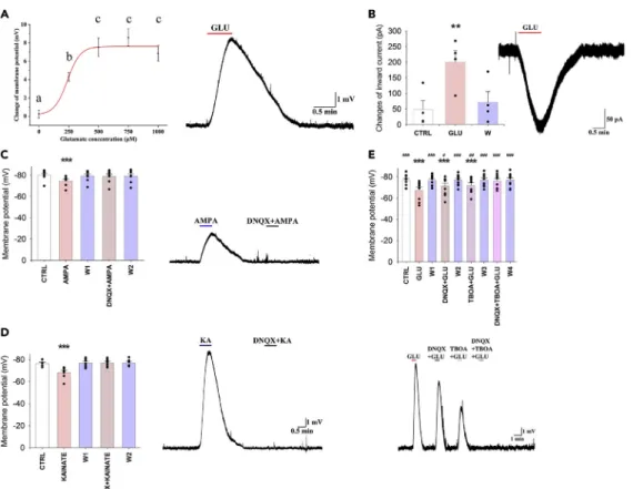
Glutamate Induces Depolarization of the Membrane Potential ofb2-Tanycytes in Mice To elucidate the effect of glutamate on theb2-tanycytes, patch clamp electrophysiology was performed.
The membrane potential of theb2-tanycytes was 77.52G0.47 mV (n = 83) under control conditions.
Four different glutamate concentrations were applied to the cells, each causing dose-dependent depolar- ization of b2-tanycytes (glutamate treatments induced change of membrane potential: control:
0.25 G0.42 mV, n = 6; 250mM: 4.29G0.46 mV, n = 11, p = 0.038; 500mM: 7.53G0.99 mV, n = 12, p < 0.001; 750mM: 8.59G0.94 mV, n = 8, p < 0.001; and 1,000mM: 6.85G0.83 mV, n = 6, p < 0.001;
Figure 3A). Treatment with 250mM glutamate caused a significant depolarization, but significantly less than the treatment with 500mM glutamate (p = 0.049). As 500mM had similar effect as that of 750 and 1,000mM concentrations, 500mM was selected for further studies.
To exclude the possibility that the effect of glutamate was mediated by tetrodotoxin (TTX; a sodium-chan- nel blocker aimed to inhibit action potential) insensitive release of transmitters from non-tanycyte cell types of the ME, an outside-out patch clamp experiment was performed. Glutamate treatment caused large inward currents ( 181.29G28.43 pA, n = 4, p < 0.01) (Figure 3B) even when the tanycyte cell body was displaced into the third ventricle. The effect of glutamate disappeared during the washout period, indi- cating that the observed effect of glutamate was exerted directly on theb2-tanycytes.
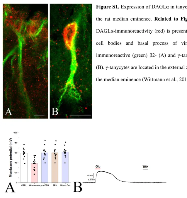
In contrast to glutamate, TRH had no effect on the membrane potential of tanycytes even in a relatively high concentration (1mM; N = 12;Figure S2).
Functional AMPA, Kainate, and GRM4 Receptors Are Present onb2-Tanycytes in Mice To investigate the receptor types that mediate glutamate-induced depolarization ofb2-tanycytes, the ef- fects of glutamate receptor agonists were studied. Similar to glutamate, both AMPA (100 mM;
Figure 2. Effects of Pharmacological Manipulation of the Endocannabinoid System on TRH Release from Median Eminence Explants
(A) TRH recovered from median eminence explants incubated in ACSF containing the TRH-DE inhibitor, P-TRH (200 nM), was measured by RIA. WIN 55,212-2 (1mM; N = 4) had no effect on the TRH release of the median eminence explants, whereas AM251 (1mM; N = 5) induced a 2-fold increase of TRH release (NControl= 4).
(B) To determine whether the absence of CB1 agonist effect is due to saturation of CB1 by endogenous cannabinoids in the explants, the DAGLainhibitor tetrahydrolipstatin (THL) was used to inhibit endocannabinoid synthesis. THL caused a marked increase of TRH release (N = 5). Although WIN 55,212-2 alone had no effect on TRH release (N = 6), it significantly decreased TRH release when 2-AG synthesis was blocked by THL (N = 7) indicating that a tonic endocannabinoid release inhibits TRH release from axons of the median eminence (NControl= 6). Data are presented as percentage of control group and as meanGSEM (N = 5). Data were analyzed by one-way ANOVA and Tukey post hoc test. * significantly different from control, *p < 0.05, **p < 0.01; # significantly different from THL treated group (p < 0.05). The amount of TRH released during 2310 min by the control groups was 80G4 pg and residual intracellular TRH was 1860G40 pg/2 ME explants.
There was no difference in residual intracellular TRH between control and drug-treated groups. Abbreviations: WIN, WIN55,212-2; THL, tetrahydrolipstatine.
Expression in Beta Tanycytes (CTgeomeanhousekeeping genes- CtgeneGSEM)
Short Name of the Gene
Description
Genes Expressed in Tanycytes
5.52G0.92 SLC1A1 Solute carrier family 1, member 1
4.50G0.34 SLC1A2 Solute carrier family 1, member 2
0.15G0.26 SLC1A3 Solute carrier family 1, member 3
2.89G0.17 GRIA1 Glutamate receptor, ionotropic, AMPA1 (alpha 1)
4.37G0.51 GRIA2 Glutamate receptor, ionotropic, AMPA2 (alpha 2)
5.97G0.68 GRIK2 Glutamate receptor, ionotropic, kainate 2 (beta 2)
0.59G0.10 GRIK3 Glutamate receptor, ionotropic, kainate 3
5.81G0.27 GRIK4 Glutamate receptor, ionotropic, kainate 4
2.57G0.35 GRIK5 Glutamate receptor, ionotropic, kainate 5 (gamma 2)
3.15G0.09 GRIN3A Glutamate receptor ionotropic, NMDA3A
5.29G0.16 GRM4 Glutamate receptor, metabotropic 4
1.57G0.25 DIO2 Deiodinase, iodothyronine, type II
5.46G0.31 DAGLA Diacylglycerol lipase, alpha
Genes Not Expressed in Tanycytes
8.07G0.61 SLC1A6 Solute carrier family 1, member 6
UD SLC1A7 Solute carrier family 1, member 7
6.83G1.68 GRIA3 Glutamate receptor, ionotropic, AMPA3 (alpha 3)
6.85G0.58 GRIA4 Glutamate receptor, ionotropic, AMPA4 (alpha 4)
10.24G0.33 GRIK1 Glutamate receptor, ionotropic, kainate 1
6.80G0.75 GRIN1 Glutamate receptor, ionotropic, NMDA1 (zeta 1)
6.88G0.30 GRIN2A Glutamate receptor, ionotropic, NMDA2A (epsilon 1)
6.54G0.21 GRIN2B Glutamate receptor, ionotropic, NMDA2B (epsilon 2)
UD GRIN2C Glutamate receptor, ionotropic, NMDA2C (epsilon 3)
8.26G0.62 GRIN2D Glutamate receptor, ionotropic, NMDA2D (epsilon 4)
UD GRIN3B Glutamate receptor, ionotropic, NMDA3B
11.77G1.98 GRM1 Glutamate receptor, metabotropic 1
UD GRM2 Glutamate receptor, metabotropic 2
6.88G1.21 GRM3 Glutamate receptor, metabotropic 3
6.38G0.73 GRM5 Glutamate receptor, metabotropic 5
UD GRM6 Glutamate receptor, metabotropic 6
9.20G0.27 GRM7 Glutamate receptor, metabotropic 7
Table 1. Expression of Glutamate Receptors and Transporters inb-Tanycytes
(Continued on next page)
5.37G1.09 mV; n = 8, p < 0.001;Figure 3C) and kainate (125mM; 8.04G2.08 mV, n = 7, p < 0.001;Figure 3D) markedly depolarized the b-tanycytes. Bath application of the AMPA and kainate receptor antagonist, DNQX (500mM), prevented these effects (Figures 3C and 3D).
In contrast to AMPA and kainate, NMDA had no effect on the membrane potential of tanycytes, even at high concentration (0.5 mM: 1.41G0.65 mV, n = 3, p = 0.42 and 4 mM: 2.12G0.3 mV, n = 3, p = 1.00.Figure S3).
Administration of the GRM4 agonist VU 0155041 caused a small, but significant, hyperpolarization (1 mM;
1.48G0.54 mV, n = 10, p < 0.01) of the membrane potential ofb2-tanycytes (Figure S3F) indicating that it is unlikely that activation of GRM4 contributes to the glutamate-induced depolarization of tanycytes.
Glutamate Depolarized theb2-Tanycytes via Both AMPA and Kainate Receptors and Also by TBOA-Sensitive Glutamate Transport in Mice
To determine whether the effect of glutamate on the membrane potential ofb2-tanycytes is mediated exclusively via AMPA and kainate receptors, hypothalamic slices were treated with glutamate in the presence of DNQX. In- hibition of the kainate and AMPA receptors caused a significant but only partial inhibition of theb2-tanycytes (glutamate: 9.18G1.55 mV, n = 9, p < 0.001 versus control; glutamate + DNQX: 5.23G1.24 mV, p < 0.001 versus control and p = 0.019 versus glutamate;Figure 3F). Therefore, we determined whether glutamate transport also contributes to the glutamate-induced depolarization. Similar to DNQX, the glutamate transporter inhibitor TBOA caused a partial inhibition of the glutamate-induced depolarization (glutamate + TBOA: 4.68G0.91 mV, n = 9, p = 0.001 versus control and p = 0.002 vs. glutamate;Figure 3E). However, the combination of DNQX and TBOA completely blocked the effect of glutamate (0.40G0.61 mV, n = 9, p = 1.00 versus control and p < 0.001 vs. gluta- mate;Figure 3E), demonstrating that glutamate depolarizes the tanycytes via a combination of AMPA and kainate receptor-mediated effects and glutamate transport.
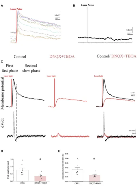
The Effect of TRH Axon Activation on Tanycytes Is Mediated Partially by Glutamate in Mice Optogenetic activation of the axon terminals of the TRH neurons around the endfeet processes ofb2-tanycytes in the external zone of the median eminence consequently caused a 0.75G0.14 mV (p < 0.001) depolarization of the patchedb2-tanycyte cell bodies in the ventricular wall of the ME (Figure 4). The peak of the depolarization was reached 51.74G2.93 ms after the start of the optogenetic activation; the velocity of this depolarization was 0.016G0.005 mV/ms. Examination of the first derivative (dV/dt) of the membrane potential showed two peaks, suggesting that the repolarization has two phases (Figure 4C). The fast depolarization was followed by a fast 0.29G0.09 mV repolarization with 126.66G10.73 ms decay time and then by a very slow repolarization (decay time: 3,127.60G446.29 ms). This suggested that the TRH axons may influence tanycytes by the release of at least two different compounds: a fast-acting transmitter and a transmitter with long-lasting effect. Simultaneous administration of DNQX and TBOA markedly decreased the optogenetic activation-induced depolarization of tanycytes (0.32G0.08 mV; p < 0.01;Figure 4). The velocity of this depolarization was significantly lower than the velocity of depolarization induced by glutamate alone (0.004G0.002; p < 0.05;Figure 4E). The first derivative (dV/dt) of the membrane potential had only one peak (Figure 4C) in the presence of antagonists, indicating that this effect has only one phase. The two antagonists prevented the first fast phase. These data demonstrate that the TRH axons influence the tanycytes with a fast-acting transmitter, glutamate, and also by a currently unknown transmitter(s) with longer-lasting effect.
Optic stimulation of slices from TRH-IRES-tdTomto mice where the TRH axons expressed tdTomato (but did not express channelrhodopsin) had no effect on the membrane potential of tanycytes (Figure 4B).
Expression in Beta Tanycytes (CTgeomeanhousekeeping genes- CtgeneGSEM)
Short Name of the Gene
Description
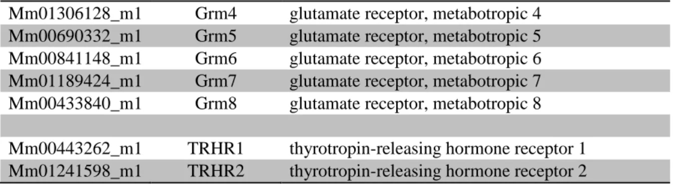
UD GRM8 Glutamate receptor, metabotropic 8
8.08G0.41 TRHR1 Thyrotropin-releasing hormone receptor 1
6.86G1.59 TRHR2 Thyrotropin-releasing hormone receptor 2
Table 1. Continued
Genes were considered to be expressed inb-tanycytes if theDCT value was lower than theDCT of GRM5. See alsoFigure S4 andTable S1.
Effect of Glutamate and TRH on the Intracellular Ca2+Levels of Tanycytes in Mice
As increase of intracellular Ca2+levels is a crucial signal to increase DAGlaactivity (Piomelli, 2003), the effect of glutamate was studied on the intracellular Ca2+level of b2-tanycytes. Glutamate treatment (500 mM) caused a robust increase of fluorescent intensity values (FIV) of b2-tanycytes (baseline:
189.19 G26.52 treatment: 416.13 G31.28; p < 0.001; Ncell= 56) increasing it to 219.95 G 27.66%
of the baseline values (Figure 5). To determine whether similar to the effect of glutamate on the mem- brane potential of tanycytes, the effect of glutamate on the intracellular Ca2+ level can also be blocked by inhibition of AMPA and kainite receptors and glutamate transport, we administered DNQX (0.5 mM) and TBOA (1 mM) during the glutamate treatment. Application of these inhibitors completely prevented the increase of FIV (baseline: 242.13 G 28.06 treatment: 250.27 G 28.67;
Ncell = 79; p = 0.99) (Figure 5).
Figure 3. Glutamate Depolarizes theb2-Tanycytes via Activation of AMPA and Kainate Receptors and via Glutamate Transport
Representative traces illustrate the effects of pharmacological treatments on theb2-tanycytes.
(A) Glutamate induced a dose-dependent depolarization ofb2-tanycytes (NControl= 6; N250mM= 11; N500mM= 12;
N750mM= 8; N1000mM= 6). A representative trace illustrates the change of the membrane potential of ab2-tanycyte in response to 500mM glutamate.
(B–D) (B) Glutamate (500mM; N = 4) evoked a large inward current onb2-tanycyte outside-out preparations,
demonstrating that glutamate directly influences the tanycytes. Similar to glutamate, both (C) AMPA (100mM; N = 8) and (D) kainate (125mM; N = 7) depolarized theb2-tanycytes, and the effects prevented by the administration of the AMPA and kainate receptor antagonist, DNQX (500mM).
(E) Although DNQX (500mM; N = 9) and TBOA (1 mM; N = 9) caused significant, but only partial, reduction of the glutamate-induced depolarization, the combination of the two inhibitors completely blocked the effect of glutamate (N = 9), indicating that the effect of glutamate on the membrane potential ofb2-tanycytes is mediated via AMPA and kainate receptors and by TBOA-sensitive glutamate transport. Data are expressed as meanGSEM and were analyzed with repeated measure ANOVA followed by Bonferroni post hoc test.
Data with different letters on (A) are significantly different (p < 0.05). * significantly different from control; # significantly different from glutamate treatment. #p < 0.05; ** and ##p < 0.01; *** and ###p < 0.001. Abbreviations: CTRL, control;
GLU, glutamate; KA, kainate;W, washout. See alsoFigures S2andS3.
Figure 4. The Optogenetic Activation of TRH Axons in the ME Induces Depolarization ofb2-Tanyctes Which Effect Is Partially Mediated by Glutamate
(A and B) The membrane potential changes of a representative tanycyte in response to 10 consecutive optic activation of TRH axons (A). The membrane potential of tanycytes is not influenced by the light impulse if the TRH axons do not express channelrhodopsin (B).
(C-E) (C) The mean response of the tanycyte membrane potential in response to 10 sweeps of optic stimulation of TRH axons. The upper traces show the membrane potential changes of the tanycyte under control conditions (black line), when the same cell was treated with a combination of DNQX (0.5mM) and TBOA (1 mM) (red) and the overlay of the two traces.
The lower graphs illustrate the first derivative (dV/dt) of the membrane potential changes. The two peaks of the dV/dt of the control trace suggest that the optic stimulation-induced membrane potential change has two phases under control conditions: an initial fast phase including a fast depolarization and a fast repolarization followed by a long-lasting phase of slow repolarization. When inhibitors are applied, the membrane potential change has only a single phase and markedly decreased amplitude. The overlay of the two traces indicates that the speed of depolarization is markedly decreased in the presence of DNQX and TBOA. Bar graphs summarize the effects of activation of TRH axons on the peak amplitude of membrane potential (D) and the depolarization velocity (E) under control condition and when the cells are treated with DNQX + TBOA.
Data are expressed as meanGSEM and were analyzed with paired Student’s t test. * significantly different (p < 0.05).
To test whether the main transmitter of the hypophysiotropic TRH neurons can influence the intracellular Ca2+level ofb2 tanycytes, the effects of TRH and its combination with glutamate were tested (Figure 5).
TRH treatment (5 mM) caused no significant changes either in the FIV of b2-tanycytes (control:
187.46 G30.58 treatment: 186.71G30.42) or in the percentage values of FIV (99.79G0.30%) when compared with the control, baseline values (p = 0.9; Ncell= 34).
Treatment ofb2-tanycytes with the combination of TRH + Glutamate (5 and 500mM, respectively) caused a robust increase of FIV (control: 154.70G26.09, treatment: 323.85G34.68) increasing it to 209.34G16.88%
of the baseline values (p = 0.002; Ncell= 37). The effect of combined TRH + glutamate treatment, however, did not differ from the effect of treatment with glutamate alone (p = 0.99).
A positive allosteric modulator of GRM4 receptor VU0155041 (1.5 mM) caused no significant changes either in the FIV ofb2-tanycytes (control: 76.01G16.56, treatment: 77.13G16.43) or the percentage values (102.90G1.39%) when compared with the control, baseline values (p = 0.9; Ncell= 21).
Effect of the Inhibition of Glutamate Action on the 2-AG Content of the Rat Median Eminence Explants
To determine whether endogenous glutamate can stimulate the 2-AG synthesis of tanycytes, the effect of glutamate receptor and transporter inhibitors was studied on the 2-AG content of ME explants. The 2-AG content of control ME explants was readily detected (0.54G0.08 ng/mg tissue). Simultaneous inhibition of AMPA and kainate receptors and the TBOA-sensitive glutamate transporters caused an approximately 50%
decrease of the 2-AG content of the ME explants (0.29G0.03 ng/mg tissue; p = 0.01) suggesting that endogenous glutamate stimulates 2-AG synthesis in tanycytes.
DISCUSSION
Endocannabinoids are well known as retrograde transmitters used by neurons to control their own neuronal inputs (Mayer et al., 1984; Ohno-Shosaku and Kano, 2014). In the current manuscript, however, Figure 5. Effects of Glutamate and TRH Treatment on the Intracellular Ca2+Level ofb2-Tanycytes
The changes of fluorescent intensity values (FIV) of the Ca2+-sensitive dye Fluo-4 AM was measured as a marker of the changes of intracellular Ca2+level ofb2-tanycytes after different treatmentsin vitro.
(A) Representative recording shows that glutamate treatment (500mM) increases FIVs in the measuredb2-tanycyte perikarya. Black arrow represents the beginning of the glutamate treatment.
(B) Bar graph summarizes effects of different treatments. GLU, glutamate (500mM, N = 56); Dn-Tb + G, DNQX + TBOA + GLU (DNQX + TBOA, 0.5 mM and 1 mM, respectively; N = 79); TRH (5mM, N = 34); T + G, TRH + glutamate (5 and 500mM, respectively; N = 37). Only glutamate and TRH + glutamate treatment increased FIV% values significantly (p < 0.001 and p = 0.002, respectively). TRH alone could not initiate any significant changes. The effects of Glutamate alone and the combined Glutamate + TRH treatment did not differ from each other (p = 0.999). Treatment of sections with DNQX + TBOA prevented the increase of intracellular Ca2+level induced by glutamate treatment (DNQX + TBOA + GLU versus control: p = 0.999). Data are shown as meanGSEM, for statistical comparison one-way ANOVA (F (4, 235) = 15.849;
p < 0.0001) was used followed by Bonferroni post hoc test. * significantly different from control p < 0.05. Scale bar, 50mm.
we demonstrate that theb-tanycytes of the ME also utilize endocannabinoids to regulate TRH release from hypophysiotropic neurons.
To establish the importance of the endocannabinoid system in regulating hypophysiotropic TRH neurons, we demonstrated that the majority of TRH neurons in the PVN expresses CB1 mRNA and that CB1 protein is present on the axon varicosities of the hypophysiotropic TRH neurons that terminate around the portal capillaries in the external zone of the ME. This observation is especially intriguing as only very few neuronal perikarya are present in the ME and the majority are located in the subependymal zone (Rethelyi, 1975). Thus, neurons are virtually absent from the external zone of the ME. As endocannabinoids can travel only extremely short distances in the neuropil before degradation (Regehr et al., 2009), the lack of neurons in the external zone of the ME excludes a neuronal origin of endocannabinoids to act on CB1 on hypophysiotropic axon terminals.
The main endocannabinoid that acts on CB1 is 2-AG (Tanimura et al., 2010) and synthesized by DAGLa (Tanimura et al., 2010). DAGLa-immunoreactivity was observed in cell bodies and basal processes of b-tanycytes in the ME. Immunoelectron microscopy further established the presence of DAGLainb-tany- cytes and demonstrated its location not only in tanycyte cell bodies, but also in their basal processes with high concentrations in the endfeet processes around portal capillaries. This is in contrast to the data of Suarez et al. (2010), who observed DAGLaimmunoreactivity only in the apical part of tanycyte cell bodies.
To determine whether the reason of this discrepancy may be species difference, DAGLaimmunostaining was performed in the ME of rats. Similar to the observation ofSuarez et al. (2010), we observed strong DAGLaimmunoreactivity in the cell bodies of tanycytes in the ME, and we also observed DAGLaimmuno- reactivity in the basal processes of tanycytes, indicating that DAGLais present in tanycyte processes in both species. The absence of DAGLaimmunoreactivity in the ME of DAGLaknockout (KO) mice clearly demonstrates the specificity of our findings. We observed CB1-IR varicosities closely associated to DAGLa-IR tanycyte processes in the external zone of the ME and, specifically, CB1-IR TRH varicosities in juxtaposition to DAGLa-IR tanycyte processes. Thus, tanycytes are in position to regulate TRH release from hypophysiotropic axon terminals in the ME.
In agreement with the morphological findings, the CB1 antagonist, AM251, increased the amount of TRH released from ME explants indicating a tonic inhibitory effect of endocannabinoids on the release of TRH. Surpris- ingly, incubation with the CB1 agonist WIN55,212-2 had no effect on TRH release from ME explants. However, when endocannabinoid synthesis was inhibited by THL, TRH release from the explants increased and this was pre- vented by WIN55,212-2. We presume that tanycytes may tonically release high concentrations of endocannabi- noids in ME explants that are sufficient to saturate CB1 receptors in hypophysiotropic TRH terminals; therefore, a further increase in agonist activity by WIN55,212-2 would not be expected to influence TRH release. This hypoth- esis is supported by the observation that, when the synthesis of endocannabinoids is blocked in the ME explants, the lower occupancy of CB1 receptors permits CB1 agonist-induced inhibition of TRH release, further demon- strating the key and dynamic role of tanycytes in the regulation of the HPT axis.
In contrast to the inhibitory effect of endocannabinoids on the TRH release, PGE2 has been shown to cause retrac- tion of tanycyte processes allowing access of hypophysiotropic axons to capillaries, thus facilitating hormone release (de Seranno et al., 2010). Intriguingly, the synthesizing enzymes of PGE2, cyclooxygenase (COX)-1 and COX-2, are synthesized in tanycytes (de Seranno et al., 2010), and arachidonic acid (AA) is similarly necessary for the synthesis of endocannabinoids and PGE2 (Malcher-Lopes and Buzzi, 2009). In the immune system, humoral signals can shift the balance between the endocannabinoid and prostaglandin system that plays, for example important role in the mediation of the effects of glucocorticoids (Malcher-Lopes and Buzzi, 2009). Thus, it is feasible to hypothesize that humoral signals may also regulate the balance of these two antagonistic signaling systems in the tanycytes to cause simultaneous regulation of hormone release and capillary access of hypophy- siotropic terminals. However, this hypothesis requires further studies.
In neuronal circuits, glutamate is an important driving force of endocannabinoid synthesis (Piomelli, 2003).
Presynaptically released glutamate binds to Grm1 and Grm5, and the activation of these metabotropic glutamate receptors stimulate the DAGLavia an increase in intracellular Ca2+levels (Katona et al., 2006;
Piomelli, 2003). In the external zone of the ME, there are no synapses between TRH axons and tanycyte processes, but glutamate that is released from the hypophysiotropic TRH axons (Hrabovszky et al., 2005) into the extracellular space is well positioned to influence tanycytes due to the close juxtaposition of the two cells. Ourin situhybridization data indicated that Grm1 and Grm5 are not present in tanycytes, but
kainate and AMPA receptor subunits have been previously described in b-tanycytes (Kawakami, 2000;
Eyigor and Jennes, 1998). In agreement with these findings, we detected the GRIA1 and GRIA2 AMPA receptor subunits and the GRIK3 kainate receptor subunit as the glutamate receptor subunits expressed inb-tanycytes. The GRIK2, GRIK4, and GRIK5 kainate receptor subunits and mGLU4 are also expressed, but at a much lower level. In contrast, expression of NMDA and the other metabotropic glutamate receptor subunits was barely detectable.
The presence of glutamate receptor subunits inb-tanycytes strongly suggested that glutamate influences b-tanycytes. Indeed, patch clamp electrophysiology experiments showed that the administration of glutamate dose-dependently depolarizes b-tanycytes. Similarly, kainate and AMPA also depolarized b-tanycytes, suggesting the importance of AMPA and/or kainate receptors in the mediation of the glutamate-induced regulation of tanycytes. Inhibition of these receptors, however, only partially inhibited glutamate-induced depolarization. Administration of GRM4 agonist caused a small hyperpolarization of b-tanycytes excluding the possibility that this receptor could be involved in the mediation of glutamate- induced depolarization of tanycytes. Since in addition to the receptor-mediated effect, glutamate also causes depolarization by uptake of glutamate via specific glutamate transporters (Kim et al., 2008), we tested the presence of glutamate transporters inb-tanycytes. A high level of expression of the glutamate transporter SLC1A3 was observed in b-tanycytes. Accordingly, simultaneous inhibition of AMPA and kainate receptors and glutamate transporters completely blocked the glutamate-induced depolarization ofb-tanycytes. Thus, the two ionotropic glutamate receptor types as well as the glutamate transporter are critical for the glutamate-induced regulation of this cell type.
To determine whether hypophysiotropic TRH axons can influence theb-tanycytes by glutamate release, an opto- genetic experiment was performed. Using combined optogenetic activation of the hypophysiotropic TRH axons and the patch clamp recording ofb2-tanycyte cell bodies, we showed that TRH axon activation causes a biphasic depolarization of tanycytes. The fast component of this depolarization can be prevented by administration of DNQX and TBOA demonstrating that the fast component of this depolarization is caused by glutamate release of the TRH axons. The slower and more prolonged component of the tanycyte depolarization was not influenced by glutamate inhibitors suggesting that the TRH axons also use other transmitters to influence tanycytes.
As peptides could have longer-lasting effect compared with glutamate and the work byMuller-Fielitz et al.
(2017)showed that TRH increases the intracellular Ca2+level ofb-tanycytes via TRHR1, we hypothesized that this long-lasting effect could be due to the TRH release of hypophysiotropic axons. Owing to the lack of TRH antagonists, we tested the effect of TRH on the membrane potential of tanycytes. Administration of a relatively high TRH dose (1mM), however, had no effect on the membrane potential of tanycytes. TRH also did not have effect on the intracellular Ca2+level of tanycytes, and we could not detect expression of any TRH receptor in the transcriptome of tanycytes isolated by laser capture microdissection. Explana- tion for the discrepancy between these two observations is uncertain, but we note thatMuller-Fielitz et al.
(2017)used an extremely high, pharmacological dose of TRH (30mM) in their studies. Our data, however, exclude the possibility that activation of TRH axons causes the long-lasting depolarization of tanycytes via release of TRH. Further studies are needed to determine the mechanism of this effect.
The activation of TRH axons resulted in less depolarization of tanycytes than the administration of gluta- mate. We have to consider, however, that exogenous glutamate can act on the entire surface of tanycytes including their cell body, whereas transmitters released from the TRH axons can act only on the endfeet processes of tanycytes that are located far, approximately 80–100mm, from the tanycyte cell bodies where the membrane potential was measured.
How depolarization regulates glial endocannabinoid synthesis is yet unknown, but an increase in intracel- lular Ca2+level is a well-known activator of DAGLa(Katona et al., 2006). Using Ca2+imaging, we demon- strated that glutamate not only depolarizesb-tanycytes, but also increases the intracellular Ca2+level of these cells. This effect of glutamate was also blocked by inhibition of the AMPA and kainite receptors and glutamate transport indicating that glutamate influences the membrane potential and the intracellular Ca2+level of tanycytes via similar mechanisms. The effect of glutamate on the intracellular Ca2+level of tanycytes suggests that glutamate can stimulate the endocannabinoid synthesis of tanycytes. Indeed, inhibition of the AMPA and kainite receptors together with inhibition of glutamate transport markedly
decreased the 2-AG content of the ME explants demonstrating that endogenous glutamate has a stimu- latory effect on the 2-AG synthesis in the ME.
Regulation of TRH release by this novel neuroglial microcircuit utilizing endocannabinoids and glutamate may be important for regulating pulsatile TRH release by synchronizing the activity of hypophysiotropic axon terminals. However, in addition to the TRH-containing CB1-IR terminals, CB1 immunoreactivity is also present in the external zone of the ME in hypophysiotropic terminals other than the axons of hypophy- siotropic TRH neurons, suggesting that regulation of hypophysiotropic terminals by tanycyte-derived endocannabinoids may play a more generalized role in the control of the neuroendocrine systems.
In summary, these data demonstrate that a regulatory microcircuit exists betweenb-tanycytes and hypo- physiotropic TRH axons, utilizing the release of endocannabinoids and glutamate (Figure 6). This circuit may contribute to controlling the release of TRH into the ME and may be an important mechanism to synchronize the activity of hypophysiotropic terminals.
Limitations of the Study
Although the morphological and electrophysiological experiments were done in mice, the explant exper- iments had to be performed in a different species, in rats, because of the very small volume of the mouse median eminence.
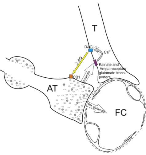
Figure 6. Schematic Illustration of the Neuro-glial Microcircuit in the External Zone of the ME
Axon terminals (AT) of the hypophysiotropic TRH neurons are closely associated to the endfeet processes of tanycytes (T) in the vicinity of fenestrated capillaries (FC) of the hypophyseal portal circulation. The TRH axons release glutamate that stimulates the DAGLaactivity of tanycytes, and therefore the 2-AG synthesis of these cells, by acting through kainite and AMPA receptors and glutamate transport and by the resulting increase of intracellular Ca2+level. The released endocannabinoids bind to the CB1 of the hypophysiotropic TRH axons and inhibit the amount of TRH released into the portal capillary.
METHODS
All methods can be found in the accompanyingTransparent Methods supplemental file.
SUPPLEMENTAL INFORMATION
Supplemental Information can be found online athttps://doi.org/10.1016/j.isci.2020.100921.
ACKNOWLEDGMENT
This work was supported by Grants from the Hungarian Science Foundation (OTKA K109710), the Hungar- ian National Brain Research Program (2017-1.2.1-NKP-2017-00002), EU H2020 THYRAGE no. 666869, CONACYT-Mexico (CB2015/254960), ERC682426, KFI-2016-0177, GINOP-2016-00979, NVKP-2016-0043 and by the BME-Biotechnology FIKP grant of EMMI (BME FIKP-BIO). The authors express their gratitude to Zso´fia La´szlo´ for her help during the preparing of some of the confocal microscopic images.
AUTHOR CONTRIBUTIONS
E.F. performed the immunofluorescent and ultrastructural studies. E.V. and B.K. designed and performed the patch clamp experiments. B.K. performed the optogenetic experiments. Z.P. deigned and performed the calcium imaging experiments. A.C.-V., J.-L.C., P.J.-B., A.S.-S., and Y.R. designed and performed the ME explant experiments and analyzed these data. M.M. synthesized, purified, and chemically characterized P-TRH. B.T. performed the 2-AG measurement. M.T. performed and analyzed thein situhybridization ex- periments. F.E., Z.M., G. S., and B.G. were involved in the generation of TRH-IRES-Cre mice. A.S.-S. and D.K. isolated the tanycytes and performed the gene expression analysis. D.Z. performed the stereotaxic AAV injections. A.K. performed immunostaining. Zs.M. and B.R. helped to design and perform the calcium imaging experiments and analyzed data. M.W. provided CB1 antibody. M.K. provided tissues of DAGLa KO mice. K.M. provided DAGLaantibody. R.M.L. analyzed data and wrote the manuscript. C.F. acquired the funding, conceptualized and interpreted the studies, and wrote the manuscript.
DECLARATION OF INTERESTS
B.R. is one of the founders of Femtonics and is a member of its scientific advisory board. The other authors have declared that no conflict of interest exists.
Received: March 25, 2019 Revised: May 20, 2019 Accepted: February 12, 2020 Published: March 27, 2020
REFERENCES
de Seranno, S., D’anglemont De Tassigny, X., Estrella, C., Loyens, A., Kasparov, S., Leroy, D., Ojeda, S.R., Beauvillain, J.C., and Prevot, V.
(2010). Role of estradiol in the dynamic control of tanycyte plasticity mediated by vascular endothelial cells in the median eminence.
Endocrinology151, 1760–1772.
Eyigor, O., and Jennes, L. (1998). Identification of kainate-preferring glutamate receptor subunit GluR7 mRNA and protein in the rat median eminence. Brain Res.814, 231–235.
Fekete, C., and Lechan, R.M. (2014). Central regulation of hypothalamic-pituitary-thyroid axis under physiological and pathophysiological conditions. Endocr. Rev.35, 159–194.
Fekete, C., Sarkar, S., Christoffolete, M.A., Emerson, C.H., Bianco, A.C., and Lechan, R.M.
(2005). Bacterial lipopolysaccharide (LPS)- induced type 2 iodothyronine deiodinase (D2) activation in the mediobasal hypothalamus (MBH) is independent of the LPS-induced fall in serum thyroid hormone levels. Brain Res.1056, 97–99.
Freitas, B.C., Gereben, B., Castillo, M., Kallo, I., Zeold, A., Egri, P., Liposits, Z., Zavacki, A.M., Maciel, R.M., Jo, S., et al. (2010). Paracrine signaling by glial cell-derived triiodothyronine activates neuronal gene expression in the rodent brain and human cells. J. Clin. Invest.120, 2206–2217.
Hrabovszky, E., Wittmann, G., Turi, G.F., Liposits, Z., and Fekete, C. (2005). Hypophysiotropic thyrotropin-releasing hormone and
corticotropin-releasing hormone neurons of the rat contain vesicular glutamate transporter-2.
Endocrinology146, 341–347.
Kano, M., Ohno-Shosaku, T., Hashimotodani, Y., Uchigashima, M., and Watanabe, M. (2009).
Endocannabinoid-mediated control of synaptic transmission. Physiol. Rev.89, 309–380.
Katona, I., Urban, G.M., Wallace, M., Ledent, C., Jung, K.M., Piomelli, D., Mackie, K., and Freund, T.F. (2006). Molecular composition of the endocannabinoid system at glutamatergic synapses. J. Neurosci.26, 5628–5637.
Kawakami, S. (2000). Glial and neuronal localization of ionotropic glutamate receptor subunit-immunoreactivities in the median eminence of female rats: GluR2/3 and GluR6/7 colocalize with vimentin, not with glial fibrillary acidic protein (GFAP). Brain Res.858, 198–204.
Kim, M.H., Uehara, S., Muroyama, A., Hille, B., Moriyama, Y., and Koh, D.S. (2008). Glutamate transporter-mediated glutamate secretion in the mammalian pineal gland. J. Neurosci.28, 10852–10863.
Lechan, R.M., and Fekete, C. (2007). Infundibular tanycytes as modulators of neuroendocrine function: hypothetical role in the regulation of the thyroid and gonadal axis. Acta Biomed.78(Suppl 1), 84–98.
Malcher-Lopes, R., and Buzzi, M. (2009).
Glucocorticoid-regulated crosstalk between arachidonic acid and endocannabinoid biochemical pathways coordinates cognitive-, neuroimmune-, and energy homeostasis-related adaptations to stress. Vitam Horm.81, 263–313.
Mayer, M.L., Westbrook, G.L., and Guthrie, P.B.
(1984). Voltage-dependent block by Mg2+ of NMDA responses in spinal cord neurones. Nature 309, 261–263.
Muller-Fielitz, H., Stahr, M., Bernau, M., Richter, M., Abele, S., Krajka, V., Benzin, A., Wenzel, J., Kalies, K., Mittag, J., et al. (2017). Tanycytes control the hormonal output of the hypothalamic- pituitary-thyroid axis. Nat. Commun.8, 484.
Ohno-Shosaku, T., and Kano, M. (2014).
Endocannabinoid-mediated retrograde modulation of synaptic transmission. Curr. Opin.
Neurobiol.29, 1–8.
Pak, T., Yoo, S., Miranda-Angulo, A.L., Wang, H., and Blackshaw, S. (2014). Rax-CreERT2 knock-in mice: a tool for selective and conditional gene deletion in progenitor cells and radial glia of the retina and hypothalamus. PLoS One9, e90381.
Piomelli, D. (2003). The molecular logic of endocannabinoid signalling. Nat. Rev. Neurosci.
4, 873–884.
Prevot, V., Dehouck, B., Sharif, A., Ciofi, P., Giacobini, P., and Clasadonte, J. (2018). The
versatile tanycyte: a hypothalamic integrator of reproduction and energy metabolism. Endocr.
Rev.39, 333–368.
Regehr, W.G., Carey, M.R., and Best, A.R. (2009).
Activity-dependent regulation of synapses by retrograde messengers. Neuron63, 154–170.
Rethelyi, M. (1975). Neurons in the subependymal layer of the rat median eminence.
Neuroendocrinology17, 330–339.
Rodriguez-Rodriguez, A., Lazcano, I., Sanchez- Jaramillo, E., Uribe, R.M., Jaimes-Hoy, L., Joseph- Bravo, P., and Charli, J.L. (2019). Tanycytes and the control of thyrotropin-releasing hormone flux into portal capillaries. Front. Endocrinol.
(Lausanne)10, 401.
Rodriguez, E.M., Blazquez, J.L., Pastor, F.E., Pelaez, B., Pena, P., Peruzzo, B., and Amat, P.
(2005). Hypothalamic tanycytes: a key component of brain-endocrine interaction. Int. Rev. Cytol.
247, 89–164.
Sanchez, E., Vargas, M.A., Singru, P.S., Pascual, I., Romero, F., Fekete, C., Charli, J.L., and Lechan, R.M. (2009). Tanycyte pyroglutamyl peptidase II
contributes to regulation of the hypothalamic- pituitary-thyroid axis through glial-axonal associations in the median eminence.
Endocrinology150, 2283–2291.
Suarez, J., Romero-Zerbo, S.Y., Rivera, P., Bermudez-Silva, F.J., Perez, J., De Fonseca, F.R., and Fernandez-Llebrez, P. (2010).
Endocannabinoid system in the adult rat circumventricular areas: an immunohistochemical study. J. Comp. Neurol.518, 3065–3085.
Tanimura, A., Yamazaki, M., Hashimotodani, Y., Uchigashima, M., Kawata, S., Abe, M., Kita, Y., Hashimoto, K., Shimizu, T., Watanabe, M., et al.
(2010). The endocannabinoid 2-arachidonoylglycerol produced by
diacylglycerol lipase alpha mediates retrograde suppression of synaptic transmission. Neuron65, 320–327.
Wittmann, G., Deli, L., Kallo, I., Hrabovszky, E., Watanabe, M., Liposits, Z., and Fekete, C. (2007).
Distribution of type 1 cannabinoid receptor (CB1)-immunoreactive axons in the mouse hypothalamus. J. Comp. Neurol.503, 270–279.
iScience, Volume23
Supplemental Information
A Glial-Neuronal Circuit in the Median Eminence
Regulates Thyrotropin-Releasing Hormone-Release via the Endocan- nabinoid System
Erzsébet Farkas, Edina Varga, Balázs Kovács, Anett Szilvásy-Szabó, Antonieta Cote- Vélez, Zoltán Péterfi, Magdalini Matziari, Mónika Tóth, Dóra Zelena, Zsolt Mezriczky, Andrea Kádár, Dóra K}ovári, Masahiko Watanabe, Masanobu Kano, Ken Mackie, Balázs Rózsa, Yvette Ruska, Blanka Tóth, Zoltán Máté, Ferenc Erdélyi, Gábor Szabó, Balázs Gereben, Ronald M. Lechan, Jean-Louis Charli, Patricia Joseph- Bravo, and Csaba Fekete