Distal radial secondary access for transcatheter aortic valve implantation:
The minimalistic approach☆
Alexandru Achim
a,b, Tamás Sz ű csborus
a, Viktor Sasi
a, Ferenc Nagy
a, Zoltán Jambrik
a, Attila Nemes
a, Albert Varga
a, Olivier F. Bertrand
c, Zoltán Ruzsa
a,⁎
a2ndDepartment of Internal Medicine, Division of Invasive Cardiology, University of Szeged, Szeged, Hungary
bMedicala 1 Invasive Cardiology Department, University of Medicine and Pharmacy“Iuliu Hatieganu”, Cluj-Napoca, Romania
cQuebec Heart-Lung Institute, University Laval, Quebec, Canada
a b s t r a c t a r t i c l e i n f o
Article history:
Received 28 September 2021
Received in revised form 16 November 2021 Accepted 17 November 2021
Available online xxxx
Background:Although not yet recommended by the guidelines, distal radial access, a new site for cardiovascular interventions, has been rapidly acknowledged and adopted by many centers due to its high rate of success, safety and fewer complications. We present our experience using secondary distal radial access during transcatheter aortic valve implantation (TAVI), proposing a new, even more minimal approach.
Methods:As of November 2020, a systematic distal radial approach as secondary access site for TAVI was adopted in our center. Primary endpoints were technical success and major adverse events (MAEs). Secondary endpoints:
the access site complication rate, hemodynamic and clinical results of the intervention, procedural related factors, crossover rate to the femoral access site, and hospitalization duration (in days).
Results:From November 2020, 41 patients underwent TAVI using this strategy. Patients had a mean age of 76 ± 11.2 years, 41% were male. Six (14.63%) patients received a balloon-expandable valve and 35 (85.37%) received a self-expandable valve. TAVI was successful in all cases. No complications occurred due to transradial access.
Puncture success, defined as completed sheath placement was maximum (N= 41/41,100%) and emergent trans- femoral secondary access was not required in any case. Primary transfemoral vascular access site complications occurred in 7 cases (17%) of which 4 (13.63%) were resolved through distal radial access: one occlusion, two flow-limiting stenoses and four perforations of the common femoral artery. There were no additional major vas- cular complications at 30 days. Overall MACE rate was 2.4%.
Conclusion:The use of the distal radial approach for secondary access in TAVI is safe, feasible and has several ad- vantages over old access sites.
© 2021 Published by Elsevier Inc.
Keywords:
TAVI
Distal radial access Distal radial artery approach Snuffbox approach Aortic stenosis Outcome of TAVI Radial access
Transcatheter aortic valve Vascular complication
1. Introduction
Vascular complications (VCs) during transcatheter aortic valve re- placement (TAVI) remain the most common procedural complications despite smaller delivery systems and improved operator experience.
The transradial approach (TRA) as secondary access in TAVI has become the preferred access site due to its significant reduction in (overall and major) vascular and bleeding periprocedural events (approximately 25% of periprocedural access VCs are related to the transfemoral second- ary access[1]). Distal radial access (dRA) at the anatomical snuffbox has been reported[2–4]as a safe and feasible unique alternative to TRA at the wrist[3]. The need for patent hemostasis with forearm radial access, to minimize the risk of radial artery occlusion (RAO) mandates close vigilance for bleeding to prevent forearm hematoma formation and re- quires prompt effective management if it develops, to stop progression to compartment syndrome. The latter risk may be lower with dRA as not only is the puncture site distal to the forearm, but it is more readily com- pressible. A typical complication of proximal radial access (pRA) is a chronic RAO caused by local hematoma, spasm, arterial wall damage or compression[4,5]and dRA was proven superior to proximal radial access in preventing proximal RAO at 24 h and 30 days after a diagnostic or interventional coronary procedure[6]; another important aspect is Cardiovascular Revascularization Medicine xxx (xxxx) xxx
Abbreviations:TAVI, transcatheter aortic valve implantation; MAEs, major adverse events; VCs, vascular complications; TRA, transradial approach; dRA, distal radial approach; pRA, proximal radial approach; RAO, radial artery occlusion; AoS, aortic stenosis; PTA, percutaneous transluminal angioplasty; PCI, percutaneous coronary intervention; COPD, chronic obstructive pulmonary disease; IDDM, insulin-dependent di- abetes mellitus; NIDDM, noninsulin-dependent diabetes mellitus; LVEF, left ventricle ejec- tion fraction.
☆ Disclosures: The authors report nofinancial relationships or conflicts of interest re- garding the content herein.Work Institution: University of Szeged, Medical Faculty, Department of Internal Medicine, Invasive Cardiology Division, Szeged, Hungary.
⁎ Corresponding author at: Szeged, Semmelweis Str 6, 6726, Hungary.
E-mail address:ruzsa.zoltan@med.u-szeged.hu(Z. Ruzsa).
https://doi.org/10.1016/j.carrev.2021.11.021 1553-8389/© 2021 Published by Elsevier Inc.
Contents lists available atScienceDirect
Cardiovascular Revascularization Medicine
Please cite this article as: A. Achim, T. Szűcsborus, V. Sasi, et al., Distal radial secondary access for transcatheter aortic valve implantation: The minimalistic approac..., Cardiovascular Revascularization Medicine,https://doi.org/10.1016/j.carrev.2021.11.021
ipsilateral pRA patency even if dRA occludes after intervention, because dRA location is after the superficial palmar branch of the radial artery (Fig. 1), thereforeflow is maintained through this branch and thrombus formation is stopped at this point. Lastly, hand position difficulties take place more often when performing pRA.
Putting forward these advantages, dRA competes with the conven- tional radial approach and may change our common access common in the near future. Several registries have been collected recently re- garding dRA for coronary angiography and interventions[7–10]but there are no data regarding dRA as a secondary site for TAVI, although it may bring significant benefit in terms of VC reduction and patient- operator ergonomics. We aimed to prospectively analyze the proce- dural performance, outcomes, safety and efficacy of dRA as a new, minimalistic option during TAVI.
2. Methods
Clinical and angiographic data from 41 consecutive patients with se- vere, symptomatic aortic stenosis (AoS) were evaluated in a retrospec- tive single-center study. Between November 2020 and April 2021 the patients were treated using dual access, one distal-radial and one femo- ral. Distal radial access was obtained using a 5 or 6 French Glidesheath (Terumo Medical Corporation, Somerset, NJ, USA). When 2 secondary access sites were needed (for periprocedural coronary protection), both distal radial arteries were cannulated. Access to the ascending aorta from the radial artery was typically obtained using a 5F pigtail on a 0.35″guidewire (J-Tip Guidewire, Terumo Medical Corporation, Somerset, New Jersey, USA). Transfemoral TAVI cases were routinely performed under local and general anesthetic with transesophageal echocardiographic guidance unless clinical reasons required otherwise.
Portico (St Jude Medical, MN, USA), Evolut (Medtronic, Inc., Minneapo- lis, Minn, USA), Acurate (Boston Scientific, Boston, MA, USA) and Myvalve (Meril Life Sciences Pvt. Ltd., Vapi, Gujarat, India) valves were used. TF primary access closure was performed with one ProGlide de- vice (Abbott Vascular, Santa Clara, CA, USA) and one AngioSeal 8F (Terumo Medical Corporation, Somerset, NJ, USA). Our Insitutional Eth- ical Review Committee approved the study (OGYÉI/50275/2017), and all patients provided written informed consent prior to study inclusion.
2.1. Inclusion criteria
We included high risk patients with symptomatic significant AoS in the study after written informed consent was obtained.
2.2. Exclusion criteria
(1) hemodynamic Preexisting aortic prosthesis, recent myocardial infarction, left ventricular or atrial thrombus, severe aortic, mitral or tri- cuspid regurgitation (grade III-IV), recent cerebrovascular event (within 3 months), carotid or vertebral arterial stenosis (>80%), active internal bleeding, thrombocytopenia (platelet count <50,000/mm3), (2) ethical:
lack of written informed consent, severe mental disorder, drug/alcohol addiction, life expectancy <1/2 year, participation in another drug or device study, pregnancy, and (3) vascular: patients with occluded, se- verely diseased or small (<1.5) mm radial or ulnar arteries on both sides were not included.
Our primary endpointswere technical success and major adverse events (MAEs).The secondary endpoints werethe access site complica- tion rate, hemodynamic and clinical results of the intervention, proce- dural related factors, crossover rate, radial occlusion rate and hospitali- zation duration (in days).
2.3. Definitions
Major adverse events (MAEs): MAEs were assessed as the composite of death, stroke, myocardial infarction, and urgent major aortic valve re- placement or implantation during the hospital stay and/or at the one- month follow-up.
Definition of vascular complications: Major vascular complications were defined as diminished or lost arterial pulse or the presence of any pseudoaneurysm or arteriovenous fistula during the clinical follow-up. Minor complications were defined as hematomas requiring no further treatment (EASY 1–2), measuring 2 cm in diameter over the radial or ulnar puncture area or measuring 5 cm in diameter over the femoral puncture site. Major bleeding was defined as a drop in the hemoglobin level of >3 g/dl, as well as any bleeding requiring blood transfusions. Major hematoma was defined as EASY 3–4 hematoma.
Technical successwas defined as successful transcatheter aortic valve implantation over the native, stenotic aortic valve.
Hemodynamic successwas defined as a 90% drop in the mean aortic gradient.
Clinical success: Primary clinical success was defined as an improve- ment of at least one clinical category in the NYHA classification.
2.4. Duplex ultrasound (US) protocol
Duplex US was used in the operating room to investigate all forearm arteries. RA, UA diameter and peak systolic velocity were measured at the wrist level. On thefirst postoperative day, all patients underwent control duplex US.
Access site selection: Two skilled operators trained in bilateral trans- radial access and TAVI performed all cases. The primary preferred access site was the right femoral artery, and for aortography the left radial ar- tery was used. The left radial artery was punctured distally, with the armflexed and pronated on the patient's abdomen, reproducing a nat- ural positon, increasing the patient's comfort and minimizing operator discomfort during the procedure (Fig. 2). The alternative access site was the ulnar artery, which was used when a Doppler ultrasound showed a small radial artery (less than 1.5 mm) or was extremely calci- fied or occluded. For rapid pacing the access site was the femoral or the jugular vein or guidewire pacing approach.
2.5. Statistical analysis
Statistical analysis was performed using commercially available GraphPad Prism 8.0 software (USA). Continuous variables were expressed as the mean ± standard deviation or as the median with in- terquartile range. Categorical variables were tabulated as percentages.
The different patient cohorts were compared using either the Mann- Fig. 1.Curtesy of P. Patelet al. Distal radial artery course through the anatomic snuffbox.
Note that the superficial palmar artery branches take off before the radial artery enters the anatomic snuffbox.
WhitneyUtest or the Kruskal-Wallis test. Probability values lower than 0.05 were considered to be significant.
3. Results
The main baseline clinical and procedural characteristics of the global population and according to the secondary approach (transfem- oral versus transradial) are shown inTable 1.
TAVI was successful in all cases. Procedural details are highlighted in Table 2. Technical success was achieved in all patients (100%). Clinical success was achieved in 40 patients (97,5%). Hemodynamic success was achieved in all patients (100%). By hemodynamic investigation, the peak-to-peak mean gradient decreased from 76.8 ± 27.2 to 10.7 ± 5.1 mmHg (p= 0.001). Balloon postdilatation was performed in 19 cases (46.3%), and the crossover to urgent surgical aortic valve im- plantation was 0%. Secondary access was achieved through the left dRA only (100%) and the crossover rate to the femoral access site was achieved in 3 cases (7.31%) of major primary femoral access perforation.
Temporary pacing was performed either through the jugular vein (n= 19, 46.3%) or femoral vein (n= 10, 24.3%) or directly on the TAVI stiff wire (n= 12, 29.2%). Thefluoroscopy time, X-ray dose, procedure
time, and contrast consumption were 18.20 ± 7.6 min, 529.8 ± 484.6 mGy, 50.3 ± 28.1 min, and 64.9 ± 28.1 ml, respectively. Mean hospitalization duration over the cohort was 4 ± 2.5 days. Ultrasonog- raphy parameters before and after the procedure are summarized in Table 2.
No complications occurred due to transradial access. Transfemoral vascular access site complications occurred in 7 cases (17.07%): 1 occlu- sion, 2flow-limiting stenoses and 4 perforations of the common femoral artery. Three complications were successfully managed using balloon dilatation and balloon tamponade from the transradial access (Fig. 3).
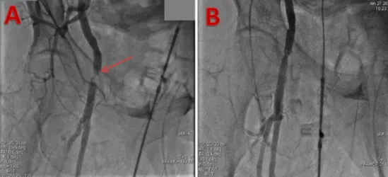
The cases of major perforation (9.75%) were successfully treated with a covered stent delivered via direct ipsilateral superficial femoral retro- grade 6F access and stent delivery with angiographic control from dRA (Fig. 4). There were no additional major vascular complications at 30 days. Five cases of PCI (12.19%) and one case of left subclavian artery PTA (2.43%) were successfully performed through dRA, in the same ses- sion. Hemostasis of the primary transfemoral access was achieved per- cutaneously (vs surgical cut-down) in all patients, using two ProGlide devices (Abbott Vascular, Santa Clara, California) in 31.82% and one ProGlide plus one Angioseal 8F device (Terumo Medical Corporation, Somerset, NJ, USA) in 68.18% of the cohort. Sheath size for secondary Fig. 2.Patient example, right femoral primary access (A) and left distal radial secondary access (B), both sheaths being in proximity of each other, enhancing better ergonomics and lowering the radiation dose.
Table 1
Demographic and clinical data.
n (%)
Demographic data Age (years) 76.0 ± 11.2
Male 17 (41)
Hypertension 34 (82.9)
Current smokers 11 (26.8)
Diabetes mellitus 15 (36.5)
IDDM 4 (9.7)
NIDDM 11 (26.8)
Weight (kg) 79.4 ± 16.7
Height (cm) 167.1 ± 9.9
COPD 9 (21.9)
Renal insufficiency 15 (36.5)
Family history 9 (21.9)
Dyslipidemia 26 (63.4)
Atrialfibrillation 17 (41.4)
Cardiac and vascular history Coronary artery disease 13 (31.7) Peripheral artery disease 7 (17.07) Previous PCI or coronary bypass 10 (24.3) Previous valve surgery 3 (7.31) Symptoms
- Angina 34 (82.9)
- Dyspnoea 41 (100)
- Syncope 2 (4.87)
Table 2 Procedural results.
Pre-interventional Post-interventional Clinical status (dyspnoe)
NYHA 1 0 1
NYHA 2 1 30
NYHA 3 30 10
NYHA 4 9 0
Vascular ultrasound (radial site)
- Radial artery (mm) 1.8 ± 0.5 1.8 ± 0.5
- Hematoma (EASY 1–2) 0 1
- Hematoma (EASY 3–4) 0 0
- Radial artery occlusion 0 0
- Pseudoaneurysm 0 0
Transthoracal ultrasound LVEF
- 50–70% 34 (82.92) 37 (90.24)
- 30–50% 6 (14.63) 4 (9.75)
- <30% 1 (2.43) 0 (0)
Aortic gradient (peak) 76.8 ± 27.2 10.7 ± 5.1
Aortic gradient (mean) 50.9 ± 18.7 12.6 ± 9.3
AVA (mm2) 0,67 ± 0.13 2.4 ± 0.27
Aortic regurgitation
- 0–2 24 (58.5) 10 (24.39)
- 3–4 0 (0) 0 (0)
access in the transradial group was 5 French (F), 6 F, and 7 F in 20 (48.7%), 19 (46.3%) and 2 (4.87%) patients, respectively. Hemostasis of the radial access was achieved via compression in all cases, using a com- bination of one StatSeal Disc (Biolife, LLC, USA) with gauze compression.
Standard hemostasis time was 3 h. The need of repeating the hemostasis was not needed in any cases. Data regarding radial occlusion were col- lected 24 h after puncture and Doppler ultrasound revealed no artery occlusion. All-cause 30-day mortality was 2.4% %, one patient suffered esophageal and tracheal bleeding after extubation with hemorrhagic shock (autopsy report revealed bleeding from the esophageal vein).
No stroke or myocardial infarction was observed.
4. Discussion
Our case series shows that TAVI can be performed safely using dRA secondary access with no increase in radiation dose, screening time or procedural duration. The occlusion rate proved to be non-existent.
This is of particular importance in order to keep access available. We also show that with appropriate equipment major vascular complica- tions related to femoral primary access can be managed entirely via the TR approach (Fig. 3).
TRA for TAVI secondary access siteshas been introduced to guide valve implantation, to treat coronary lesions, and to control or treat vascular access site complications. Coronary angioplasties, even complexs such as LM-stenting could be simply and elegantly completed using dRA.
Puncturing the left dRA brings the left hand very close to the primary
femoral access, thus keeping the radiation dose low and enhancing the comfort of the operator. Easy closure shortens the procedural time and keeps the pRA available for emergency second-look at any time. A total of 13.63% of our patients necessitated pre- or post-PCI during TAVI, which was performed via dRA only. It has been reported that up to a quarter of vascular complications during TAVI result from the use of femoral secondary arterial access[1]. The introduction of TRA for cor- onary angiography and angioplasty has reduced bleeding and access site complications in coronary interventions[11]. Logically, the same may be true during TAVI procedures, especially as the elderly patient co- hort currently eligible for TAVI is at high risk of developing femoral ar- terial complications. Because old and frail adults often present with lim- ited arm motion, our approach demonstrated greater patient tolerabil- ity and resulted in short hospital stays. Additionally, the procedural time was shorter than that in other reports[1,12].
Transradial secondary access readily allows aortography to guide valve implantation. For non-TF TAVI cases, this is the sole purpose of secondary arterial access and thus TR access can be considered the de- fault approach. Changing from conventional pRA to dRA, thus heading toward an even more minimally invasive approach, can achieve further VC reduction and longer radial lumen patency for possible future inter- ventions. Various small observational studies have shown similar radia- tion doses between pRA and dRA on coronary interventions[6,9,10], be- cause the two access sites are very close to each other. However, we be- lieve this concept could be different in TAVI, as in this case the operator could receive the“femoral”radiation dose, lower than the radial dose, Fig. 3.Thrombotic subocclusion of femoral artery treated percutaneously.
A - Angiography via MPA guide catheter in the iliac artery, showing extensive thrombus and poorflow. B - Angiography showing restoration of distalflow following balloon dilatation.
Technical details: 7.5 F sheatless guiding catether, V18 0.14″guidewire (Boston Scientific, Natick, MA, USA), Passeo 6 × 40 mm balloon (Biotronik AG, Buelach, Switzerland), 2 min inflation at 8 atm.
Fig. 4.Stent graft placement from direct femoral access route.
Balloon tamponade via dRA route (A), with unsuccessful hemostasis and active bleeding (B, arrow) andfinal stent graft implantation over the common and superficial right femoral artery, with subsequent deep femoral artery occlusion (C).
by bringing the left hand very close to the femoral access. Won-Keun Kim et al. recently described a novel technique using only a single arte- rial access technique[12](valve deployment without contrast injection but using a guiding wire for valve positioning that is introduced through the same femoral access but outside the large bore sheath), heading for the same minimalistic approach and stating that secondary access com- plications were eliminated together with the whole secondary access concept but we believe aortography is necessary for the valve position- ing and further postdilatation decisions or ad-hoc PCI; therefore, sec- ondary access should be kept and dRA may be the solution for minimiz- ing all possible secondary access complications.
dRA for coronary angioplastyis becoming the primary access site in many catheterization laboratories. The main advantage of this access site is the low rate of radial artery occlusion and the better comfort for the patient during left transradial cases[3–10]. The main disadvantage of this access site is the small size of the RA in this location, the angulated course of the RA and the need of ultrasonography for the puncture. It is essential to know that the dRA diameter is 80% less than the proximal diameter, which may affect suitable French sheaths that can be used, and it varies according to sex, race, and other factors.
The diameter of the dRA in females tends to be smaller than that in males. Some studies have shown that hypertensive patients have larger radial artery diameters than normal patients. Other studies show that the diameter of dRA is positively correlated with both body weight and basal metabolic index. Ulnar arteries were slightly larger than radial arteries and may be used as an alternative to dRA[8,13,14]. We did not find a correlation between body weight and vessel diameter in our group. A blind puncture increases the risk of tendon damage. At the same time, the double-wall technique can irritate the underlying peri- osteum and increase the risk of hematoma formation. Ultrasound (US) allows the identification of anatomical landmarks, and enables accurate vessel access, especially in impalpable dRA. The probe can be used to perform a compressibility test to confirm that the target vessel corre- sponds to the radial artery rather than the cephalic vein. Accurate scan- ning can identify the superficial branch of the radial nerve, thus avoiding potential injury. A further benefit of US guidance is that the op- erator can measure the vessel size before puncturing. A vessel diameter under 2.0 mm was associated with higher occlusion rate, spasm and pa- tient pain[13,14,15]. The success rate of sheath placement was found to be 100% (41/41), without differences between the side (left dRA or right dRA) or site (anatomical snuffbox or distal dorsal of the tendon of the extensor pollicis longus) of the dRA. However, we must admit that an adequately powered randomized trial is necessary to confirm this suc- cess rate, as our study was a single-center study; moreover, it was a rel- atively small study and we did not include acute, unstable patients re- quiring emergency TAVI.
dRA for iliac and femoral angioplastyhas been published recently with high technical success and a very low complication rate[16]. Our center is a dedicated dRA center; thus most of our operators are trained to perform this puncture procedure. The limitation of the dRA for the treatment of access site complications is the distance between the dRA and the common femoral artery, therefore small bleedings or dissec- tions can be treated from this access site with available devices (sheaths, balloons, stents) (Fig. 3), but perforations need distal access if stent graft implantation is needed. In this case series, stent graft placement was needed in two cases and both were implanted from direct superficial femoral artery access (Fig. 4). Spasm was infrequent because of the use of a long transradial slender sheath, although we did not administer any intraarterial nitroglycerine due to aortic stenosis.
4.1. Limitations
This registry included neither a randomization nor a control group and although we used uniformly prespecified endpoints for the analysis it was a retrospective data collection. It is therefore possible that some patients with complications or crossover may have been overlooked.
We tried to counter this problem by double rechecking all cath lab pro- tocols in the participating centers during the period of data acquisition.
The lack of a control group with proximal access certainly limits our conclusions; our goal was to implement this promising new access site in other cardiac interventions. Additionally, no data were collected regarding the occurrence of radial occlusion. Although this is usually a silent complication with no clinical consequences, future studies will have to evaluate the impact of this complication in the TAVI population.
More and more positive data on (left) distal radial access are being published[10–17]. In fairness, the more publications appear, the more local complications will be described as well. Simultaneously, many of the benefits associated with distal access are becoming less and less the- oretical at this point of time and as more practitioners adopt this tech- nique, new nuances, indications, and technical aspects will assuredly continue to emerge.
5. Conclusion
The initial results of this novel, minimalistic approach demonstrate its feasibility and potential beneficial effects without compromising procedural safety. The main advantages are less arterial obstruction and short hemostasis. Larger randomized trials are needed to further evaluate the advantages of dRA over pRA.
CRediT authorship contribution statement
Alexandru Achim MD: Contributing Author, Writing- Original Draft and Editing, ConceptualizationTamás Szűcsborus MD: Data Curating, Software, ValidationViktor Sasi MD PhD: Data Curating, Validation Ferenc Nagy MD PhD: Project Administration, Data CurationZoltán Jambrik MD PhD: Methodology, ConceptualizationAttila Nemes MD DsC: Data Validation, Reviewing, SoftwareAlbert Varga MD DsC: Con- ceptualizationOlivier F Bertrand MD PhD: Resources, Formal Analysis Zoltán Ruzsa MD PhD: Supervision, Writing- Reviewing and Editing, Investigation.
Declaration of competing interest
The authors declare that they have no known competingfinancial interests or personal relationships that could have appeared to influ- ence the work reported in this paper.
References
[1]Junquera L, Urena M, Latib A, Munõz-Garcia A, Nombela-Franco L, Veiga-Fernandez Faurie B, et al. Comparison of transfemoral versus transradial secondary access in transcatheter aortic valve replacement. Circ Cardiovasc Interv. 2020;24:334–42.
[2] Kaledin A, Kochanov IN, Podmetin PS, Seletsky SS, Ardeev VN. Distal radial ar- tery in endovascular interventions; 2017.https://doi.org/10.13140/RG.2.2.
13406.33600https://www.researchgate.net/.
[3]Kiemeneij F. Left distal transradial access in the anatomical snuffbox for coronary an- giography (ldTRA) and interventions (ldTRI). EuroIntervention. 2017;13:851–7.
[4]Lee JW, Park SW, Son JW, Ahn SG, Lee SH. Real-world experience of the left distal transradial approach for coronary angiography and percutaneous coronary inter- vention: a prospective observational study (LeDRA). EuroIntervention. 2018;14 995-03.
[5]Sciahbasi A, Rigattieri S, Sarandrea A, et al. Radial artery occlusion and hand strength after percutaneous coronary procedures: results of the HANGAR study. Catheter Cardiovasc Interv. 2016;87:868–74.
[6]Eid-Lidt G, Rivera Rodríguez A, Jimenez Castellanos J, Farjat Pasos JI, Estrada López KE, Gaspar J. Distal radial artery approach to prevent radial artery occlusion trial.
JACC Cardiovasc Interv. 2021;14:378–85.
[7]Aoi S, Htun WW, Freeo S, Lee S, Kyaw H, Alfaro V, et al. Distal transradial artery ac- cess in the anatomical snuffbox for coronary angiography as an alternative access site for faster hemostasis. Catheter Cardiovasc Interv. 2019;94:651–7.
[8]Kim Y, Ahn Y, Kim I, Lee DH, Kim MC, Sim DS, et al. Feasibility of coronary angiogra- phy and percutaneous coronary intervention via left snuffbox approach. Korean Circ J. 2018;48:1120–30.
[9]Liontou C, Kontopodis E, Oikonomidis N, Maniotis C, Tassopoulos A, Tsiafoutis I, et al.
Distal radial access: a review article. Cardiovasc Revasc Med. 2020;21:412–6.
[10]Oliveira MDP, Navarro EC, Kiemeneij F. Distal transradial access as default approach for coronary angiography and interventions. Cardiovasc Diagn Ther. 2019;9:513–9.
[11]Brueck M, Bandorski D, Kramer W, Wieczorek M, Holtgen R, Tillmanns H. A randomised comparison of transradial versus transfemoral approach for coronary angiograms and angioplasty. J Am Coll Cardiol Int. 2009;2:1047–54.
[12]Kim WK, Doerr O, Renker M, Choi YH, Liakopoulos O, Hamm CW, et al. Initial expe- rience with a novel, modular, minimalistic approach for transfemoral aortic valve implantation. Int J Cardiol. 2021;332:54–9.
[13]Beniwal S, Bhargava K, Kausik SK. Size of distal radial and distal ulnar arteries in adults of southern Rajasthan and their implications for percutaneous coronary inter- ventions. Indian Heart J. 2014;66:506–9.
[14]Ashraf T, Panhwar Z, Habib S, Memon MA, Shamsi F, Arif J. Size of radial and ulnar artery in local population. J Pak Med Assoc. 2010;60:817–9.
[15]Loh YJ, Nakao M, Tan WD, Lim CH, Tan YS, Chua YL. Factors influencing radial artery size. Asian Cardiovasc Thorac Ann. 2007;15:324–6.
[16]Ruzsa Z, Csavajda Á, Nemes B, Deák M, Sótonyi P, Bertrand OF, et al. Distal radial ar- tery access for superficial femoral artery interventions. J Endovasc Ther. 2021;28:
255–61.
[17]Al-Azizi KM, Grewal V, Gobeil K, Maqsood K, Haider A, Mohani A, et al. The left distal transradial artery access for coronary angiography and intervention: a US experi- ence. Cardiovasc Revasc Med. 2019;20:786–9.