Corresponding author:
Levente Molnár MD, Heart and Vascular Center, Semmelweis University, Budapest, Hungary, e-mail: mlevente@yahoo.com Received: 29.07.2021, accepted: 4.11.2021.
Safety and feasibility of transradial aortic valve valvuloplasty (TRAV study)
Levente Molnár1, Roland Papp1, Tímea Szigethi1, István F. Édes1, Dávid Becker1, Olivier F. Bertrand2, Béla Merkely1, Zoltán Ruzsa3
1Heart and Vascular Center, Semmelweis University, Budapest, Hungary
2Cardiology Department, University Laval, Quebec, Canada
3Invasive Cardiology Division, Internal Medicine Department, University of Szeged, Szeged, Hungary
Adv Interv Cardiol 2021; 17, 4 (66): 381–388 DOI: https://doi.org/10.5114/aic.2021.111341
A b s t r a c t
Introduction: The importance of balloon aortic valvuloplasty (BAV) in the transcatheter aortic valve implantation (TAVI) era emerged in the past decades, but the access site related complication rate remained significant.
Aim: To establish the safety and technical success of transradial balloon aortic valvuloplasty (trBAV). The secondary objective was to determine the effectiveness and appropriate role of trBAV.
Material and methods: Between 2017 and 2019, 36 consecutive patients with symptomatic aortic stenosis (AoS) were treated with trBAV in this prospective, single-center study. During the procedure, the efficacy and the aortic valve insufficiency were con- trolled by hemodynamic measurements and later by echocardiography. The primary end-points were technical success and major adverse events (MAE). Secondary end-points were the access site complication rate, hemodynamic and clinical result of the inter- vention, procedure-related factors, crossover rate to the femoral access site and hospitalization duration.
Results: Clinical and technical success was achieved in all cases. Invasively measured peak-to-peak gradient decreased from 76.8 ±27.2 to 54.7 ±21.1 mm Hg (p = 0.001), and the aortic-valve area increased from 0.69 ±0.2 to 0.91 ±0.3 cm2 (p = 0.001). No major adverse cardiac or cerebrovascular events or vascular complications (according to VARC 2 criteria) occurred during the proce- dures. The perioperative death rate was 2.7% (n = 1).
Conclusions: According to our study, radial artery access is a safe and effective option for balloon aortic valvuloplasty in patients with severe aortic valve stenosis.
Key words: vascular complications, aortic valve valvuloplasty, transradial approach.
Introduction
Percutaneous balloon aortic valvuloplasty (BAV) has been widely used in high-risk patients since it was first introduced by Cribier et al. [1]. The technique has been questioned in many trials due to the limited hemodynam- ic effect and a high rate of restenosis [2–4]. Nowadays, the role of BAV in the management of severe symptomat- ic aortic stenosis (AoS) has come under the spotlight fol- lowing the development of the transcatheter aortic valve implantation (TAVI) technique. From palliative and bridge to aortic valve implantation therapy, the indications have been widened to include symptomatic improvement be- fore the consideration of definitive TAVI intervention [5, 6]. The primary access for BAV is still the femoral artery access, but also subclavian, brachial, and radial artery (RA)
access has been reported in the literature [7–14]. The aim of the study was to investigate the efficacy and safety of the BAV procedure performed from transradial access.
Aim
The aim of our study was to establish the safety and the technical success of transradial balloon aortic valvu- loplasty (trBAV). The secondary objective was to deter- mine the effectiveness and appropriate role of trBAV.
Material and methods Methods
Clinical and angiographic data from 36 consecutive patients with symptomatic AoS were evaluated in a pro-
spective pilot single-center study. Four patients were excluded from the study due to the small size or occlu- sion of the radial artery. Between 2017 and 2019, the patients were treated utilizing bilateral radial or radial and contralateral ulnar artery access with 6–10 F com- patible balloons. The decision of the BAV was made by the Semmelweis University Heart and Vascular Cen- ter Valve Team and all the procedures were performed in a hybrid operating room. The impact of the learning curve was analyzed after 10 trBAV cases. In 21 cases tr- BAV was used as a bridge to TAVI or surgical aortic valve replacement (SAVR) and in 12 patients to palliate the symptoms as destination therapy. In 2 cases trBAV was performed before acute non-cardiac surgery. Our Hungar- ian State Ethical Review Committee approved the study (OGYÉI/50275/2017), and all patients provided written informed consent prior to study inclusion.
Inclusion criteria
We included high-risk patients with symptomatic sig- nificant AoS in the study after written informed consent.
Exclusion criteria
Preexisting aortic prosthesis, recent myocardial in- farction, left ventricular or atrial thrombus, severe aor- tic, mitral or tricuspid regurgitation (grade III–IV), recent cerebrovascular event (within 3 months), carotid or ver- tebral artery stenosis (> 80%), active internal bleeding, thrombocytopenia (platelet count < 50,000/mm3), lack of written informed consent, severe mental disorder, drug/
alcohol addiction, life expectancy < 1/2 year, participation in another drug or device study, pregnancy, and subopti- mal vascular anatomy: patients with occluded, severely diseased or small (< 1.5 mm) radial or ulnar arteries on both sides were not included.
Endpoints
Our primary endpoints were technical success and major adverse events (MAE). The secondary endpoints were the access site complication rate, hemodynamic and clinical result of the intervention, procedure-related factors, and crossover rate to the femoral access site.
Duplex ultrasound (US) protocol
Duplex US was used in the operating room to investi- gate all forearm arteries. Radial artery diameter, ulnar ar- tery (UA) diameter, and peak systolic velocity were mea- sured at the wrist level. On the first postoperative day, all patients underwent follow-up Duplex US.
Access site selection
Two skilled operators trained in bilateral transradial access, BAV and peripheral vascular intervention per- formed all cases. The preferred access site for trBAV was
the right RA (n = 21), the alternative access site was the left RA (n = 14) and in 1 case the right UA, which was used when Doppler ultrasound showed a small (less than 1.5 mm) and extremely calcified radial artery. Aortogra- phy was performed from the contralateral RA approach, except 1 case, when UA was used. Rapid pacing was per- formed via the femoral or jugular vein.
Antithrombotic regimen
All patients with known coronary artery disease were treated with daily 100 mg aspirin; the patients who underwent coronary stenting received dual antiplate- let therapy (aspirin 100 mg and clopidogrel 75 mg) for 6 months. In addition, 5000 IU heparin sodium, and 250 µg nitroglycerine were administered directly into the radial artery through the sheath. Additional Na-heparin was given until reaching 70 IU/kg. Routine ACT was not measured during the intervention.
TrBAV technique (Figure 1)
Puncture: After local anesthesia, the RA or UA was punctured under ultrasonography guidance with a dedi- cated transradial needle and sheath (Terumo Co., Japan, 5F). Aortography was performed using a pigtail catheter in a left lateral 30-degree view with 15 ml of contrast. RA puncture was carried out in the conventional or distal ra- dial artery access (snuff box). The TrBAV procedure was started with a US based puncture of the right contralater- al radial or ulnar artery. The aortic valve was passed with a Terumo Guidewire using Amplatz left 1 or 2 diagnostic catheters and then the Terumo GW was replaced with a 260 cm J tip 0.035’’ guidewire. The Amplatz catheter was replaced with a diagnostic pigtail catheter and the baseline pressure gradient was measured. After the baseline pres- sure recordings, the Amplatz Super stiff or Safari guide- wire (Boston Sci, USA) was advanced into the left ventricle.
At this point, the radial sheath was removed and a short hydrophilic femoral sheath (Terumo, Japan) was advanced in the RA through the 0.035 GW. The size of the introducer was selected as specified by the US measurement of the access site and the entry profile of the preferred balloon.
According to the preoperative US parameters, there were no access site/introducer disproportion. The procedure was performed with 16–22 mm balloons under conven- tional rapid pacing. The balloon diameter was calculated from the preoperative thoracic CTA or echocardiography measurements (balloon to annulus diameter ratio 0.8).
After balloon removal, the pressure measurements were made with the pigtail catheter again. Invasive hemody- namic measurement is presented in Figure 2. The second balloon dilatation was the operator’s decision.
Postoperative treatment
After the procedure, the sheath was immediately re- moved, and hemostasis was achieved by applying the
Terumo Band dedicated compression device (Terumo, Ja- pan) to the radial access site for 6 h. The femoral venous sheath was removed immediately and a compression bandage was applied for 6 h. During the postprocedural care, overnight intensive care unit observation was con- ducted.
Post-procedural follow-up
After the procedure, all patients underwent physi- cal examination and echocardiography measurements.
All patients were scheduled for a detailed clinical fol- low-up examination at 3, 6, and 12 months after the procedure.
Definitions
Major adverse events (MAE): MAE were assessed as the composite of death, stroke, myocardial infarction, and urgent major aortic valve replacement or implanta- tion during the hospital stay or at the 1-month follow-up.
Definition of vascular complications: Major vascular complication was defined as diminished or lost arterial pulse or the presence of any pseudo-aneurysm or ar- teriovenous fistula during the clinical follow-up. Minor complications were defined as hematomas requiring no further treatment (EASY (Early Discharge After Transradi- al Stenting of Coronary Arteries Study) 1–2), measuring Figure 1. Single balloon angioplasty: A – TrBAV setup in hybrid operation room, B–D – Sequential angiographic images demonstrating the procedure and the invasive hemodynamic measurements, using the retrograde technique (B), followed by balloon dilatation (C) and invasive measurements (D)
D C
A B
2 cm in diameter over the radial or ulnar puncture area, or measuring 5 cm in diameter over the femoral puncture site. Major bleeding was defined as a drop in the hemo-
globin level of > 3 g/dl, as well as any bleeding requir- ing blood transfusions. Major hematoma was defined as EASY 3–4 hematoma [7].
Technical success was defined as successful valvu- loplasty balloon inflation at the aortic valve.
Hemodynamic success was defined as at least 25%
drop in peak aortic gradient.
Clinical success: Primary clinical success was defined as an improvement of at least one clinical category in the NYHA classification.
Statistical analysis
Statistical analysis was performed using the com- mercially available Graph Pad Prism 8.0 software (USA).
Continuous variables were expressed as the mean ± standard deviation or as the median with inter-quartile range. Categorical variables were tabulated as percentag- es. The different patient cohorts were compared using ei- ther the Mann-Whitney U test or the Kruskal-Wallis test.
Probability values lower than 0.05 were considered to be significant.
Results
TrBAV was performed in 36 patients with significant AoS, using bi-radial RA (n = 34, 94.4%) or hybrid (radi- al and ulnar) (n = 2, 5.6%) access. The access site for pacemaker insertion was the femoral vein in 3 (8.3%) or jugular vein in 20 (55.5%) patients. In 10 cases the rapid pacing was done through the left ventricular guidewire.
The indication for trBAV was the bridge to TAVI in 20 pa- tients (55.5%), bridge to surgical valve replacement in 1 (2.7%) case, cardiogenic shock in 1 (2.7%) case, before acute non-cardiac surgery in 2 (5.5%) cases and destina- tion therapy in 12 (33.3%) cases. Demographic and clini- cal data are summarized in Table I.
Figure 2. Demonstration of invasive aortic valve mean pressure gradient measurement before the procedure (A – 74.32 Hg mm) and after TrBAV (B – 61.05 Hg mm)
A B
Table I. Demographic and clinical data
Parameter N (%) or mean ± SD
Demographic data:
Age [years] 73.0 ±11.5
Male 18 (50)
Hypertension 29 (80.5)
Current smokers 7 (19.4)
Diabetes mellitus 15 (41.6)
IDDM 4 (11.1)
NIDDM 11 (30.5)
COPD 5 (13.8)
Renal insufficiency 15 (41.6)
Weight [kg] 79.4 ±16.7
Height [cm] 167.1 ±9.9
EuroSCORE II (%) 16.9 ±11.6
STS Score (%) 5.6 ±5.0
Cardiac and vascular history:
Coronary artery disease 15 (41.6)
Peripheral artery disease 7 (19.4) Previous PCI or coronary bypass 15 (41.6)
Previous valve surgery 1 (2.7)
Symptoms:
Angina 24 (66.6)
Dyspnoea 36 (100)
Collapse 7 (19.4)
Indication for the intervention:
20 (55.5) 20 (55.5)
1 (2.7) 1 (2.7)
12 (33.3) 12 (33.3)
2 (5.5) 2 (5.5)
1 (2.7) 1 (2.7)
Procedural data are summarized in Table II. Technical success was achieved in all patients (100%). Clinical suc- cess was achieved in 36 (100%) patients. Hemodynamic success was achieved in 20 (55.6%) patients. In hemody- namic investigation, the peak-to-peak gradient decreased from 76.8 ±27.2 to 54.7 ±21.1 mm Hg (p = 0.001). Balloon angioplasty was performed in all cases, and the cross- over to urgent TAVI or surgical aortic valve implantation was 0%. Single balloon and dual trBAV were performed in 35 (97.2%) and 1 (2.7%) cases, respectively. Addition- al coronary angioplasty was done in the same session in 4 (11.1%) cases. The cross-over rate to the femoral access site was 0%. The fluoroscopy time, X-ray dose, procedure time, and contrast consumption were 10.10 ±7.3 min, 319.8 ±484.6 mGy, 50.3 ±28.1 min, and 64.9 ±28.1 ml, re- spectively. Ultrasonography parameters before and after the procedure are summarized in Table II.
Complications after the procedure and during fol- low-up are summarized in Table III.
Major procedural complications were not detected.
RA or UA access site complications were encountered in 2 (5.5%) patients (1 asymptomatic RA occlusion and 1 forearm hematoma successfully treated with forearm
bandage). Left distal radial access was used in 18 patients without any complication (0%). There was no significant difference in the complication rate between proximal radi- al or ulnar (n = 54) and distal radial access (n = 18) (com- plication rate 3.7% and 0%). The cumulative incidence of MAE at 3- and 12-month follow-up was 16.6% and 30.5%, respectively. The cumulative incidence of death at 3- and 12-month follow-up was 13.9% and 27.7%. The patients from the bridge to TAVI list underwent successful TAVI procedures in 11 (30.5 %) cases. One-year mortality in the successful bridge to TAVI or SAVR group vs. other patients was 25% vs. 33.3% (p = NS).
Discussion
At the beginning of the valvulotomy era, the acute hemodynamic and clinical results of BAV were opti- mistic [1–4], although the limitations of the procedure were identified soon after. At 6 months, the gradients are between baseline and post-BAV values, and the val- vular restenosis rate is high, up to 50–70%, at 1 year, but symptomatic relief can extend up to 1.5–3 years [9].
Francesco Saia et al. analyzed 405 patients who under- went elective and emergency BAV and reported that cu- Table II. Procedural results
Parameter Pre-interventional
n (%)
Post-interventional n (%) Clinical status (dyspnea):
NYHA I 0 1 (2.7)
NYHA II 1 (2.7) 24 (66.6)
NYHA III 25 (69.4) 11 (30.5)
NYHA IV 10 (27.7) 0
Vascular ultrasound (radial site):
Radial artery diameter [mm] 1.9 ±0.4 1.9 ±0.4
Ulnar artery diameter [mm] 2 ±0.3 2 ±0.3
Hematoma (EASY 1-2) 0 1 (2.7)
Hematoma (EASY 3-4) 0 0
Radial artery occlusion 0 1 (2.7)
Pseudoaneurysm 0 0
Transthoracic ultrasound:
LVEF:
50–70% 13 (36.1) 13 (36.1)
30–50% 13 (36.1) 15 (41.6)
< 30% 10 (27.7) 8 (22.2)
Peak aortic gradient [mm Hg] 81.7 ±27.6 61.9 ±17.1
Mean aortic gradient [mm Hg] 51.4 ±19.7 39.6 ±12.3
AVA [cm2] 0.69 ±0.17 0.9 ±0.27
Aortic regurgitation:
0–2 36 (100%) 34 (94.4)
3–4 0 (0) 2 (5.6)
Hemodynamic measurement:
Peak to peak gradient [mm Hg] 76.8 ±27.2 54.7 ±21.1
Table III. Complications and clinical follow-up
Parameter 3-month follow-up 12-month follow-up
Cardiac complications:
Death 5 (13.9%) 10 (27.7%)
Myocardial infarction 0 (0%) 2 (5.5%)
Worsening heart failure 1 (2.7%) 2 (5.5%)
Aortic dissection 0 (0%) 0 (0%)
Severe aortic regurgitation 2 (5.5%) 2 (5.5%)
Neurological complications:
TIA 0 (0%) 0 (0%)
Stroke 0 (0%) 0 (0%)
Vascular complications:
Minor:
Radial artery occlusion 1 (2.7%) 1 (2.7%)
Severe spasm 0 (0%) 0 (0%)
Hematoma (EASY 1-2) 1 (2.7%) 1 (2.7%)
Major:
Radial artery pseudoaneurysm 0 (0%) 0 (0%)
Hematoma (EASY 3-4) 0 (0%) 0 (0%)
Radial artery rupture 0 (0%) 0 (0%)
Vascular complications (venous site):
Minor 0 (0%) 0 (0%)
Major 0 (0%) 0 (0%)
Summary of vascular complications 2 (5.5%) 2 (5.5%)
Major adverse events:
Death 5 (13.9%) 10 (27.7%)
Stroke 0 (0%) 0 (0%)
Myocardial infarction 1 (2.7%) 1 (2.7%)
Major emergency surgery 0 (0%) 0 (0%)
TAVI or SAVR procedure:
Successful TAVI 3 (8.3 %) 11 (30.5%)
Alive with TAVI 3 (8.3 %) 6 (16.6%)
Alive with SAVR 0 (0%) 1 (2.7%)
Alive and waiting for TAVI 1 (2.7%) 1 (2.7%)
Alive and TAVI is not planned 15 (41.6%) 15 (41.6%)
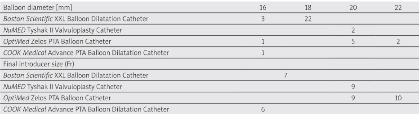
Table IV. Distribution of the applied balloon types, balloon diameters and final introducer sizes
Balloon diameter [mm] 16 18 20 22
Boston Scientific XXL Balloon Dilatation Catheter 3 22
NuMED Tyshak II Valvuloplasty Catheter 2
OptiMed Zelos PTA Balloon Catheter 1 5 2
COOK Medical Advance PTA Balloon Dilatation Catheter 1 Final introducer size (Fr)
Boston Scientific XXL Balloon Dilatation Catheter 7
NuMED Tyshak II Valvuloplasty Catheter 9
OptiMed Zelos PTA Balloon Catheter 9 10
COOK Medical Advance PTA Balloon Dilatation Catheter 6
mulative 1-year and 2-year mortality rates were 33.2%
and 57.4%, respectively, with the highest incidence in the cardiogenic shock group (70.7% and 80.4%) and the low- est in the bridge to AVR group (21.7% and 38.4%). Other
events were stroke (0.5%), major vascular complications (2.2%), and life-threatening bleeding (1.5%) [5].
In the past decade with the rise of the TAVI proce- dures, the need for BAV emerged by reason of bridge to
TAVI indications [12]. The fragility of the patients who undergo BAV before TAVI is high; in addition, most of the procedures are performed in acute settings. Improve- ment of the different tools and techniques used to per- form BAV is remarkable, and the procedure-related com- plication rates were reduced during the past decades;
nevertheless, most of the problems are related to the access site of the procedure.
Vascular complications at the puncture site are the most common issues associated with BAV [13]. Vascular complications in the literature are reported at the rate of 5–10%, even after the initiation of vascular closure devices. In order to avoid femoral artery access site com- plications during BAV and TAVI, the ultrasound or angiog- raphy-guided femoral artery puncture, the use of closure devices [13, 14], and the use of alternative access sites were introduced.
Alternative access sites for BAV are the brachial, ax- illary, and radial arteries [15, 16]. The brachial and axil- lary access sites can accommodate large access sites for BAV and TAVI and the puncture site can be closed with a Perclose device, but brachial access for endovascular procedures is associated with a high rate of thrombot- ic and bleeding complications in large series and these complications need surgical repair in most cases [16, 17].
The transradial approach for BAV has been presented in case reports and pilot studies [18–21]. The most com- mon advantage of the technique is the lower rate of ma- jor vascular access complications and fast mobilization.
The limitations of trBAV technique are mainly anatomi- cal. The size and pathology of the radial artery must be investigated by vascular ultrasound. In many cases, the radial artery cannot accommodate big sheaths, due to calcification, size, or stenosis/occlusion. In these cases, the alternative access site can be the brachial or the fem- oral artery. The size of the balloon is very important to select the size of the sheath. The size of the balloon is de- termined by the perimeter of the annulus. The diameter of the delivery sheath can be decreased with the use of peripheral balloons or by decreasing the size of the bal- loon. Operators can select from semi-compliant (Tyshak II, Braun International Systems) or non-compliant aortic balloons (Z-Med, Braun International Systems; Maxi LD Balloon, Cordis Corporation; XXL balloon, Boston Scientif- ic). Semicompliant balloons tend to have a lower profile and therefore require smaller vascular access sheaths which help with reduced vascular complications in this elderly cohort. The trade-off, however, is that they have less predictable inflation diameters than the non-compli- ant balloons and have lower rated burst pressures. Dedi- cated BAV balloons have a larger entry profile, but periph- eral balloons can be used in smaller sheaths (Table IV).
Smaller balloons can have a smaller hemodynamic effect than larger balloons. In our study, the 0.8 annulus to bal- loon ratio was safe and had a satisfactory clinical effect,
but the hemodynamic effect was not always improved. In some cases, the use of 7F sheathless guiding catheters allows the use of balloons when the delivery of the bal- loons is difficult.
In our patient population, we did not experience any major vascular event and the rate of minor events was 5.5%. We have used distal radial access in 18 patients without any complications (0%). Our data suggest that the vascular complication rate can be decreased by tran- sradial access preference and it can improve the mortali- ty and morbidity of the acute phase.
Study limitations: The primary limitation of the study is the small number of patients and the lack of random- ization with the femoral approach. Another limitation of the study was the ultrasound-based balloon size selec- tion.
Conclusions
The main findings of the study are the following:
(1) trBAV is a safe and technically feasible procedure under the US control of the access artery size; (2) the vascular complication rate of the procedure is low;
(3) 1-year mortality remains high in patients who are not candidates for TAVI and the procedure cannot signifi- cantly improve the mortality rate.
According to our study, bilateral radial artery access is a safe and effective option for balloon aortic valvuloplas- ty in patients with severe aortic valve stenosis and an optimal RA diameter. Long-term mortality is still high due to comorbidities and associated diseases.
Acknowledgments
Levente Molnar and Roland Papp contributed equally to this work.
Project no. NVKP_16-1–2016-0017 (’National Heart Program’) has been implemented with the support pro- vided from the National Research, Development and In- novation Fund of Hungary, financed under the NVKP_16 funding scheme. The research was financed by the The- matic Excellence Programme (2020-4.1.1.-TKP2020) of the Ministry for Innovation and Technology in Hungary, within the framework of the Therapeutic Development and Bioimaging thematic programmes of Semmelweis University.
Conflict of interest
The authors declare no conflict of interest.
References
1. Cribier A, Savin T, Saoudi N, et al. Percutaneous transluminal valvuloplasty of acquired aortic stenosis in elderly patients: an alternative to valve replacement? Lancet 1986; 1: 63-7.
2. Letac B, Cribier A, Koning R, Bellefleur JP. Results of percutane- ous transluminal valvuloplasty in 218 adults with valvular aortic stenosis. Am J Cardiol 1988; 62: 598-605.
3. Safian RD, Berman AD, Diver DJ, et al. Balloon aortic valvuloplas- ty in 170 consecutive patients. N Engl J Med 1988; 319: 125-30.
4. Bashore TM, Berman AD, Davidson CJ, et al. Percutaneous bal- loon aortic valvuloplasty. Acute and 30-day follow-up results in 674 patients from the NHLBI Balloon Valvuloplasty Registry.
Circulation 1991; 84: 2383-97.
5. Saia F , Marrozzini C, Ciuca C, et al. Emerging indications, in-hos- pital and long-term outcome of balloon aortic valvuloplasty in the transcatheter aortic valve implantation era. EuroInterven- tion 2013; 8: 1388-97.
6. McKay RG. The Mansfield Scientific Aortic Valvuloplasty Reg- istry: overview of acute hemodynamic results and procedural complications. J Am Coll Cardiol 1991; 17: 485-91.
7. Bertrand OF. Acute forearm muscle swelling post transradial catheterization and compartment syndrome: prevention is bet- ter than treatment! Catheter Cardiovasc Interv 2010; 75: 366-8.
8. Ben-Dor I, Pichard AD, Satler LF, et al. Complications and out- come of balloon aortic valvuloplasty in high-risk or inoperable patients. JACC Cardiovasc Interv 2010; 3: 1150-6.
9. Lieberman EB, Bashore TM, Hermiller JB, et al. Balloon aortic val- vuloplasty in adults: failure of procedure to improve long-term survival. J Am Coll Cardiol 1995; 26: 1522-8.
10. Otto CM, Mickel MC, Kennedy JW, et al. Three-year outcome af- ter balloon aortic valvuloplasty. Insights into prognosis of valvu- lar aortic stenosis. Circulation 1994; 89: 642-50.
11. Ben-Dor I, Maluenda G, Dvir D, et al. Balloon aortic valvuloplasty for severe aortic stenosis as a bridge to transcatheter/surgical aortic valve replacement. Catheter Cardiovasc Interv 2013; 82:
632-7.
12. Frerker C, Schewel D, Kuck KH, et al. Ipsilateral arterial access for management of vascular complication in transcatheter aortic valve implantation. Catheter Cardiovasc Interv 2013; 81: 592- 602.
13. Sharp AS, Michev I, Maisano F, et al. A new technique for vascu- lar access management in transcatheter aortic valve implanta- tion. Catheter Cardiovasc Interv 2010; 75: 784-93.
14. Armario X, Rosseel L, McGrath B, Mylotte D. Balloon aortic valvu- loplasty with two simultaneous balloons via ipsilateral transra- dial and transbrachial access. EuroIntervention 2021; 17: 88-9.
15. Alvarez-Tostado JA, Moise MA, Bena JF, et al. The brachial artery:
a critical access for endovascular procedures. J Vasc Surg 2009;
49: 378-85.
16. Madden NJ, Calligaro KD, Zheng H, et al. Outcomes of brachi- al artery access for endovascular interventions. Ann Vasc Surg 2019; 56: 81-6.
17. Molnár L, Papp R, Nagy A, et al. TCT-596 transradial access for bal- loon aortic valvuloplasty. J Am Coll Cardiol 2017; 70 (18 Suppl):
B246.
18. Tumscitz C, Campo G, Tebaldi M, et al. Safety and feasibility of transradial mini-invasive balloon aortic valvuloplasty: a pilot study. JACC Cardiovasc Interv 2017; 10: 1375-7.
19. Vassileva A, Valsecchi O, Guagliumi G, et al. Bilateral slender transradial aortic balloon valvuloplasty. J Invasive Cardiol 2018;
30: E48-9.
20. Gómez-Peña F, Sanmartín-Pena X, López-Canoa N, et al. Bilateral transradial aortic valvuloplasty with double balloon angioplasty:
our first experience. Rev Esp Cardiol 2020; 73: 175-6.
21. Di Cesare A, Tonet E, Campo G, Tumscitz C. Snuffbox approach for balloon aortic valvuloplasty: a case series. Catheter Cardio- vasc Interv 2021; 97: E743-7.