C A S E R E P O R T
An alternative treatment strategy for large vessel coronary perforations
Iosif Xenogiannis MD
1| Peter Tajti MD
1,2| M. Nicholas Burke MD
1| Emmanouil S. Brilakis MD, PhD
11Minneapolis Heart Institute and Minneapolis Heart Institute Foundation, Abbott Northwestern Hospital, Minneapolis, Minnesota
2Division of Invasive Cardiology, Second Department of Internal Medicine and Cardiology Center, University of Szeged, Szeged, Hungary
Correspondence
Emmanouil S. Brilakis, MD, PhD, Minneapolis Heart Institute, 920 E 28th Street #300, Minneapolis, MN 55407.
Email: esbrilakis@gmail.com
Abstract
The standard treatment for large vessel coronary perforations is implantation of a covered stent.
Antegrade attempts for crossing a right coronary artery chronic total occlusion resulted in guide- wire and microcatheter exit with pericardial bleeding. A balloon was inflated proximal to the per- foration site to achieve temporary hemostasis. Retrograde crossing of the chronic total occlusion was achieved through an epicardial collateral using the reverse controlled antegrade and retrograde tracking technique. Stent implantation resulted in hemostasis, likely due to crea- tion of a subintimal flap that sealed the perforation site. If technically feasible, subintimal recan- alization can be an alternative treatment strategy for coronary perforations occurring during chronic total occlusion percutaneous coronary intervention.
K E Y W O R D S
chronic total occlusion, percutaneous coronary intervention, perforation
1 | I N T R O D U C T I O N
Coronary artery perforation is one of the most feared complications in chronic total occlusion (CTO) percutaneous coronary intervention (PCI).1Perforation occurs in 1–9% of CTO PCIs leading to tamponade and need for pericardiocentesis in approximately 1%.2–4
The initial management of coronary perforation is inflation of a balloon proximally to stop pericardial bleeding. Subsequent treatment depends on the type of perforation. Large vessel perforations are usu- ally treated with covered stents, whereas distal vessel or collateral vessel perforation is usually treated with fat or coil5,6or occasionally thrombus embolization.7 Covered stent delivery can, however, be challenging due to high crossing profile and low flexibility, especially through tortuous and calcified vessels, which are more prone to devel- opment of perforations. We describe a novel strategy for treating a large vessel perforation of the right coronary artery (RCA) CTO PCI using subintimal CTO crossing and stenting.
2 | C A S E R E P O R T
A 69-year-old woman presented with lifestyle-limiting angina. She had hypertension, dyslipidemia, carotid artery disease and was a
current smoker. A prior computed tomography angiography had dem- onstrated severe coronary artery calcification. Diagnostic coronary angiography demonstrated 70% stenosis in the left anterior coronary artery (LAD) and a CTO in the mid RCA (Figure 1, Panel A). She con- tinued to be symptomatic despite recanalization of the LAD lesion and was referred for PCI of the RCA CTO. The RCA CTO had a blunt proximal cap and diffusely diseased distal vessel that was filling via an epicardial collateral from the circumflex artery (CX). It was severely calcified and moderately tortuous.
We obtained access through the right radial and right femoral artery. The left main was engaged with a 6-Fr 3.5 XB (Cordis, Fremont, CA) guide catheter and the RCA with an 8-Fr AL 1 (Medtronic, Minneapolis, MN) guide catheter. Antegrade wire escalation was attempted by advancing a Turnpike LP (Teleflex, Wayne, PA) micro- catheter to the proximal cap and using a Gaia 3rd (Asahi Intecc, Nagoya, Japan), followed by a Confianza Pro 12 (Asahi Intecc) guide- wire. The Confianza Pro 12 wire appeared to enter the subintimal space and the Turnpike LP was advanced slightly into the occlusion followed by insertion of a Fielder XT (Asahi Intecc) guidewire for forming a knuckle. However, an orthogonal projection revealed that the Turnpike LP was outside the vessel (Figure 1, Panel B). The micro- catheter was removed and an antegrade injection demonstrated an DOI: 10.1002/ccd.28034
Catheter Cardiovasc Interv.2019;93:635–638. wileyonlinelibrary.com/journal/ccd © 2018 Wiley Periodicals, Inc. 635
Ellis class 3 perforation (Figure 1, Panel C), (Video 1). A 2.5 mm bal- loon was advanced through the 8-Fr AL1 guide catheter and was inflated in the proximal RCA stopping the pericardial bleeding. The patient remained hemodynamically stable (Figure 1, Panel D).
The contralateral epicardial collateral was successfully crossed with a Sion blue (Asahi Intecc) and a Suoh 03 (Asahi Intecc) guidewire that was followed by the Turnpike LP microcatheter (Asahi Intecc) (Figure 1, Panel D). Attempts for retrograde CTO crossing using a Pilot 200 guidewire (Abbott Vascular, Santa Clara, CA) failed and resulted in loss of the Turnpike LP position due to poor guide catheter support.
We obtained left femoral arterial access and engaged the left main with an 8-Fr EBU 3.5 (Medtronic) guide catheter. We re-crossed the epicardial collateral with a Suoh 03 guidewire and crossed the CTO subintimally using a knuckled Fielder XT guidewire. Likely due to subintimal crossing the perforation was successfully sealed (Video 2).
After substituting the antegrade balloon (that remained inflated throughout this time) for a 3.5 mm balloon we successfully re-entered into the proximal true lumen using the reverse controlled antegrade and retrograde tracking (reverse CART) (Figure 1, Panel E). An R350 guidewire (Vascular Solutions, Maple Grove, MN) was externalized followed by predilation of the proximal, mid and distal RCA. Due to the extreme RCA calcification a 6 Fr guide catheter extension was used to facilitate stent delivery. After stenting the perforation remained sealed. Intravascular ultrasound demonstrated stent under- expansion at the level of the perforation, hence multiple additional
high-pressure balloon inflations were performed with a 3.0 mm non- compliant balloon (Figure 2). A small aorto-coronary dissection was visible and was treated by placing a 3.5×8 mm ostial stent that was FIGURE 1 Panel A: chronic total occlusion in mid-right coronary artery (RCA). Filling by an epicardial collateral from circumflex artery (CX). Panel B: using two orthogonal projections, the knuckled fielder XT (Asahi Intecc, Nagoya, Japan) guidewire and the turnpike LP (Teleflex, Wayne, PA) microcatheter proved to be outside the vessel architecture. Panel C: contrast extravasation after ipsilateral injection compatible with Ellis class 3 perforation (arrow). Panel D: a balloon was inflated proximal to the point of perforation (black arrow). At the same time, the epicardial collateral was crossed with a Sion guidewire (yellow arrow) and a turnpike LP microcatheter (arrowhead). Panel E: reverse controlled antegrade and retrograde tracking allowed retrograde CTO crossing and created a flap that sealed the perforation. Panel F: after stent deployment a small aorto- ostial dissection was noticed. Panel G: an additional stent was implanted at the ostium of RCA, followed by ostial flash balloon inflation (arrow).
Panel H: final result [Color figure can be viewed at wileyonlinelibrary.com]
FIGURE 2 Intravascular ultrasound at the level of perforation after stent deployment, demonstrating mild stent under-expansion [Color figure can be viewed at wileyonlinelibrary.com]
636 XENOGIANNISET AL.
postdilated with an Ostial Flash balloon (Figure 1, Panels F,G). An excellent final angiographic result was achieved with sealing of the perforation (Figure 1, Panel H), (Video 3). Echocardiography did not show a pericardial effusion.
The patient remained hemodynamically stable and asymptomatic throughout the procedure and was discharged home the following day.
3 | D I S C U S S I O N
To the best of our knowledge, this is the first description of coronary perforation treatment using subintimal CTO PCI techniques.
Dissection and re-entry techniques have been successfully used in acute vessel closure to regain access into the distal true lumen and re-open the occluded vessel.8–10 In this case, we demonstrate how dissection and re-entry techniques can also be used to treat a large vessel perforation. The subintimal tissue flaps created after retrograde crossing with the reverse CART technique sealed the site of perfora- tion achieving hemostasis.
Guidewire exit during CTO (and non-CTO) PCI very rarely leads to pericardial effusion, likely due to the small size of the opening created in the affected vessel. When, however, a microcatheter or balloon is advanced over the perforated guidewire, significant perforation can occur, as happened in our case. Confirmation of initial guidewire posi- tion in two projections before advancing the microcatheter is critical for preventing perforation.
Once a microcatheter is advanced over the perforated vessel into the pericardium, it may be best to not remove it, until after definitive plans have been made about how to treat the perfora- tion. The microcatheter prevents bleeding into the pericardium and the development of tamponade. In our case, the microcath- eter was removed, which allowed confirmation of the perforation, but also required inflation of a balloon proximally to stop bleeding.
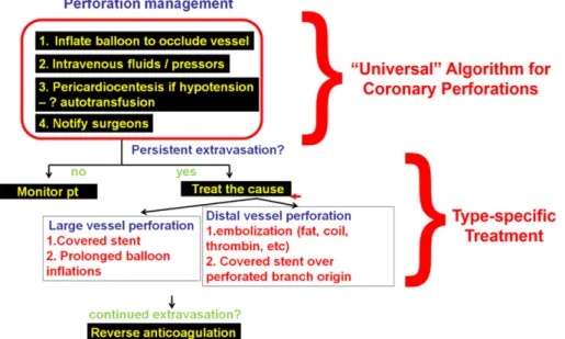
The very first step in the management of a coronary perforation is to advance and inflate a balloon proximal to the perforation. Intra- venous fluids are administered, pericardiocentesis is performed if tamponade develops, and the cardiac surgeons are notified, in case surgery is required. Subsequent treatment depends on the perforation type, with large vessel perforations usually treated by implantation of a covered stent and distal vessel or epicardial vessel perforations usu- ally treated with fat or coil embolization. However, covered stent delivery can be very challenging, especially through tortuous and calcified vessels (which are the ones more likely to perforate) and covered stents have high rates of restenosis and stent thrombosis.11 Anticoagulation is not reversed until after removal of all intracoron- ary equipment (Figure 3).
In our patient, the perforation could have been treated by coil embolization, given that it was located in the proximal CTO cap, but this would likely have hindered subsequent recanalization attempts.
Inflation of a balloon proximal to the perforation prevented formation of pericardial effusion and tamponade. Subintimal retrograde recanali- zation was feasible through a large epicardial collateral, highlighting the importance of careful pre-procedural preparation and planning of alternative crossing strategies. Creation of subintimal dissection flaps likely sealed the perforation, obviating the need for coiling or other treatment. Had the perforation not been sealed by implantation of a standard drug-eluting stent, subsequent placement of a covered stent could be performed, with the added likely benefit of lower restenosis due to drug-eluting stent implantation.
Although radial access reduces the risk for access complications and provides enough support for most cases, sometimes cross-over to femoral access may be needed, as in our patient. Obtaining a second femoral access with an 8 French sheath and guide was instrumental in providing enough support for retrograde CTO crossing and successful completion of the procedure.
Use of dissection/re-entry techniques for treating a perforation has limitations. First, it requires significant expertise in subintimal
FIGURE 3 Algorithm for the management of coronary perforations. Reproduced with permission from Brilakis ES. Manual of chronic total occlusion interventions a step-by-step approach. Second edition. Ed. London: Elsevier/academic press; 2018 [Color figure can be viewed at wileyonlinelibrary.com]
techniques, such as retrograde crossing and antegrade dissection and re-entry. Second, it may require a long period of time, hence it is not suitable for unstable patients. Third if may not always be successful, or might even worsen the perforation, by inflicting additional injury to the coronary artery wall. It should, therefore, only be used by experi- enced operators when the anticipated benefits appear to exceed the potential risks.
4 | C O N C L U S I O N S
In summary, dissection and re-entry strategies, such as the reverse CART can be used to seal large vessel coronary perforations during CTO PCI, obviating the need for covered stent or embolization.
C O N F L I C T O F I N T E R E S T Dr. Xenogiannis: nothing to disclose.
Dr. Tajti: nothing to disclose.
Dr. Burke: consulting and speaker honoraria from Abbott Vascular and Boston Scientific.
Dr. Brilakis: consulting/speaker honoraria from Abbott Vascular, ACIST, American Heart Association (associate editor Circulation), Amgen, Asahi, Cardiovascular Innovations Foundation (Board of Directors), CSI, Elsevier, GE Healthcare, and Medtronic; research sup- port from Boston Scientific and Osprey. Shareholder: MHI Ventures.
Board of Trustees: Society of Cardiovascular Angiography and Interventions.
O R C I D
Iosif Xenogiannis https://orcid.org/0000-0002-5004-6635 M. Nicholas Burke https://orcid.org/0000-0002-0547-9976 Emmanouil S. Brilakis https://orcid.org/0000-0001-9416-9701
R E F E R E N C E S
1. Tajti P, Burke MN, Karmpaliotis D, et al. Update in the percutaneous management of coronary chronic total occlusions. JACC Cardiovasc Interv. 2018;11:615-625.
2.Danek BA, Karatasakis A, Tajti P, et al. Incidence, treatment, and out- comes of coronary perforation during chronic total occlusion percuta- neous coronary intervention. Am J Cardiol. 2017;120:1285-1292.
3.Sapontis J, Salisbury AC, Yeh RW, et al. Early procedural and health status outcomes after chronic total occlusion angioplasty: A report from the OPEN-CTO registry (outcomes, patient health status, and efficiency in chronic total occlusion hybrid procedures). JACC Cardio- vasc Interv. 2017;10:1523-1534.
4.Kinnaird T, Anderson R, Ossei-Gerning N, et al. Legacy effect of coro- nary perforation complicating percutaneous coronary intervention for chronic total occlusive disease: An analysis of 26 807 cases from the British cardiovascular intervention society database. Circ Cardiovasc Interv. 2017;10:e004642.
5.Kotsia AP, Brilakis ES, Karmpaliotis D. Thrombin injection for sealing epicardial collateral perforation during chronic total occlusion percuta- neous coronary interventions. J Invasive Cardiol. 2014;26:E124-E126.
6.Shemisa K, Karatasakis A, Brilakis ES. Management of guidewire-induced distal coronary perforation using autologous fat particles versus coil embolization. Catheter Cardiovasc Interv. 2017;89:253-258.
7.Tokuda T, Hirano K, Yamawaki M, et al. Efficacy and safety of a coagu- lated thrombus injection for peripheral artery perforation: The coagu- lated thrombus hemostasis method. Catheter Cardiovasc Interv. 2018;
91:302-307.
8.Martinez-Rumayor AA, Banerjee S, Brilakis ES. Knuckle wire and sting- ray balloon for recrossing a coronary dissection after loss of guidewire position. JACC Cardiovasc Interv. 2012;5:e31-e32.
9.Shaukat A, Mooney M, Burke MN, Brilakis ES. Use of chronic total occlu- sion percutaneous coronary intervention techniques for treating acute vessel closure. Catheter Cardiovasc Interv. 2018. Epud ahead of print.
10. Brilakis ES. Manual of Chronic Total Occlusion Interventions: A Step- By-Step Approach. 2nd ed. London: Elsevier/Academic Press; 2018.
11. Yang Q, Liu W, Fang F, Yu CM, Zhou YJ. In-stent restenosis in a poly- tetrafluoroethylene covered stent combined with drug eluting stents:
Potential pathogenesis revealed by optical coherence tomography. Int J Cardiol. 2015;198:42-44.
S U P P O R T I N G I N F O R M A T I O N
Additional supporting information may be found online in the Sup- porting Information section at the end of the article.
How to cite this article: Xenogiannis I, Tajti P, Nicholas Burke M, Brilakis ES. An alternative treatment strategy for large vessel coronary perforations.Catheter Cardiovasc Interv.
2019;93:635–638.https://doi.org/10.1002/ccd.28034
638 XENOGIANNISET AL.
![FIGURE 2 Intravascular ultrasound at the level of perforation after stent deployment, demonstrating mild stent under-expansion [Color figure can be viewed at wileyonlinelibrary.com]](https://thumb-eu.123doks.com/thumbv2/9dokorg/1317556.106149/3.892.74.823.71.451/figure-intravascular-ultrasound-perforation-deployment-demonstrating-expansion-wileyonlinelibrary.webp)
