Percutaneous Endovascular Treatment of Innominate Artery Lesions: A Single-centre Experience on 77 Lesions
T.M. Paukovits
a,*, L. Luka ´cs
a, V. Be ´rczi
b, K. Hirschberg
a, B. Nemes
a, K. Hu ¨ttl
aaHeart Center, Faculty of Medicine, Semmelweis University, 1122 Gaal Jozsef Street 9, Budapest, Hungary
bDepartment of Radiology and Oncotherapy, Faculty of Medicine, Semmelweis University, Budapest, Hungary
Submitted 1 November 2009; accepted 12 March 2010 Available online 1 May 2010
KEYWORDS Angioplasty;
Brachiocephalic trunk;
Innominate artery;
Neurological complications;
Patency rate;
Percutaneous intervention
Abstract Purpose:To assess primary success and safety of percutaneous transluminal angio- plasty and/or stenting of innominate artery lesions and to compare its 30-day stroke/mortality level with the literature data.
Methods:A total of 72 patients (77 stenoses, five recurrent, 58 symptomatic and 39 female) with seven innominate vessel occlusions, nine subocclusive lesions and 61 significant (>60%) stenoses of innominate artery treated between 2000 and 2009 were retrospectively reviewed.
With the exception of seven, all procedures were performed using a transfemoral approach. A stent was implanted in 49 (63.6%) cases. Follow-up included neurological examination, carotid duplex scan and office/telephone interview.
Results:Primary technical success was 93.5% (72/77). There was neither periprocedural (<48 h) death, nor major neurological complication. Minor periprocedural neurological complications con- sisted of 2/72 (2.6%) ipsilateral TIAs. Access site complications included 4 (5.2%) access site bleed- ings. Follow-up was achieved in 65/72 (90.3%) of all patients and 68 (88.3%) of all procedures for a mean of 42.3 months and revealed neither major neurological complication, nor additional TIA.
The cumulative primary patency rate was 100% at 12 months, 981.6% at 24 months, and 69.9 8.5% at 96 months. The cumulative secondary patency rate was 100% at 12 and at 24 months, and 81.57.7% at 96 months. Log-rank test showed no significant difference (pZ0.79) in primary cumulative patencies between PTA alone (nZ28) or PTA/stent (nZ49).
Conclusion:Transfemoral PTA with or without stent appears to be a safe treatment option for innominate artery lesions.
ª2010 European Society for Vascular Surgery. Published by Elsevier Ltd. All rights reserved.
* Corresponding author at: Heart Center, Faculty of Medicine, Semmelweis University, Varosmajor utca 68, 1023 Budapest, Hungary.
Tel.:þ47 94250042.
E-mail address:tomirko@t-online.hu(T.M. Paukovits).
1078-5884/$36ª2010 European Society for Vascular Surgery. Published by Elsevier Ltd. All rights reserved.
doi:10.1016/j.ejvs.2010.03.017
Before the era of endovascular approach, open surgery had been the sole operative treatment for the stenotic and occlusive supra-aortic trunk lesions.1 Although surgical revascularisation results in long lasting patency, these procedures are associated with high morbidity and mortality rates.2e6 During the last two decades, balloon angioplasty with or without stent deployment has become an increasingly accepted treatment for stenotic and occlusive supra-aortic arterial disease. Extrathoracic bypasses and transpositions continue to be favoured by surgeons, but they tend to be used for cases when endo- vascular therapy failed. Data on endovascular treatment of solely innominate artery lesions are scarce; most of these studies focussed on the treatment of the combination of several supra-aortic arteries simultaneously, and only limited number of innominate artery lesions are evaluated, mostly with short-time follow-up. Therefore, any conclu- sions from these studies are limited. Natural history data and treatment guidelines for carotid bifurcation stenting7e9 do not exist for innominate lesions. The aim of the current study was to retrospectively evaluate the primary technical and clinical success rate, safety and efficacy of the angio- plasty (PTA) with or without stent placement in the treat- ment of innominate artery lesions.
Patients and Methods
We present our experience on 72 consecutive patients who underwent 77 angioplasty of the innominate artery. Our retrospective cohort study has been carried out at a tertial referral centre, between the 1 January 2000 and 31 February 2009. There were 39 women (54.2%) and 33 men (45.8%).
Patients had a mean age of 56.6 (range 28e82 years) (Table 1).
Intervention was performed when the patient had clinical symptoms and/or clinically significant stenosis or occlusion of the brachiocephalic vessel. Preprocedural symptoms con- sisted of 16 (20.8%) hemisensory or motor deficit, eight (10.4%) amaurosis fugax. 40 (52%) patients suffered of right-upper limb claudication and 28 (36.4%) patients complained of dizziness. There were 19 asymptomatic patients (24.7%) whose angiographies were carried out under the suspicion of stenoses on other supra-aortic arteries, revealing the lesions in the innominate artery.
Concerning the patients’ co-morbidity, 25 patients (34.7%) had previous myocardial infarction, 47 patients (65.3%) had hypertension, 13 (18.6%) person suffered from type II diabetes mellitus, 30 (41.7%) had hyperlipidaemia (cholesterol >5.2 mmol l1), 36 patients (50%) had symp- tomatic arterial occlusion in their lower extremities and 33 (45.8%) smoked (Table 1).
The initial diagnosis of innominate artery stenosis or occlusion was based on the clinical data, pulse palpation of the radial arteries and blood pressure measurement on both arms. Preprocedural examination consisted of colour duplex scan (CDS) of the extracranial arteries in all patients. Post-stenotic flow pattern in the common carotis artery (CCA) was considered indicative of a likely proximal stenosis of the subclavian artery. Retrograde flow in the right vertebral artery on duplex scan (14 patients; 18.2%) defined subclavian steal syndrome. Stenoses and occlusions were verified by preprocedural diagnostic angiography in all cases, done in the same session with the treatment.
The lesions were measured according to NASCET study.
There were seven innominate vessel occlusions (9.1%), nine subocclusive lesions (11.7%) and 61 greater than 60%
stenoses (79.2%). Angiography revealed 13 (16.9%) simul- taneous supra-aortic stenoses, which have been treated in one stage with the innominate artery lesions. These con- sisted of seven (9.1%) subclavian stenoses (five left, one right, one bilateral), one (1.3%) left subclavian occlusion, three (3.9%) ICA (two left, one right) stenoses and two (2.6%) left CCA stenoses.
The patients have signed informed consent form. Before the intervention, the patients were on 125 mg day1aspirin per os, in case of contraindication for aspirin, the patients received 2250 mg ticlopidin. During the procedure, 5000 U heparin i.v. was given. All procedures were performed in the angiography suite with the patient under local anaes- thesia. Diagnostic angioplasty was performed through a femoral approach with a 4F sheath, which was replaced with 7e8-F, 90-cm long introduction sheath in case of intervention (Advanti plus; Cordis Johnson & Johnson, Roden, the Netherlands) in all, but seven (9.1%) cases. In seven cases brachial approach for intervention were gained through 4e5-F introducer. Angle-tip guide wires (Terumo guide wire; Terumo Medical, Tokyo, Japan) with a diameter of 0.032 or 0.035 inch were used to pass the stenosis in the brachiocephalic artery under fluoroscopic control.
In case of tortuosity the hydrophilic wire was replaced by a stiff Amplatz wire (Boston Scientific Natick, MA USA). The selection of the balloon, and/or stent size was based on
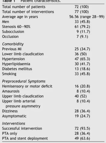
Table 1 Patient characteristics.
Total number of patients 72 (100) Total number of interventions 77 (100)
Average age in years 56.56 (range 28e99)
Men 33 (45.8)
Stenosis 60e90% 61 (79.2)
Subocclusion 9 (11.7)
Occlusion 7 (9.1)
Comorbidity
Previous MI 25 (34.7)
Lower limb claudication 36 (50)
Hypertension 47 (65.3)
Hyperlipidaemia 30 (41.7)
Diabetes mellitus 13 (18.6)
Smoking 33 (45.8)
Preprocedural Symptoms
Hemisensory or motor deficit 16 (20.8)
Amaurosis 8 (10.4)
Upper limb claudication 40 (52)
Upper limb arterial pressure asymmetry
8 (10.4)
Dizziness 28 (36.4)
Asymptomatic 19 (24.7)
Interventions
Successful intervention 72 (93.5)
PTA only 28 (36.4)
PTA and stent deployment 49 (63.6) Values in parenthes indicate percentage.
angiographic measurements of the innominate artery beyond the lesions as a reference (9e12 mm) with a length appropriate to the lesion (usually 2 or 4 cm). The balloon was inflated for 10e15 s until satisfactory dilation with abolition of the ‘waisting’ of the balloon was achieved (6e10 atm).
Stent deployment has been done in 49 cases (63.6%).
Different type of stents were used, depending on the personal preference of the interventional radiologist.
Balloon expandable stents: nZ22 Genesis 10 19 mm, nZ15 Genesis 1029 mm,nZ5 Genesis 919 mm,nZ2 Genesis 815 mm,nZ1 Genesis 818 mm,nZ1 Genesis 929 mm (Cordis Corporation, Miami Lakes, FL, USA). Self expandable stents: n Z1 S.M.A.R.T 10 25 mm (Cordis Corporation, Miami Lakes, FL, USA),nZ1 BARD 1220 mm (Murray Hill, NJ, USA),nZ1 Carotid Wallstent 1024 mm (Boston Scientific, Natic, MA, USA) stents were deployed. For angioplasty, Wanda and Pheron balloons were used: Wanda 940 mm, in 46 (59.7%) cases, Wanda 1040 mm, in 22 (28.6%) cases, Wanda 8 40 mm, in seven (9.1%) cases, Pheron 840 mm (BIOTRONIK GmbH, Berlin, Germany) in one (1.3%) and Pheron 740 mm in one (1.3%) case. In case of stent deployment, Vista Brite Tip (7F) guiding sheath was utilized. In five patients (5.6%) the re-canalisation of the stenotic lesion could not be done via femoral access. In these cases, 4- or 5-F sheaths (Advanti Plus) were used to gain additional access through the right brachial artery. Following successful re-canalisation, stent employment was per- formed through the same access site using 6- or 7-F sheaths.
There were no puncture site occlusion in any of these cases.
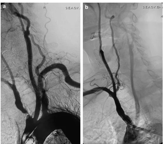
A control post-angioplasty angiogram was performed in each case (Figs. 1 and 2). No neuroprotection device was utilized during procedures. Angioplasty was regarded as initially technically successful, when a residual diameter stenosis remained of less than 30%. In cases of upper arm claudication or dizziness, clinical success was defined as relief or substantial reduction of the preprocedural symptoms.
Aspirin (125 mg daily) was continued (provided there was no contraindication) for at least 6 months after PTA.
Sixteen (20.8%) patients were treated with dual anti- platelet therapy, depending the individual decision of the radiologist. Patients were usually discharged 1 day after the procedure.
Postoperative clinical evaluation and follow-up exami- nation was carried out 6 weeks, 3 months and 6 months after PTA, and annually thereafter. Besides regular controls, patients were advised to come immediately to our clinic if they had any acute symptoms. Clinical examination included pulse palpation; blood pressure measurements on both arms; subclavian, common carotid and vertebral artery CDS on both sides and completion of a check list containing routine questions regarding residual or new symptoms and co-morbidities.
During the follow-up examinations, the innominate artery was considered to be patent without significant stenosis if the right radial pulse was palpable, the blood pressure difference between the two arms was 30 mmHg or less and the flow in the vertebral artery was antegrade.
Restenosis was defined as higher than 30 mm Hg difference in blood pressure between the two arms, recurrent symp- toms, retrograde flow in the right vertebral artery and/or more than 50% lumen reduction of the treated vessel at follow-up.
For statistical analysis, the KaplaneMeier method was employed to calculate cumulative primary and secondary patency rates (SPSS, version 13.0, Chicago, IL, USA).
This retrospective study has been done according to the institutional ethical guidelines.
Results
All the lesions but 5 (6.5%) were crossed with balloon catheter and treated successfully with PTA. All five attempts failed in cases of occluded innominate artery (nZ7) that could not be crossed using either a femoral or brachial approach. Twenty-eight (36.4%) successful balloon angioplasties, and 49 (63.6%) PTAs with stent deployment were carried out. The initial technical success rate was 93.5% for procedures. Examples of successful innominate artery PTAs without and with stent deployment are shown inFigs. 1 and 2, respectively In six (7.8%) cases, after stent deployment, control DSA showed unsatisfactory morphology and/or higher than 50% residual stenosis. This was eliminated with repeated PTA. Other supra-aortic PTAs in the same session with the innominate artery intervention were carried out in 13 (16.9%) cases.
Clinical primary success was achieved in all but one of the 58 symptomatic cases, resulting a 98.3% clinical success rate: 32 patients have become symptomless and 25 patients showed moderate reductions of the preprocedural symp- toms. None of the nineteen asymptomatic patients became symptomatic during or after the procedure within 48 h.
There was neither lethal nor major neurologic compli- cation nor myocardial infarction during the procedures or within 30 days of the intervention. Dissection of aortic arch, or supra-aortic arteries was not observed. There were two (2.6%) minor neurologic complications: one left-sided transient hemiparesis occurred during the procedure and one patient lost consciousness for a short period of time 2 h following the procedure. Access site complications included four (5.2%) access site bleedings, resulting in a groin hematoma, one (1.3%) of them required surgical evaluation an suture. In one (1.3%) case, palmar and finger pain was experienced, suggesting transient microemboli.
All but the seven patients with complications were dis- charged the following day.
The mean follow-up time was 42.3 months (range 2e103 months). Clinical follow-up including both measured data (blood pressure on both upper extremities, CDS of CCA with peak sytolic velocity (PSV), end diastolic velocity (EDV) and systolic rise time (SRT) measurements) and patient history data (progression of the symptoms, new symptoms and data on actual medication) is available for 65/72 (90.3%) of all patients and 68/77 (88.3%) of all procedures. All but five (6.5%) patients, who were available for follow-up, had bilateral satisfactory radial pulse, and anterograde flow was visualised in the right vertebral artery. The difference between the arms decreased to less than 10 mmHg in all but 10 (13.9%) patients. In these patients the blood pressure difference was>30 mmHg, and nine (11.7%) of them showed relevant symptoms, fulfilling the inclusion criteria for control angiography. Significant restenosis were identified in four (5.2%) cases and re-occlusion in one case (1.3%). Successful re-PTA was done in all five cases, in one patient with stent deployment. Further follow-up showed open innominate
vessels. Cumulative primary and secondary patency rates were analyzed with the KaplaneMeier method; initial tech- nical failure was includedFig. 3. Cumulative primary patency was 100% at 12 months, 981.6% at 24 months, and 69.98.5% at 96 months. Number of patients at risk was 56 at 12 months, 47 patients at 24 and one patient at 96 months.
The cumulative secondary patency was 100% at 12 and 24 months, and 81.57.7% at 96 months. Number of patients at risk calculated for secondary patency was 56 at 12 months, 47 patients at 24 and one patient at 96 months. Log-rank test showed no significant difference (pZ0.79) in primary cumulative patencies between PTA alone and PTA/stent. Six patients died during follow-up in non-procedure-related diseases; three deaths caused by gastrointestinal malignant tumour 14, 17 and 21 months postprocedural, one patient died of myocardial infarct 3 months postprocedural and one patient in septic shock 27 months after intervention.
Discussion
Innominate artery lesions are uncommon. They represent 0.5e2% of all vascular lesions (Table 2). Before the era of endovascular therapy, surgical treatment was the only option in the management of occlusive disease of innomi- nate artery. However, it represented only 1.7% of all supra- aortic operations.10Lesions of the innominate artery have been treated via transthoracic or extrathoracic surgical
route. Mortality of the transthoracic group varies between 3% and 16%; extrathoracic surgical procedures have an associated lower mortality (0e10%), but have a high (15e25%) complication rate,2e6therefore, in recent prac- tice, surgery is indicated only in the case of unsuccessful PTA.2,11e13PTA/stenting of innominate artery has become the treatment of choice over direct surgical intervention owing to its lower complication and mortality rates and shorter hospital stay13 (Table 1). There are only two other studies on solely innominate artery intervention (Table 2).
In these studies the paraprocedural neurological compli- cations rate represented 4% TIA, reported by vanHattumet al., and 2% left occipital infarction beside 6% TIA in our previous study.14 Primary success rates were 83.3% and 96.4%, respectively.14,15
In contrast to the male predominance in other athero- matous lesions in other supra-aortic stenoses, innominate vessel stenoses and occlusions occur with equal frequency in both sexes, and usually develop after the age of 60.16e18 Our patient group reflected similar gender and age proportion (Table 1).
Atherosclerosis remains among the leading causes of the supra-aortic lesions, less common causes may consist of aortoarteritis (Table 2). The aetiology of the lesions was atherosclerotic in all cases except one. The youngest patient had steroid-treated autoimmune aortoarteritis (Takayasu’s disease) in her previous medical history. This Figure 1 a and b. Successful PTA (Wanda 940 mmWanda,Boston Scientific, Natick, MA) of the innominate artery of a 75-year- old male patient with hemisymptoms and claudication of the right-upper limb. The patients claudication improved throughout the follow-up period. The last follow-up duplex scan at 14 months suggested patent innominate artery.
patient was treated in a non-acute stage of her disease.
Hypertension, diabetes mellitus and smoking are the most important risk factors for these patients.14,16e18Our series reflect similar proportion: 65.3% of all patients suffered from hypertension, 18.6% had diabetes mellitus and 45.8%
of our patients were smokers.
The indication of angioplasty of innominate vessel is not well established. There are no data at all on the natural history. Previous papers evaluate a small number of innominate artery stenoses, with various inclusion criteria for PTA (Table 2). Indications for endovascular treatment are as follows:10,13,19e22 neurologic symptoms (frequency 5e90%), upper limb claudication or digital embolisation, asymptomatic patients with multivessel disease with or without subclavian steal syndrome. Due to the above- mentioned advantages of PTA compared to surgery, at our clinic all innominate artery stenoses, occlusions are currently treated with PTA/stenting. Surgery is restricted to cases of failed endovascular treatment.
In the largest series of innominate artery endovascular treatment, femoral artery access was gained; the axillary artery and brachial artery were used less frequently for access. The latter was associated with higher access site complication rate.13,14,23In our series, in seven cases of brachial approach 4 and 5-F-sized sheaths were used No access site bleeding, nor distal hand/digital embolisation occurred.
Figure 2 a and b. Successful PTA and stent (1019 mm Genesis, Cordis Corp, Miami, Fla) deployment in the innominate artery of a 67-year-old symptomatic female patient with upper limb claudication. Follow-up duplex scan suggested no restenosis after 57 months follow-up. a, Angiography revealed subocclusion of the innominate artery. b, Control angiography after stent implantation.
Figure 3 KaplaneMeier analysis of cumulative patency rate for 77 innominate artery interventions, including excluding initial failures. Since the non-invasive direct visualisation of the innominate arteries by color duplex scan was not always possible during follow-up, we refer to “patency rate” rather than “restenosis-free patency rate” in our analysis.
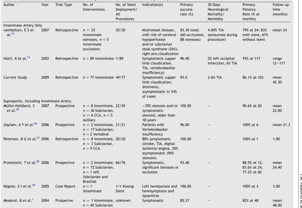
Table 2 Endovascular treatment of innominate artery lesions in the literature over the past 16 years.
Author Year Trial Type No. of
Interventions
No. of Stent Deployment/
All Procedures
Indication(s) Primary success rate (%)
30 Days Neurological Mortality/
Morbidity
Primary Patency Rate (% at months)
Follow-up time (months)
Innominate Artery Only vanHattum, E Set
al.15
2007 Retrospective nZ25 Innominate stenoses,nZ5 Innominate occlusions
20/30 Multivessel disease, with risk of cerebral hypoperfusion and/or subclavian steal syndrome (26%), right arm claudication
83.30 total:
(60 occlusions, 88 stenoses)
4.00% TIA (amaurosis during procedure)
79% at 24: 83%
with stent, 67%
without stent
mean 24
Hu¨ttl, Ket al.14 2002 Retrospective nZ89 Innominate 1/89 Symptomatic (upper limb claudication, TIA, vertebrobasilar insufficiency)
96.40 2% left occipital infarction, 6% TIA
93% at 117 range 12e117
Current Study 2009 Retrospective nZ77 Innominate 49/77 Symptomatic (upper limb claudication, dizziness),
asymptomatic in 24%
of cases
93.5 2.6% TIA 86.1% at 103 mean
42.30
Supraaortic, including Innominate Artery Mu¨ller-Hu¨lsbeck, S
et al.25
2007 Prospective nZ8 Innominate, nZ36 Subclavian, nZ6 CCA,nZ5 Axillary
22/55 >70% stenosis and/or symptomatic
stenosis, older than 18 years
100.00 e 90.6% at 20 mean
22.00
Zaytsev, A Y et al.26 2006 Prospective nZ2 Innominate, nZ17 Subclavian, nZ2 Vertebral
21/21 Patients with Vertebrobasilar insufficiency
96.00 e 100% at 6 mean 21.3
Peterson, B G et al.27 2006 Retrospective nZ8 Innominate, nZ3 Subclavian, nZ9 CCA
20/20 80% symptomatic (stroke, TIA, digital ischemia/angina, 20%
asymptomatic (90%
stenosis)
100.00 e 100% at 1 1.00
Przewlocki, T et al.28 2006 Prospective nZ2 Innominate, nZ72 Subclavian, nZ1 left
Subclavian and Brachial
66/76 Symptomatic,
significant stenoses or occlusion
93.40 e 88.5% at 12,
83.6% at 24, 77.2% at 60
mean 24.40
Nagata, S-i et al.29 2005 Case Report nZ1 Innominate
1/1 Kissing Stent
Left hemiparesis and hemisymptoms and dysarthria
100.00 e 100% at 3 3.00
Modarai, B et al.1 2004 Prospecive nZ1 Innominate, nZ40 Subclavian
unknown Symptomatic 85.37 e 82% at 48 mean
48.00
T.M.Paukovitsetal.
Gonza´les, A et al. 2002 Prospective nZ2 Innominate, nZ7 Subclavian
8/9 Symptomatic
occlusions
100.00 e 77.8% at 40 mean
37.40 Azzarone, M et al. 2000 Case Report nZ1 Innominate 1/1 Symptomatic
(amaurosis fugax) subtotal occlusion
100.00 e 100% at 12 12
Ko¨rner, J et al.31 1999 Prospective nZ4 Innominate, nZ38 Subclavian, nZ1 Subclaviane Subclavian bypass
0/43 Symptomatic
(subclavian steal sy, upper extremity arterial insufficiency, peripheral
thromboembolism, PRIND/stroke)
84.00 9% cerebrovascular thromboembolism
72% at 100 mean 15
Sullivan, T M et al.12 1998 Prospective nZ7 Innominate, nZ66 Subclavian, nZ14 CCA
87/87 Symptomatic 94.30 2.3% ischemic
stroke in CCA interventions
85% at 35 mean 14.3
Motarjeme, A et al.32 1993 Prospective nZ9 Innominate, nZ66 Subclavian, nZ3 Axillary, nZ3 Brachial, nZ35 Vertebral, nZ6 CCA,nZ7 ICA,nZ2 ECA
0/131 Symptomatic 93.00 total,
(98 for stenotic lesions, vs. 40 for total occlusions)
0.76% TIA (temporary bilateral blindness)
96.3% at 60 mean 60
Selby J B et al.33 1992 Prospecive nZ2 Innominate, nZ26 Subclavian, nZ4 Axillary
0/32 Arm Claudication, Vertebrobasilar Insufficiency, Graft Dilatation
100.00 e 96.9% at 90 mean
36.00
ArteryPTA41
Our series reflects similarly excellent primary success rate as the relevant literature (Table 2). In conclusion, high occlusions/stenoses rate results poor primary outcome. The use of new, lower profile balloons and stents might obviate this problem.
The principal advantage of stent placement in the treatment of innominate artery stenoses is its impact on the immediate outcome. However, there is no evidence that stenting is superior to angioplasty alone for innominate artery lesions; current practice includes primary stenting when highly calcified lesions are present and stenting lesions with significant residual stenosis after balloon angioplasty. Only one stent was depleted during 89 inter- ventions in the largest retrospective study on the topic, carried out at our institution between 1989 and 2000.14
Compared to the present study, in which 49/77 (63.6%) stents were used, primary and secondary patency rates are highly similar. However, the comparison of the two patient groups is limited, because of inhomogeneous patient pop- ulations, regarding availability of stents and varying indi- cations for stent deployment in the two studies.
Due to the anatomic position of the treated lesions, the non-invasive, direct visualisation of innominate vessel is mostly not possible; thus, we cannot exclude the chance of a restenosis in symptomless patients in whom no angiog- raphy was performed. We refer to open, patent innominate artery in all the asymptomatic patients with a present radial pulse and blood pressure difference less than 30 mmHg between the two arms. Asymptomatic restenoses might therefore be missed and the patency rate may be lower than reported. Literature on solely innominate artery intervention describes 79e93% primary patency at 24 and 117 months, respectively (Table 2).
During our study, no periprocedural neuroprotection device was used. The previous relevant cohort studies do not report the use of neuroprotection devices during the treatment of innominate artery either.24e33 According to the largest series on transluminar endovascular treatment of proximal common carotid artery (pACC), in which no neuroprotection was used, the use of neuroprotection devices would be technically difficult in innominate artery as well, without an established indication.34 Dual anti- platelet therapy was not routinely used in many of the published studies of innominate artery endovascular procedures, including this report. However, use of dual anti-platelet therapy may further reduce the number of neurological complications associated with innominate endovascular treatment.
The main limitation of this study is its retrospective nature. In addition, due to the long period studied (2000e09), the population was not homogeneous as to whether or not stents, and/or dual anti-platelet therapy were used. For the same reason, follow-up is missing for 9.7% of the procedures; however, procedural and in-hospital clinical outcomes were available for all 77 treatments. The number of restenoses, especially those below 70%, may also be underestimated due to the limitations of duplex scan examination in visual- isation of innominate artery. The lack of significant differences of patency rate between angioplasty alone and angioplasty stenting may also be due to the rela- tively small sample size.
In conclusion, this is one of the three studies published on angioplasty/stenting of solely the innominate artery.
The primary technical success rate is high (93.5%) with a 0%
30-day all stroke/death rate. Our results should help vascular surgeons and interventional radiologists to consider risk versus benefit when deciding treatment options for innominate artery lesions.
Conflict of Interest/Funding
None.
References
1 Modarai B, Ali T, Dourado R, Reidy J, Taylor P, Burnand K.
Comparison of extra-anatomic bypass grafting with angioplasty for atherosclerotic disease of the supra-aortic trunks.Br J Surg 2004;91:1453e7.
2 Dorros G, Lewin RF, Jamnadas P, Mathiak LM. Peripheral transluminal angioplasty of the subclavian and innominate arteries utilizing the brachial approach: acute outcome and follow-up.Catheter Cardiovasc Diagn1990;19:71e6.
3 Melliere D, Becquemin J-P, Benyahia NE, Ecollan P, Fitoussi M.
Atherosclerotic disease of the innominate artery: current management and results.J Cardiovasc Surg1992;33:319e23.
4 Cherry KJ, McCullough JL, Hallet JW, Pairolero PC, Gloviczki P.
Technical principles of direct innominate artery revasculariza- tion: a comparison of endarterectomy and bypass grafts.J Vasc Surg1989;9:718e24.
5 Brewster DC, Moncure AC, Darling RC, Ambrosino JJ, Abbot WM.
Innominate artery lesions: problems encountered and lessons learned.J Vasc Surg1985;2:99e112.
6 Herring M. The subclavian steal syndrome: a review.Am Surg 1977;43:220e8.
7 North American Symptomatic Carotid Endarterectomy Trial (NAS- CET) investigators. Clinical alert: benefit of carotid endarterec- tomy for patients with high-grade stenosis of the internal carotid artery. National Institute of Neurological Disorders and Stroke Stroke and Trauma Division.Stroke1991;22:816e7.
8 Endovascular versus surgical treatment in patients with carotid stenosis in the carotid and vertebral artery transluminal angioplasty study (CAVATAS): a randomized trial.Lancet2001;
357:1729e37.
9 Halliday A, Mansfield A, Marro J, Peto C, Peto R, Potter J, et alA Asymptomatic Carotid Surgery Trial (ACST) Collaborative Group. Prevention of disabling and fatal strokes by successful carotid endarterectomy in patients without recent neurological symptoms: randomized controlled trial. Lancet 2004;363:
1491e502.
10 Wylie EJ, Effeney DJ. Surgery of the aortic arch branches and vertebral arteries.Surg Clin North Am1979;59:660e80.
11 Insall RL, Lambert D, Chamberlain J, Proud G, Murthy LNS, Loose HWC. Percutaneous transluminal angioplasty of the innominate, subclavian and axillary arteries.Eur J Vasc Surg 1990;4:591e5.
12 Sullivan TM, Gray BH, Bacharach JM, Perl II J, Childs MB, Modzelewski L, et al. Angioplasty and primary stenting of the subclavian, innominate, and common carotid ateries in 83 patients.J Vasc Surg1998;28:1059e65.
13 Motarjeme A. Percutaneous transluminal angioplasty of supra- aortic vessels.J Endovasc Surg1996;3:171e81.
14 Hu¨ttl K, Nemes B, Simonffy A, Entz L, Be´rczi V. Angioplasty of the innominate artery in 89 patients: experience over 19 years.
Cardiovasc Intervent Radiol2002;25:109e14.
15 van Hattum ES, de Vries J-P, Lalezari F, van den Berg J, Moll F.
Angioplasty with or without stent placement in the
brachiocephalic artery: feasible and durable? A retrospective cohort study.J Vasc Interv Radiol2007;18:1088e93.
16 Patterson JR, Grabois M. Locked-in syndrome: a review of 139 cases.Stroke1986;17:758e64.
17 Bockenheimer ST, Reinhuber F, Mohs C. Intraarterielle throm- bolyse hirnversorgender gefase.Radiologe1991;31:210e5.
18 Thompson JR, Simmons CR, Hasso AN, Hinshaw Jr DB. Occlusion of the intradural vertebrobasilar artery.Neuroradiology1978;4:
219e29.
19 Henry M, Amor M, Henry I, Ethevenot G, Tzvetanov K, Chati Z.
Percutaneous transluminal angioplasty of the subclavian arteries.J Endovasc Surg1999;6:33e41.
20 Queral LA, Criado FJ. The treatment of focal aortic branch lesions with palmaz stents.J Vasc Surg1996;23:368e75.
21 Vitek JJ, Keller FS, Duvall ER, Gupta KL, Chandra-Sekar B.
Brachiocephalic artery dilatation by percutaneous transluminal angioplasty.Radiology1986;158:779e85.
22 Lyon RD, Shonnard KM, McCarter DL, Hammond SL, Ferguson D, Rholl KS. Supra-aortic arterial stenoses: management with palmaz-balloon expandable intraluminal stents. J Vasc Interv Radiol1996;7:825e35.
23 Brountzos E, Malagari K, Kelekis D. Endovascular treatment of occlusive lesions of the subclavian and innominate arteries.
Cardiovasc Intervent Radiol2006;29:503e10.
24 Rodriguez-Lopez JA, Werner A, Martinez R, Torruella LJ, Ray LI, Diethrich EB. Stenting for atherosclerotic occlusive disease of the subclavian artery.Ann Vasc Surg2006;13:254e60.
25 Mu¨ller-Hu¨lsbeck S, Both M, Charalambous N, Scha¨fer P, Heller M, Jahnke T. Endovascular treatment of atherosclerotic arterial stenoses and occlusions of the supraaortic arteries:
mid-term results from a single center analysis.Rontgenpraxis 2007;56:119e28.
26 Zaytsev A, Stoyda A, Smirnov V, Scherbyuk A, Kondrashin S, Artukchina E, Kikevitch V. Endovascular treatment of supra-
aortic extracranial stenoses in patients with vertebrobasilar insufficiency symptoms. Cardiovasc Intervent Radiol;29:
731e8.
27 Peterson B, Resnick S, Morasch M, Hassoun H, Eskandari M.
Aortic arch vessel stenting: A single-center experience using cerebral protection. Arch Surg 2006;141:560e3. discussion 563e564.
28 Przewlocki T, Kablak-Ziembicka A, Pieniazek P, Musialek P, Kadzielski A, Zalewski J, et al. Determinants of immediate and long-term results of subclavian and innominate artery angio- plasty.Catheter Cardiovasc Interv2006;67:519e26.
29 Nagata S, Kazekawa K, Matsubara S, Sugata S. Percutaneous reconstruction of the innominate bifurcation using the retro- grade ‘kissing stents’ technique. Neuroradiology 2006;48:
537e40.
30 Gonza´lez A, Gil-Peralta A, Gonza´lez-Marcos J, Mayol A. Angio- plasty and stenting for total symptomatic atherosclerotic occlusion of the subclavian or innominate arteries.Cerebrovasc Dis2002;13:107e13.
31 Ko¨rner M, Baumgartner I, Do D, Mahler F, Schroth G. Pta of the subclavian and innominate arteries: long-term results. Vasa 1999;28:117e22.
32 Motarjeme A, Gordon G. Percutaneous transluminal angioplasty of the brachiocephalic vessels: guidelines for therapy. Int Angiol1993;12:260e9.
33 Selby JJ, Matsumoto A, Tegtmeyer C, Hartwell G, Tribble C, Daniel T, et al. Balloon angioplasty above the aortic arch:
immediate and long-term results.AJR Am J Roentgenol1993;
160:631e5.
34 Paukovits TM, Haa´sz J, Molna´r A, Szeberin Z, Nemes B, Varga D, et al. Transfemoral endovascular treatment of proximal common carotid artery lesions: a single-center experience on 153 lesions.J Vasc Surg2008 Jul;48(1):80e7. Erratum in: J Vasc Surg. 2008 Sep;48(3):778. Le´ra´nt, Gergely [added].