Application of Fractional Flow Reserve in the diagnostics of coronary artery disease
PhD thesis
Gábor G Tóth
Semmelweis University – Doctoral School of Clinical Medicine
Consultant: Peter Andréka, MD, PhD
Official reviewers: Dániel Aradi, MD, PhD
András Komócsi, MD, DSc
Head of the Final Examination Committee: Mátyás Keltai, MD, PhD Members of the Final Examination Committee: Zoltán Ruzsa, MD, PhD
Tamás Ferenci, PhD
Budapest 2016
INTRODUCTION
Although cardiovascular disease is still the leading causes of death globally, one has to realize that it is mainly well treatable when detected on time. Accordingly, cardiovascular death is well preventable with careful and appropriate medical care.
Therefore, accurate definition and diagnosis of significant or clinically relevant coronary artery disease has been in the focus of interest since the first coronary stenosis was diagnosed by angiography.
Note, when aiming to change prognosis, the main determinant factor is not the angiographic severity of a coronary stenosis by itself, but the true presence and extent of ischemia. Therefore benefit from revascularization can be only expected when it eliminates ischemic risk.
The angiographic cut-off value to determine significant coronary artery disease is derived from early animal experiences, linking morphologic severity to reduction of coronary blood flow. As angiographic judgment is really simple and straight- forward to obtain, it is generally applied to guide clinical decision-making. However, angiography allows only indirect evaluation of functional importance, and so its accuracy on individual basis can be uncertain. This limitation of pure angiographic diagnosis has been circumvented by the development of pressure measuring guide wires and the invention of the concept of fractional flow reserve.
Fractional flow reserve is defined as the ratio of maximal hyperemic myocardial blood flow in the presence of a stenosis to the physiologic maximal hyperemic myocardial blood flow in the same territory but in the absence of any stenosis. Thus, the value of fractional flow reserve quantifies to what extent hyperemic flow is reduced by the presence of the epicardial narrowing. The value of fractional flow reserve is calculated as the ratio of distal coronary artery pressure and aortic pressure during maximal hyperemia.
Thanks to all technical and conceptual advantages, after 20 years fractional flow reserve became the standard of reference to define the ischemic potential of
OBJECTIVES
Despite powerful outcome data and the highest level of recommendation by European revascularization guidelines the adaptation of fractional flow reserve by the interventional cardiologists community is still limited. Indeed, there might be some important questions regarding the concept, causing potential hesitation against its unlimited clinical applicability.
(1) In daily practice the vast majority of decisions about revascularization are based on diameter stenosis as gauged by visual estimation or computerized quantification on coronary angiogram. Anatomic severity on quantitative coronary angiography is an oversimplified measure of stenosis severity that do not account for all aspects of severity, especially in case of ‘intermediate’ stenosis. Accordingly, the first goal of the present work is to analyze the concordance or discordance between stenosis severity by quantitative coronary angiography and by fractional flow reserve in a large unselected patient cohort. (Quantitative Coronary Angiography versus Fractional Flow Reserve study)
(2) The formula of fractional flow reserve calculation is simplified by excluding the right atrial pressure, considered to be negligibly low compared to the arterial values and so it would have limited impact on the calculated fractional flow reserve value. Ever since, this assumption has been confirmed and supported by the excellent clinical outcome data. However, as field of application got wider, debates have risen again about fractional flow reserves’ universal applicability and validity including patients with elevated right atrial pressures, such as patients with myocardial or valvular heart failure. Accordingly, the second goal of this work was to assess the impact, if any, of a wide range of right atrial pressures on fractional flow reserve assessment (myocardial fractional flow reserve) and on fractional flow reserve-guided clinical decision-making. (Fractional Flow Reserve vs Myocardial Fractional Flow Reserve study)
stenotic and normal conditions, the cornerstone of fractional flow reserve measurement is the reliable and stable maximal hyperemia. Despite the widespread adoption of intracoronary adenosine, a recurring debate still exists regarding its optimal dose, as no prior study has created sufficiently detailed and convincingly extensive dose-response curve in humans. The sense or non-sense of applying extreme dosages of adenosine is a recurring issue, requiring clarification. Therefore, the third goal of this work is to define the dose-response relationship between intracoronary adenosine and its resulting hyperemia. (Dose-response study)
METHODS
Quantitative Coronary Angiography versus Fractional Flow Reserve study
Study population
2.986 patients underwent both quantitative coronary angiography and fractional flow reserve measurements in at least one stable coronary artery stenosis.
Quantitative coronary angiography
Quantitative coronary angiography was performed based on computerized technology (Siemens Healthcare Axiom Artis, Siemens Healthcare ACOM.PC 5.01 or General Electric AW VolumeShare 6E). The contrast-filled catheter was used for calibration. From an end-diastolic still-frame, reference diameter, minimum luminal diameter, percent diameter stenosis and lesions length were calculated.
Fractional flow reserve measurement
Fractional flow reserve was measured after intracoronary administration of isosorbide dinitrate (200 µg). A pressure monitoring guide wire was advanced distal to the coronary artery stenosis. Hyperemia was obtained after administration of intravenous adenosine (continuous infusion of 140 µg/kg/min), intracoronary adenosine (bolus of 50-150 µg) or intracoronary papaverine (bolus of 10-20 mg). A fractional flow reserve value ≤0.80 was considered ‘positive’, i.e. likely to induce reversible myocardial ischemia. A fractional flow reserve value >0.80 was considered ‘negative’, i.e. unlikely to induce reversible myocardial ischemia.
Fractional Flow Reserve versus Myocardial Fractional Flow Reserve study
Study population
1.235 patients underwent both left- and right heart catheterization and fractional flow reserve measurement in at least one coronary stenosis.
Left and right heart catheterization
Diagnostic catheters were used for obtaining pressure values in the left heart, including aortic and left ventricular pressures. Swan-Ganz catheter was used for obtaining pressure values in the right heart, including right atrial-, right ventricular-, pulmonary arterial- and pulmonary capillary wedge pressures.
Fractional flow reserve measurement
Fractional flow reserve measurement was performed as described above. Fractional flow reserve was defined as the ratio of the simultaneously recorded mean arterial pressure distal to the stenosis and the mean aortic pressure at the tip of the guiding catheter during stable, steady state hyperemia. Myocardial fractional flow reserve was defined as the ratio of mean distal coronary pressure minus mean right atrial pressure, and the mean aortic pressure minus mean right atrial pressure during stable, steady state hyperemia.
Dose-response study
Study population
Patients with stable coronary artery disease undergoing routine diagnostic coronary angiography for a variety of indications were approached for participation. All patients had documented coronary atherosclerosis, but the measurements were performed in vessels free of any stenosis with more than 20% diameter reduction.
Intracoronary Doppler velocity measurement
Coronary flow velocity measurements were performed with Doppler sensor tipped guide wire. Flow velocity was measured in under following conditions: (1) resting conditions; then after administration of 8 mL (2) arterial blood; (3) pure saline at room temperature; (4) contrast medium (iodixanol 270 mg/mL); then after administration of (5-13) 9 escalating doses of adenosine [4, 12, 20, 60, 100, 160, 200, 300, and 500 µg / 8 mL]; and finally after administration of (10) 200 µg of adenosine plus contrast medium in 8 mL.
Statistical Analysis
All analyses were performed with Prism GraphPad 5.0 (GraphPad Software Inc., California, US), SPSS 20.0 (IBM Inc., New York, US) and R version 3.1.2 (R Foundation for Statistical Computing, Vienna, Austria) with standard summary statistics. p<0.05 was considered statistically significant. Normal distribution was tested with the D’Agostino-Pearson omnibus K2 test. Unpaired t-test or Mann- Whitney test were used to compare two independent groups, as appropriate. To compare multiple groups one-way ANOVA or Kruskal-Wallis test were used, as appropriate. Correlation among variables was determined by Pearson or Spearman correlation tests, as appropriate and expressed in r value. Sensitivity, specificity, diagnostic accuracy, and optimal diagnostic cut-off value were defined from the calculated receiver operator characteristic curves, as appropriate. Logistic regression analysis was performed to assess the impact of various characteristics on the accuracy of 50% diameter stenosis cut-off value in predicting FFR ≤ 0.80.
RESULTS
Quantitative Coronary Angiography versus Fractional Flow Reserve study
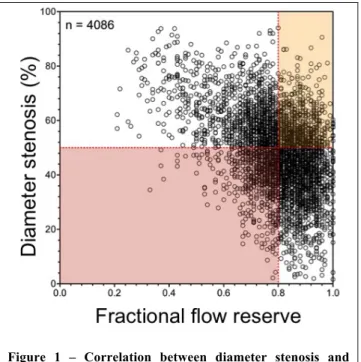
Data from 4.086 coronary artery stenoses in 2.986 patients were analyzed. Fractional flow reserve was in median 0.82 (0.74; 0.88) diameter stenosis was 48% (39; 57).
Overall relationship between angiographic metrics and fractional flow reserve The relationship between diameter stenosis and fractional flow reserve was only modest but statistically significant (-0.38 [95% CI: -0.41; -0.36]; p<0.001) with marked scatter around the regression line. Figure 1. A diameter stenosis ≥50%
correctly identified a fractional flow reserve value ≤ 0.80 with a sensitivity of 61%
[95% CI: 59; 63] and a specificity of 67% [95% CI: 65; 69], associated with a diagnostic accuracy of 0.64 [95% CI: 0.56; 0.72].
Figure 1 – Correlation between diameter stenosis and fractional flow reserve.
Influence of patients’ characteristics
Two parameters, namely male gender (p=0.017) and presence of diabetes (p=0.005) were found to influence negatively the value of 50% diameter stenosis cut-off in predicting significant fractional flow reserve.
Diagnostic performance of 50% versus 70% diameter stenosis
The overall diagnostic performance of angiography is significantly weaker when a 70% diameter stenosis is considered as cut-off value (Youden index 0.30 (0.28;
0.32) vs 0.08 (0.06; 0.12), respectively; p=0.004).
Optimal angiographic cut-off values for percent diameter stenosis
The optimal cut-off values of diameter stenosis for predicting fractional flow reserve
≤0.80 were markedly different: 43% for the left main stem, 51% for the global population, 55% for the distal segments.
Fractional Flow Reserve versus Myocardial Fractional Flow Reserve study
Data from 1.676 coronary artery stenoses in 1.235 patients were analyzed. Indication for catheterization was heart failure with NYHA Class II-IV in 914 patients (74%), ischemic heart disease in 642 patients (52%) and concomitant valve heart disease in 593 patients (48%).
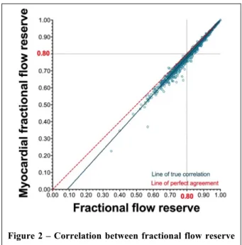
Average fractional flow reserve value was 0.85 (0.78; 0.91), while average myocardial fractional flow reserve was 0.83 (0.76; 0.90). Correlation and agreement between the two parameters were excellent (r2=0.987; slope 1.096±0.003). The median difference was 0.01 (0.01; 0.02). (Figure 2)
Relationship between FFR and FFRmyo
In patients, having normal right atrial pressure (Pra ≤5mmHg) median difference between fractional flow reserve and myocardial fractional flow reserve was minimal: 0.01 (0.00; 0.01). When grouping the patients into tertiles of Pra, a
and myocardial fractional flow reserve over the three groups [0.01 (0.00; 0.01) vs.
0.01 (0.01; 0.02) vs. 0.02 (0.01; 0.03), respectively; p<0.001].
Figure 2 – Correlation between fractional flow reserve and myocardial fractional flow reserve.
Out of 1.146 stenoses with fractional flow reserve above 0.80, none had a myocardial fractional flow reserve equal to or below 0.75; and 110 (9%) stenoses had a myocardial fractional flow reserve equal to or below 0.80. In the latter group the difference was 0.02 (0.02; 0.03), yet with right atrial pressure significantly higher than in the overall population [9 (7; 12) mmHg; p<0.001].
Dose-response study
30 patients were investigated. Coronary flow reserve varied from 1.42 to 4.88. The baseline flow velocity was higher in patients with a low coronary flow reserve than in patients with a high coronary flow reserve (29±11 cm/s versus 16±7 cm/s;
p<0.001). Hyperemic flow velocity was similar in both groups (61±26 cm/s versus 55±17 cm/s; p=0.41).
Dose-response analysis
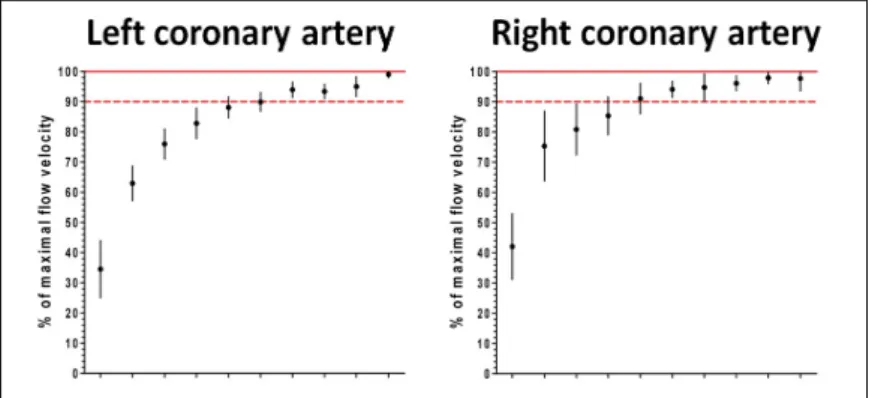
Figure 3 summarizes the dose-response relationships of intracoronary adenosine in the left- and the right coronary artery.
Figure 3 – Adenosine dose-response relation in the left- and in the right coronary artery.
For the right coronary artery baseline-to-maximal flow did not increase significantly at any higher dose than 60 µg. For the left coronary artery baseline-to-maximal flow did not increase significantly at any higher dose than 160 µg.
Effect of blood, saline and contrast medium
Doppler flow velocity varied among 8 mL intracoronary boluses of arterial blood, saline and contrast (p<0.001 by ANOVA), and all pairwise comparisons were significant (p<0.001 for blood and contrast; p=0.041 for saline and blood; p=0.013 for saline and contrast). Contrast increased Doppler flow velocity the most (+38±52% over blood, p<0.001 by paired t-test; +17±28% over saline, p=0.019) and saline was superior to blood (+21±43%, p=0.008). Flow velocity after contrast
CONCLUSIONS
Present work reports data from the largest patient population in the topic so far about the comparison of angiographic metrics and functionality. Our results emphasize that reliance on the angiogram needs to be modified by physiologic measures of severity for a wide range of intermediate stenoses. Explaining the results of randomized trials, showing outcome benefit associated with fractional flow reserve- guidance, we found that as much as one third of the decisions based angiographic metrics are discordant with the fractional flow reserve. Data show that increasing the threshold to 70% improved the specificity (i.e. will decrease the trend of coronary angiography to overestimate lesion severity, resulting potentially in unnecessary revascularization) but decreased sensitivity (i.e. will increase the number of stenoses underestimated by coronary angiography, resulting potentially in untreated risk left behind). Summed, increasing the threshold to 70% decreases the overall diagnostic performance of diameter stenosis in predicting significant fractional flow reserve, as compared to 50% cut-off value.
In this work we investigated on the largest patient population so far, affected by various degree of heart failure of different etiologies, whether incorporating the value of right atrial pressure into the formula (myocardial fractional flow reserve) has any clinical impact on fractional flow reserve measurement. Although these patients had a right atrial pressure often markedly above the normal range, the correlation and the agreement between measured fractional flow reserve and calculated myocardial fractional flow reserve was still excellent, with a difference as minimal as 0.01. In no case a fractional flow reserve value above 0.80 turned to a myocardial fractional flow reserve below 0.75. Summarizing, our findings indicate that accounting for actual value of right atrial pressure induces only minimal differences in the calculations of FFR, on average within the limits of the test-retest repeatability. In addition, these differences have negligible clinical consequences even in patients with elevated right atrial pressure.
The present dose-response study of intracoronary adenosine on intracoronary Doppler flow velocity suggests that optimal bolus to induce maximal hyperemia consistently, reliably and safely is 60-100 µg for the right coronary artery and 160- 200 µg for the left coronary artery. Sequential doses above the indicated amounts showed no statistically significant further increase in flow. Notably, we observed an increased incidence of AV-block at higher doses. Based on the findings of our dose- response analysis a clear recommendation can be made for the optimal dosing of adenosine for the measurement of fractional flow reserve. These doses allow achieving >95% of maximum hyperemia and are clinically indistinguishable from higher dosages. While lower doses are less reliable to reach maximal hyperemia, therefore inducing inaccuracy of our measurement with potential underestimation of lesion severity.
Summary
This work investigated important topics, and we believe, a couple of crucial questions have been answered, facilitating an even broader acceptance of fractional flow reserve:
This work confirms on the largest population so far what massive discrepancies can be observed between angiographic severity of a stenosis and its true ischemic potential.
Dose-response data gives recommendation for optimal hyperemia induction, and confirms that any decrease in adenosine dosages impacts negatively the accuracy, while further increase dosages does not have any benefit, therefore can be considered as non-sense.
Data confirm that right atrial pressure has no relevant impact on the fractional flow reserve value, not even in patients with pathologically elevated central venous pressures.
We believe these findings have the potential to impact future interventional practices.
BIBLIOGRAPHY OF THE CANDIDATE’S PUBLICATIONS – related to the thesis –
1. Toth G,* Hamilos M,* Pyxaras S, Mangiacapra F, Nelis O, De Vroey F, Di Serafino L, Muller O, Van Mieghem C, Wyffels E, Heyndrickx GR, Bartunek J, Vanderheyden M,Barbato E, Wijns W, De Bruyne B. (2014) Evolving concepts of angiogram: fractional flow reserve discordances in 4000 coronary stenoses. Eur Heart J. 35: 2831-8. * shared first authorship due to equal contribution
2. Toth GG, De Bruyne B, Rusinaru D, Di Gioia G, Bartunek J, Pellicano M, Vanderheyden M, Adjedj J, Wijns W, Pijls NH, Barbato E. (2016) Impact of Right Atrial Pressure on Fractional Flow Reserve Measurements: Comparison of Fractional Flow Reserve and Myocardial Fractional Flow Reserve in 1,600 Coronary Stenoses. JACC Cardiovasc Interv. 9: 453-9.
Adjedj J,* Toth GG,* Johnson NP, Pellicano M, Ferrara A, Floré V, Di Gioia G, Barbato E, Muller O, De Bruyne B. (2015) Intracoronary Adenosine: Dose- Response Relationship With Hyperemia. JACC Cardiovasc Interv. 8: 1422-30.
* shared first authorship due to equal contribution
BIBLIOGRAPHY OF THE CANDIDATE’S PUBLICATIONS – not related to the thesis –
1. Toth GG, Ntalianis A, Ntarladimas Y, de Booij M, De Winter O, Barbato E, Pilet B, Van Mieghem C, Wijns W, De Bruyne B. (2015) Effective radiation doses associated with non-invasive versus invasive assessment of coronary anatomy and physiology. Catheter Cardiovasc Interv. 85: 1173-81.
2. Toth G, De Bruyne B, Casselman F, De Vroey F, Pyxaras S, Di Serafino L, Van Praet F, Van Mieghem C, Stockman B, Wijns W, Degrieck I, Barbato E. (2013) Fractional flow reserve-guided versus angiography-guided coronary artery bypass graft surgery. Circulation. 128: 1405-11.
3. Toth GG, Toth B, Johnson NP, De Vroey F, Di Serafino L, Pyxaras S, Rusinaru D, Di Gioia G, Pellicano M, Barbato E, Van Mieghem C, Heyndrickx GR, De Bruyne B, Wijns W. (2014) Revascularization Decisions in Patients With Stable Angina and Intermediate Lesions: Results of the International Survey on Interventional Strategy. Circ Cardiovasc Interv. 7: 751-9.
4. Toth GG, Pyxaras S, Mortier P, De Vroey F, Di Gioia G, Adjedj J, Pellicano M, Ferrara A, De Schryver T, Van Hoorebeke L, Verhegghe B, Barbato E, De Bruyne B, De Beule M, Wijns W. (2015) Single String Technique for Coronary Bifurcation Stenting: Detailed Technical Evaluation and Feasibility Analysis. JACC Cardiovasc Interv. 8: 949-59.
5. Toth GG, Kala P, Lansky A, Baumbach A. (2014) Will this trial change my practice? PRAMI - treatment of bystander coronary lesions in patients undergoing primary PCI for acute STEMI. EuroIntervention. 10: 411-2.
6. Tóth GG, Yamane M, Heyndrickx GR. (2015) How to select a guidewire:
technical features and key characteristics. Heart. 101: 645-52.
7. Toth GG, Vanderheyden M, Bartunek J. (2016) Novel device-based interventional strategies for advanced heart failure. Adv Interv Cardiol. 1: 13–16 8. Toth GG, Vanderheyden M, Bartunek J. Novel Device-based Strategies in Treatment of Chronic Heart Failure: Translational Approach to Heart Failure.
9. Di Gioia G, Pellicano M, Toth GG, Casselman F, Adjedj J, Van Praet F, Ferrara A, Stockman B, Degrieck I, Bartunek J, Trimarco B, Wijns W, De Bruyne B, Barbato E. (2016) Fractional Flow Reserve-Guided Revascularization in Patients With Aortic Stenosis. Am J Cardiol. pii: S0002-9149(16)30236-3
10. Adjedj J, De Bruyne B, Floré V, Di Gioia G, Ferrara A, Pellicano M, Toth GG, Bartunek J, Vanderheyden M, Heyndrickx GR, Wijns W, Barbato E. (2016) Significance of Intermediate Values of Fractional Flow Reserve in Patients With Coronary Artery Disease. Circulation. 133: 502-8.
11. Fearon WF, Yong AS, Lenders G, Toth GG, Dao C, Daniels VD, Pijls NHJ, De Bruyne B. (2015) The Impact of Downstream Coronary Stenosis on Fractional Flow Reserve Assessment of Intermediate Left Main Coronary Artery Disease: Human Validation. JACC Cardiovasc Interv. 8: 398-403.
12. Di Serafino L, Pyxaras SA, Mangiacapra F, Dierickx K, Toth G, Bartunek J, De Bruyne B, Van Mieghem C, Wijns W, Barbato E. (2013) Influence of transradial versus transfemoral diagnostic heart catheterisation on peripheral vascular endothelial function. EuroIntervention. 8: 1252-8.
13. Pyxaras SA, Mangiacapra F, Wijns W, Di Serafino L, De Vroey F, Toth G, Sinagra G, De Bruyne B, Heyndrickx GR, Barbato E. (2014) ACEF and clinical SYNTAX score in the risk stratification of patients with heavily calcified coronary stenosis undergoing rotational atherectomy with stent implantation. Catheter Cardiovasc Interv. 83: 1067-73.
14. Pyxaras SA, Tu S, Barbato E, Barbati G, Di Serafino L, De Vroey F, Toth G, Mangiacapra F, Sinagra G, De Bruyne B, Reiber JH, Wijns W. (2013) Quantitative angiography and optical coherence tomography for the functional assessment of nonobstructive coronary stenoses: comparison with fractional flow reserve. Am Heart J. 166: 1010-1018.
15. Pyxaras SA, Mangiacapra F, Verhamme K, Di Serafino L, De Vroey F, Toth G, Perkan A, Salvi A, Bartunek J, De Bruyne B, Wijns W, Sinagra G, Barbato E. (2013) Synergistic effect of thrombus aspiration and abciximab in primary percutaneous coronary intervention. Catheter Cardiovasc Interv. 82: 604-11.
16. Pyxaras SA, Sinagra G, Mangiacapra F, Perkan A, Di Serafino L, Vitrella G, Rakar S, De Vroey F, Santangelo S, Salvi A, Toth G, Bartunek J, De Bruyne B, Wijns W, Barbato E. (2013) Contrast-induced nephropathy in patients undergoing primary percutaneous coronary intervention without acute left ventricular ejection fraction impairment. Am J Cardiol. 111: 684-8.
17. Ruzsa Z, Ungi I, Horváth T, Sepp R, Zimmermann Z, Thury A, Jambrik Z, Sasi V, Tóth G, Forster T, Nemes A. (2009) Five-year experience with transradial coronary angioplasty in ST-segment-elevation myocardial infarction. Cardiovasc Revasc Med. 10: 73-9.
18. Pellicano M, Toth G, Di Gioia G, Rusinaru D, Wijns W, Barbato E, De Bruyne B, Degrieck I, Van Mieghem C. (2015) Unrecognized anomalous left circumflex coronary artery arising from right sinus of Valsalva: a source of perioperative complication. J Cardiovasc Med (Hagerstown). [Epub ahead of print]
19. Adjedj J, Toth GG, De Bruyne B. (2015) Invasive Measures of Myocardial Perfusion and Ischemia. Prog Cardiovasc Dis. pii: S0033- 0620(15)00014-6.
20. Capodanno D, Ducrocq G, Toth G, Dörler J, Iversen AZ, Dobric M, Schurtz G, Morice MC, Windecker S, Gonçalves L, Gilard M, Naber CK, Fajadet J.
(2014) Unmet needs of young interventional cardiologists: proceedings from the 2nd summit of the European association of percutaneous cardiovascular interventions.
EuroIntervention. 10: 47-9.
21. Tu S, Echavarria-Pinto M, von Birgelen C, Holm NR, Pyxaras SA, Kumsars I, Lam MK, Valkenburg I, Toth GG, Li Y, Escaned J, Wijns W, Reiber JH.
(2015) Fractional flow reserve and coronary bifurcation anatomy: a novel quantitative model to assess and report the stenosis severity of bifurcation lesions.
JACC Cardiovasc Interv. 8: 564-74.