Journal of Proteomics 245 (2021) 104293
Available online 10 June 2021
1874-3919/© 2021 Elsevier B.V. All rights reserved.
High-throughput rat immunoglobulin G N-glycosylation profiling revealed subclass-specific changes associated with chronic stress
Sini ˇ sa Habazin
a,*,1, Dra ˇ zen Mlinarevi ´ c
b,1, Marta Balog
c, Ana Bardak
c, Robert Gaspar
d, K ´ alm ´ an Ferenc Sz ucs ˝
d, Sandor G. Vari
e, Frano Vu ˇ ckovi ´ c
a, Gordan Lauc
a,f, Mislav Novokmet
a,*,2, Marija Heffer
c,2aGlycoscience Research Laboratory, Genos Ltd., Zagreb, Croatia
bUniversity Hospital Centre Osijek, Osijek, Croatia
cLaboratory of Neurobiology, Department of Medical Biology and Genetics, Faculty of Medicine Osijek, Josip Juraj Strossmayer University of Osijek, Osijek, Croatia
dDepartment of Pharmacology and Pharmacotherapy, Faculty of Medicine, Interdisciplinary Excellence Centre, University of Szeged, Szeged, Hungary
eCedars-Sinai Medical Center, International Research and Innovation in Medicine Program, LA, California, United States
fDepartment of Biochemistry and Molecular Biology, Faculty of Pharmacy and Biochemistry, University of Zagreb, Zagreb, Croatia
A R T I C L E I N F O Keywords:
Immunoglobulin G Rat N-glycans Glycoproteomics Chronic stress HPA axis
A B S T R A C T
Immunoglobulin G (IgG) glycosylation corresponds well with immune system changes, so it can potentially be used as a biomarker for the consequences of chronic stress such as low-grade inflammation and enhanced immunosenescence in older animals. Here we present a high-throughput glycoproteomic workflow, including IgG enrichment, HILIC glycopeptide purification, and nano-LC-MS analysis of tryptic glycopeptides applied for the analysis of rat IgG. A cohort of 80 animals was exposed to seven stressors in a customized chronic stress protocol with blood and tissue sampling in three timepoints. Young female rats experienced an increase in agalactosylated glycoforms on IgG2a and IgG2c accompanied by a decrease in monogalactosylation. Among old females, increased galactosylation was observed in the IgG2b subclass, pointing to an anti-inflammatory activity of IgG. Additionally, IgG Fc N-glycosylation patterns in Sprague Dawley rats were analyzed, quantified, and reported for the first time. Our findings emphasize age-, sex- and subclass-dependent differences in IgG glyco- sylation related to chronic stress exposure, confirming the relevance of newly developed methods for further research in glycobiology of rodent immune response.
Significance: In this study, we showed that a high-throughput streamlined methodology based on protein L 96- well monolithic plates for efficient rat IgG immunoaffinity enrichment from blood plasma, paired with appro- priate tryptic glycopeptide preparation, HILIC-SPE enrichment, and nano-LC-MS methods was suitable for quick processing of large sample sets. We report a subclass-specific profiling and changes in rat IgG Fc galactosylation and adrenal gland immunohistochemistry of male and female animals exposed to a customized chronic stress protocol.
1. Introduction
Glycosylation is an elaborate form of co- and post-translational protein modification indirectly encoded by the genome and greatly influenced by environmental factors. Immunoglobulin G (IgG) is the most abundant plasma glycoprotein, containing a glycan attached to a conserved Asn-297 residue at the fragment crystallizable (Fc) domain of
both heavy chains. Between 15% and 20% of IgG molecules are also glycosylated at the fragment antigen-binding (Fab) domain [1]. Minor alterations in IgG glycan composition significantly affect IgG effector functions, disturbing the pro- and anti-inflammatory equilibrium [1–4].
The structural complexity of glycans and their biological functions have only begun to unravel since the advent of high-throughput glyco- analytics. Rats are the animal model of choice for cancer, diabetes,
* Corresponding authors.
E-mail addresses: shabazin@genos.hr (S. Habazin), mnovokmet@genos.hr (M. Novokmet).
1 These authors contributed equally to this work.
2 Equally contributing last authors.
Contents lists available at ScienceDirect
Journal of Proteomics
journal homepage: www.elsevier.com/locate/jprot
https://doi.org/10.1016/j.jprot.2021.104293
Received 24 November 2020; Received in revised form 3 May 2021; Accepted 26 May 2021
investigated. Comprehensive glycomics already confirmed that the rat serum glycan profile was more similar to the human glycan profile than was the mouse profile. The rat serum glycan profile shows an unusually high diversity of structures, including N-glycans with O-acetylated N- acetyl- (Neu5Ac) and N-glycolylneuraminic acid (Neu5Gc), typically not present in human and mouse sera [10]. A single study on rat IgG glycans using MALDI-TOF-MS reported relative proportions of N-linked glycans and confirmed the presence of both Neu5Ac and Neu5Gc but did not structurally characterize them any further [11]. Rats have four IgG subclasses (IgG1, IgG2a, IgG2b and IgG2c) [12], with unknown subclass-specific glycosylation variability. IgG1 and IgG2a subclasses are considered essential for the chronic infection onset, while IgG2b and IgG2c participate in the protection against infection [13]. Although IgG glycans are essential in the modulation of immune system function, data on rat immunoglobulins glycosylation are scarce. This encouraged us to test the hypothesis that IgG glycosylation patterns were interconnected with chronic stress [14].
N-linked glycosylation plays a vital role in endoplasmic reticulum stress coping response, priming cellular defense against apoptosis [15].
To the best of our knowledge, IgG N-glycans have never been studied in diseases presenting as hyperresponsive or insufficient system-level stress response. However, a number of changes in glycoproteins and lectins have been described in conditions associated with chronic mental, physical, or combined stress [16]. Chronic stress is associated with premature senescence and induces maladaptive responses in virtually all organ systems [17–21]. It dysregulates the neuroendocrine system and results in chronic inflammation, contributing to early thrombotic com- plications of atherosclerosis and insulin resistance leading to diabetes [22–24]. Stressors disturb homeostasis by activating the hypothalamic- pituitary-adrenal (HPA) axis and the autonomic nervous system (ANS), but also by changing the glycome [25,26]. A recent study identified three potential N-glycan biomarkers that could predict stress suscepti- bility in a rat post-traumatic stress disorder model [27]. The stress response significantly depends on age and sex, which is reflected in different circulating glucocorticoid (GC) levels and alterations in glucocorticoid receptor (GR) signaling after chronic stress exposure [28–30]. The various effects of glucocorticoids are modulated by the density and activity of GRs, but also by corticosterone synthesis from 11- deoxycorticosterone, which is performed by 11β-hydroxylase (CYP11B1), and by intracellular interconversion of active corticosterone into inactive 11-dehydrocorticosterone, controlled by 11β-hydroxyste- roid dehydrogenase (HSD11B) [31]. Animal models of chronic stress should demonstrate behavioral changes caused by alterations in specific brain regions, and indicate activation of low-grade inflammatory pro- cesses as well as overall alterations of the HPA axis [27,28,30].
We aimed to determine the effects of chronic stress on rat IgG Fc- specific N-glycosylation patterns by establishing and validating a high- throughput glycoproteomic workflow that allows for quick processing of large-sample cohorts. This study for the first time reported a site- specific N-glycosylation diversity of rat IgG. Additionally, immunohis- tochemistry of rat adrenal glands was performed to demonstrate the HPA axis activation as a biological effect of the chronic stress protocol.
2. Materials and methods
2.1. Animals and chronic stress protocol
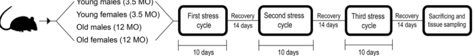
The chronic stress protocol was performed from May until December 2016. Sprague Dawley CR rats (Charles River, Wilmington, MA, USA) were assigned to eight groups, with 10 animals in each. Four
10-day stress cycles, using seven different stressors, with a 14-day re- covery period between each stress cycle. The complete protocol lasted for 10 weeks (Fig. 1). After the third stress cycle had ended, the animals were given 14 days to recover from the effects of acute stress. This enabled us to identify only the consequences of long-lasting chronic stress. The control group was subjected to sham stress – no stressors were applied but the animals were handled in the same fashion and in the same surroundings as the experimental animals, thereby equilibrating the stress caused by handling and surroundings in both groups. After the final recovery period, the animals were sacrificed, and tissue samples and plasma were obtained.
2.2. Rat adrenal glands
Adrenal glands were extirpated from the male and female rats during tissue collection at the ages of 6 and 14.5 months, fixed in 10% form- aldehyde solution, and embedded in paraffin. The tissues were cut into 6-μm sections with a microtome (SM2000R, Leica).
2.3. Antibodies
Highly specific rabbit polyclonal primary antibody to CYP11B1 (Biorbyt, Cambridge, UK, cat. no: orb5937) and mouse monoclonal antibody to glucocorticoid receptor (GR), specifically the membrane receptor (kindly provided by prof. Peter Balogh, School of Medicine, University of P`ecs, Hungary), were used. Secondary donkey-anti-rabbit CY3 (Jackson Immunoresearch, West Grove, PA, USA, cat.no: 711-165- 152) and donkey-anti-mouse CY2 antibodies (Jackson Immunoresearch, West Grove, PA, USA, cat.no: 715-175-150) were appropriately com- bined with the primary antibodies.
2.4. Single immunofluorescence analysis
The slides were thoroughly de-paraffinized and immersed into cit- rate buffer (pH 6) in a steamer (96 ◦C) for 30 min. Nonspecific staining was blocked with 5% donkey serum, 5% goat serum and 1% BSA in PBS buffer (pH 7.4) for 1 h at room temperature. The slides were then incubated in antibody solution, diluted to 1:20 in the case of CYP11B1 and 1:3000 in the case of GR antibody, overnight in blocking solution at +4 ◦C. In the next step, the samples were washed in PBS buffer, incu- bated in the dark for 4 h at +4 ◦C with appropriate secondary antibodies diluted to 1:500. After washing in PBS, coverslips were mounted using Vector Laboratories Vectashield® mounting media with DAPI (Vector Labs, Burlingame, CA, USA).
2.5. Immunohistochemical evaluation of immunofluorescence
At least 5 biological replicates were used for microscopy with the Olympus Fluoview FV1000 confocal microscope. The images were ob- tained with Olympus FV10-ASW 4.2 Viewer software. CYP and GR expression in the fascicular layer of the adrenal glands was quantified in at least seven representative images, 512 pixels in size. All images were analyzed in the Fiji software by measuring the integrated density value according to the protocol [33].
2.6. Blood plasma sampling and sample randomization
Rat plasma was collected at three time points: at the beginning of the study, one day before the stress protocol started (BASE), at the end of the 3rd chronic stress cycle (END) and after sacrifice (SAC), and stored at
− 20 ◦C. Prior to IgG enrichment, the samples were thawed at +4 ◦C, vortexed and centrifuged at 1620 ×g for 10 min. One hundred micro- liters of each sample was transferred to a corresponding well of a 96-well 2-mL collection plate (Waters, Milford, MA, USA) predetermined by randomization. The sample list was randomized using Microsoft Excel 2010 software (Microsoft Corp., Redmond, WA, USA). Each of the three plates contained additional eight blanks and five standards (100 μL each) made from the pooled rat plasma of the same strain.
2.7. High-throughput protein L immunoaffinity rat IgG enrichment IgG from rat plasma was enriched using newly developed 96-well plates (Suppl. Fig. 5) with recombinant protein L immobilized to monolithic supports (BIA Separations Ltd., Ajdovˇsˇcina, Slovenia). First, the samples were diluted with 1×PBS (pH 7.4) and vacuum-filtered through 0.45 μm 96-well Acroprep® filter plate (Pall Corp., NY, USA) into a clean 96-well 2-mL collection plate (Waters, Milford, MA, USA).
The filtered samples were applied to preconditioned monoliths. The unbound portion was washed away with 1×PBS, and IgG was eluted with 0.1 M formic acid (Merck KgaA, Darmstadt, Germany) and immediately neutralized with 1.0 M ammonium bicarbonate (Acros Organics, Geel, Belgium). IgG concentration was measured with Nano- Drop™ 8000 spectrophotometer (Thermo Scientific, Waltham, MA, USA) at 280 nm using a built-in program for IgG quantification. Protein L monolithic plate was regenerated with 0.1 M formic acid, 10×PBS (pH 7.4), 1×PBS, and stored in 20% ethanol (v/v) (Honeywell, Seelze, Germany) containing 20 mM TRIS (Sigma-Aldrich, St. Louis, MO, USA) and 0.1 M NaCl (Gram-mol Ltd. Zagreb, Croatia), pH 7.4. The eluate was frozen at − 20 ◦C until further use. The entire protocol was performed on a vacuum manifold (Pall Corp., NY, USA), and a detailed procedure is described in Suppl. File 2.
2.8. Tryptic glycopeptides preparation
One hundred and fifty microliters of IgG eluate, corresponding to
~40 μg of protein, was transferred to a 1-mL round-bottom 96-well collection plate (Waters, Milford, MA, USA), dried down in vacuo at 37 ◦C and redissolved in 40 μL 25 mM ammonium bicarbonate (Sigma- Aldrich). For digestion, sequencing grade trypsin (Promega Corp., Madison, WI, USA) at 1:20 protease:protein ratio (w/w) was added, and the samples were incubated for 18 h at 37 ◦C in a foil-sealed plate.
2.9. High-throughput HILIC glycopeptide enrichment
Glycopeptide enrichment following trypsin digestion was performed with Chromabond® HILIC silica beads (Macherey-Nagel GmbH & Co., Düren, Germany) with zwitterionic modification. A 50 mg/mL suspen- sion of HILIC beads was first prepared in 0.1% (v/v) TFA (Sigma- Aldrich) on a magnetic stirrer, and 100 μL of suspension was added to each well of the Orochem OF1100 filter plate (Orochem Technologies Inc., Naperville, IL, USA). Beads were washed twice with 0.1% TFA (v/v) and thrice with 0.1% TFA in 90% ACN (v/v) (Honeywell). The samples were brought to 90% ACN final concentration with 0.1% TFA in ACN, applied to the preconditioned beads, and unbound analytes were washed away with two portions of 0.1% TFA in 90% ACN.
Glycopeptides were eluted with 0.1% TFA into a clean skirted PCR plate by low-speed centrifugation. The eluates were dried in a vacuum centrifuge and stored at − 20 ◦C until analysis. For complete protocol, refer to Suppl. File 2.
2.10. Nano-LC-ESI-QqTOF analysis of glycopeptides
Glycoproteomic analysis was performed with ACQUITY UPLC M- class nano-LC system (Waters, Milford, MA, USA) coupled to Compact™
QqTOF mass spectrometer via CaptiveSpray™ ESI interface supported with nanoBooster™ dopant addition technology (Bruker Daltonik GmbH, Bremen, Germany). Dried glycopeptides were redissolved in 40 μL ultrapure water and 15 μL was injected onto C18 Acclaim™ PepMap™ trap column (5 ×0.3 mm, 5 μm, 100 Å, Thermo-Scientific), desalted for 5 min with a 40 μL/min flowrate of 0.1% TFA (v/v) from the auxiliary pump and diverted to an analytical column. The separation was per- formed on the HALO C18 column (150 ×0.1 mm, 2.7 μm, Advanced Materials Technology, Inc., Wilmington, USA) heated to 30 ◦C. Before and after each sample was loaded, the trap column was washed by injecting 20 μL of i-PrOH/ACN (25:75, v/v) followed by the same vol- ume of 95% ACN (v/v). A linear 15-min gradient, at a 1 μL/min flowrate, starting from 0% eluent B (0.1% TFA in 80% ACN, v/v) and 100% eluent A (0.1% TFA, v/v) and finishing at 35% eluent B, was employed for efficient separation of rat IgG glycopeptides. After separation, the col- umn was equilibrated with 100% eluent A for 4 min.
The mass spectrometer was operated in a reflectron positive ion mode with profile spectra acquisition rate at 2 ×0.5 Hz and in m/z range 600–3000. ACN-doped nebulizing nitrogen gas was provided at 0.2 bar.
Drying gas heated to 150 ◦C was introduced at 4 L/min, and capillary voltage was set to 1.5 kV. Ion transfer time was stepped up from 70 to 150 μs, and pre-pulse storage time was set to 10 μs. Nano-LC-MS system was operated under otofControl, MassLynx and Compass HyStar soft- ware (all v.4.1).
This method was also used for MS/MS structural characterization of the most abundant IgG glycopeptides from all three subclasses. The method modifications and spectra annotation are described in the Suppl.
File 6. For manual interpretation of fragmentation spectra, Compass DataAnalysis (v.4.1) and GlycoWorkbench 2.0 software [34] were employed. Glycan structures were depicted using the symbols and rules proposed by the Consortium for Functional Glycomics.
2.11. Method validation
The workflow was validated for intra-day (repeatability) and inter- day (within-laboratory reproducibility) precision for the same glyco- forms quantified from the cohort samples. The experimental design is presented in the Suppl. File 5.
2.12. Protein L eluate proteomic analysis
To confirm the presence of rat IgG subclasses and other potentially co-enriched proteins in the protein L eluate, we performed a simple peptide mapping for protein identification. The details on sample preparation, analysis, and database search are given in the Suppl. File 7.
Fig. 1.Chronic stress protocol scheme. Young and old Sprague Dawley CR rats were assigned to groups based on sex. The experimental group was exposed to a customized chronic stress protocol (see Section 2.1 and Suppl. File 1 for details), and the control group was exposed to sham stress.
ture data on glycan structures comprising murine IgG and plasma glycome (Suppl. File 3). Three chromatographic peak clusters were defined, and the LaCy Tools data processing package (v.1.0.1, b.5) was used for fast automatic peak integration. A detailed description of this software and its use has been previously published [35,36]. Original data files were first converted to mzXML open data format by Proteo- Wizard msConvert [37], and chromatograms were aligned based on the retention times of three calibrants, i.e., the most abundant glycopeptide signals in each cluster. Summed spectra were integrated to cover 95% of the theoretical isotopic pattern. The data were manually curated to exclude the analytes with an S/N ratio below 9, isotopic pattern quality (deviation from theoretical isotopic pattern) above 35%, and mass de- viation higher than 35 ppm. Only the charge states compliant with these three criteria were summed and quantified. Finally, absolute areas were normalized to the total area for each of the three IgG subclasses.
2.14. Statistical analyses 2.14.1. Immunostaining
Statistical analysis of immunostaining data was performed with Statistica 12 software (TIBCO, Palo Alto, CA, USA). A p-value of <0.05 was considered significant. The interaction between stress exposure, age, sex and CYP and GR expression was analyzed with a three-way factorial ANOVA.
2.14.2. Glycoproteomic data analysis
In order to minimize experimental variation from the measurements, LC-MS glycopeptide data were normalized and batch corrected. To make measurements across samples comparable, normalization by total area was performed. Before batch correction, normalized glycan measure- ments were log-transformed due to the right-skewness of their distri- butions and the multiplicative nature of batch effects. The batch correction was performed on log-transformed measurements with the ComBat method (R package sva), where the technical source of variation (which sample was analyzed on which plate) was modeled as batch covariate. To correct the measurements for experimental noise, esti- mated batch effects were subtracted from log-transformed measure- ments. Longitudinal analysis of samples through their observation period was performed by implementing a linear mixed-effects model (LMM), where time was modeled as a fixed effect while individual ID was modeled as a random effect. The effects were estimated using the LMM approach, where an effect is defined as the difference between two slopes – the first slope represents a change of glycopeptide abundance through time in the control group, while the second slope represents a change through time in the stress group. Before the analyses, glycan variables were transformed to a standard normal distribution by the inverse transformation of ranks to normality (R package “GenABEL”, function rntransform). Using rank-transformed variables makes the estimated effects of different glycopeptides comparable as these will have the same standardized variance. False discovery rate (FDR) was controlled by the Benjamini-Hochberg procedure at a specified level of 0.05. The p-values were corrected for multiple testing. The data was analyzed and visualized using the R programming language (version 3.5.2).
3. Results and discussion
3.1. Glycoproteomic workflow and rat IgG-Fc glycoform diversity Glycoproteomic analyses offer an in-depth overview of IgG subclass
Affinity capture and purification of most antibody classes from the sera of different species, including rats, is made possible by a unique interaction of five peptostreptococcal protein L (PpL) immunoglobulin (Ig)-binding domains with human Ig kappa, but not lambda, light chains (subtypes VκI, VκIII and VκIV). As more than 90% of rat Igs carry kappa light chain, all four rat IgG subclasses bind strongly to PpL [41,42], offering the possibility for immunoaffinity enrichment for a subsequent subclass-specific glycopeptide analysis.
High-throughput enrichment of rat IgG tryptic glycopeptides was performed using zwitterionic Chromabond® HILIC stationary phase to ensure optimal capture efficiency for neutral and sialylated glycopep- tides when combined with TFA as an ion-pairing reagent [43,44]. We translated and upscaled this enrichment strategy to a 96-well filter plate packed with HILIC beads slurry to perform the clean-up step for three plates in a single working day. Although the IgG1 subclass was present in the protein L eluate (see Suppl. File 7 for the results of peptide mapping experiment), we did not observe its very large (over 5 kDa) tryptic glycopeptides, even with longer separation LC gradients or different glycopeptide enrichment modes (data not shown). Further subclass- specific glycosylation analysis was then made on rat IgG2a, IgG2b and IgG2c.
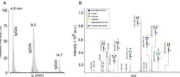
After fast 15-min nano-LC separation (Fig. 2A; Suppl. Figs. 3 and 4), spectra processing and integration, four, nine and three glycoforms for IgG2a, IgG2b and IgG2c (Fig. 3), respectively, met the criteria for manual data curation (see Section 2.13). Other glycoforms were either undetected or below the quantification limit (Fig. 2B, Suppl. Fig. 2). All three rat IgG subclasses featured exclusively core-fucosylated mono- and biantennary structures. An umbrella feature in each subclass were abundant glycoforms FA1, FA2 and FA2G1. Their peptide and glycan sequences were further confirmed with MS/MS. We also detected afu- cosylated, oligomannose and disialylated glycoforms but did not quan- tify them. Terminally α1,3-galactosylated glycans were not detected, leading us to the conclusion that these glycans might not be Fc-linked in rat IgG or that their proportion was below the detection limit. Addi- tionally, the only bisected structure observed on IgG2a was FA2B. IgG2c showed the least diversity, with only three glycoforms quantified, arguably because the concentration of this subclass in serum is generally low [45,46] and many glycopeptides were not detected rather than not being present (Suppl. Fig. 2). The same glycoforms were confirmed previously in a functionally homologous mouse IgG3 [8,47]. The IgG2b levels in normal rat serum are highest among other Ig isotypes [48], and this was mirrored in the highest number of glycoforms that we reliably quantified, including two digalactosylated (FA2BG2, FA2G2) and three monosialylated (FA2BG1Z1, FA2G1Z1, FA2G2Z1) structures (Fig. 3).
This subclass shares sequence homology and Fc-linked N-glycan di- versity with mouse IgG2a and IgG2b [8,48]. A single different glyco- pattern for rat IgG2b was the presence of bisected glycan FA2BG2.
To assess the technical variation of our glycoproteomic sample preparation workflow, we assessed the intra- and inter-day precision for the glycoforms quantified in cohort samples and obtained the co- efficients of variation (CV) below 12% for both parameters and for all quantified glycopeptides. Increased variation was observed for the least abundant bisected and sialylated glycoforms (Suppl. File. 5). Zaytseva et al. [9] made a similar observation when analyzing mouse IgG gly- copeptides. Higher CVs of bisected glycoforms could also be explained by the shielding effect of antennary monosaccharide residues, which preclude consistent interaction of bisecting GlcNAc with the HILIC sta- tionary phase during enrichment [48].
3.2. Chronic stress induces subclass-specific changes in rat IgG glycosylation
Heat shock proteins, acute-phase proteins, lipid peroxidation, malondialdehyde (MDA), cortisol level and various biofluid metabolites have been proposed as valuable biomarkers of stress-related conditions and diseases [49], yet stress-related changes in protein glycosylation remain largely unaddressed. Rodents have already been proven a useful animal model of chronic stress. Gokul et al. [50] found increased serum glutamic-oxaloacetic transaminase, pyruvic transaminase, MDA, corti- costerone and total antioxidant level in Wistar rats exposed to chronic unpredictable mild stress (CUMS) that developed comorbid depression accompanied by an inflammatory response. A different, glycomics- centered study of CUMS in BALB/c mice revealed altered N-glycan patterns of serum glycome [51]. Specifically, a potential biomarker of CUMS-induced depression was reported to be the disialylated glycans (A2G2S2 and A2G2Z2) ratio, signalizing changes in sialic acids expression. These discoveries indicate that protein post-translational modifications are affected by chronic stress, allowing the possibility that stress triggers changes in rat IgG glycosylation.
In our study, chronic stress affected rat IgG Fc-linked glycosylation only in IgG2a and IgG2c for young females and in IgG2b for old females (Fig. 4, Table 1, Suppl. File 4). The most notable glycofeature
differentiating between control and stress groups was altered gal- actosylation. IgG2a exhibited an increased level of agalactosylated biantennary glycoform FA2, while monogalactosylated FA2G1 showed a strong negative group effect. Monoantennary glycoform FA1 was also in decline, probably because it is a substrate to which a second GlcNAc residue on a Man α1,6 arm of the glycan tri-mannosyl core is attached, yielding FA2. A similar situation was observed on IgG2c, where FA1 decrease was the only significant quantified change. Although a higher expected level of biantennary FA2 was not found to exhibit any signif- icant group effect for this subclass, we cannot exclude its importance because it represents a logical step in the N-glycan processing pathway leading to a synthesis of core-fucosylated and galactosylated glycan structures. A positive group effect for IgG2b FA2G2 digalactosylated glycoform was the only quantified glycofeature in old female rats, emphasizing contrasts in IgG subclass-specific glycosylation.
In healthy humans, IgG galactosylation depends on age, with high inter-population variability; the relative number of agalactosylated structures declines slowly, reaching a minimum at around 25 years of age and then increases again [14,52]. In acute systemic inflammation, rapid changes in IgG galactosylation resembling “on-and-off mode” were described [14,53]. Low total IgG galactosylation was traditionally believed to indicate a pro-inflammatory state [54,55], although it is now known that not only pathogenic but also bulk endogenous antibody Fig. 2. Chromatographic separation of rat IgG-Fc glycoforms for high-throughput glycoprofiling. (A) Representative base peak chromatogram of rat IgG glyco- peptides forming three peak clusters for each subclass upon reversed-phase nano-LC separation in a fast 15-min gradient. (B) Representative summed mass spectrum (4.3–5.1 min) of rat IgG2a peak cluster with annotated doubly, [M +2H]2+, or triply, [M +3H]3+, charged glycopeptide ions. For corresponding summed spectra of rat IgG2b and IgG2c glycopeptide peak clusters, refer to Suppl. Fig. 2.
Fig. 3. Relative site-specific abundances of rat IgG2a (A), IgG2b (B) and IgG2c (C) glycoforms in the analyzed chronic stress cohort samples. Boxes represent the interquartile range between 25th and 75th percentile, median (solid line) and mean (cross) values. Whiskers represent the minimum and maximum values. Tryptic peptide sequences with an N-glycosylation site marked in red are given below each panel.
galactosylation levels affect binding to FcγRs and C1q, which disputes the previous oversimplified concepts. Studies where the opposite was found to be true [56–58] verify that antibody galactosylation effect on complement activation is not as straightforward as previously believed.
In our chronic stress protocol, young females generally exhibited a low- galactose IgG glycosylation pattern, which points to a potentially increased immune response. In addition, at the end of the study young animals were 6 months old, which translates to adolescence in humans (15–20 years) [59], characterized by a relatively high agalactosylated glycan profile [14,48]. Therefore, an alternative interpretation would be that chronic stress relatively delays the maturation of the IgG glycome in female animals.
IgG galactosylation is also affected by estrogens and other sex hor- mones. Menopausal increase in agalactosylation, reflected primarily through high FA2 levels [60], confirms the existence of a hormonal route of IgG glycosylation and immune response modulation. This possibly explains why galactosylation changes we observed were pro- nounced in female rats only. Reproductive senescence of female Sprague Dawley rats starts at 8–10 months of age and is characterized by a decreased number of developing follicles and changes in estrous cyclcity [59,61,62]. Thus, the group of old females (12 months) used in our study
resembles the state of premenopause. Interestingly, chronically stressed animals had a higher level of FA2G2 compared with the control group.
Since in all three subclasses sialylation differences between stressed and control groups were not significant, we can only speculate that this in- crease in FA2G2 level on IgG2b was to be followed by an increase in anti- inflammatory sialylated glycoforms, but without conclusive confirma- tion. This might explain why mature females responded to stress differently and experienced a consequent overall increase in IgG2b galactosylation, suggesting an anti-inflammatory response.
While IgG glycosylation in young rats points to a pro-inflammatory pattern, indicating probable IgG glycan profile immaturity, older pre- menopausal rats showed an anti-inflammatory pattern as well, charac- teristic for a younger and reproductively capable organism still protected by sufficient sex hormone levels. In this group we expected an increase in agalactosylated glycoforms characteristic for human meno- pause. Therefore, to properly translate the rat chronic stress model to the human system in terms of age, future studies should be performed on older animals.
Specific stressors can also induce different levels of response in males and females, which might explain the observed sex-related differences [63]. Furthermore, aged rodents might have an impaired stress Fig. 4. Chronic stress-related differences in five glycoforms between control and stress groups of female rats across three timepoints. Plasma sampling: BASE =one day before protocol start, END =end of 3rd stress cycle, SAC =after sacrificing. Relative abundances and standard deviations for all the glycoforms quantified are listed in the Suppl. File 5.
Table 1
Five IgG glycoforms had significant positive or negative group effects estimated using the LMM approach in female chronically stressed rats. Standard error (SE) – estimated standard deviation of the sampling distribution of the estimated effect. For glycan symbol nomenclature, refer to Fig. 2B.
IgG subclass Glycoform Depiction Age Sex Group effect Group SE p-value Adjusted p-value
IgG2a FA1 Young Female −1.301 0.377 3.54E-03 4.90E-02
IgG2a FA2 Young Female 2.006 0.497 7.49E-04 1.70E-02
IgG2a FA2G1 Young Female −2.318 0.509 5.08E-05 3.45E-03
IgG2b FA2G2 Old Female 1.861 0.504 6.14E-04 1.70E-02
IgG2c FA1 Young Female −2.021 0.573 3.60E-03 4.90E-02
perception because of sensorial loss [64], and the effect of some stressors is likely to be diminished.
3.3. Hypothalamic-pituitary-adrenal (HPA) axis activation
Glycosylation has all the necessary characteristics of an efficient stress response system – glycans can be modified extensively and quickly in a non-template-driven mode. The importance of glycosylation in stress response is visible in the fact that chronic stress affects lymphocyte frequency and spleen morphology, evident as an expansion of the red pulp compartment and involution of the white pulp, with a loss of the prominent marginal zone. Specifically, chronic stress increases mouse T lymphocyte frequency, reduces B lymphocyte frequency, and increases B-lymphocyte irregularity in the spleen [65,66]. However, the role of spleen B lymphocytes in the systemic immune response remains unknown.
The chronic stress protocol (Suppl. File 1) in our study was evaluated at the level of the adrenal gland in both sexes and two age groups. For this purpose, expressions of CYP11B1 and GR in the fascicular layer of the adrenal gland were quantified by immunofluorescence (Suppl.
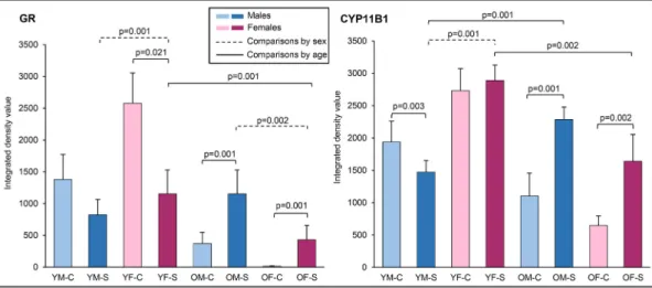
Fig. 1). A three-way factorial ANOVA revealed that the interaction of three independent variables (age, sex and chronic stress exposure) contributed significantly (F(1,54) =7.881, p <0.007) to CYP expression (Fig. 5), as well as the interactions of stress and age (F(1,54) =73.118, p
<0.001) and age and sex (F(1,54) =130.391, p <0.001). Age (F(1,54) = 133.397, p <0.001), sex (F(1,54) =14.616, p <0.001) and chronic stress exposure (F(1,54) =41.249, p <0.001) also showed statistically signifi- cant main effects.
Three-way factorial ANOVA for GR expression in the adrenal gland (Fig. 5) yielded a significant interaction between stress and age (F(1,54)
=92.058, p <0.001) and between age and sex (F(1,54) =140.916, p <
0.001). We also observed significant main effects of stress (F(1,54) = 11.021, p =0.002) and age (F(1,54) =296.485, p <0.001). These results indicate that the chronic stress protocol activated the HPA axis, with age-specific and sex-specific patterns.
Chronically stressed young animals of both sexes had similar or lower levels of CYP11B1 and GR expression, while old animals had a higher expression of both proteins. Typically, chronic stress exposure increases glucocorticoid levels and should down-regulate CYP11B1 and GR [5,6,67]. In our study, the stress protocol ended with a two-week
recovery phase. During that time, stress response in young rats decreased, resulting in a lower expression of both proteins – a good sign of self-limiting stress response. In older rats, on the other hand, a high stress response was maintained, which is a maladaptive strategy, possibly inducing low-grade inflammation.
We also observed a higher CYP11B1 and GR expression in young females in comparison with young males. Old females, in comparison with old males, showed a trend towards lower expression levels of both proteins. These findings indicate a sex-specific difference in stress response, which may be mediated by sex hormone levels or some other sex-specific mechanism [7,8].
4. Conclusions
We designed and validated a simple high-throughput glyco- proteomic workflow for rat IgG Fc N-glycosylation analysis and demonstrated its applicability and performance on a cohort of male and female rats of different age exposed to chronic stress. The subclass- specific analysis exhibited satisfactory repeatability and reproduc- ibility and revealed N-glycosylation diversity of rat IgG2a, IgG2b and IgG2c subclasses.
The most prominent glycofeatures of animals exposed to chronic stress were galactosylation alterations. IgG2a exhibited an increased level of agalactosylated biantennary glycoforms and decreased levels of its monoantennary precursors, as well as monogalactosylated glyco- forms. Together with a decline in IgG2c galactosylation, these effects indicated a pro-inflammatory state in young females. Conversely, old females showed an increased digalactosylation on IgG2b, exhibiting an increase in galactosylation and consequently the anti-inflammatory ac- tivity of IgG. Even though old animals – especially females – were incapable of coping with stress, a reaction specific for pre- and post- menopausal age, their IgG glycan profile did not show an expected pro-inflammatory pattern. This suggested that animals in our study were still not old enough and might have been at least partially protected by sex hormones. The observed glycofeatures indicate an important effect of chronic stress on IgG N-glycosylation, which may trigger low-grade inflammation and highlight the sex-specific differences in rat IgG sub- class glycosylation.
Despite the overfrequent use of rats as animal models, their appli- cation to human biology is not straightforward and remains challenging.
Fig. 5. The quantitative analysis of GR (left) and CYP11B1 (right) immunoreactivity using integrated density value revealed significant sex-specific changes in the expression of both proteins upon chronic stress and aging. Significant differences are marked by lines, with exact p-values indicated below or above the lines. Black solid lines indicate significant differences in the expression of both between the animals of the same sex, while dashed-dotted lines indicate significant differences between sexes. Dashed lines represent significant age-specific differences in the expression of both proteins. Three-way factorial ANOVA was used for statistical comparison of different animal groups, with significant p-values for post hoc test shown. Abbreviations: GR =glucocorticoid receptor, OM-C =control group of old males, OM-S =stressed group of old males, OF-C =control group of old females, OF-S =stressed group of old females, YM-C =control group of young males, YM-S = stressed group of young males, YF-C =control group of young females, YF-S =stressed group of young females.
glycosylation related to chronic stress will contribute to the growing field of glycoimmunology and glycan biomarker discovery.
Data availability statement
The following datasets related to this paper are publicly available and stored in Mendeley Data cloud repository (DOI: 10.17632/h3z p6mfxdx.5): (i) batch-corrected glycoproteomic data, (ii) raw glyco- proteomic data extracted using LaCy Tools processing package, (iii) summed mass spectra of rat IgG2a/b/c peak clusters and (iv) MaxQuant search results for protein L eluate peptide mapping.
Funding
This work was supported by the Scientific center of excellence for personalized healthcare (ZCI) [grant number KK.01.1.1.01.0010]; the Croatian Science Foundation [grant number IP-2014-09-2324]; the Cedars-Sinai Medical Center’s International Research and Innovation in Medicine Program; and the Association for Regional Cooperation in the Fields of Health, Science and Technology (RECOOP HST Association) [grant numbers RECOOP #001 BMYS RG, #8220 and #8221].
Declaration of Competing Interest
Professor Gordan Lauc is a founder and CEO of Genos Ltd., a pri- vately held company specialized in commercial high throughput glyco (proteo)mics. SH, FV and MN are employees of Genos Ltd. Other authors declare no competing interest.
Appendix A. Supplementary data
Supplementary data to this article can be found online at https://doi.
org/10.1016/j.jprot.2021.104293.
References
[1] I. Trbojevi´c-Akmaˇci´c, M. Vilaj, G. Lauc, High-throughput analysis of immunoglobulin G glycosylation, Expert Rev. Proteomics 13 (2016) 523–534, https://doi.org/10.1080/14789450.2016.1174584.
[2] S. B¨ohm, I. Schwab, A. Lux, F. Nimmerjahn, The role of sialic acid as a modulator of the anti-inflammatory activity of IgG, Semin. Immunopathol. (2012) 443–453, https://doi.org/10.1007/s00281-012-0308-x.
[3] Y. Kaneko, F. Nimmerjahn, J.V. Ravetch, Anti-inflammatory activity of immunoglobulin G resulting from Fc sialylation, Science (80-.) 313 (2006) 670–673, https://doi.org/10.1126/science.1129594.
[4] A. Schaffert, M. Hani´c, M. Novokmet, O. Zaytseva, J. Kriˇsti´c, A. Lux, L. Nitschke, M. Peipp, M. Pezer, R. Hennig, E. Rapp, G. Lauc, F. Nimmerjahn, Minimal B cell extrinsic IgG glycan modifications of pro- and anti-inflammatory IgG preparations in vivo, Front. Immunol. 10 (2020), https://doi.org/10.3389/fimmu.2019.03024.
[5] P.M. Iannaccone, H.J. Jacob, Rats!, DMM Dis, Model. Mech. 2 (2009) 206–210, https://doi.org/10.1242/dmm.002733.
[6] Y. Yoshida, J. Furukawa, S. Naito, K. Higashino, Y. Numata, Y. Shinohara, Quantitative analysis of total serum glycome in human and mouse, Proteomics 16 (2016) 2747–2758, https://doi.org/10.1002/pmic.201500550.
[7] J. Kriˇsti´c, O.O. Zaytseva, R. Ram, Q. Nguyen, M. Novokmet, F. Vuˇckovi´c, M. Vilaj, I. Trbojevi´c-Akmaˇci´c, M. Pezer, K.M. Davern, G. Morahan, G. Lauc, Profiling and genetic control of the murine immunoglobulin G glycome, Nat. Chem. Biol. 14 (2018) 516–524, https://doi.org/10.1038/s41589-018-0034-3.
[8] N. de Haan, K.R. Reiding, J. Kriˇsti´c, A.L.H. Ederveen, G. Lauc, M. Wuhrer, The N- glycosylation of mouse immunoglobulin G (IgG)-fragment crystallizable differs between IgG subclasses and strains, Front. Immunol. 8 (2017) 31, https://doi.org/
10.3389/fimmu.2017.00608.
[9] O.O. Zaytseva, B.C. Jansen, M. Hani´c, M. Mrˇcela, G. Razdorov, R. Stojkovi´c, J. Erhardt, I. Brizi´c, S. Jonji´c, M. Pezer, G. Lauc, MIgGGly (mouse IgG glycosylation analysis) - a high-throughput method for studying Fc-linked IgG N-glycosylation in mice with nanoUPLC-ESI-MS, Sci. Rep. 8 (2018) 13688, https://doi.org/10.1038/
s41598-018-31844-1.
nih.gov/10764836/ (accessed July 22, 2020).
[12] G.A. Medgyesi, K. Miklos, J. Kulics, G. Füst, J. Gergely, H. Bazin, Classes and ´ subclasses of rat antibodies: reaction with the antigen and interaction of the complex with the complement system, Immunology 43 (1981) 171–176. htt p://www.ncbi.nlm.nih.gov/pubmed/7251048 (accessed July 23, 2020).
[13] C. C´etre, C. Pierrot, C. Cocude, S. Lafitte, A. Capron, M. Capron, J. Khalife, Profiles of Th1 and Th2 cytokines after primary and secondary infection by Schistosoma mansoni in the semipermissive rat host, Infect. Immun. 67 (1999) 2713–2719, https://doi.org/10.1128/iai.67.6.2713-2719.1999.
[14] I. Gudelj, G. Lauc, M. Pezer, Immunoglobulin G glycosylation in aging and diseases, Cell. Immunol. 333 (2018) 65–79, https://doi.org/10.1016/j.
cellimm.2018.07.009.
[15] M.A. Lehrman, Stimulation of N-linked glycosylation and lipid-linked
oligosaccharide synthesis by stress responses in metazoan cells, Crit. Rev. Biochem.
Mol. Biol. 41 (2006) 51–75, https://doi.org/10.1080/10409230500542575.
[16] G. Lauc, S. Dabelic, S. Supraha, M. Flogel, Glycobiology of stress, Ann. N. Y. Acad.
Sci. 851 (1998) 397–403, https://doi.org/10.1111/j.1749-6632.1998.tb09013.x.
[17] H.M. Lagraauw, J. Kuiper, I. Bot, Acute and chronic psychological stress as risk factors for cardiovascular disease: insights gained from epidemiological, clinical and experimental studies, Brain Behav. Immun. 50 (2015) 18–30, https://doi.org/
10.1016/j.bbi.2015.08.007.
[18] K. Bisht, K. Sharma, M.`E. Tremblay, Chronic stress as a risk factor for Alzheimer’s disease: roles of microglia-mediated synaptic remodeling, inflammation, and oxidative stress, Neurobiol. Stress 9 (2018) 9–21, https://doi.org/10.1016/j.
ynstr.2018.05.003.
[19] B. Brzozowski, A. Mazur-Bialy, R. Pajdo, S. Kwiecien, J. Bilski, M. Zwolinska- Wcislo, T. Mach, T. Brzozowski, Mechanisms by which stress affects the experimental and clinical inflammatory bowel disease (IBD): role of brain-gut Axis, Curr. Neuropharmacol. 14 (2016) 892–900, https://doi.org/10.2174/
1570159x14666160404124127.
[20] M. Murata, Inflammation and cancer, Environ. Health Prev. Med. 23 (2018) 50, https://doi.org/10.1186/s12199-018-0740-1.
[21] G.P. Chrousos, The concepts of stress and stress system disorders. Overview of physical and behavioral homeostasis, JAMA (1992) 1244–1252.
[22] K.E. Wellen, G.S. Hotamisligil, Inflammation, stress, and diabetes, J. Clin. Invest.
115 (2005) 1111–1119, https://doi.org/10.1172/jci25102.
[23] M. Fioranelli, A.G. Bottaccioli, F. Bottaccioli, M. Bianchi, M. Rovesti, M.G. Roccia, Stress and inflammation in coronary artery disease: a review
psychoneuroendocrineimmunology-based, Front. Immunol. 9 (2018), https://doi.
org/10.3389/fimmu.2018.02031.
[24] E.S. Miller, C.G. Apple, K.B. Kannan, Z.M. Funk, J.M. Plazas, P.A. Efron, A.
M. Mohr, Chronic stress induces persistent low-grade inflammation, Am. J. Surg.
218 (2019) 677–683, https://doi.org/10.1016/j.amjsurg.2019.07.006.
[25] R.G. Hunter, Epigenetic effects of stress and corticosteroids in the brain, Front.
Cell. Neurosci. (2012) 1–20, https://doi.org/10.3389/fncel.2012.00018.
[26] M. Konjevod, L. Tudor, D. Svob Strac, G. Nedic Erjavec, C. Barbas, N. Zarkovic, M. Nikolac Perkovic, S. Uzun, O. Kozumplik, G. Lauc, N. Pivac, Metabolomic and glycomic findings in posttraumatic stress disorder, Prog. Neuro Psychopharmacol.
Biol. Psychiatry 88 (2019) 181–193, https://doi.org/10.1016/j.
pnpbp.2018.07.014.
[27] C.L. Fazekas, E. Sipos, T. Klaric, B. T¨or¨ok, M. Bellardie, G. Nedic Erjave, M. Nikolac Perkovic, G. Lauc, N. Pivac, D. Zelena, Searching for glycomic biomarkers for predicting resilience and vulnerability in a rat model of posttraumatic stress disorder, Stress (2020) 1–30, https://doi.org/10.1080/10253890.2020.1795121.
[28] S.O. Reber, D.A. Slattery, Editorial: using stress-based animal models to understand the mechanisms underlying psychiatric and somatic disorders, Front. Psychiatry 7 (2016) 192, https://doi.org/10.3389/fpsyt.2016.00192.
[29] V.K. Patchev, A.V. Patchev, Experimental models of stress, Dialogues Clin.
Neurosci. 8 (2006) 417–432. www.dialogues-cns.org (accessed July 22, 2020).
[30] A. Sequeira-Cordero, A. Salas-Bastos, J. Fornaguera, J.C. Brenes, Behavioural characterisation of chronic unpredictable stress based on ethologically relevant paradigms in rats, Sci. Rep. 9 (2019) 1–21, https://doi.org/10.1038/s41598-019- 53624-1.
[31] K. Chapman, M. Holmes, J. Seckl, 11β-hydroxysteroid dehydrogenases intracellular gate-keepers of tissue glucocorticoid action, Physiol. Rev. 93 (2013) 1139–1206, https://doi.org/10.1152/physrev.00020.2012.
[32] S.G. Sotocinal, R.E. Sorge, A. Zaloum, A.H. Tuttle, L.J. Martin, J.S. Wieskopf, J.C.
S. Mapplebeck, P. Wei, S. Zhan, S. Zhang, J.J. McDougall, O.D. King, J.S. Mogil, The Rat Grimace Scale: A partially automated method for quantifying pain in the laboratory rat via facial expressions, Mol. Pain 7 (2011), https://doi.org/10.1186/
1744-8069-7-55.
[33] J. Schindelin, I. Arganda-Carreras, E. Frise, V. Kaynig, M. Longair, T. Pietzsch, S. Preibisch, C. Rueden, S. Saalfeld, B. Schmid, J.Y. Tinevez, D.J. White, V. Hartenstein, K. Eliceiri, P. Tomancak, A. Cardona, Fiji: an open-source platform for biological-image analysis, Nat. Methods 9 (2012) 676–682, https://doi.org/
10.1038/nmeth.2019.
[34] A. Ceroni, K. Maass, H. Geyer, R. Geyer, A. Dell, S.M. Haslam, GlycoWorkbench: a tool for the computer-assisted annotation of mass spectra of glycans, J. Proteome Res. 7 (2008) 1650–1659, https://doi.org/10.1021/pr7008252.
[35] B.C. Jansen, D. Falck, N. De Haan, A.L. Hipgrave Ederveen, G. Razdorov, G. Lauc, M. Wuhrer, LaCyTools: a targeted liquid chromatography-mass spectrometry data processing package for relative quantitation of glycopeptides, J. Proteome Res. 15 (2016) 2198–2210, https://doi.org/10.1021/acs.jproteome.6b00171.
[36] D. Falck, B.C. Jansen, N. de Haan, M. Wuhrer, High-throughput analysis of IgG Fc glycopeptides by LC-MS, in: Methods Mol. Biol., Humana Press Inc., 2017, pp. 31–47, https://doi.org/10.1007/978-1-4939-6493-2_4.
[37] M.C. Chambers, B. MacLean, R. Burke, D. Amodei, D.L. Ruderman, S. Neumann, L. Gatto, B. Fischer, B. Pratt, J. Egertson, K. Hoff, D. Kessner, N. Tasman, N. Shulman, B. Frewen, T.A. Baker, M.Y. Brusniak, C. Paulse, D. Creasy, L. Flashner, K. Kani, C. Moulding, S.L. Seymour, L.M. Nuwaysir, B. Lefebvre, F. Kuhlmann, J. Roark, P. Rainer, S. Detlev, T. Hemenway, A. Huhmer, J. Langridge, B. Connolly, T. Chadick, K. Holly, J. Eckels, E.W. Deutsch, R.
L. Moritz, J.E. Katz, D.B. Agus, M. MacCoss, D.L. Tabb, P. Mallick, A cross-platform toolkit for mass spectrometry and proteomics, Nat. Biotechnol. 30 (2012) 918–920, https://doi.org/10.1038/nbt.2377.
[38] K.B. Chandler, N. Mehta, D.R. Leon, T.J. Suscovich, G. Alter, C.E. Costello, Multi- isotype glycoproteomic characterization of serum antibody heavy chains reveals isotype-and subclass-specific n-glycosylation profiles, Mol. Cell. Proteomics 18 (2019) 686–703, https://doi.org/10.1074/mcp.RA118.001185.
[39] M.E. Sonneveld, C.A.M. Koeleman, H.R. Plomp, M. Wuhrer, C.E. van der Schoot, G. Vidarsson, Fc-glycosylation in human IgG1 and IgG3 is similar for both total and anti-red-blood cell anti-k antibodies, Front. Immunol. 9 (2018) 31, https://doi.org/
10.3389/fimmu.2018.00129.
[40] E. Maverakis, K. Kim, M. Shimoda, M.E. Gershwin, F. Patel, R. Wilken, S. Raychaudhuri, L.R. Ruhaak, C.B. Lebrilla, Glycans in the immune system and the altered glycan theory of autoimmunity: a critical review, J. Autoimmun. 57 (2015) 1–13, https://doi.org/10.1016/j.jaut.2014.12.002.
[41] G. Rodrigo, M. Gruvegård, J. Van Alstine, Antibody fragments and their purification by protein L affinity chromatography, Antibodies 4 (2015) 259–277, https://doi.org/10.3390/antib4030259.
[42] M. De Chˆateau, B.H.K. Nilson, M. Erntell, E. Myhre, C.G.M. Magnusson, B. Åkerstr¨om, L. Bjorck, On the interaction between protein L and ¨
immunoglobulins of various mammalian species, Scand. J. Immunol. 37 (1993) 399–405, https://doi.org/10.1111/j.1365-3083.1993.tb03310.x.
[43] T. Keser, T. Pavic, G. Lauc, O. Gornik, Comparison of 2-aminobenzamide, procainamide and RapiFluor-MS as derivatizing agents for high-throughput HILIC- UPLC-FLR-MS N-glycan analysis, Front. Chem. 6 (2018) 324, https://doi.org/
10.3389/fchem.2018.00324.
[44] Y. Xue, J. Xie, P. Fang, J. Yao, G. Yan, H. Shen, P. Yang, Study on behaviors and performances of universal: N -glycopeptide enrichment methods, Analyst 143 (2018) 1870–1880, https://doi.org/10.1039/c7an02062g.
[45] J. Rousseaux, M.T. Picque, H. Bazin, G. Biserte, Rat IgG subclasses: differences in affinity to protein A-sepharose, Mol. Immunol. 18 (1981) 639–645, https://doi.
org/10.1016/0161-5890(81)90035-3.
[46] M. Johnson, Rat antibody, Mater. Methods 3 (2013), https://doi.org/10.13070/
mm.en.3.154.
[47] H. Bazin, Revival: Rat Hybridomas and Rat Monoclonal Antibodies (1990), 1st ed., CRC Press Inc., Boca Raton, FL, USA, 1990. https://www.routledge.com/Rat-Hybr idomas-and-Rat-Monoclonal-Antibodies-1990/Bazin/p/book/9781138561649 (accessed July 24, 2020).
[48] M.J. Badgett, E. Mize, T. Fletcher, B. Boyes, R. Orlando, Predicting the hilic retention behavior of the n-linked glycopeptides produced by trypsin digestion of immunoglobulin gs (Iggs), J. Biomol. Tech. 29 (2018) 98–104, https://doi.org/
10.7171/jbt.18-2904-002.
[49] K. Dhama, S.K. Latheef, M. Dadar, H.A. Samad, A. Munjal, R. Khandia, K. Karthik, R. Tiwari, M.I. Yatoo, P. Bhatt, S. Chakraborty, K.P. Singh, H.M.N. Iqbal, W. Chaicumpa, S.K. Joshi, Biomarkers in stress related diseases/disorders:
diagnostic, prognostic, and therapeutic values, Front. Mol. Biosci. 6 (2019) 91, https://doi.org/10.3389/fmolb.2019.00091.
[50] M. Gokul, N.A. Kumar, R.D. Kini, V. Blossom, B. Kodavanji, A. Noojibail, N. Murali, S.P.V. Rai, Evaluation of biomarkers of stress in chronic stress-exposed comorbid depression model Wistar rats, J. Basic Clin. Physiol. Pharmacol. 30 (2020), https://
doi.org/10.1515/jbcpp-2018-0215.
[51] M.E. Mahmoud, I.F. Rehan, K. El-Dawy Ahmed, A. Abdelrahman, S. Mohammadi, A.F. Abou-Elnaga, M. Youssef, H.M. Diab, D. Salman, A. Elnagar, H.H. Mohammed, O. Shanab, R.M. Ibrahim, E.K.H. Ahmed, A.E.L. Hesham, A. Gupta, Identification of serum N-glycoproteins as a biological correlate underlying chronic stress response in mice, Mol. Biol. Rep. (2019), https://doi.org/10.1007/s11033-019-04717-7.
[52] R. Parekh, I. Roitt, D. Isenberg, R. Dwek, T. Rademacher, Age-related
galactosylation of the N-linked oligosaccharides of human serum IgG, J. Exp. Med.
167 (1988) 1731–1736, https://doi.org/10.1084/jem.167.5.1731.
[53] M. Novokmet, E. Luki´c, F. Vuˇckovi´c, ˇZ. Duri´c, T. Keser, K. Rajˇsl, D. Remondini, G. Castellani, H. Gaˇsparovi´c, O. Gornik, G. Lauc, Changes in IgG and total plasma protein glycomes in acute systemic inflammation, Sci. Rep. 4 (2014) 1–10, https://
doi.org/10.1038/srep04347.
[54] I. Trbojevic Akmacic, N.T. Ventham, E. Theodoratou, F. Vuˇckovi´c, N.A. Kennedy, J. Kriˇsti´c, E.R. Nimmo, R. Kalla, H. Drummond, J. ˇStambuk, M.G. Dunlop, M. Novokmet, Y. Aulchenko, O. Gornik, H. Campbell, M. Puˇci´c Bakovi´c, J. Satsangi, G. Lauc, Inflammatory bowel disease associates with proinflammatory potential of the immunoglobulin G glycome, Inflamm. Bowel Dis. 21 (2015) 1237–1247, https://doi.org/10.1097/MIB.0000000000000372.
[55] F. Vuˇckovïc, J. Kriˇstïc, I. Gudelj, M. Teruel, T. Keser, M. Pezer, M. Puˇcïc-Bakovïc, J.
ˇStambuk, I. Trbojevïc-Akmaˇcïc, C. Barrios, T. Pavïc, C. Menni, Y. Wang, Y. Zhou, L.
Cui, H. Song, Q. Zeng, X. Guo, B.A. Pons-Estel, P. McKeigue, A. Leslie Patrick, O.
Gornik, T.D. Spector, M. Harjaˇcek, M. Alarcon-Riquelme, M. Molokhia, W. Wang, G. Lauc, Association of systemic lupus erythematosus with decreased
immunosuppressive potential of the IgG glycome, Arthritis Rheumatol. 67 (2015) 2978–2989. doi:https://doi.org/10.1002/art.39273.
[56] G. Dekkers, T. Rispens, G. Vidarsson, Novel concepts of altered immunoglobulin G galactosylation in autoimmune diseases, Front. Immunol. 9 (2018), https://doi.
org/10.3389/fimmu.2018.00553.
[57] I. Quast, C.W. Keller, M.A. Maurer, J.P. Giddens, B. Tackenberg, L.X. Wang, C. Münz, F. Nimmerjahn, M.C. Dalakas, J.D. Lünemann, Sialylation of IgG Fc domain impairs complement-dependent cytotoxicity, J. Clin. Invest. 125 (2015) 4160–4170, https://doi.org/10.1172/JCI82695.
[58] B. Peschke, C.W. Keller, P. Weber, I. Quast, J.D. Lünemann, Fc-galactosylation of human immunoglobulin gamma isotypes improves C1q binding and enhances complement-dependent cytotoxicity, Front. Immunol. 8 (2017), https://doi.org/
10.3389/fimmu.2017.00646.
[59] Pallav Sengupta, The laboratory rat: relating its age with human’s, Int. J. Prev.
Med. 4 (2013) 624–630. /pmc/articles/PMC3733029/?report=abstract (accessed September 27, 2020).
[60] A. Ercan, W.M. Kohrt, J. Cui, K.D. Deane, M. Pezer, E.W. Yu, J.S. Hausmann, H. Campbell, U.B. Kaiser, P.M. Rudd, G. Lauc, J.F. Wilson, J.S. Finkelstein, P.
A. Nigrovic, Estrogens regulate glycosylation of IgG in women and men, JCI Insight 2 (2017), https://doi.org/10.1172/jci.insight.89703.
[61] R.D. Brinton, Minireview: translational animal models of human menopause:
challenges and emerging opportunities, Endocrinology 153 (2012) 3571–3578, https://doi.org/10.1210/en.2012-1340.
[62] G. Cruz, D. Fernandois, A.H. Paredes, Ovarian function and reproductive senescence in the rat: role of ovarian sympathetic innervation, Reproduction 153 (2017) R59–R68, https://doi.org/10.1530/REP-16-0117.
[63] Y. Sze, P.J. Brunton, Sex, stress and steroids, Eur. J. Neurosci. 52 (2020) 2487–2515, https://doi.org/10.1111/ejn.14615.
[64] A. Novais, S. Monteiro, S. Roque, M. Correia-Neves, N. Sousa, How age, sex and genotype shape the stress response, Neurobiol. Stress 6 (2017) 44–56, https://doi.
org/10.1016/j.ynstr.2016.11.004.
[65] M. Kadmiel, J.A. Cidlowski, Glucocorticoid receptor signaling in health and disease, Trends Pharmacol. Sci. 34 (2013) 518–530, https://doi.org/10.1016/j.
tips.2013.07.003.
[66] L. Guo, Y.X. Chen, Y.T. Hu, X.Y. Wu, Y. He, J.L. Wu, M.L. Huang, M. Mason, A.
M. Bao, Sex hormones affect acute and chronic stress responses in sexually dimorphic patterns: consequences for depression models,
Psychoneuroendocrinology 95 (2018) 34–42, https://doi.org/10.1016/j.
psyneuen.2018.05.016.
[67] R. Pasquali, The hypothalamic-pituitary-adrenal axis and sex hormones in chronic stress and obesity: pathophysiological and clinical aspects, Ann. N. Y. Acad. Sci.
1264 (2012) 20–35, https://doi.org/10.1111/j.1749-6632.2012.06569.x.