https://doi.org/10.1007/s00392-021-01809-y REVIEW
Pathomechanisms and therapeutic opportunities in radiation‑induced heart disease: from bench to bedside
Márta Sárközy1 · Zoltán Varga2 · Renáta Gáspár1 · Gergő Szűcs1 · Mónika G. Kovács1 · Zsuzsanna Z. A. Kovács1 · László Dux3 · Zsuzsanna Kahán2 · Tamás Csont1
Received: 29 October 2020 / Accepted: 16 January 2021
© The Author(s) 2021
Abstract
Cancer management has undergone significant improvements, which led to increased long-term survival rates among cancer patients. Radiotherapy (RT) has an important role in the treatment of thoracic tumors, including breast, lung, and esophageal cancer, or Hodgkin’s lymphoma. RT aims to kill tumor cells; however, it may have deleterious side effects on the surrounding normal tissues. The syndrome of unwanted cardiovascular adverse effects of thoracic RT is termed radiation-induced heart disease (RIHD), and the risk of developing RIHD is a critical concern in current oncology practice. Premature ischemic heart disease, cardiomyopathy, heart failure, valve abnormalities, and electrical conduct defects are common forms of RIHD.
The underlying mechanisms of RIHD are still not entirely clear, and specific therapeutic interventions are missing. In this review, we focus on the molecular pathomechanisms of acute and chronic RIHD and propose preventive measures and pos- sible pharmacological strategies to minimize the burden of RIHD.
Keywords Onco-cardiology · Radiation heart sequelae · Molecular pathomechanisms of radiation-induced heart disease · Prevention and therapy of radiation-induced heart disease
Introduction
Cardiovascular and cancerous diseases are the leading causes of mortality worldwide [1, 2]. The most common cancerous diseases are thoracic malignancies, including lung and breast cancers among adults [2]. Recently, cancer management has
undergone significant improvement, leading to increased long- term survival rates among cancer survivors. Consequently, age-related chronic diseases and cardiovascular risk factors, including hypertension, diabetes mellitus, hyperlipidemia, chronic kidney disease, and smoking, are often aggravated by chronic side effects of multimodality cancer therapy, accelerat- ing the progression of atherosclerosis and increasing the bur- den of cardiovascular diseases (CVDs) in cancer survivors [3].
Zsuzsanna Kahán and Tamás Csont authors contributed equally to the manuscript.
* Márta Sárközy
sarkozy.marta@med.u-szeged.hu
* Zsuzsanna Kahán
kahan.zsuzsanna@med.u-szeged.hu
* Tamás Csont
csont.tamas@med.u-szeged.hu Zoltán Varga
varga.zoltan@med.u-szeged.hu Renáta Gáspár
gaspar.renata@med.u-szeged.hu Gergő Szűcs
szucs.gergo@med.u-szeged.hu Mónika G. Kovács
kovacs.monika.gabriella@med.u-szeged.hu
Zsuzsanna Z. A. Kovács
kovacs.zsuzsanna@med.u-szeged.hu László Dux
dux.laszlo@med.u-szeged.hu
1 MEDICS Research Group, Department of Biochemistry, Interdisciplinary Center of Excellence, University of Szeged, Szeged 6720, Hungary
2 Department of Oncotherapy, Faculty of Medicine, University of Szeged, Szeged 6720, Hungary
3 Department of Biochemistry, Faculty of Medicine, University of Szeged, Szeged H-6720, Hungary
While high-energy ionizing radiation successfully destroys cancer cells, at the same time, it may have harm- ful effects on the surrounding healthy tissues leading to various side effects [4]. RT has been frequently used in thoracic malignancies, including breast, lung, esophageal cancer, thymoma, and Hodgkin’s lymphoma, which could be in close anatomical proximity to the heart [5]. Depending on the RT technique and dose, the heart may be at risk of being exposed to ionizing radiation resulting in radiogenic sequelae in a dose-dependent manner [6]. The syndrome of unwanted cardiovascular side effects of thoracic RT is called radiation-induced heart disease (RIHD), and the risk of development of RIHD is a critical concern in current oncology practice. RIHD includes structural and functional abnormalities of the pericardium, coronary vessels, myo- cardium, valves, and conduction system [7, 8]. Much of our current knowledge on radiation-induced cardiovascular complications in cancer survivors is based on the patients’
data coming from the era of the 1980s or before that, with less developed RT techniques, extended RT fields, and high radiation doses [4, 9]. The earliest data indicating the pres- ence of RIHD originate from follow-up studies of lymphoma and breast cancer patients due to the high incidence and high cure rate of these diseases [10, 11]. In clinical prac- tice, the consequences of RIHD mostly emerge in breast cancer patients receiving left-sided postoperative RT and less frequently in esophageal cancer patients treated with preoperative chemo-radiotherapy [6].
The prevention or management of radiogenic CVDs has become a challenge in clinical practice since RIHD can worsen the outcome, quality of life, and health care costs in long-term cancer survivors [12, 13]. These factors have recently contributed to the emergence of a new specialty termed onco-cardiology or cardio-oncology. Unfortunately, therapeutic options for RIHD are currently insufficient.
Therefore, understanding the exact molecular mechanisms in the progression of RIHD is necessary for developing pre- ventive and therapeutic strategies without attenuating the effect of RT on cancer cells. Tailored surveillance of patients according to their risk status serves early intervention if nec- essary [12, 13].
In this review, we summarize our current knowledge on the molecular pathomechanisms of the development and progression of RIHD and overview those potential pharma- cological and other strategies that may be suitable for the prevention or management of RIHD.
Clinical manifestations of RIHD
RIHD may manifest in a broad spectrum of cardiovascu- lar complications, including acute and chronic pericarditis, ischemic heart disease (IHD), cardiomyopathy and heart
failure (HF), valvular heart disease, and cardiac conduc- tion abnormalities [4, 14] (Fig. 1). Complications typically appear years to decades after the irradiation, showing a median of 10–15 years. The overall absolute risk of cardiac death is related to the mean heart dose (MHD) of RT [9, 15].
Radiation-induced cardiovascular complications are more severe with (i) higher total radiation dose [6], (ii) extended target volume exposure with closer tumor localization to the heart [16], (iii) younger age at the radiation exposure [9, 15], (iv) longer follow-up duration [17], (v) concomitant therapy with cardiotoxic chemotherapeutic drugs such as anthracyclines and biological agents [18, 19], (vi) presence of genetic factors, and (vii) comorbidities and cardiovascular risk factors [3].
Acute forms of RIHD Acute pericarditis
Acute pericarditis could be the earliest form of RIHD [20]
(Fig. 1). Before the era of selective RT-techniques in the late 1980s, 80% of patients receiving thoracic irradiation suffered from acute pericarditis [20]. In these cases, high MHD (> 36 Gy) administered to > 30% of the heart was responsible for the development of acute exudative pericar- ditis [5, 20]. Nowadays, its occurrence is rare (< 5%) due to alertness and the modern heart-sparing RT-techniques [13, 21]. Acute pericarditis causes chest pain, fever, and ECG abnormalities. Approximately half of the affected patients suffer from hemodynamic abnormalities due to significant pericardial effusion. Even cardiac tamponade can develop necessitating intervention [20].
Acute conduction system abnormalities
In the acute phase, some patients develop mostly revers- ible asymptomatic ECG repolarization abnormalities, which rarely persists (the incidence is unknown due to the lack of screening during or shortly after RT) [14]. These events may occur in response to transitory circulatory/metabolic changes, or if irradiation induces the development of defini- tive structural changes in the conduction system (e.g., via increasing oxidative/nitrosative/nitrative stress or inflam- mation), that probably affects an irrelevant part of the heart only.
Chronic forms of RIHD Chronic pericarditis
The presence of pericardial effusion in the acute phase might predispose patients to chronic pericarditis of delayed onset from months to years [14] (Fig. 1). Certain chemotherapeutic
drugs (e.g., anthracyclines, cyclophosphamide, and bleo- mycin) may enhance the risk of radiogenic pericarditis development [20]. Up to 20% of patients develop chronic constrictive pericarditis with severe symptoms requiring pericardiectomy at a median time of 11 years post-irradi- ation [22].
Ischemic heart disease (IHD)
Our understanding concerning the burden of radiogenic IHD is based the most on epidemiological studies [6, 8, 9, 11, 15]. Nevertheless, estimations may be given using nor- mal tissue complication probability models [23]. However, prospectively collected data with careful follow-up obser- vations are needed [24]. In the various studies, different end-points, dose parameters, and statistical methods were used (Table 1). The studied populations also differed: some included all irradiated patients [8, 9, 11, 15] while others, only patients with major IHD events such as the need for coronary intervention/acute myocardial infarction (AMI) or IHD-related death [25, 26]. The risk of coronary artery dis- ease (CAD) is clearly radiation dose-dependent. The tradi- tional reference dose parameter is MHD [8, 9]. Nevertheless,
Bogaard et al. showed that among other dose parameters, the volume of the left ventricle receiving at least 5 Gy (LV- V5) is the most sensitive predictor of coronary events [27].
Most studies agree that beyond radiation dose, risk factors for CAD or the history of IHD, and young age are also deter- minants of outcome (Table 1) [8, 9, 11, 15, 27].
Breast cancer patients receiving radiation doses of < 2 Gy, 2–4 Gy, 5–9 Gy, and > 10 Gy had a dose-dependent excess risk for angina pectoris, AMI, and sudden cardiac death of 10%, 30%, 40%, and 116%, respectively [8, 9, 27] (Table 1;
Fig. 1). The risk for IHD increased linearly with the MHD by 7.4% per 1 Gy absorbed dose with no apparent lower thresh- old [8]. Similar dose–response results were found by Jacobse et al. in a nested case–control study: every 1 Gy increase in MHD was associated with a 6.4% increase in the risk of AMI; in MHD > 20 Gy cases, the risk of AMI was 3.4 times higher (Table 1) [15]. Based on modern three-dimensional conformal radiation therapy (3DCRT) dose-volume data, Bogaard et al. also confirmed these results. They introduced the LV-V5 parameter indicating a 1.7% increase in coronary events incidence by every 5 Gy increase of dose to the left ventricle [27]. Among lymphoma patients, identical results were found on MHD data and long-term AMI incidence as
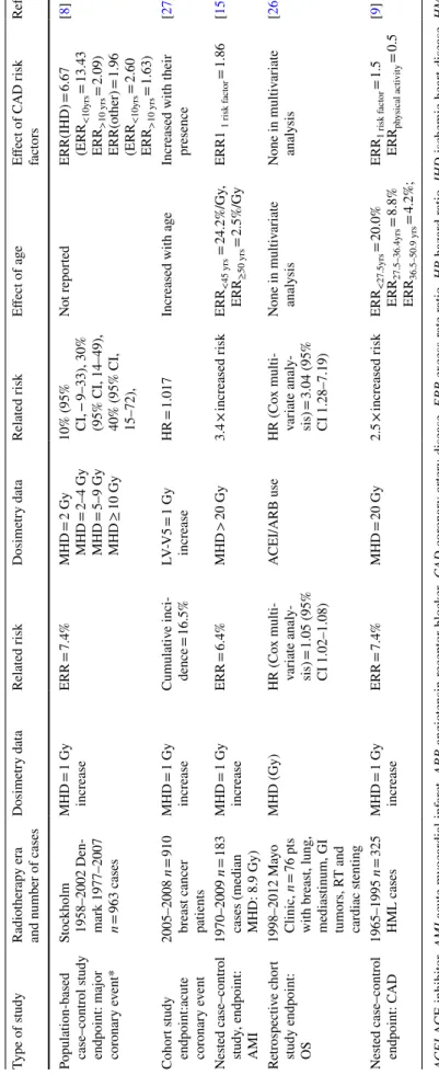
Fig. 1 Possible clinical manifestations of RIHD. RIHD is a progres- sive disease that covers a broad spectrum of cardiac pathology. RIHD may manifest as acute or chronic pericarditis, conduction system abnormalities, ischemic heart disease, cardiomyopathy, heart failure
including HFpEF and HFrEF, or valvular heart disease, according to the site of damage. LAD left descending coronary artery, LVH left ventricular hypertrophy, HFpEF heart failure with preserved ejection fraction, HFrEF heart failure with reduced ejection fraction
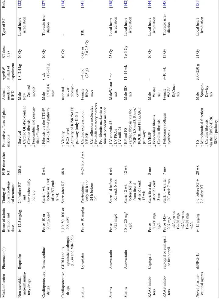
Table 1 The effect of radiation dose on the risk of coronary artery disease (CAD): studies with various dosimetry data ACEI ACE inhibitor, AMI acute myocardial infarct, ARB angiotensin receptor blocker, CAD coronary artery disease, ERR excess rate ratio, HR hazard ratio, IHD ischemic heart disease, HML Hodgkin lymphoma, LV-V5 the volume of the left ventricle receiving at least 5 Gy, MHD mean heart dose, OS overall survival, RR relative risk, *Major coronary event: AMI/coronary revascu- larization/death from IHD Type of studyRadiotherapy era and number of casesDosimetry dataRelated riskDosimetry dataRelated riskEffect of ageEffect of CAD risk factorsRefs.
Population-based case–contr
ol study endpoint: major coronary event*
Stockholm 1958–2002 Den- mark 1977–2007 n = 963 cases
MHD = 1 Gy increaseERR = 7.4%MHD = 2 Gy MHD = 2–4 Gy MHD = 5–9 Gy MHD ≥ 10 Gy
10% (95% CI,
− 9–33), 30%
(95% CI, 14–49), 40% (95% CI, 15–72),
Not reportedERR(IHD) = 6.67 (ERR<10yrs = 13.43 ERR>10 yrs = 2.09) ERR(other) = 1.96 (ERR<10yrs = 2.60 ERR>10 yrs = 1.63)
[8] Cohort study endpoint:acute coronary event
2005–2008 n = 910 breast cancer patients
MHD = 1 Gy increaseCumulative inci- dence = 16.5%LV-V5 = 1 Gy increaseHR = 1.017Increased with ageIncreased with their presence[27] Nested case–control study, endpoint: AMI
1970–2009 n = 183
cases (median MHD: 8.9 Gy)
MHD = 1 Gy increaseERR = 6.4%MHD > 20 Gy3.4 × increased riskERR<45 yrs = 24.2%/Gy, ERR≥50 yrs = 2.5%/GyERR1 1 risk factor = 1.86[15] Retrospective chort study endpoint: OS
1998–2012 Mayo Clinic, n = 76 pts with breast, lung, mediastinum, GI tumors, RT and cardiac stenting
MHD (Gy)HR (Cox multi- variate analy- sis) = 1.05 (95% CI 1.02–1.08)
ACEI/ARB useHR (Cox multi- variate analy- sis) = 3.04 (95% CI 1.28–7.19)
None in multivariate analysisNone in multivariate analysis[26] Nested case–control endpoint: CAD1965–1995 n = 325 HML casesMHD = 1 Gy increaseERR = 7.4%MHD = 20 Gy2.5 × increased riskERR<27.5yrs = 20.0% ERR27.5–36.4yrs = 8.8% ERR36.5–50.9 yrs = 4.2%;
ERR1 risk factor = 1.5 ERRphysical activity = 0.5[9]
in the classic Darby-study; in MHD > 20 Gy cases, 2.5 times higher AMI risk was detected [9]. The dose-dependent and irradiated volume-dependent nature of RIHD-related sur- vival were demonstrated in a retrospective cohort study in lymphoma patients with CAD requiring coronary interven- tion [26]. The strongest predictor of a major coronary event is left-sided RT [28, 29]. Boekel et al. showed a significantly increased risk of IHD when the chest wall or the internal mammary nodes were irradiated independent of laterality in breast cancer patients [16]. The association between the radiation dose to a cardiac segment and the injury of the affected structure was demonstrated by Taylor et al. [30]. In this study, the highest RT doses were detected in the distal part of the left anterior descending (LAD) artery [30].
Cardiomyopathy and heart failure (HF)
Radiation-induced cardiomyopathy and consequent HF are progressive multifactorial diseases, which may evolve over several years (Fig. 1). They are often aggravated by medi- cal anti-cancer therapies, concomitant radiation-induced valvular heart disease (VHD), or IHD. RT-induced cardio- myopathy and HF cover a spectrum of cardiac pathological conditions among which a typical initial phase is HF with preserved ejection fraction (HFpEF). HFpEF is character- ized by diastolic dysfunction and compensatory left ventric- ular hypertrophy (LVH) (Fig. 1). [31–34]. Later on, progres- sive interstitial fibrosis develops, separating and replacing the cardiomyocytes, which ultimately results in HF with reduced ejection fraction (HFrEF) [20, 35]. A tolerance dose of 40 Gy has been estimated for the human myocardium for the end-point of diffuse myocardial injury [20]. Indeed, dif- fuse myocardial injury is more common in patients who have received higher RT doses (> 60 Gy) and/or anthracycline chemotherapy [14, 29]. Radiation-induced cardiomyopathy is often asymptomatic. Its risk increases 5 years after RT but may develop even decades after RT [20].
Valvular heart disease (VHD)
Within the first 10 years after RT, the earliest morphological changes appear to be leaflet thickening, fibrosis, shortening and calcification, and consequent regurgitation preferentially at the mitral or aortic valves [20] (Fig. 1). The progression to fibrotic thickening and calcification of the valves may lead to stenosis, which develops mainly in the aortic valve approximately 20 years after RT [20]. The rate of VHD cor- relates better with the RT dose to the affected [36] valve than to the mediastinal dose [37]. Myocardial ischemia and hypoxia caused by IHD and fibrosis also play a role in the development of VHD that may contribute to HF [14].
Chronic conduction system abnormalities
In the chronic phase after irradiation, in about 5% of the cases, radiogenic conduction system abnormalities develop [37, 38]. Among these, bundle branch blocks and first- degree atrioventricular (AV) block occur most commonly [39], but pathological sinus node syndrome, QTc prolonga- tion, supraventricular arrhythmias, and ventricular extrasys- tole or tachycardia may also develop [14, 39, 40] (Fig. 1).
Circulatory changes such as autonomic dysfunction with tachycardia and blunted blood pressure, rarely syncope or even sudden death related to the denervation-like status of the heart may occur; (nevertheless, similar symptoms due to neck irradiation with injury of the vessels and baroreceptors should be distinguished) [38, 41, 42].
Late conduction anomalies may be explained with various pathomechanisms. Most obviously, fibrotic lesions resulted from decreased microvessel density, chronic hypoxia, com- pensatory hypertrophy are behind the abnormalities [35, 43–46]. In other cases, RT-related valvular disease and increased right atrial pressure cause atrial arrhythmias [38], or exercise-induced ischemia of the atrioventricular node due to the stenosis of the right coronary artery results in AV block [40, 47].
Pathomechanisms of RIHD
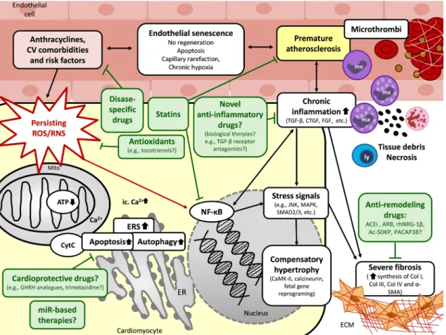
RIHD is a progressive multifactorial disease that has overlapping common and different molecular pathways in the acute and chronic phases. RT simultaneously causes damage to the macrovasculature (i.e., coronary arteries), the microvasculature, and the myocardium (i.e., diffuse injury) [7]. Key questions relating to the precise molecu- lar mechanisms of the disease progression in RIHD from acute to chronic heart diseases remained unanswered. The chain of biological events from acute to chronic forms is more likely a complex interaction between molecular pro- cesses. A substantial body of evidence suggests that the radiation-induced immediate oxidative/nitrosative/nitra- tive damage of macromolecules, including DNA, proteins, and lipids, is the initiating event in RIHD. At this early phase, the increased oxidative/nitrative/nitrosative stress triggers other biological processes, including endothelial cell injury, acute inflammation, and the various forms of cell death [35, 43, 44] (Fig. 2). In the early chronic phase of RIHD, several compensatory mechanisms, including endothelial cell proliferation and cardiac hypertrophy, develop in the sublethally damaged surviving cells [35, 43, 44] (Fig. 3). If these compensatory mechanisms are exhausted, chronic inflammatory processes, fibrosis, and endothelial senescence play the central role in the disease progression [35, 43, 44] (Fig. 3). The exact molecular
transition mechanisms and time points between the acute to compensated or decompensated chronic forms of RIHD are yet unknown. Moreover, several pathomechanisms, including oxidative/nitrative/nitrosative stress, cell death, and inflammatory processes, overlap during the acute and chronic phases of RIHD. These mechanisms could activate and potentiate each other leading to a vicious cycle in the RIHD progression. Therefore, the precise understanding of the complex interplay of these acute and chronic molecular mechanisms would help to develop strategies to counteract the progression of RIHD. In this section, we briefly sum- marize the predominating pathomechanisms in which have a role in developing the acute and chronic forms of RIHD.
Pathomechanisms in the acute phase of RIHD Mechanisms of increased oxidative/nitrosative/nitrative stress in the acute phase of RIHD
Increased oxidative/nitrosative/nitrative stress plays a cru- cial role in developing both the desired anticancerous effects and the undesired side effects of RT. Absorption of ionizing radiation used for RT may induce both direct and indirect effects in all cell types [48] (Fig. 2). Ionizing radiation can directly disrupt atomic structures, leading to further chemi- cal and biological changes [49] (Fig. 2). Approximately 80%
of the cells is water. Therefore, the initial radiation-induced
Fig. 2 Putative mechanisms in the acute phase of RIHD and potential pharmacological interventions. RT could induce immediate oxida- tive/nitrosative/nitrative damage of macromolecules, including DNA, proteins, and lipids, in all cardiac cell types. The increased oxidative/
nitrative/nitrosative stress triggers other biological processes, includ- ing acute inflammation, and cell death forms in the acute phase of RIHD in the different cell types. Parallel, hypertrophic, and fibrotic gene programs start in the surviving cardiomyocytes as compensa- tory mechanisms. Potential preventive and therapeutic pharmaco- logic agents are depicted in green boxes targeting different molecular mechanisms. ACEi angiotensin-converting enzyme inhibitors, Ac- SDKP N-acetyl-Ser-Asp-Lys-Pro, ARB angiotensin receptor block- ers, α-SMA α-smooth muscle actin, ATP adenosine triphosphate,
Ca2+ calcium ion, Col collagen, CTGF connective tissue growth factor, CytC cytochrome C, ERS endoplasmic reticulum stress, FGF fibroblast growth factor, GHRH growth hormone-releasing hormone, ICAM intercellular adhesion molecules, IL interleukin, JNK c-Jun N-terminal kinases, ly lymphocyte, ma macrophage, MAPK mitogen- activated protein kinase, miR microRNA, mo monocyte, Mito mito- chondrion, NF-κB nuclear factor-κB, ng neutrophil granulocyte, NO nitric oxide, PACAP38 pituitary adenylate cyclase-activating poly- peptide 38, PARP1poly-ADP-ribose-polymerase 1, PECAM platelet endothelial cell adhesion molecule, rhNRG-1β recombinant human neuregulin-1β, ROS/RNS reactive oxygen and nitrogen species, RT radiotherapy, TGF-β tissue growth factor-β, TNF-α tumor necrosis factor- α, VCAM vascular cell adhesion molecule
cellular damage is mostly caused by the direct radiolysis of water generating reactive species leading to indirect effects [48]. The major reactive species produced in the radiolysis of water are superoxide (O2•−), hydroxyl radical (•OH), elec- trons (e−), and hydrogen peroxide (H2O2). Organic radicals (R•) are also formed by H-abstraction reactions initiated by
•OH radicals. These carbon-centered radicals usually react rapidly with O2 to give peroxyl radicals (RO2•), which are stronger oxidizing agents than their parent radicals. The RO2• radicals can abstract H• from other molecules to form hydroperoxides (ROOH) [48]. Ionizing radiation-induced tissue injury may up-regulate inducible nitric oxide syn- thase (iNOS), thereby generating a large amount of nitric oxide (•NO), which can react with O2•− to form peroxyni- trite (ONOO−) and secondarily other reactive nitrogen spe- cies (RNS) [48–50] (Fig. 2). Other enzymatic sources for reactive oxygen species (ROS) include NADPH oxidases (NOX isoforms), lipoxygenases (LOX), cyclooxygenases (COX), peroxidases in inflammatory cells, and xanthine oxidase, which can be activated by RT-induced tissue injury [35, 48]. Ionizing radiation may also disrupt the mitochon- drial respiratory chain contributing to persistent oxidative/
nitrosative/nitrative stress. The removal of ROS/RNS during RT via the key antioxidant enzymatic systems, including, e.g., superoxide dismutase (SOD), catalase, glutathione per- oxidase, glutathione reductase, and heme oxygenase, may also be insufficient [48]. Accumulated ROS/RNS during and shortly after irradiation may cause macromolecular damage including, lipid peroxidation, protein oxidation/
nitration, inactivation of enzymes, DNA damage, interac- tion with both DNA repair enzymes (e.g., poly-ADP-ribose polymerase 1 [PARP1], p53) and transcription factors (e.g., nuclear factor-κB [NF-κB]) [14, 20, 35, 43, 48, 51] (Fig. 2).
Oxidative/nitrative stress can also induce acute inflamma- tion and cell death via different mechanisms. The macro- molecular and cellular damage may result in the activation of the inflammatory response (interleukins [IL] including, e.g., IL-1, IL-6, IL-8, IL-10, tumor necrosis factor-alpha [TNF-α], and transforming growth factor-beta [TGF-β]), stress signals (e.g., c-Jun N-terminal kinase [JNK], and p38- MAPK) or cell death (e.g., apoptosis and necrosis), and dys- regulation of autophagy. The oxidation/nitration of proteins involved in excitation–contraction coupling, contractility, Ca2+-handling, elements of the mitochondrial electron trans- port chain and Krebs cycle, metabolism, and extracellular matrix might result in acute and chronic deleterious events [14, 20, 35, 43, 48, 51] (Fig. 2).
Endothelial cell injury and acute inflammation in RIHD Endothelial cell injury is considered the primary cause of radiation damage in cardiac tissue [14, 35] (Fig. 2).
Within minutes after RT, increased vascular permeability
and vasodilation are present [5, 35, 43]. In the first few hours and days after RT, activated NF-κB [52, 53] may induce the secretion of adhesion molecules including, e.g., E-selectin [54], P-selectin [54], intercellular adhesion mol- ecule-1 (ICAM-1) [54], vascular cell adhesion molecule-1 (VCAM-1) [55], platelet endothelial cell adhesion mol- ecule-1 (PECAM-1) [55], and cytokines (e.g., IL-6, IL-8) [56, 57] in the damaged endothelial cells thereby activating leukocyte rolling, arrest, and transmigration [35, 43, 52].
The predominant inflammatory cells in the acute phase are neutrophil granulocytes. They infiltrate the endocar- dium, myocardium, and epicardium of the irradiated heart.
They are the first responders releasing pro-inflammatory cytokines (e.g., monocyte chemotactic protein [MCP], TNF- α, and IL-8) to recruit other inflammatory cells [5, 43, 59]
(Fig. 2). Recruited inflammatory cells may release further pro-inflammatory (e.g., IL-1, and IL-6) and pro-fibrotic cytokines including, e.g., TGF-β, connective tissue growth factor (CTGF), platelet-derived growth factor (PDGF), and fibroblast growth factor (FGF) [43, 58, 60–62]. Additionally, adhesion molecules, inflammatory and pro-fibrotic cytokines (e.g., ICAM-1, IL-6, and FGF) can also be produced by the microvascular endothelial cells suggesting their role in the maintenance of the pro-inflammatory state [35, 63–65].
Matrix metalloproteinases (MMPs), including MMP-1 and MMP-2, could immediately be activated by RT in endothe- lial cells, possibly via the increased oxidative/nitrosative/
nitrative stress or inflammatory mechanisms [43, 58, 66, 67]. These proteases may degrade the endothelial basement membrane, allowing effective recruitment of neutrophils and macrophages to cellular injury sites in order to phagocyte tissue debris [43, 68] (Fig. 2). The recruited inflammatory cells and the damaged endothelial cells can produce a large amount of O2•− and •NO, the latter via iNOS, which results in further ONOO− formation [58, 69]. The decreased bioa- vailability of •NO could lead to endothelial damage, vascular dysfunction, vasoconstriction, and tissue hypoxia [35]. The mechanisms described above may lead to focal endothelial denudation and endothelial dysfunction, triggering initial arteriosclerotic lesions in larger coronary arteries [35].
Increased thrombogenicity and acute inflammation in RIHD Initial endothelial damage in the microvasculature could also activate the coagulation cascade leading to fibrin deposition [43] (Fig. 2). Notably, it can be a result of (i) RT-induced endothelial damage itself [20], (ii) the thrombomodulin inhi- bition caused by TGF-β [70], and (iii) increased release of von Willebrand factor (vWF) from endothelial cells [35].
Several coagulation factors (e.g., thrombin) may induce the endothelial release of IL-8 and MCP, promoting the expres- sion of adhesion molecules and chemotaxis of neutrophil granulocytes [71]. RT can also activate COX and LOX
enzymes, which produce bioactive eicosanoids from ara- chidonic acid, including, e.g., prostaglandins, prostacyclin, thromboxanes, and leukotrienes in different cell types [72].
These bioactive molecules are well-known mediators of inflammation via vasodilation or vasoconstriction, vascular permeability, extravasation of leukocytes, and microthrom- bus formation [72]. Decreased bioavailability of •NO can lead to vasoconstriction, aggravating thrombogenicity [35].
Endoplasmic reticulum stress and apoptosis in RIHD
Irradiation can induce endoplasmic reticulum stress (ERS) and cell death in the different cell types of cardiac tissue, including cardiomyocytes, endothelial cells, fibroblasts, and cells of the conducting system [35] (Fig. 2). RT-induced irre- versible damage in the structure of cellular compartments and molecules, mitochondrial dysfunction, and ERS are the critical components in cell death pathways in RIHD [14].
During ERS, the ER is overwhelmed with incorrectly folded or unfolded proteins [73]. The protein overload induces dis- ruption of protein homeostasis and activates the unfolded protein response (UPR). The UPR leads to apoptotic cell death via three major pathways [73]. These are (i) the pro- tein kinase R-like endoplasmic reticulum kinase (PERK)- regulated, (ii) the activating transcription factor 6 (ATF6), and (iii) the inositol-requiring enzyme 1 (IRE1) pathways [74]. PERK inhibits protein translation via phosphorylation and subsequent inactivation of the eukaryotic translation initiator factor 2α (eIF2-α) to avoid further misfolded pro- tein accumulation. The ATF6 and IRE1 pathways activate transcription of genes involved in ER-associated protein degradation, protein folding, and ER membrane expansion.
IRE can also inhibit the anti-apoptotic activity of Bcl-2 and Bcl-XL [74]. After irradiation of cardiomyocytes, the stimu- lated ER releases calcium ions into the cytoplasm, leading to mitochondrial calcium overload, cytochrome-C release into the cytoplasm, and activation of the pro-apoptotic Bax [75, 76] (Fig. 2). The translocation of Bax from the cyto- plasm to the mitochondrial outer membrane induces mito- chondrial membrane permeability transition (MPT), leading to mitochondrial swelling, depolarization of the membrane, uncoupled electron transport, and oxidative phosphorylation [75] (Fig. 2).
Necrosis in RIHD
Several death initiators, signaling pathways, and effector molecules are common key mediators in both apoptosis and necrosis [77]. Triggered by elevated oxidative/nitrosative/
nitrative stress and calcium ion toxicity after RT, the MPT is a causative event in cell death mechanisms, including both apoptosis and necrosis in cardiomyocytes [14]. Extrinsic stimuli via cell surface death receptors, such as TNF-α, Fas,
and TNF-related apoptosis-inducing ligand (TRAIL) recep- tors, can also stimulate both types of cell death [77] (Fig. 2).
Necrotic cells release factors like endogenous mitochondrial damage-associated molecular patterns (DAMPs), including high mobility group box 1 (HMGB1), ATP, and IL-1α pro- duced by stressed cells to evoke an inflammatory response [78, 79]. These signals are sensed by the nucleotide-binding domain and leucine-rich-repeat-containing family pyrin 3 (NLRP3), a core protein of the inflammasome. NLRP3 acti- vates and releases the pro-inflammatory cytokine IL-1β and IL-18. RT may activate NLRP3 inflammasome via multiple other mechanisms, including increased oxidative/nitrosative/
nitrative stress, calcium ion influx, and potassium ion efflux [78, 79]. NLRP3 inflammasome was suggested to play a critical role in the development of RIHD via its complex relationship with cell death and inflammatory processes [78, 79].
Autophagy in RIHD
Autophagy, a predominantly cytoprotective catabolic pro- cess, has been linked to apoptosis and necrosis, providing either a pro-survival or pro-death function [80]. The homeo- static role of autophagy is particularly critical in terminally differentiated cells, including cardiomyocytes. Cytosolic components or cell organelles are packed into double mem- braned autophagic vesicles that fuse with lysosomes. It results in the degradation and recycling of cellular compo- nents, thereby promoting survival [77]. However, the over- activation of autophagy could be harmful under pathologi- cal conditions. RT can directly or indirectly damage DNA, which activates repairing signaling pathways. Many proteins participating in DNA damage repairing signaling pathways, such as p53, ATM, PARP1, FOXO3a, mTOR, and SIRT1, are involved in the regulation of autophagy (Fig. 2). Irradia- tion may also damage extranuclear targets such as plasma membrane, mitochondria, and ER, leading to increased cera- mide, ROS/RNS, and calcium ion concentrations, which can activate many autophagic pathways [81, 82].
Pathomechanisms in the chronic phase of RIHD Mechanisms of the increased oxidative/nitrosative/nitrative stress in the chronic phase of RIHD
The early biochemical modifications, which occur during or shortly after the radiation exposure, were thought to be responsible for most of the effects of ionizing radiation in cells [48]. The initial oxidative/nitrative/nitrosative stress is caused by the radiolysis of water induced by ionizing radia- tion (see also Sect. “Mechanism of the increased oxidative/
nitrosative/nitrative stress in the chronic phase of RHID).
However, oxidative and nitrative changes might continue to
present months or years after the initial radiation exposure, presumably due to continuously increased generation of ROS/RNS via different mechanisms including, e.g., mito- chondrial damage, inflammatory and cell death processes (see also Sect.“The interplay of oxidative /nitrosative/nitra- tive stress with chronic inflammatory pathway in RIHD”.), overexpression of ROS-generating enzymes in cardiac tissue and insufficient antioxidant mechanisms [20, 48] (Fig. 3).
Remarkably, these processes occur both in the irradiated cells and their progeny [48, 49]. It is also well-known that aging, cardiovascular risk factors (e.g., hypertension, dia- betes mellitus, hypercholesterolemia, and chronic kidney
disease) [83–85], and concomitant anthracycline therapy [86] are also associated with increased oxidative/nitrosative/
nitrative stress and low-grade chronic inflammation (Fig. 3).
These factors might further aggravate the progression of RIHD.
The interplay of oxidative/nitrosative/nitrative stress with chronic inflammatory pathways in RIHD
In the chronic phase of inflammation, the elevated ROS/
RNS levels may result in increased expression and activity of TGF-β [58]. TGF-β and other growth factors (e.g., CTGF
Fig. 3 Putative mechanisms in the chronic phase of RIHD and poten- tial pharmacological interventions. Several pathomechanisms in the chronic phase of RIHD including oxidative/nitrative/nitrosative stress, cell death, and inflammatory processes, overlap during the acute and chronic phases of RIHD. These mechanisms could activate and potentiate each other in the different cardiac cell types leading to a vicious cycle. In the early chronic phase of RIHD, compensa- tory mechanisms including manifest left ventricular hypertrophy and endothelial cell proliferation are predominant. If these compensatory mechanisms are exhausted, fibrosis and endothelial senescence play the central role in the late phase of disease progression. The exact molecular transition points from acute to compensated and decom- pensated chronic forms of RIHD are unknown yet. Potential pre- ventive and therapeutic pharmacologic agents are depicted in green boxes targeting different molecular mechanisms. ACEi angiotensin-
converting enzyme inhibitors, ARB angiotensin receptor blockers, α-SMA α-smooth muscle actin, ATP adenosine triphosphate, Ca2+
calcium ion, CaMK Ca2+/calmodulin-dependent protein kinase, CKD chronic kidney disease, Col collagen, CTGF connective tissue growth factor, CytC cytochrome C, CV cardiovascular, ERS endoplasmic reticulum stress, ETC electron transport chain, FGF fibroblast growth factor, GHRH growth hormone-releasing hormone, JNK c-Jun N-ter- minal kinases, ly lymphocyte, ma macrophage, MAPK mitogen-acti- vated protein kinase, miR microRNA, mo monocyte, Mito mitochon- drion, NF-κB nuclear factor-κB, ng neutrophil granulocyte, PACAP38 pituitary adenylate cyclase-activating polypeptide 38, PARP1 poly- ADP-ribose-polymerase 1, PECAM platelet endothelial cell adhesion molecule, ROS/RNS reactive oxygen and nitrogen species, TGF-β tis- sue growth factor-β, TNF-α tumor necrosis factor-α
and PDGF) promote myofibroblast differentiation, cardio- myocyte hypertrophy, the proliferation of endothelial cells and fibroblasts, leading to compensatory hypertrophy with increased collagen deposition and remodeling in the heart and vessel walls, and also stenosis in the vessel lumen [58].
With the exacerbated atherosclerosis and reduced capillary network, these processes may lead to myocardial hypoxia and chronic ischemia, potentially resulting in cell death and, ultimately, HF or IHD [87] (Fig. 3). Evidence suggests that chronic activation of the renin–angiotensin–aldosterone system (RAAS) and the sympathetic nervous system in HF also stimulates the inflammation and oxidative/nitrosative/
nitrative stress, which factors further aggravate each other [88, 89]. Angiotensin II has been reported to activate car- diac NADPH oxidase and, subsequently, the overproduction of ROS/RNS. The increased oxidative/nitrosative/nitrative stress triggers the production of pro-inflammatory media- tors, including, e.g., IL-1, IL-6, TNF-α, and TGF-β, con- tributing to cardiac remodeling and HF [90, 91] (Fig. 3).
Compensatory cardiac hypertrophy in RIHD
After RT-induced acute cell damage and death, a compen- satory hypertrophy is initiated in the surviving cardiomyo- cytes to compensate for declined cardiac function due to the loss of cardiomyocytes. Chronic hypoxia, inflamma- tory pathways, and repetitive ischemia could play a role in the development of compensatory hypertrophy [31–34]
(Figs. 1 and 3). Irradiation induces a significant increase in left ventricular wall thicknesses accompanied by reduced left ventricular inner diameters. In this early phase of HF, diastolic dysfunction develops with elevated left ventricular filling pressures and preserved ejection fraction (HFpEF) [33, 34, 92, 93]. It has also been reported that miR-212 [34], increased oxidative stress, TGF-β signaling, and exchange protein activated by cAMP (Epac) could play an essential role in the development of cardiac hypertrophy after RT [32, 33]. Epac was shown to increase intracellular Ca2+ flux, activate hypertrophic signals such as the Ca2+/calmodulin- dependent protein kinase II (CaMKII) and calcineurin, and induce fetal gene reprogramming independently of TGF-β- mediated fibrotic pathways [32].
Cardiac fibrosis in RIHD
Cardiac fibrosis is considered the main late cardiac side effect of RT, leading ultimately to HF with reduced ejection fraction (HFrEF) in the decompensated phase of HF (Figs. 1 and 3). It begins early after RT in parallel with the compen- satory hypertrophy and might remain asymptomatic for years [39] (Figs. 2 and 3). Cardiac fibrosis is the result of abnor- mally increased extracellular deposition of collagen. The initiation stage of fibrogenesis is driven by the RT-induced
primary vascular endothelial cell injury. The acute changes, occurring within a few hours after RT, are related to cell death and the resulting release of acute-phase inflammatory response molecules (e.g., PDGF, TGF-β, basic FGF, insu- lin-like growth factor [IGF], CTGF, IL-4, IL-13, IL-8, and MCP) [20, 43, 58]. The duration of the acute phase may be up to several days after the RT. Within 2–3 weeks after RT, fibrogenic effector cells, including fibroblasts, fibrocytes, tissue-specific pericytes, and myofibroblasts, are activated to differentiate into mature myofibroblasts in the second phase of fibrogenesis [58, 94]. The activated and terminally differentiated myofibroblasts secrete a high amount of type I, III, and IV collagens, as well as α-smooth muscle actin, into the extracellular matrix [43]. In this process, TGF-β is considered a key factor in promoting the differentiation and mesenchymal cells to myofibroblasts. TGF-β can activate the canonical SMAD2/3 and the non-canonical Rho/Rack pro- fibrotic pathways, inhibit the collagenases, and stimulate the production of CTGF [20]. In the third phase of fibrogenesis, myofibroblasts produce a large range of extracellular pro- teins, primarily in an autocrine manner, which may last sev- eral weeks or months after the second phase. Myofibroblasts are permanently activated in the irradiated tissues even after repair of the initial injury. This process is driven mainly by TGF-β and PDGF [95]. Another factor is the ROS/RNS- induced activation of the transcription factor NF-κB. Its acti- vation results in increased adhesion molecule, cytokine, and chemokine production [43] (Fig. 3). NF-κB was shown to be chronically upregulated in irradiated human arterial vascular cells from 4 to 500 weeks after RT suggesting that it might play a critical role in the transition from acute to chronic inflammation and fibrosis [52]. The last phase of fibrogen- esis is the manifest myocardial fibrosis developing years or decades after the RT. The progressive and diffuse interstitial fibrosis leads to decreased tissue elasticity and contractility as well as chronic hypoxia by separating and replacing the cardiomyocytes. [20, 35]. Cardiac fibrosis ultimately results in cell death mechanisms, organ dysfunction, and HF lead- ing to a decompensated stage (Fig. 3).
Cellular senescence in RIHD
Cellular senescence was traditionally considered a process to inhibit uncontrolled replication in proliferative cells [96].
Nowadays, it is thought that post-mitotic cells also develop a senescent-like phenotype [96]. Generally, senescent cells become flattened, enlarged, and irreversibly lose the abil- ity of proliferation [96]. Senescent cells produce increased levels of ROS/RNS, which represent increased oxidative/
nitrosative/nitrative stress to neighboring cells. Typical senescent cells secrete a plethora of inflammatory media- tors (e.g., cytokines and chemokines) and extracellular pro- teases, and the entity is named the senescence-associated
secretory phenotype. This phenotype leads to chronic ster- ile inflammation and contributes to tissue remodeling [96, 97]. The role of senescent endothelial cells seems to be crucial in the development of RIHD (Fig. 3). RT-induced endothelial senescence may involve the activation of IGF1/
phosphatidylinositol-3-kinase (PI3K)/Akt-mTOR pathway acting upstream of p53/p21, p38, NF-κB, and TGF-β type 1 receptor ALK5. The induction of ERS and repression of telomerase reverse transcriptase are also characteristics of senescent endothelial cells [96, 97]. Due to the decreased NO bioavailability, senescent endothelial cells are incapable of regulating vasodilation, resulting in accelerated athero- sclerosis and hypertension (Fig. 3). They are pro-inflam- matory, pro-thrombotic, and pro-atherogenic due to their increased production of various inflammatory cytokines, adhesion molecules, and plasminogen activator inhibitor-1 (PAI-1), and decreased levels of thrombomodulin (Fig. 3).
They are also incapable of regeneration, leading to reduced density of cardiac capillaries and small coronary arterioles.
The capillary rarefaction can lead to chronic cardiac hypoxia contributing to the RT-induced HF and IHD [96, 97].
Diagnosis and follow‑up of RIHD in patients
If needed, the early diagnosis and control of RIHD are essen- tial since medical therapy or intervention may be of benefit.
Control of RIHD should start with identifying individuals at risk for RIHD by registering heart and coronary artery dosimetry data. The existence of other cardiac risk factors such as history, age, and the use of chemotherapy, should be registered. In individuals at risk, clinical history and base- line measurement of cardiac function should be recorded.
Regular monitoring of symptoms/signs and cardiac function should start after RT, while in non-risk patients, the patient’s and physician’s alertness is sufficient [13, 24]. There has been an interest in testing various biomarkers of myocardial injury or HF, including troponin I, troponin T, B-type natriu- retic peptide, or inflammatory cytokines such as growth dif- ferentiation factor-15 and C-reactive protein. These may be used for the detection and follow-up of HF most often related to medical therapies, but only as a complementing tool of other diagnostic tools [98]. Cardiac imaging meth- ods include echocardiography, nuclear imaging, cardio-CT, or MRI to serve better diagnosis [13]. Nevertheless, 2D speckle tracking echocardiography seems to be a sensitive and highly specific clinical approach for the detection of early subclinical heart abnormalities [98]. Cardiac SPECT perfusion scanning was reported to detect injury as early as a few months after RT [28]. The choice of the method may also depend on availability. For consistency reasons, the same expert is preferred to follow the case using the same diagnostic method. Different guidelines exist for the
management of RIHD, including the follow-up of cancer patients after RT [12, 13, 21, 24, 99, 100]. During follow- up, careful exploration of symptoms (notably, radiogenic IHD presents relatively often silent), repeated ECG, stress and contrast echocardiography, 3D echocardiography, stress perfusion imaging, tissue Doppler imaging, and screening for coronary calcium deposits might be applied.
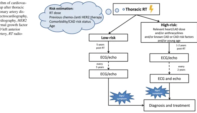
Onco-cardiology follow-up in everyday practice is shown in Fig. 4. All guidelines recommend the specification of the risk-status, preferably before starting oncological treatments or before outlining individual follow-up strategies [12, 13, 24]. For that, heart dose, the use of toxic oncological treat- ments, comorbidities, and age should be considered. Accord- ing to the risk level, follow-up should start immediately or many years after the RT. In high-risk patients (a mediastinal dose of > 30 Gy, or if anthracyclines were also given, etc.) monitoring should start 1–2 years post-irradiation and con- tinue every 2 years thereafter; a great emphasis should be given to correcting risk factors. In low-risk cases, regular follow-up performed every 2–5 years started 5 years after the RT is considered sufficient. For the routine follow-up of asymptomatic patients, ECG and echocardiography are used. During follow-up, careful attention should be paid to medical history; if symptoms or suspicious signs develop, imaging and functional studies should be performed as pre- viously described [12, 13, 24].
Prevention and therapy of RIHD
Currently, there are two main strategies to lessen the bur- den caused by radiogenic heart sequelae. One is to prevent heart exposure as much as possible by applying new RT technologies and protocols [101]. Second, identifying those risk patients whose surveillance after RT is crucial, some- times with comprehensive multimodality imaging-based screening protocols if necessary; the early diagnosis and non-specific management of radiation heart damage may effectively improve outcomes. A third possibility would be the application of pharmacons to protect the heart from radiation-induced damage. However, at present, no specific pharmaceutical agent is approved for the prevention or treat- ment of RIHD in the clinics. Nevertheless, several lines of evidence obtained in preclinical or clinical studies suggest that a number of pharmacological agents might be effective for the treatment of RIHD.
Prevention of RIHD by cardiac dose‑sparing techniques
It is mandatory to individually estimate the net benefit of RT before its start by balancing the gains against the greatness of cardiac dose and the patient’s background cardiovascular
risk in the absence of RT [102]. There are many approaches to protect the heart from radiation exposure in breast can- cer patients [102]. Prone positioning during RT reduces heart doses in about 2/3 of the patients [103–105], while the deep-inspirational breath-holding (DIBH) technique is advantageous in an even higher proportion of patients [106, 107]. Both methods operate by separating the heart and the radiation fields. Intensity-modulated radiation therapy (IMRT) and proton irradiation are advanced techniques not yet widely applied while reducing the volume to be irradi- ated during partial breast irradiation (PBI), or the omittance of RT are options in low-risk of cancer recurrence cases.
For the best risk–benefit ratio, the selection of individually tailored techniques and RT modality is needed [103, 104].
In non-breast cancer patients needing RT to the chest, devel- oped techniques such as the IMRT/volumetric modulated arc radiotherapy (VMAT), cyberknife, or stereotactic radiosur- gery are all based on image-guidance and particle RT. Some- times breathing-control/gating ensure precise targeting and best protection of normal tissues [106]. Special consensus guidelines stressing the use of modern techniques, and selec- tive RT have been elaborated for the modern RT of lym- phoma patients [108]. RT guidelines with similar concepts have been published for lung cancer [109] and oesophageal cancer [110].
Prevention and therapy of RIHD with pharmacological agents
Although many agents have been tested for the prevention of radiation damage, none of them yet gained registration
with this indication. Most of our knowledge in this field is experimental only. The group of the so-called radioprotec- tors has anti-oxidant and/or anti-inflammatory properties being administered as molecular preventive strategies before radiation exposure [44]. The so-called mitigators are admin- istered during or shortly after the irradiation with the aim of ameliorating the radiation injury of normal tissues [44]. A third approach is starting the protectant several weeks after the radiation exposure. The pharmacological treatment of RIHD initiated after the completion of the RT has a clear benefit with the advantage of not interfering with the effi- cacy of cancer therapy but, only a few preclinical studies tested this approach. Despite the lack of specific treatments for RIHD in clinical practice, some recommendations exist on using standard therapies in the radiation heart sequelae indication (e.g., HF or IHD) [12, 13]; furthermore, some promising novel approaches also exist. This section collects well-known drugs and promising novel agents mostly tested in rodent RIHD models. The description of the potential side effects of the well-known drugs is out of the scope of this review.
Anti‑oxidants
Since the production of ROS/RNS is a crucial element in the development of acute and chronic RIHD, testing anti- oxidants seems logical.
α-tocopherol (vitamin E)
Administration of a single dose of tocotrienols 24 h before the cardiac irradiation preserved the Bax/Bcl2 ratio and prevented mitochondrial permeability transition and
Fig. 4 Algorithm of cardiovas- cular follow-up after thoracic RT. CAD coronary artery dis- ease, ECG electrocardiography, echo: echocardiography, HER2 human epidermal growth factor receptor, LAD left anterior descending artery, RT radio- therapy
RT-induced alterations in the mitochondrial respiration in rats 2 weeks after RT [111] (Table 2). However, the single dose of tocotrienols could not improve the cardiac remod- eling 28 weeks after RT [111]. In another study, the phos- phodiesterase inhibitor pentoxifylline plus α-tocopherol given daily and started 1 week before the RT or 3 months after the RT reduced the collagen deposition in rats 6 months after RT [93] (Table 2). Daily administration of α-tocopherol plus pentoxifylline for 6 months started 3 days before the RT reduced collagen deposition and TGFβ1 levels in a rat model 6 months after the RT [112] (Table 2). However, the daily administration of α-tocopherol plus pentoxifylline for 3 months started 3 days before the RT could not reduce the cardiac remodeling 6 months after the RT [112] (Table 2).
In another study, the daily application of pentoxifylline and α-tocopherol 3 months after the RT did not alter cardiac fibrosis and left ventricular expression of vWF, neuregu- lin-1, hypertrophic, and fibrotic signal mediators in rats 6 months after the RT [113]. However, the cardiac number of macrophages and mast cells was reduced [113] (Table 2).
Amifostine (Ethyol)
The inactive prodrug amifostine (WR-2721) is a phos- phorylated thiol, which can be converted to its active form (WR-1065) by alkaline phosphatase-catalyzed dephospho- rylation in the vascular endothelial cells [114]. It has been demonstrated that amifostine protects normal tissues from both acute and chronic damage without interfering with the effects of RT on the tumor [115, 116]. Several preclinical studies have shown that a single dose of amifostine given 15–30 min before irradiation could be protective against RT-induced cardiac fibrosis, myocardial dysfunction, and vascular damage 100 days or 6 months after RT [114, 117, 118] (Table 2).
Natural products
Several natural products, including hesperidin, curcumin, melatonin, caffeic acid phenylethyl ester, black grape juice, and the ginger component zingerone, have been proposed as radioprotective agents due to their antioxidant and/or anti- inflammatory properties against RIHD as reviewed recently [119].
Anti‑inflammatory drugs
Inflammation plays a major role in the development of RIHD, so it is not surprising that attenuation of the inflam- matory response may beneficially affect the cardiac conse- quences of RT.
Colchicine
Colchicine is known to inhibit microtubule polymeriza- tion. Therefore, it can inhibit mitosis, neutrophil motility, and decrease platelet aggregation. The anti-inflammatory and platelet aggregation inhibiting properties of colchicine are suggested to be protective against RIHD [120]. However,
there is no experimental or clinical evidence available in the literature for its use in RIHD.
Steroidal anti-inflammatory drugs
Reeves et al. reported that the steroidal anti-inflammatory drugs, dexamethasone, and methylprednisolone given 2 h prior to heart irradiation and every 24 h for 3 consecutive days reduced the cardiac fibrosis and hydroxyproline content in male rabbits 100 days after RT [121, 122] (Table 2).
Non-steroidal anti-inflammatory drugs (NSAIDs) Administration of the non-selective NSAID ibuprofen administered 2 h prior to heart irradiation and for 2 days thereafter reduced fibrosis, pericarditis, pericardial effu- sions, and improved survival in male rabbits 100 days after RT [122] (Table 2). It has been reported that the use of vari- ous several non-selective COX inhibitors (e.g., ibuprofen, diclofenac, and naproxen) and selective COX-2 inhibitors (e.g., celecoxib) increased the risk of AMI [123]. There- fore, several NSAIDs are contraindicated among patients with CVDs. However, there is limited data available about the effects of NSAIDs on the risk of AMI in patients treated with thoracic RT. Uehara et al. investigated the effects of NSAIDs, including diclofenac, etodolac, indomethacin, ketoprofen, meloxicam, and rofecoxib, on RT-induced expression of ICAM-1, VCAM-1, E-selectin, and COX-2 in human umbilical vein endothelial cells (HUVECs) [124].
They found that indomethacin, diclofenac, and meloxicam given 1 h before RT highly upregulated the RT-induced expression of ICAM-1 and COX-2 in HUVECs, suggest- ing the potentiating effects of these NSAIDs on RT and the increased risk for AMI after thoracic RT [124]. The COX-2 inhibitor celecoxib has been reported to act synergistically with RT in cancer cells since it attenuates tumor growth and expression of cell proliferation markers and induces apoptosis in tumor cells [125]. The increased expression of adhesion molecules and apoptotic effects of celecoxib or several NSAIDs might be responsible for the increased risk for AMI after RT. However, further preclinical and clinical studies are needed to evaluate the effects of NSAIDs on the development of RIHD.
Cardioprotective drugs
Cardioprotective drugs are used to lower the severity of consequences due to CVD risk factors under various stress conditions. The application of such drugs in the case of chest RT may be useful.
Trimetazidine
Trimetazidine is an antianginal drug that inhibits the beta-oxidation of fatty acids by blocking mitochondrial long- chain 3-ketoacyl coenzyme A thiolase [126]. Trimetazidine also has anti-oxidant, anti-apoptotic, and inflammatory effects and may improve endothelial function [127]. Daily trimetazidine treatment started 1 week before or after chest
![Fig. 1). The risk for IHD increased linearly with the MHD by 7.4% per 1 Gy absorbed dose with no apparent lower thresh-old [8]](https://thumb-eu.123doks.com/thumbv2/9dokorg/998269.61783/3.892.123.765.88.534/fig-risk-increased-linearly-absorbed-apparent-lower-thresh.webp)