Full Terms & Conditions of access and use can be found at
https://www.tandfonline.com/action/journalInformation?journalCode=irnf20
Renal Failure
ISSN: 0886-022X (Print) 1525-6049 (Online) Journal homepage: https://www.tandfonline.com/loi/irnf20
The value of combined hemodynamic, respiratory and intra-abdominal pressure monitoring in
predicting acute kidney injury after major intraabdominal surgeries
Csaba Kopitkó, László Medve & Tibor Gondos
To cite this article: Csaba Kopitkó, László Medve & Tibor Gondos (2019) The value of combined hemodynamic, respiratory and intra-abdominal pressure monitoring in predicting acute kidney injury after major intraabdominal surgeries, Renal Failure, 41:1, 150-158, DOI:
10.1080/0886022X.2019.1587467
To link to this article: https://doi.org/10.1080/0886022X.2019.1587467
© 2019 The Author(s). Published by Informa UK Limited, trading as Taylor & Francis Group.
Published online: 25 Mar 2019.
Submit your article to this journal
Article views: 394
View Crossmark data
CLINICAL STUDY
The value of combined hemodynamic, respiratory and intra-abdominal pressure monitoring in predicting acute kidney injury after major intraabdominal surgeries
Csaba Kopitkoa , Laszlo Medvea and Tibor Gondosb
aIntensive Care Unit, Dr. Kenessey Albert Hospital, Balassagyarmat, Hungary;bFaculty of Health Sciences, Department of Clinical Studies, Semmelweis University, Budapest, Hungary
ABSTRACT
Background: The incidence of postoperative acute kidney injury (AKI) is predominantly deter- mined by renal hemodynamics. Beside arterial blood pressure, the role of factors causing a deterioration of venous congestion (intraabdominal pressure, central venous pressure, mechan- ical ventilation) has emerged. The value of combined hemodynamic, respiratory and intra- abdominal pressure (IAP) monitoring in predicting postoperative acute kidney injury has received only limited exploration to date.
Methods: Data were collected for adult patients admitted after major abdominal surgery at nine Hungarian ICUs. Hemodynamic parameters were compared in AKI vs. no-AKI patients at the time of admission and 48 h thereafter. Regarding ventilatory support, we tested mean airway pressures (Pmean). Effective renal perfusion pressure (RPP) was calculated as MAP(IAPþCVPþPmean). The Mann–WhitneyU and the chi-square tests were carried out for statistical analysis with forward stepwise logistic regression for AKI as a dependent outcome.
Results: A total of 84 patients (34 ventilated) were enrolled in our multicenter observational study. The median values of MAP were above 70 mmHg, IAP not higher than 12 mmHg and CVP not higher than 8 mmHg at all time-points. When we combined those parameters, even those belonging to the‘normal’range with Pmean, we found significant differences between no-AKI and AKI groups only at 12 h after ICU admission (median and IQR: 57 (42–64) vs. 40 (36–52);
p<.05). Below it’s median (40.7 mmHg) on admission, AKI developed in all patients. If above 40.7 mmHg on admission, they were protected against AKI, but only if it did not decrease within the first 12 h.
Conclusions: Calculated effective RPP with the novel formula MAP(IAPþCVPþPmean) may predict the onset of AKI in the surgical ICU with a great sensitivity and specificity. Maintaining effective RPP appears important not only at ICU admission but during the next 12 h, as well.
Additional, larger studies are needed to explore therapeutic interventions targeting this parameter.
ARTICLE HISTORY Received 6 December 2018 Revised 20 February 2019 Accepted 20 February 2019 KEYWORDS
Postoperative acute kidney injury; central venous pressure; intraabdominal pressure; mean airway pressure
Introduction
Acute kidney injury (AKI) associates with poor prognosis in critically ill patients [1–3]. There are well-known risk factors for postoperative AKI: (1) sepsis especially due to intraabdominal infections; (2) deterioration of renal blood supply; (3) type of surgery (intraabdominal, car- diac, emergency interventions); (4) mechanical ventila- tion; (5) vasopressor therapy; (6) preexisting kidney disease [1,4–9]. The deterioration of arterial blood sup- ply as indicated by a mean arterial blood pressure (MAP) less than 65 mmHg clearly leads to worsening of
kidney function, but until now only limited animal and human studies have been conducted for highlighting the role of elevated renal venous pressures [10–12].
Abdominal perfusion pressure (APP), recommended by the WSACS – the Abdominal Compartment Society for estimating the adequacy of renal perfusion, is com- puted by MAP – intraabdominal pressure (IAP) [13].
Several studies have been published on this method [14–18]. Elevated IAP with or without a decreased APP has been shown to be present in connection with a higher incidence of AKI [13]. On the other hand, other possible contributors to impair venous return and net
CONTACT Csaba Kopitko kopcsab2@gmail.com Intensive Care Unit, Dr. Kenessey Albert Hospital, Rakoczi fejedelem ut 125-127, H-2660 Balassagyarmat, Hungary
ß2019 The Author(s). Published by Informa UK Limited, trading as Taylor & Francis Group.
This is an Open Access article distributed under the terms of the Creative Commons Attribution License (http://creativecommons.org/licenses/by/4.0/), which permits unrestricted use, distribution, and reproduction in any medium, provided the original work is properly cited.
2019, VOL. 41, NO. 1, 150–158
https://doi.org/10.1080/0886022X.2019.1587467
renal perfusion pressures, such as central venous pres- sure (CVP) or transmitted airway pressures induced by mechanical ventilation have not been sufficiently con- sidered to date. Several other intensive care investiga- tors use the term‘transrenal pressure’, defined as MAP –CVP as a predictor of AKI, albeit without considering the impact of IAP as a contributing factor [10–12]. To date, only one study has been conducted to investigate the relationship between elevated CVP and the rise of IAP; however, this study found poor correlation between elevated CVP and IAP in decompensated heart failure patients [19]. The WSACS – the Abdominal Compartment Society proposed the filtration gradient (FG¼MAP2IAP) for computing the renal perfusion pressure in the Society’s former guidelines, but this rec- ommendation was subsequently withdrawn in 2013 [13,20].
In daily practice, mechanical ventilation has a well- known association with a higher risk of AKI, but none of the individual respiratory parameters, such as positive end-expiratory pressure (PEEP), tidal volume, mean air- way pressure (Pmean), either alone or combined with any circulatory pressure parameters has proven predict- ive with regard to AKI until now [21].
Proper hemodynamic monitoring improves out- comes after major abdominal surgery [22]. For this rea- son, the continuous measurement of MAP and IAP is generally accepted and widely practiced [13]. The majority of postoperative AKI cases are attributable to the impairment of the effective vascular perfusion to the kidney; therefore, the optimization of macro-hemo- dynamics remains critically important [9].
Most recently, we reported [MAP(IAPþ CVPþPmean)], a novel formula for predicting renal per- fusion pressure (RPP) and calculated on admission to the ICU after major abdominal surgery, appears to be the most precise way to predict AKI in critically ill post- operative patients from the twelve investigated formu- las [23]. The aim of our study was to test the hypothesis whether the formula, we termed ‘effective RPP’ that includes MAP, IAP, CVP and Pmean are appropriate for predicting the onset of AKI in surgical ICU patients after major abdominal surgery.
Materials and methods Patients
Data were collected prospectively in adult patients admitted after major abdominal surgery in nine Hungarian Intensive Care Units (ICU) (4 universities, 4 regional and 1 city hospitals) in our multicenter obser- vational between 5 January 2015 and 6 October in
2015 [23]. Ethical approval for this study was provided by the Hungarian Health Registration and Training Center, the national ethical committee (Ethical Committee N 039320/2014/OTIG). Written informed consent was obtained from all study participants or granted by their legal guardians or next-of-kin. The eth- ical standards of the experiments were in accordance with the guidelines provided by the World Medical Association Declaration of Helsinki on Ethical Principles for Medical Research Involving Humans for Studies.
Patients were enrolled after major abdominal surgery requiring a minimum of 48-h ICU postoperative ther- apy. Patients under 18 years of age, those with an end- stage kidney disease, those having surgery with supra- renal cross-clamping or undergoing kidney or urinary bladder operations were excluded from the study.
Patients enrolled in other clinical studies were also excluded. To collect data, we used an Excel-based data collection file and, included demographic and peri- operative information, severity of illness on admission (Simplified Acute Physiology Score version II (SAPS II), Sepsis-related Organ Failure Assessment Score (SOFA)) and the outcome data (presence of AKI at 48 h) [24,25].
The internationally accepted Acute Kidney Injury Network (AKIN) criteria (both creatinine and urine out- put) were used for defining AKI [26]. We considered a preoperative serum creatinine level at baseline [23].
Measurements
MAP, CVP and IAP were recorded by commercially avail- able patient monitors at the time of admission and at 6, 12, 24 and 48 h thereafter. The measurement of IAP was performed by nursing staff via indwelling urinary cathe- ters and according to the WSACS – the Abdominal Compartment Society guidelines. RPP was calculated at the given time points according to the novel formula of MAP(IAPþCVPþPmean) and the onset of AKI was detected at 48 h after ICU admission. The measurement of CVP was performed at the end of expiration. All pres- sures were given in mmHg (1 mmHg = 1.36 cmH2O).
Statistical analysis
All values were presented as median with interquartile range (IQR). The median values of the different groups were compared using the Mann–Whitney U-test and the occurrence rates using the chi-square test. To test the sensitivity and specificity of changes of RPP between 6 and 12 h, a Receiver Operating Curve (ROC) analysis was performed for parameters discriminating AKI and no-AKI groups [Group 1: patients without AKI;
RENAL FAILURE 151
Group 0: patients with AKI (grade 1, 2 or 3)]. Confidence intervals (95%) were calculated according to the for- mula described by Hosmer and Lemeshow. While CVP and airway pressures can influence IAP, their independ- ency has been analyzed by linear regression analysis, as shown in our previous work [23]. Data were dichotom- ized regarding effective RPP, which demonstrated the best predictivity with reference to its previously pub- lished median of 40.7 mmHg (measured on admission) when comparing no-AKI and AKI groups at later time points.
To explore the relationship between abdominal per- fusion pressure and the incidence of AKI, forward step- wise logistic regression was performed. All the basic hemodynamic parameters, Pmean and the effective RPP on admission, 6 h and 12 h, as well as all the changes between these time points were included. All the pressures (MAP, CVP, IAP, Pmean and RPP) were considered as independent predictors measured at the same time or interval, and the presence of AKI as the outcome parameter.
Our study had an 80% statistical power of effective RPP 12 h after admission between the AKI and no-AKI
groups at a significance level of 0.05. For estimating the effect size of our study, we calculated Hedges’ g modi- fied for small samples. For MAP(IAPþCVPþPmean) Hedges’ g values were 0.74 on admission, 1.02 at 12 h after admission, and 0.93 at the change between 6 and 12 h. Hedges’g is considered to be high above 0.8. All differences were deemed to be significant ifp.05. All analyses were performed by the SPSS statistical soft- ware package 23.0 (IBM SPSS, Armonk, NY, US).
Results
A total of 84 patients were admitted to the ICU during the study period (type of surgery and number of cases –gastric: 9; bowel: 28; rectal: 19; hepatic: 3; pancreatic:
4; aortic: 13; trauma: 5 and miscellaneous others: 3).
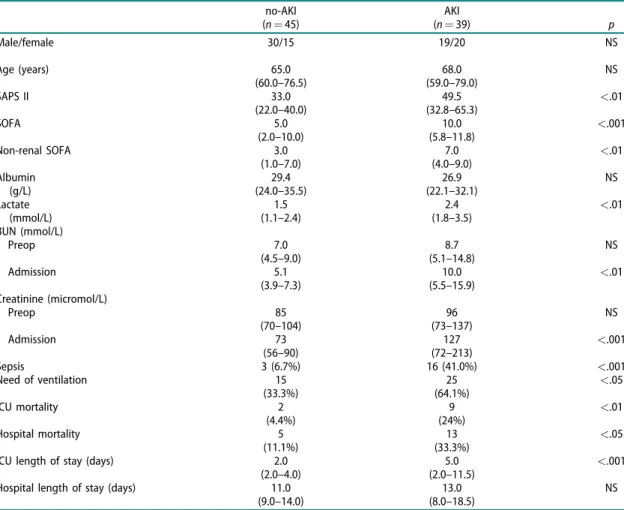
Between the AKI and no-AKI groups, there were no sig- nificant differences in age, gender and serum albumin levels. A higher incidence of sepsis, as well as higher serum lactate levels and severity-of-disease scores were found in the AKI group (Table 1).
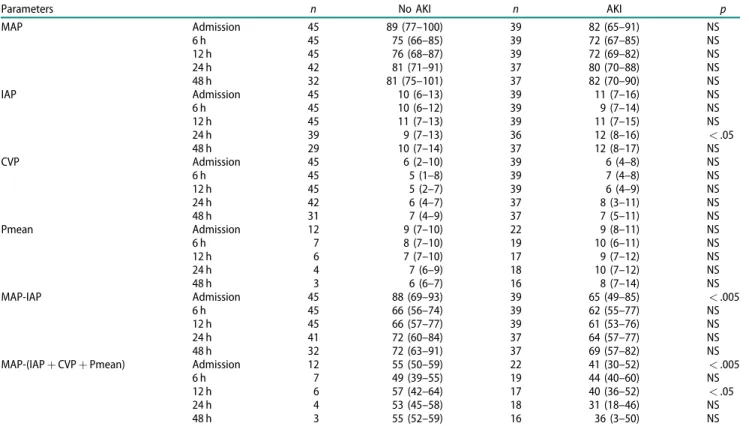
The MAP and the calculated effective RPP values were significantly lower in the AKI group (Table 2) on
Table 1. Demographics of patients at admission (median and IQR; case numbers and percentage).
no-AKI (n¼45)
AKI
(n¼39) p
Male/female 30/15 19/20 NS
Age (years) 65.0
(60.0–76.5)
68.0 (59.0–79.0)
NS
SAPS II 33.0
(22.0–40.0)
49.5
(32.8–65.3) <.01
SOFA 5.0
(2.0–10.0)
10.0
(5.8–11.8) <.001
Non-renal SOFA 3.0
(1.0–7.0)
7.0
(4.0–9.0) <.01
Albumin (g/L)
29.4 (24.0–35.5)
26.9 (22.1–32.1)
NS Lactate
(mmol/L)
1.5 (1.1–2.4)
2.4
(1.8–3.5) <.01
BUN (mmol/L)
Preop 7.0
(4.5–9.0)
8.7 (5.1–14.8)
NS
Admission 5.1
(3.9–7.3)
10.0
(5.5–15.9) <.01
Creatinine (micromol/L)
Preop 85
(70–104)
96 (73–137)
NS
Admission 73
(56–90)
127
(72–213) <.001
Sepsis 3 (6.7%) 16 (41.0%) <.001
Need of ventilation 15
(33.3%)
25
(64.1%) <.05
ICU mortality 2
(4.4%)
9
(24%) <.01
Hospital mortality 5
(11.1%)
13
(33.3%) <.05
ICU length of stay (days) 2.0
(2.0–4.0)
5.0
(2.0–11.5) <.001
Hospital length of stay (days) 11.0
(9.0–14.0)
13.0 (8.0–18.5)
NS AKI: Acute Kidney Injury; BUN: Blood Urea Nitrogen; SAPS II: Simplified Acute Physiology Score version II; SOFA: Sepsis-related Organ Failure Assessment Score.
admission. No differences were found in airway pres- sures between the two groups. The change of RPP between time points is presented inTable 3.
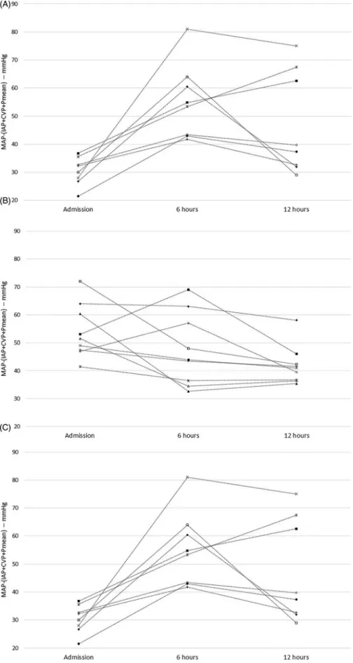
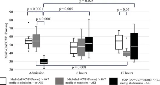
Below the median of this parameter AKI developed in all cases. If effective RPP was above the median on admission and increased in the first 12 h in the ICU, none of the study participants developed AKI, whereas most patients with decreasing effective RPP did (Figure 1). In most AKI patients, effective RPP decreased between 6 and 12 h. Even though perfusion improved both at 6 and 12 h in the poor RPP group, AKI remained unavoidable (Figure 2). ROC analysis of changes of RPP between 6 and 12 h revealed moderate probability of the RPP for AKI according to both positive and negative likelihood ratios (LR) (1-AUC: 0.804, sensitivity: 86%, spe- cificity: 81%, LRþ: 4.50, LR: 0.18) (Figure 3).
After logistic regression analysis, the change of effective RPP between 6 and 12 h was the only param- eter that remained in the equation and that showed an almost significant result (B: 0.091, SE: 0.048, Wald:
3.641, Sig: 0.056, Exp (B): 0.913, 95% CI for Exp (B):
0.813–1.002). The logistic regression analysis with the
parameters at other time points or intervals has not revealed significant relationship between basic pres- sures or RPP and the presence of AKI at 48 h.
Discussion
The role of arterial blood supply is clear and well- studied, but venous congestion is underestimated in different guidelines, and currently, only the measure- ment of IAP is emphasized [13,27]. In this paper, having analyzed parameters recorded within 12 h after admis- sion in our study, we are reporting the value of the for- mula of MAP(IAPþCVPþPmean) in predicting AKI beyond ICU admission, incorporating elements of hemodynamic, as well as ventilator-derived and intra- abdominal pressures.
Human studies for investigating the optimal MAP were conducted mostly in patients with septic shock.
The generally accepted target is 65 mmHg, a number extrapolated from intraoperative observations and from clinical experience derived from monitoring septic patients’hemodynamics. Keeping MAP above this value Table 2. The values of parameters (mmHg, median (IQR)).
Parameters n No AKI n AKI p
MAP Admission 45 89 (77–100) 39 82 (65–91) NS
6 h 45 75 (66–85) 39 72 (67–85) NS
12 h 45 76 (68–87) 39 72 (69–82) NS
24 h 42 81 (71–91) 37 80 (70–88) NS
48 h 32 81 (75–101) 37 82 (70–90) NS
IAP Admission 45 10 (6–13) 39 11 (7–16) NS
6 h 45 10 (6–12) 39 9 (7–14) NS
12 h 45 11 (7–13) 39 11 (7–15) NS
24 h 39 9 (7–13) 36 12 (8–16) <.05
48 h 29 10 (7–14) 37 12 (8–17) NS
CVP Admission 45 6 (2–10) 39 6 (4–8) NS
6 h 45 5 (1–8) 39 7 (4–8) NS
12 h 45 5 (2–7) 39 6 (4–9) NS
24 h 42 6 (4–7) 37 8 (3–11) NS
48 h 31 7 (4–9) 37 7 (5–11) NS
Pmean Admission 12 9 (7–10) 22 9 (8–11) NS
6 h 7 8 (7–10) 19 10 (6–11) NS
12 h 6 7 (7–10) 17 9 (7–12) NS
24 h 4 7 (6–9) 18 10 (7–12) NS
48 h 3 6 (6–7) 16 8 (7–14) NS
MAP-IAP Admission 45 88 (69–93) 39 65 (49–85) <.005
6 h 45 66 (56–74) 39 62 (55–77) NS
12 h 45 66 (57–77) 39 61 (53–76) NS
24 h 41 72 (60–84) 37 64 (57–77) NS
48 h 32 72 (63–91) 37 69 (57–82) NS
MAP-(IAPþCVPþPmean) Admission 12 55 (50–59) 22 41 (30–52) <.005
6 h 7 49 (39–55) 19 44 (40–60) NS
12 h 6 57 (42–64) 17 40 (36–52) <.05
24 h 4 53 (45–58) 18 31 (18–46) NS
48 h 3 55 (52–59) 16 36 (3–50) NS
AKI: Acute Kidney Injury; CVP: Central Venous Pressure; IAP: Intraabdominal Pressure; MAP: Mean Arterial Pressure.
Table 3. The change of RPP in the first 12 h after admission to ICU (mmHg, median (IQR)).
Parameter n No AKI n AKI p
D[MAP-(IAPþCVPþPmean)] 0–6 h 8 5 (41 to–6) 23 6 (9 to–18) NS
6–12 h 7 9 (2–13) 21 4 (10 to–1) <.05
0–12 h 7 3 (28 to–14) 21 6 (14 to–11) NS
RENAL FAILURE 153
Figure 1. The patients’data with (A) below 40.7 mmHg of MAP(IAPþCVPþPmean) and above it (B) with or (C) without AKI in the first 12 h.
is protective against AKI [28]. From the parameters affecting renal congestion, the elevated IAP (>12 mmHg) is associated with a higher risk of AKI [13].
The CVP (but not the cardiac index) has been shown to be a risk factor of renal failure in cardiac patients and is proved to be independent of IAP [10]. In septic ICU patients, higher CVP and lower diastolic pressure have
been associated with AKI [11]. In another study, mean perfusion pressure deficit (defined as MAPCVP) and diastolic perfusion pressure deficit have been shown to be predictors of worsening kidney function [12].
Against these results, in our prospective study, the median values of MAP were above 70 mmHg, IAP not higher than 12 mmHg and CVP not higher than Figure 2. The course of MAP(IAPþCVPþPmean) in the first 12 h in ICU after major abdominal surgery. Thepvalues within or between groups not shown were not statistically significant.
Figure 3. ROC analysis of the changes of RPP between 6 and 12 h after admission.
RENAL FAILURE 155
8 mmHg at all time-points. To reach these hemo- dynamic parameters, many patients needed vasopres- sor therapy and various amounts of intravenous fluids.
The aim of our study was to find the simplest, continu- ously recordable and proper parameter for determining the adequacy of renal perfusion pressure in patients requiring ICU admission after major abdominal surgery.
Mechanical ventilation can increase the risk of AKI, but in our study, we found no differences in the Pmean values between the groups [23]. Because no unique and simple parameter is suitable for monitoring and selecting patients who are at risk of AKI after major abdominal surgery, we tried to find the best combin- ation. In our previous paper, we reported that the MAP(IAPþCVPþPmean) formula was the best pre- dictor of AKI on admission to ICU [23]. Moreover, when we combined those parameters even with parameters of ‘normal’ range, we found significant differences between groups in effective RPP at 12 h. We should emphasize that this effect was not due to a change of arterial blood supply as MAP remained unchanged. The factors affecting the venous congestion of the kidney (IAP, CVP, Pmean) have been proved to be independent of each other [23].
The physiological basis of computing these equa- tions was the previously mentioned data concerning venous congestion [10,19]. Medians of all parameters were in so-called‘normal’range, and also the CVP was there. There were no significant differences in medians of AKI and of no-AKI groups. The IQRs of MAP are the same across groups at 6 and 12 h, as you can see in Table 2. The IQR of IAP is lower in no-AKI group at 6 h, and this difference disappears at 12 h. The medians and IQRs of CVP are higher in AKI group at both 6 and 12 h, although this difference is not significant statistically.
The medians of Pmean are higher in AKI group at both 6 and 12 h (difference is not significant). The net driving pressure of filtration in the glomeruli is about 10 mmHg under physiological condition. The medians and IQRs of the parameters mentioned above are the same range.
Therefore, the cumulation of seemingly small changes can result in AKI. CVP represents not only extracapsular, but also the intracapsular pressure of kidney, which can differ because of the tight fibrous capsule of it.
The predictivity of effective RPP was found to be suitable not only on admission but at later time points as well: our prediction model has a sensitivity of 86%
and specificity of 81%. When this pressure fell below 40.7 mmHg on admission to the ICU, AKI developed in every case, regardless of any further improvement in perfusion. On the other hand, keeping the effective RPP above its median (40.7 mmHg) on admission and the
subsequent 12 h in patients having undergone major abdominal surgery protected against AKI. The individual course of RPP above its median measured on admission to ICU is an important tool in prediction of postopera- tive AKI and can be a part of the patient’s personalized care of the patient. Moreover, monitoring this param- eter is simpler and may be much easier than using the points of different scoring systems or models.
Our study has several limitations: first, we had only a relatively small-sized cohort with a low number of sub- jects recruited from the individual centers. The propor- tion of patients receiving mechanical ventilation was even smaller and likely received heterogeneous and somewhat different from center-to-center care. Details regarding preexisting or total body fluid excess, vaso- pressors types and doses or intraoperative blood pres- sures were not available. In our study, only moderately elevated IAP and a normal range of CVP were found, representing both relative strength and weakness of the study, and our conclusions need to be verified with even sicker patients with higher IAP, Pmean or massive volume overload. The value of effective RPP during renal replacement therapy in the course of AKI remains fully unexplored and likely a fertile territory for future investigations.
Conclusions
Continuous measurement of MAP, IAP and CVP can be useful in patients with shock and potential intrabdomi- nal hypertension. We were able to demonstrate that com- puting the effective RPP as [MAP(IAPþCVPþPmean)]
and combining values even ’near-to-normal’ values of hemodynamic and respiratory parameters can be a pre- dictor of AKI in the early postoperative period.
Accordingly, this formula may represent a valuable addition to the care of ICU patients at-risk for AKI.
Even if there is a modest elevation of venous pressures then MAP should be kept correspondingly higher to keep RPP above 40.7 mmHg. Additionally, larger stud- ies are needed to explore therapeutic interventions targeting this parameter.
We sincerely appreciated the assistance of Mr. Attila Lenart-Muszka (Debrecen, Hungary) and of Richard Mann during editing and grammar review.
Acknowledgements
List of investigators: Anita Zsirmik, Domonkos Trasy MD, Ildiko Laszlo MD, Zsolt Molnar (Department of Anesthesiology and Intensive Therapy, Faculty of Medicine, University of Szeged); Gabriella Kecskes, Bela Gartner (Petz Aladar Hospital, Gy}or); Zoltan Petro, Zoltan F€ulep (Bacs-
Kiskun Provincial Hospital, Kecskemet); Istvan Laszlo, Bela Fulesdi€ (Department of Anesthesiology and Intensive Therapy, Faculty of Medicine, University of Debrecen); Miklos Siptar, Barbara Benkovics, Lajos Bogar (Institute of Anesthesiology and Intensive Therapy, Faculty of Medicine, University of Pecs); Judit Lorincz, L} aszlo Nagy (Central Military Hospital, Budapest); Emese Helbocsanyi, Gabor Bencsik (Hetenyi Geza Hospital, Szolnok); Gabor Szigligeti, Csaba Antek (Josa Andras Hospital, Nyıregyhaza). We sin- cerely appreciated the assistance of Mr. Richard Mann (Budapest, Hungary) and of Mr. Attila Lenart-Muszka (Debrecen, Hungary) during editing and grammar review.
Disclosure statement
No potential conflict of interest was reported by the authors.
Data availability
The data that support the findings of this study are available from the corresponding author, CsK, upon reason- able request.
ORCID
Csaba Kopitko http://orcid.org/0000-0002-1922-7294 Laszlo Medve http://orcid.org/0000-0003-1911-7615 Tibor Gondos http://orcid.org/0000-0003-0282-0785
References
[1] Medve L, Antek C, Paloczi B, et al. Epidemiology of acute kidney injury in Hungarian intensive care units:
a multicenter, prospective, observational study. BMC Nephrol. 2011;12:43.
[2] Wu HC, Wang WJ, Chen YW, et al. The association between the duration of postoperative acute kidney injury and in-hospital mortality in critically ill patients after non-cardiac surgery: an observational cohort study. Renal Failure. 2015;37:985–993.
[3] Liu Y, Xue FS, Liu GP, et al. Assessing association between duration of postoperative acute kidney injury and in-hospital mortality after noncardiac surgery.
Renal Failure. 2016;38:342–343.
[4] F€ul€op T, Pathak MB, Schmidt DW, et al. Volume- related weight gain and subsequent mortality in acute renal failure patients treated with continuous renal replacement therapy. Asaio J. 2010;
56:333–337.
[5] Brienza N, Giglio MT, Marucci M, et al. Does periopera- tive hemodynamic optimization protect renal function in surgical patients? A meta-analytic study. Crit Care Med. 2009;37:2079–2090.
[6] Medve L, Gondos T. Epidemiology of postoperative acute kidney injury in Hungarian intensive care units:
an exploratory analysis. Renal Failure. 2012;34:
1074–1078.
[7] Moore EM, Bellomo R, Nichol AD. The meaning of acute kidney injury and its relevance to intensive care
and anaesthesia. Anaesth Intensive Care. 2012;40:
929–948.
[8] Makris K, Spanou L. Acute kidney injury: definition, pathophysiology and clinical phenotypes. Clin Biochem Rev. 2016;37:85–98.
[9] Romagnoli S, Ricci Z, Ronco C. Perioperative acute kid- ney injury: prevention, early recognition, and support- ive measures. Nephron. 2018;140:105–110.
[10] Mullens W, Abrahams Z, Francis GS, et al. Importance of venous congestion for worsening of renal function in advanced decompensated heart failure. J Am Coll Cardiol. 2009;53:589–596.
[11] Legrand M, Dupuis C, Simon C, et al.
Association between systemic hemodynamics and septic acute kidney injury in critically ill patients: a retrospective observational study. Crit Care. 2013;17:
R278.
[12] Saito S, Uchino S, Takinami M, et al. Postoperative blood pressure deficit and acute kidney injury pro- gression in vasopressor dependent cardiovascular sur- gery patients. Crit Care. 2016;20:74.
[13] Kirkpatrick AW, Roberts DJ, De Waele J, Pediatric Guidelines Sub-Committee for the World Society of the Abdominal Compartment Syndrome, et al.
Intra-abdominal hypertension and the abdominal compartment syndrome: updated consensus defini- tions and clinical practice guidelines from the World Society of the Abdominal Compartment Syndrome.
Intensive Care Med. 2013;39:1190–1206.
[14] Kirkpatrick AW, Brenneman FD, McLean RF, et al. Is clinical examination an accurate indicator of raised intraabdominal pressure in critically injured patients.
Can J Surg. 2000;43:207–211.
[15] Cheatham ML, White MW, Sagraves SG, et al.
Abdominal perfusion pressure: a superior parameter in the assessment of intra-abdominal hypertension.
J Trauma. 2000;49:621–627.
[16] Cheatham ML. Nonoperative management of intraab- dominal hypertension and abdominal compartment syndrome. World J Surg. 2009;33:1116–1122.
[17] Ouellet JF, Leppaniemi A, Ball CG, et al. Alternatives to formal abdominal decompression. Am Surg. 2011;
77:S51–S57.
[18] De Keulenaer BL, De Waele JJ, Malbrain ML.
Nonoperative management of intra-abdominal hyper- tension and abdominal compartment syndrome:
evolving concepts. Am Surg. 2011;77:S34–S41.
[19] Mullens W, Abrahams Z, Skouri HN, et al.
Elevated intra-abdominal pressure in acute decom- pensated heart failure: a potential contributor to wor- sening renal function? J Am Coll Cardiol. 2008;51:
300–306.
[20] Cheatham ML. Abdominal Compartment Syndrome:
pathophysiology and definitions. Scand J Trauma Resusc Emerg Med. 2009;17:10.
[21] van den Akker JP, Egal M, Groeneveld AB. Invasive mechanical ventilation as a risk factor for acute kidney injury in the critically ill: a systematic review and meta-analysis. Crit Care. 2013;17:R98.
[22] Salzwedel C, Puig J, Carstens A, et al. Perioperative goal-directed hemodynamic therapy based on radial arterial pulse pressure variation and continuous RENAL FAILURE 157
cardiac index trending reduces postoperative compli- cations after major abdominal surgery: a multi-center.
prospective. randomized study. Crit Care. 2013;17:
R191.
[23] Kopitko C, Medve L, Gondos T. Renoprotective post- operative monitoring: what is the best method for computing renal perfusion pressure? – an observa- tional, prospective, multicentre study. Nephron. 2018;
139:228–236.
[24] Le Gall JR, Lemeshow S, Saulnier F. A new Simplified Acute Physiology Score (SAPS II) based on a European/North American multicenter study. JAMA.
1993;270:2957–2963.
[25] Vincent JL, Moreno R, Takala J, et al. The SOFA (Sepsis-related Organ Failure Assessment) score to
describe organ dysfunction/failure. On behalf of the Working Group on Sepsis-Related Problems of the European Society of Intensive Care Medicine.
Intensive Care Med. 1996;22:707–710.
[26] Mehta RL, Kellum JA, Shah SV, the Acute Kidney Injury Network, et al. Acute Kidney Injury Network: report of an initiative to improve outcomes in acute kidney injury. Crit Care. 2007;11:R31.
[27] Kidney Disease: Improving Global Outcomes (KDIGO) Acute Kidney Injury Work Group. KDIGO clinical prac- tice guideline for acute kidney injury. Kidney Int Suppl. 2012;2:1–138.
[28] Leone M, Asfar P, Radermacher P, et al.
Optimizing mean arterial pressure in septic shock: a crit- ical reappraisal of the literature. Crit Care. 2015;19:101.