High expression of DNA methyltransferases in primary human medulloblastoma
Tímea Pócza1,2, Tibor Krenács3,4, Eszter Turányi3, János Csáthy1, Zsuzsanna Jakab1, Peter Hauser1
12nd Department of Pediatrics, Semmelweis University, Budapest, 2Department of Molecular Genetics, National Institute of Oncology, Budapest, 31st Department of Pathology and Experimental Cancer Research, Semmelweis University, Budapest,
4Tumor Progression Research Group, Joint Research Organization of the Hungarian Academy of Sciences and Semmelweis University, Budapest, Hungary
Folia Neuropathol 2016; 54 (2): 105-113 DOI: 10.5114/fn.2016.60365
A b s t r a c t
Epigenetic alterations have been implicated in cancer development. DNA methylation modulates gene expression, which is catalyzed by DNA methyltransferases (DNMTs). The objective of our study was to evaluate expression of DNMTs in medulloblastoma and analyze its correlation with clinical features. Nuclear expression of DNMT1, DNMT3A and DNMT3B was analyzed in human primary medulloblastoma of 44 patients using immunohistochem- istry. Correlation of expression of DNMT levels with classical histological subtypes, novel molecular subgroups and survival of patients was analyzed. Elevated expression of DNMT1, DNMT3A and DNMT3B was observed in 63.64%, 68.18% and 72.73% of all cases, respectively. None of them showed a correlation with classical histology or survival.
Concerning molecular subtypes, significantly higher expression of DNMT1 was observed in the SHH group compared to non-SHH samples (p = 0.02), but without significant difference in DNMT3A or DNMT3B levels between any sub- types. In conclusion, DNMT1, DNMT3A and DNMT3B are highly expressed in human medulloblastoma samples, sug- gesting that promoter hypermethylation may play a role in medulloblastoma development. Demethylation of tumor suppressor gene promoters may be considered as a possible future target in therapy of medulloblastoma.
Key words: medulloblastoma, DNA methyltransferases, survival, SHH.
Communicating author:
Peter Hauser, 2nd Department of Pediatrics, Semmelweis University, Tűzoltó utca 7-9, H-1094 Budapest, Hungary, phone: +36(1)2151380, fax: +36(1)2151381, e-mail: hauserpeti@yahoo.com
Introduction
Medulloblastoma (MB) is the most frequent malignant brain tumor in childhood, with various histological appearances. There are non-overlapping histological (classic, desmoplastic, extensive nodular, large cell/anaplastic) and molecular classifications (SHH, WNT, Group 3 and 4) for medulloblastoma [3,6,11,23,25,27,34]. Despite multimodal therapies, the 70-75% long-term survival rate has not been
exceeded yet in high-risk patients [13,29,34]. A more precise classification of patients would help to devel- op a more effective treatment strategy and reduce side effects [3,11,23].
There is emerging evidence that epigenetic chang- es contribute to carcinogenesis. The most important mechanisms of epigenetic alteration are DNA meth- ylation, histone modification and microRNA regula- tion. DNA methyltransferases (DNMTs) are enzymes transferring methyl groups to cytosine in CpG dinu-
cleotides, resulting in gene silencing. The DNMT family has three active members: DNMT1, DNMT3A and DNMT3B. DNMT3A and DNMT3B are regarded as de novo methyltransferases, whilst DNMT1 reacts on hemimethylated DNA, copying the methylation pattern during replication. Generally, tumor cells are characterized by global hypomethylation and hyper- methylation of certain gene promoters. Therefore, repetitive elements are hypomethylated and spe- cific tumor suppressor genes are repressed due to hypermethylation. These may contribute to chromo- somal instability and abnormal gene silencing that could promote tumor-initializing steps [2,9,14,28,38].
Promoter hypermethylation of tumor suppressor genes and DNA repair genes has been observed in a wide variety of tumors, such as colorectal, breast cancer, glioma and medulloblastoma [19,22,36].
Overexpression of DNMTs was detected at the mRNA or protein level. Increased DNMT1 expression was described in gastric cancers, along with DNMT3A in breast and pancreatic cancer, or along with DNMT3B in glioma [8,16,30,38,39]. Although there is emerging evidence that epigenetic events play a remarkable role in the development of adult cancers, our knowl- edge about their role in pediatric brain tumors is still very limited.
DNA methylation studies in medulloblastoma have focused on the role of tumor suppressor genes in the development of brain tumors, and shed light on contradictory results in the majority of genes, except for RASSF1A [37]. Recent studies indicate that the DNA methylation profile can be used for MB sub- grouping and disease risk assessment [18,35].
The aims of our study are to characterize the expression of DNMTs in human medulloblastoma samples and their potential prognostic role in surviv- al of patients. Possible correlations of DNMT proteins and clinicopathological features are also analyzed.
Material and methods
Medulloblastoma tissue samples
Formalin-fixed, paraffin-embedded MB tumor samples were collected from the National Insti- tute of Neurosciences (Budapest, Hungary) and 1st Institute of Pathology and Experimental Cancer Research, Semmelweis University (Budapest, Hun- gary). Forty-four primary medulloblastoma samples removed surgically between 2004 and 2010 and related clinical data of patients derived from the
Hungarian Pediatric Cancer Registry were used.
The study was approved by the Ethics Committee of the Institutional Ethical Review Board of Sem- melweis University (TUKEB No. 30/2015).
Tissue microarray (TMA) construction and immunohistochemistry
Hematoxylin and eosin (HE) stained sections were reviewed by a neuropathologist to select rep- resentative areas for producing TMA. TMA blocks were created by a computer-controlled TMA Mas- ter (3DHISTECH, Budapest, Hungary) instrument.
Depending on sample quality one or two cores of representative areas with a diameter of 2 mm were taken from each tumor sample. From the TMA blocks 4 μm sections were created. Sections were depar- affinized, rehydrated, and endogenous peroxidase activity was blocked. Heat-induced antigen retriev- al was performed with Tris-EDTA buffer (pH = 9.0) for 45 min in a microwave oven. Sections were incubated overnight using anti-DNMT1 antibody (dilution 1 : 200; Ab19905, Abcam, Cambridge, UK), anti-DNMT3A antibody (dilution 1 : 600; Ab13888, Abcam, Cambridge, UK) and anti-DNMT3B antibody (dilution 1 : 200; Ab13604, Abcam, Cambridge, UK) diluted in 5% BSA containing Tris-buffered saline (TBS; pH = 7.4). Antigen development was per- formed by Novolink Polymer Detection System (Novocastra, Wetzlar, Germany) following the man- ufacturer’s instructions. Reactions were visualized by 3,3’-diaminobenzidine (DAB) (DAKO, Denmark) as a substrate, and then slides were counterstained by hematoxylin. Tonsil for DNMT1, and kidney for DNMT3A and DNMT3B were used as positive tissue controls.
For determination of the WNT subgroup anti-β-catenin antibody (dilution 1 : 150; M-3539, DAKO, Denmark) and of the SHH subgroup anti- secreted frizzled-related protein 1 (SFRP1) antibody (dilution 1 : 1500; ab-4193, Abcam, Cambridge, UK), characteristic proteins of these pathways, were applied. Immunohistochemical reactions were exe- cuted as described above, but antigen retrieval was performed in TE buffer boiled in a pressure cooker for 20 minutes.
Evaluation of immunostaining
Slides were digitalized by a Pannoramic Scan instrument and examined by Pannoramic Viewer
software (3DHISTECH, Budapest, Hungary). To eval- uate DNMT expression only nuclear staining was considered. Intensity of staining and rate of positive cells were calculated from 1,000 cells in a represen- tative area of each sample. According to the intensi- ty of staining, four values were created: 0 (negative), 1 (weak), 2 (moderate) and 3 (strong). The proportion of positive cells was ranked in five groups: 0 (neg- ative), 1 (1-25%), 2 (26-50%), 3 (51-75%) and 4 (76- 100%). The products of the two values (0-12) were divided into two groups: 0-3 regarded as no/weak expression and 4-12 regarded as moderate/strong expression. Samples were classified into WNT or SHH subgroups when the proportion of positive cells with nuclear β-catenin or negative nuclear β-catenin and positive SFRP1 staining was above 10% [7,27].
Statistical analysis
All statistical analysis was performed using Sta- tistica software (StatSoft Inc., Tulsa, OK, USA). Sur- vival of patients was calculated by the Kaplan-Meier method. Correlation of expression of DNMTs and survival was examined by the log-rank test. Correla- tion of expression of each DNMT was analyzed by the Spearman rank order test. Correlation of expres- sion of DNMTs and age, gender, histological sub- type or molecular subgroups was examined by the Mann-Whitney U-test and Fisher’s exact test, when appropriate. Results were considered statistically significant when the p value was less than 0.05.
Results
Patients’ data
Demographic characteristics of the studied cohort are summarized in Table I. Median age of 44 patients was 8.5 years (range 1.1-28.7 years).
The proportion of male patients was 55%. Histologi- cally, 84.1%, 11.4% and 4.5% of patients showed clas- sic, desmoplastic and large cell/anaplastic (L/A) type of medulloblastoma, respectively. Survival data were available in 40 patients (90.9%). Median follow-up time was 5.6 years (range 0-9.7 years). Patients were treated according to the Hungarian MBL2004/2008 schedules [15]. There is no standard treatment for adults with medulloblastoma in Hungary. Patients are treated according to the individual decision of each treating physician, and these treatment details are poorly accessible in Hungary. Therefore, adult patients were omitted from the survival analysis.
Expression of DNMTs in human medulloblastoma
Elevated (moderate/strong) expression of DNMT1, DNMT3A and DNMT3B was observed in 63.64%, 68.18% and 72.73% of patients, respectively (Table I).
Representative stains are shown in Figure 1. The re- lationship of histology and DNMT expression was evaluated only in the classic and desmoplastic type.
Moderate/strong expression of DNMT1 was detect- ed in 62.2% of classic and 60.0% of desmoplastic, of DNMT3A in 70.3% of classic and 60.0% of desmo- plastic, and of DNMT3B in 70.3% of classic and 80.0%
of desmoplastic samples. We could analyze only two samples of L/A subtype. Both of them showed mod- erate/strong expression for DNMT1 and DNMT3B, but only one for DNMT3A.
Correlation between DNMT expression and patients’ data
There was no correlation between the expression of DNMT1, DNMT3A and DNMT3B and age at disease onset or gender or histological subtype (Table II).
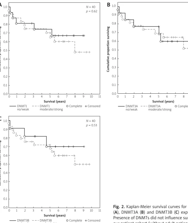
Kaplan-Meier curves, based on different expres- sion of DNMTs, did not show a significant differ- ence in overall survival (Fig. 2). None of the DNMTs could be used as a prognostic marker for medul- loblastoma in our cohort. DNMT1 and DNMT3A (p = 0.08), or DNMT1 and DNMT3B (p = 0.17) or DNMT3A and DNMT3B (p = 0.69) did not show a co-expression pattern. Expression of nuclear β-cat- enin was shown only in one patient (2.3%) with weak staining. There were 12 (27.3%) patients belonging to SHH subgroup according to their cytoplasmic, mem- branous or secreted SFRP1 expression and lack of nuclear β-catenin (Table I). Expression of DNMT1 in the SHH subgroup compared to the non-WNT/non- SHH subgroup of patients was significantly higher (p = 0.02), whereas expression of DNMT3A and DNMT3B did not differ significantly between different groups (p = 0.78 and p = 0.17, respectively, Table II).
Discussion
In recent years a new prognostic classification of medulloblastoma was introduced by molecular find- ings, whilst there are still several twists in this clas- sification system. It is still motivating to find further prognostic markers [3,6,11,23,25,27,34]. Nowadays, an exact diagnosis of pediatric brain tumors can be established based on the methylation profile of dif-
Table I. Characteristics of patients and results of immunohistochemical staining of DNMTs, β-catenin, and SFRP1 Gender Histology Age at disease
onset (years)
State Survival (years)
DNMT1* DNMT3A* DNMT3B* β-catenin SFRP1
1 F Classic 1.18 Dead 0.18 0 0 0 Negative Negative
2 M L/A 7.84 Dead 1.47 9 4 6 Negative Positive
3 F Classic 5.99 Dead 4.56 4 9 3 Negative Negative
4 F Classic 14.04 Dead 0.00 4 6 8 Negative Negative
5 M Classic 10.81 Dead 7.98 4 8 8 Negative Positive
6 F Classic 3.31 Alive 9.38 6 8 12 Negative Negative
7 M Classic 13.54 Alive 9.74 8 12 12 Negative Negative
8 M Classic 17.09 Alive 8.99 2 0 8 Negative Positive
9 F Classic 10.09 Dead 3.07 2 8 8 Negative Negative
10 M Desmoplastic 14.54 Alive 9.08 1 2 3 Negative Negative
11 M Desmoplastic 5.14 Dead 0.59 4 4 6 Negative Negative
12 F Classic 17.90 Dead 5.02 4 2 8 Negative Negative
13 M Classic 3.11 Dead 0.92 2 1 4 Negative Negative
14 F Classic 4.71 Alive 8.54 4 6 12 Negative Negative
15 M Classic 15.30 Alive 7.80 2 4 2 Positive Negative
16 F Classic 8.48 Alive 7.24 12 2 2 Negative Negative
17 F Classic 14.28 Alive 8.09 4 8 8 Negative Negative
18 M Desmoplastic 6.63 Alive 7.90 3 12 8 Negative Positive
19 M L/A 13.84 Dead 1.94 4 3 12 Negative Positive
20 M Desmoplastic 4.34 Dead 0.09 9 12 4 Negative Negative
21 M Classic 8.56 Alive 7.00 2 4 12 Negative Negative
22 M Classic 15.46 Alive 6.52 2 8 12 Negative Negative
23 M Desmoplastic 2.59 Alive 6.52 4 2 4 Negative Positive
24 M Classic 4.50 Dead 1.92 4 12 3 Negative Negative
25 F Classic 9.30 Dead 5.54 8 12 12 Negative Negative
26 M Classic 6.81 Alive 6.63 4 2 3 Negative Negative
27 F Classic 8.50 Alive 6.07 8 0 12 Negative Negative
28 M Classic 7.70 Alive 6.02 2 8 8 Negative Negative
29 F Classic 1.77 Alive 6.76 9 8 8 Negative Positive
30 F Classic 4.92 Alive 6.56 6 8 6 Negative Negative
31 M Classic 22.90 NA NA 6 4 8 Negative Positive
32 M Classic 10.76 Dead 5.22 2 2 4 Negative Negative
33 F Classic 21.43 NA NA 6 0 12 Negative Positive
34 F Classic 16.71 Alive 5.67 2 12 1 Negative Negative
35 M Classic 7.92 Alive 5.48 1 1 4 Negative Negative
36 F Classic 10.75 Alive 5.65 4 4 3 Negative Negative
37 F Classic 1.78 Dead 0.48 2 8 6 Negative Negative
38 M Classic 4.63 Alive 4.99 4 3 4 Negative Negative
39 M Classic 28.25 NA NA 9 4 6 Negative Positive
40 F Classic 1.07 Alive 3.23 6 12 3 Negative Negative
41 F Classic 2.84 Alive 3.12 2 8 2 Negative Negative
42 M Classic 13.18 Alive 2.80 2 8 8 Negative Negative
43 F Classic 28.69 NA NA 9 12 2 Negative Positive
44 M Classic 9.68 Alive 2.12 9 12 12 Negative Positive
*Product of intensity (0-3) and rate of positive cells (0-4), maximal value: 12 SFRP1 – secreted frizzled-related protein 1, DNMT – DNA methyltransferase, M – male, F – female, L/A – large cell/anaplastic, NA – not available
A B
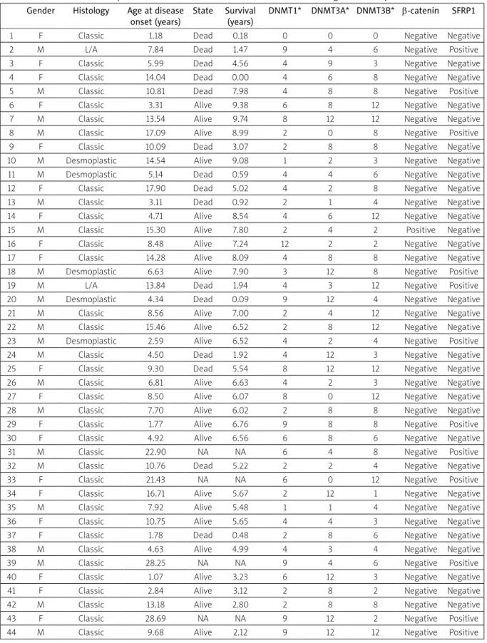
Fig. 1. Representative immunohistochemical staining of DNMTs in medulloblastoma. A) Negative staining of DNMT3A. B, C, D) Moderate/strong immunopositivity of DNMT1, DNMT3A and DNMT3B, respectively.
C D
Table II. Evaluation of patients’ characteristics and expression of different DNMTs. Correlation of DNMTs and clinical features are shown by probability value (p)
All DNMT1 p DNMT3A p DNMT3B p
No/
Weak Moderate/
Strong No/
Weak Moderate/
Strong No/
Weak Moderate/
Strong Number of patients 44 16
(36.36%)
28
(63.64%) 0.91a 14 (31.82%)
30
(68.18%) 0.91a 12 (27.27%)
32
(72.73%) 0.65a Median age
of patients (years)
8.53 9.33 8.49 8.49 8.93 7.65 8.93
Gender
Male 24 11 13
0.21b 9 15
0.52b 4 20
0.10b
Female 20 5 15 5 15 8 12
Histological subtype
Classic 37 14 23
1.00b 11 26
0.64b 11 26
1.00b
Desmoplastic 5 2 3 2 3 1 4
Large cell/anaplastic 2 0 2 # 1 1 # 0 2 #
Molecular subtype
Wnt 1 1 0 # 0 1 # 1 0 #
Shh 12 2 10
0.02a * 4 8
0.78a 1 11
0.17a
Non-Wnt/non-Shh 21 13 18 10 21 10 21
aMann-Whitney U-test, bFisher’s exact test, two-tailed, # – not applicable, * – statistically significant
Fig. 2. Kaplan-Meier survival curves for DNMT1 (A), DNMT3A (B) and DNMT3B (C) expression.
Presence of DNMTs did not influence survival in our patient cohort (without adult patients).
A
Cumulative proportion surviving
1.0 0.9 0.8 0.7 0.6 0.5 0.4 0.3 0.2 0.1
0.00 1 2 3 4 5 6 7 8 9 10 11 N = 40 p = 0.62
Survival (years)
Complete Censored DNMT1
no/weak DNMT1 moderate/strong
C
Cumulative proportion surviving
1.0 0.9 0.8 0.7 0.6 0.5 0.4 0.3 0.2 0.1
0.00 1 2 3 4 5 6 7 8 9 10 11 N = 40 p = 0.53
Survival (years)
Complete Censored DNMT3B
no/weak DNMT3B moderate/strong
B
Cumulative proportion surviving
1.0 0.9 0.8 0.7 0.6 0.5 0.4 0.3 0.2 0.1
0.00 1 2 3 4 5 6 7 8 9 10 11 N = 40 p = 0.99
Survival (years)
Complete Censored DNMT3A
no/weak DNMT3A moderate/strong
ferent tumor types [18,21,35]. DNMTs are respon- sible for DNA methylation, an important epigenetic regulation process, which contributes to develop- ment of cancer. DNMT1 is responsible for mainte- nance of methylation patterns during DNA repli- cation. DNMT3A and DNMT3B are regarded as de novo methyl-transferases and abundantly expressed during embryonic development [2,9,14,19,22,28,38].
In our study, expression of three members of the DNMT family (DNMT1, DNMT3A, DNMT3B) in me-
dulloblastoma samples and their possible effect on disease outcome were examined. During the devel- opment and progression of MB, the biological role of DNMTs, crucial regulators of the methylation pat- tern, is not completely understood yet. In our study, the correlation of expression of these enzymes at the protein level and clinical data of MB patients were investigated.
In our experiments elevated expression of all DNMTs was observed in most of the MB samples.
Correlation of expression of DNMTs and histologi- cal subtype of MB, age at disease onset or gender was not proven in our experience. Increased DNMT1 expression indicates worse outcomes in several types of cancers, such as gastric cancer, hepatocel- lular carcinomas and pancreatic ductal adenocarci- noma [10,26,33]. In contrast, our analysis showed no difference in disease outcome when comparing patients with no/weak and moderate/strong expres- sion of three DNMTs, so DNMTs are not predictive markers for medulloblastoma survival. The different result may be due to the different biological nature of medulloblastoma and above-mentioned tumor types. While there are some common dysregulated signaling pathways (WNT, SHH, Notch) in medullo- blastoma and in these other cancer types, differenc- es could also be observed in regulation of the follow- ing pathways: in gastric cancer altered EGFR, ErbB, mTOR, VEGF, HGF/MET pathways, etc.; in hepatocel- lular carcinoma altered Ras/Raf/MAPK, mTOR, etc.;
and in pancreatic ductal adenocarcinoma FGF, Notch, TGF-β, EGF and retinoid signaling [1,31,32].
The SHH subgroup of MB is characterized by the expression of SFRP1 and lack of nuclear β-catenin [17,27]. Our results indicate that SHH pathway acti- vation is associated with elevated DNMT1 expres- sion, which could be a consequence of upregulation of DNMT1 by SHH in SHH-associated medulloblas- tomas. Similar observations were made in pancre- atic cancer and in myelodysplastic syndrome (MDS) [16,40]. DNMT1 and DNMT3A could be regulated by Gli1, an effector of the SHH signaling pathway.
The SHH pathway was inhibited in pancreatic cancer and MDS cell lines, resulting in decreased DNMT1 expression, whereas induction of the SHH pathway resulted in higher DNMT1 expression [16,40]. In contrast, DNMT1 inhibition resulted in increased expression of SFRP1 in MB cell lines [20].
One explanation could be that investigated MB cell lines do not belong to the SHH subgroup, since they showed lower levels of SFRP1 compared to the SHH subgroup. Among non-WNT/non-SHH patients, other mechanisms could result in DNMT1 activa- tion. Further studies are needed to clarify the rela- tionship between regulation of DNA methylation and SHH signaling pathway.
Theoretically, inhibition of DNA methylation is a therapeutic opportunity by reversing gene silenc- ing [9,12,14,38]. An in vitro experiment showed that the DNMT inhibitor 5-aza-2’-deoxycytidine (decit-
abine) reactivates the tumor suppressor PTCH1 gene in the MB cell line [4]. A combination of DNMT and histone deacetylase (HDAC) inhibitors was effective to prevent MB development in Ptch knockout mice, while in advanced stage tumors this therapy was less efficient [5]. Epigenetic modulators combined with multi-kinase inhibitor could enhance apoptosis in vitro in medulloblastoma cells [24].
In conclusion, this is the first study to analyze DNMT expression in medulloblastoma in terms of expression level and correlation with clinical data.
Elevated expression of DNMTs in human medullo- blastoma, and association of DNMT1 and SHH sub- groups were observed, without an effect on survival.
Based on increased expression of DNMTs in me dul- loblastoma, their inhibition has potential for further investigation to optimize therapy of medullobla- stoma.
Acknowledgments
We thank Edit Parsch and Zita Bratu for excellent technical assistance, Miklós Garami for assistance in performing experiments and Balázs Hauser for help in preparation of the manuscript.
Disclosure
Authors report no conflict of interest.
References
1. Chen C, Wang G. Mechanisms of hepatocellular carcinoma and challenges and opportunities for molecular targeted therapy.
World J Hepatol 2015; 7: 1964-1970.
2. Choi JD, Lee JS. Interplay between epigenetics and genetics in Cancer. Genomics Inform 2013; 11: 164-173.
3. DeSouza RM, Jones BR, Lowis SP, Kurian KM. Pediatric medullo- blastoma – update on molecular classification driving targeted therapies. Front Oncol 2014; 4: 176.
4. Diede SJ, Guenthoer J, Geng LN, Mahoney SE, Marotta M, Olson JM, Tanaka H, Tapscott SJ. DNA methylation of devel- opmental genes in pediatric medulloblastomas identified by denaturation analysis of methylation differences. Proc Natl Acad Sci U S A 2010; 107: 234-239.
5. Ecke I, Petry F, Rosenberger A, Tauber S, Mönkemeyer S, Hess I, Dullin C, Kimmina S, Pirngruber J, Johnsen SA, Uhmann A, Nitzki F, Wojnowski L, Schulz-Schaeffer W, Witt O, Hahn H. Antitumor effects of a combined 5-aza-2’deoxycytidine and valproic acid treatment on rhabdomyosarcoma and medulloblastoma in Ptch mutant mice. Cancer Res 2009; 69: 887-895.
6. Ellison DW. Childhood medulloblastoma: novel approaches to the classification of a heterogeneous disease. Acta Neuropathol 2010; 120: 305-316.
7. Ellison DW, Dalton J, Kocak M, Nicholson SL, Fraga C, Neale G, Kenney AM, Brat DJ, Perry A, Yong WH, Taylor RE, Bailey S, Clif- ford SC, Gilbertson RJ. Medulloblastoma: clinicopathological correlates of SHH, WNT, and non-SHH/WNT molecular sub- groups. Acta Neuropathol 2011; 121: 381-396.
8. Etoh T, Kanai Y, Ushijima S, Nakagawa T, Nakanishi Y, Sasako M, Kitano S, Hirohashi S. Increased DNA methyltransferase 1 DNMT1; protein expression correlates significantly with poor- er tumor differentiation and frequent DNA hypermethylation of multiple CpG islands in gastric cancers. Am J Pathol 2004;
164: 689-699.
9. Gal-Yam EN, Saito Y, Egger G, Jones PA. Cancer epigenetics:
modifications, screening, and therapy. Annu Rev Med 2008; 59:
267-280.
10. Gao J, Wang L, Xu J, Zheng J, Man X, Wu H, Jin J, Wang K, Xiao H, Li S, Li Z. Aberrant DNA methyltransferase expression in pan- creatic ductal adenocarcinoma development and progression.
J Exp Clin Cancer Res 2013; 32: 86.
11. Gerber NU, Mynarek M, von Hoff K, Friedrich C, Resch A, Rut- kowski S. Recent developments and current concepts in medul- loblastoma. Cancer Treat Rev 2014; 40: 356-365.
12. Goffin J, Eisenhauer E. DNA methyltransferase inhibitors-state of the art. Ann Oncol 2002; 13: 1699-1716.
13. Gottardo NG, Gajjar A. Chemotherapy for malignant brain tumors of childhood. J Child Neurol 2008; 23: 1149-1159.
14. Gros C, Fahy J, Halby L, Dufau I, Erdmann A, Gregoire JM, Aus- seil F, Vispé S, Arimondo PB. DNA methylation inhibitors in can- cer: recent and future approaches. Biochimie 2012; 94: 2280- 2296.
15. Hauser P, Jakab Z, Kiss C, Szegedi I, Bárdi E, Batyik K, Ottóffy G, Kajtár P, Szűcs R, Nagy K, Cservák J, Masát P, Bálint K, Kordás M, Bognár L, Kocsis B, Vízkeleti J, Kriván G, Kállay K, Benyó G, Schu- ler D, Garami M. Előzetes eredmények a medulloblastoma/prim- itív neuroektodermális tumor PNET; kezelésében a magyar MBL 2004 kezelési sémával. Magyar Belorvosi Archívum 2009; 62:
196-201.
16. He S, Wang F, Yang L, Guo C, Wan R, Ke A, Xu L, Hu G, Xu X, Shen J, Wang X. Expression of DNMT1 and DNMT3a are regulated by GLI1 in human pancreatic cancer. PLoS One 2011; 6: e27684.
17. He J, Sheng T, Stelter AA, Li C, Zhang X, Sinha M, Luxon BA, Xie J.
Suppressing Wnt signaling by the hedgehog pathway through sFRP-1. J Biol Chem 2006; 281: 35598-35602.
18. Hovestadt V, Remke M, Kool M, Pietsch T, Northcott PA, Fischer R, Cavalli FM, Ramaswamy V, Zapatka M, Reifenberger G, Rutkow- ski S, Schick M, Bewerunge-Hudler M, Korshunov A, Lichter P, Taylor MD, Pfister SM, Jones DT. Robust molecular subgroup- ing and copy-number profiling of medulloblastoma from small amounts of archival tumour material using high-density DNA methylation arrays. Acta Neuropathol 2013; 125: 913-916.
19. Jin B, Robertson KD. DNA methyltransferases, DNA damage repair, and cancer. Adv Exp Med Biol 2013; 754: 3-29.
20. Kongkham PN, Northcott PA, Croul SE, Smith CA, Taylor MD, Rutka JT. The SFRP family of WNT inhibitors function as novel tumor suppressor genes epigenetically silenced in medullo- blastoma. Oncogene 2010; 29: 3017-3024.
21. Korshunov A, Ryzhova M, Hovestadt V, Bender S, Sturm D, Cap- per D, Meyer J, Schrimpf D, Kool M, Northcott PA, Zheludkova O,
Milde T, Witt O, Kulozik AE, Reifenberger G, Jabado N, Perry A, Lichter P, von Deimling A, Pfister SM, Jones DT. Integrated anal- ysis of pediatric glioblastoma reveals a subset of biologically favorable tumors with associated molecular prognostic mark- ers. Acta Neuropathol 2015; 129: 669-678.
22. Lahtz C, Pfeifer GP. Epigenetic changes of DNA repair genes in cancer. J Mol Cell Biol 2011; 3: 51-58.
23. Li KK, Lau KM, Ng HK. Signaling pathway and molecular sub- groups of medulloblastoma. Int J Clin Exp Pathol 2013; 6: 1211- 1222.
24. Marino AM, Frijhoff J, Calero R, Baryawno N, Ostman A, Johnsen JI.
Effects of epigenetic modificators in combination with small molecule inhibitors of receptor tyrosine kinases on medullo- blastoma growth. Biochem Biophys Res Commun 2014; 450:
1600-1605.
25. Massimino M, Giangaspero F, Garrè ML, Gandola L, Poggi G, Biassoni V, Gatta G, Rutkowski S. Childhood medulloblastoma.
Crit Rev Oncol Hematol 2011; 79: 65-83.
26. Mutze K, Langer R, Schumacher F, Becker K, Ott K, Novotny A, Hapfelmeier A, Höfler H, Keller G. DNA methyltransferase 1 as a predictive biomarker and potential therapeutic target for che- motherapy in gastric cancer. Eur J Cancer 2011; 47: 1817-1825.
27. Northcott PA, Korshunov A, Witt H, Hielscher T, Eberhart CG, Mack S, Bouffet E, Clifford SC, Hawkins CE, French P, Rutka JT, Pfister S, Taylor MD. Medulloblastoma comprises four distinct molecular variants. J Clin Oncol 2011; 29: 1408-1414.
28. Okano M, Bell DW, Haber DA, Li E. DNA methyltransferases Dnmt3a and Dnmt3b are essential for de novo methylation and mammalian development. Cell 1999; 99: 247-257.
29. Packer RJ, Zhou T, Holmes E, Vezina G, Gajjar A. Survival and secondary tumors in children with medulloblastoma receiving radiotherapy and adjuvant chemotherapy: results of Children’s Oncology Group trial A9961. Neuro Oncol 2013; 15: 97-103.
30. Rajendran G, Shanmuganandam K, Bendre A, Muzumdar D, Mujumdar D, Goel A, Shiras A. Epigenetic regulation of DNA methyltransferases: DNMT1 and DNMT3B in gliomas. J Neuro- oncol 2011; 104: 483-494.
31. Rhim AD, Stanger BZ. Molecular biology of pancreatic ductal adenocarcinoma progression: aberrant activation of develop- mental pathways. Prog Mol Biol Transl Sci 2010; 97: 41-78.
32. Riquelme I, Saavedra K, Espinoza JA, Weber H, García P, Nervi B, Garrido M, Corvalán AH, Roa JC, Bizama C. Molecular classifi- cation of gastric cancer: towards a pathway driven targeted therapy. Oncotarget 2015; 6: 24750-24779.
33. Saito Y, Kanai Y, Nakagawa T, Sakamoto M, Saito H, Ishii H, Hirohashi S. Increased protein expression of DNA methyltrans- ferase DNMT 1 is significantly correlated with the malignant potential and poor prognosis of human hepatocellular carcino- mas. Int J Cancer 2003; 105: 527-532.
34. Schroeder K, Gururangan S. Molecular variants and mutations in medulloblastoma. Pharmgenomics Pers Med 2014; 7: 43-51.
35. Schwalbe EC, Williamson D, Lindsey JC, Hamilton D, Ryan SL, Megahed H, Garami M, Hauser P, Dembowska-Baginska B, Per- ek D, Northcott PA, Taylor MD, Taylor RE, Ellison DW, Bailey S, Clifford SC. DNA methylation profiling of medulloblastoma allows robust subclassification and improved outcome predic-
tion using formalin-fixed biopsies. Acta Neuropathol 2013; 125:
359-371.
36. Sciuscio D, Hegi ME. Epigenetics and Brain Cancer. In: Emerg- ing Concepts in Neuro-Oncology. Watts C (ed.). Springer-Verlag, London 2013; pp. 21-40.
37. Sexton-Oates A, MacGregor D, Dodgshun A, Saffery R. The po- tential for epigenetic analysis of paediatric CNS tumours to improve diagnosis, treatment and prognosis. Ann Oncol 2015;
26: 1314-1324.
38. Subramaniam D, Thombre R, Dhar A, Anant S. DNA methyl- transferases: a novel target for prevention and therapy. Front Oncol 2014; 4: 80.
39. Yu Z, Xiao Q, Zhao L, Ren J, Bai X, Sun M, Wu H, Liu X, Song Z, Yan Y, Mi X, Wang E, Jin F, Wei M. DNA methyltransferase 1/3a overexpression in sporadic breast cancer is associated with reduced expression of estrogen receptor-alpha/breast cancer susceptibility gene 1 and poor prognosis. Mol Carcinog 2015;
54: 707-719.
40. Zou J, Hong Y, Tong Y, Wei J, Qin Y, Shao S, Wang C, Zhou K.
Sonic hedgehog produced by bone marrow-derived mesen- chymal stromal cells supports cell survival in myelodysplastic syndrome. Stem Cells Int 2015; 2015: 957502.