DOI 10.1007/s00401-013-1198-2 OrIgINAl PAPer
TERT promoter mutations are highly recurrent in SHH subgroup medulloblastoma
Marc Remke · Vijay Ramaswamy · John Peacock · David J. H. Shih · Christian Koelsche · Paul A. Northcott · Nadia Hill · Florence M. G. Cavalli · Marcel Kool · Xin Wang · Stephen C. Mack · Mark Barszczyk ·
A. Sorana Morrissy · Xiaochong Wu · Sameer Agnihotri · Betty Luu · David T. W. Jones · Livia Garzia · Adrian M. Dubuc · Nataliya Zhukova · Robert Vanner · Johan M. Kros · Pim J. French · Erwin G. Van Meir · Rajeev Vibhakar · Karel Zitterbart · Jennifer A. Chan · László Bognár · Almos Klekner · Boleslaw Lach · Shin Jung · Ali G. Saad · Linda M. Liau · Steffen Albrecht · Massimo Zollo · Michael K. Cooper · Reid C. Thompson ·
Oliver O. Delattre · Franck Bourdeaut · François F. Doz · Miklós Garami · Peter Hauser · Carlos G. Carlotti · Timothy E. Van Meter · Luca Massimi · Daniel Fults · Scott L. Pomeroy · Toshiro Kumabe · Young Shin Ra · Jeffrey R. Leonard · Samer K. Elbabaa · Jaume Mora · Joshua B. Rubin · Yoon‑Jae Cho · Roger E. McLendon · Darell D. Bigner · Charles G. Eberhart · Maryam Fouladi · Robert J. Wechsler‑Reya · Claudia C. Faria · Sidney E. Croul · Annie Huang · Eric Bouffet · Cynthia E. Hawkins · Peter B. Dirks · William A. Weiss ·
Ulrich Schüller · Ian F. Pollack · Stefan Rutkowski · David Meyronet · Anne Jouvet · Michelle Fèvre‑Montange · Nada Jabado · Marta Perek‑Polnik · Wieslawa A. Grajkowska · Seung‑Ki Kim · James T. Rutka · David Malkin · Uri Tabori · Stefan M. Pfister · Andrey Korshunov · Andreas von Deimling · Michael D. Taylor
received: 2 September 2013 / Accepted: 15 October 2013 / Published online: 31 October 2013
© The Author(s) 2013. This article is published with open access at Springerlink.com
analyzed the TERT promoter by direct sequencing and genotyping in 466 medulloblastomas. The mutational dis- tributions were determined according to subgroup affilia- tion, demographics, and clinical, prognostic, and molecu- lar features. Integrated genomics approaches were used to identify specific somatic copy number alterations in TERT promoter-mutated and wild-type tumors. Over- all, TERT promoter mutations were identified in 21 % of medulloblastomas. Strikingly, the highest frequencies of Abstract Telomerase reverse transcriptase (TERT)
promoter mutations were recently shown to drive telom- erase activity in various cancer types, including medul- loblastoma. However, the clinical and biological impli- cations of TERT mutations in medulloblastoma have not been described. Hence, we sought to describe these muta- tions and their impact in a subgroup-specific manner. We
Electronic supplementary material The online version of this article (doi:10.1007/s00401-013-1198-2) contains supplementary material, which is available to authorized users.
M. remke · V. ramaswamy · J. Peacock · D. J. H. Shih · N. Hill · F. M. g. Cavalli · X. Wang · S. C. Mack · M. Barszczyk · A. S. Morrissy · X. Wu · S. Agnihotri · B. luu · l. garzia · A. M. Dubuc · N. Zhukova · r. Vanner · M. D. Taylor The Arthur and Sonia labatt Brain Tumour research Centre, The Hospital for Sick Children, Toronto, ON, Canada
M. remke · V. ramaswamy · J. Peacock · D. J. H. Shih · N. Hill · F. M. g. Cavalli · X. Wang · S. C. Mack · A. S. Morrissy · X. Wu · B. luu · l. garzia · A. M. Dubuc · M. D. Taylor
Developmental and Stem Cell Biology Program, The Hospital for Sick Children,
Toronto, ON, Canada
M. remke · V. ramaswamy · J. Peacock · D. J. H. Shih · X. Wang · S. C. Mack · A. M. Dubuc · S. e. Croul · M. D. Taylor (*)
Department of laboratory Medicine and Pathobiology, University of Toronto, Toronto, ON, Canada
e-mail: mdtaylor@sickkids.ca
C. Koelsche · A. Korshunov · A. von Deimling (*)
Department of Neuropathology, University Hospital Heidelberg, Heidelberg, germany
e-mail: andreas.vondeimling@med.uni-heidelberg.de C. Koelsche · A. Korshunov · A. von Deimling
Clinical Cooperation Unit Neuropathology, german Cancer research Center (DKFZ), Heidelberg, germany
P. A. Northcott · M. Kool · D. T. W. Jones · S. M. Pfister Division of Pediatric Neurooncology, german Cancer research Center (DKFZ), Heidelberg, germany
J. M. Kros
Department of Pathology, erasmus Medical Center, rotterdam, The Netherlands
P. J. French
Department of Neurology, erasmus Medical Center, rotterdam, The Netherlands
TERT mutations were observed in SHH (83 %; 55/66) and WNT (31 %; 4/13) medulloblastomas derived from adult patients. group 3 and group 4 harbored this alteration in
<5 % of cases and showed no association with increased patient age. The prognostic implications of these mutations were highly subgroup-specific. TERT mutations identified a subset with good and poor prognosis in SHH and group 4 tumors, respectively. Monosomy 6 was mostly restricted to WNT tumors without TERT mutations. Hallmark SHH focal copy number aberrations and chromosome 10q dele- tion were mutually exclusive with TERT mutations within SHH tumors. TERT promoter mutations are the most com- mon recurrent somatic point mutation in medulloblas- toma, and are very highly enriched in adult SHH and WNT tumors. TERT mutations define a subset of SHH medul- loblastoma with distinct demographics, cytogenetics, and outcomes.
Keywords TerT promoter mutations · SHH pathway · Adult · Medulloblastoma
Introduction
Medulloblastoma is a highly malignant embryonal brain tumor located in the posterior fossa [6, 29, 33, 35]. While this tumor comprises the most common malignant brain tumor in children, it only accounts for approximately 1 % of primary CNS tumors in adults [18, 20]. The cur- rent consensus recognizes four core molecular subgroups (WNT, SHH, group 3, and group 4) with distinct molecu- lar, demographic, clinicopathological, and prognostic char- acteristics [5, 15, 16, 26, 27, 37, 38, 41, 42]. The defining features of medulloblastoma subgroups differ dramatically according to age at diagnosis [15, 27, 41]. Specifically, group 3 tumors are largely confined to non-adults, SHH tumors are most frequent in infants and adults, while WNT and group 4 medulloblastomas are mostly observed in pediatric cohorts [15, 24, 27, 38, 41]. Particularly within SHH tumors, age-associated heterogeneity was observed regarding the transcriptional characteristics, somatic copy number alterations (SCNA), and the prognostic impli- cations of biomarkers [15, 18, 38, 40]. Delineation of tumorigenic features characteristic for these age-related
e. g. Van Meir
Departments of Neurosurgery and Hematology and Medical Oncology, School of Medicine and Winship Cancer Institute, emory University, Atlanta, gA, USA
r. Vibhakar
Department of Pediatrics, University of Colorado Denver, Aurora, CO, USA
K. Zitterbart
Department of Pediatric Oncology, School of Medicine, Masaryk University and University Hospital Brno, Brno, Czech republic
J. A. Chan
Department of Pathology and laboratory Medicine, University of Calgary, Calgary, AB, Canada l. Bognár · A. Klekner
Department of Neurosurgery, Medical and Health Science Centre, University of Debrecen, Debrecen, Hungary B. lach
Division of Anatomical Pathology, Department of Pathology and Molecular Medicine, McMaster University,
Hamilton, ON, Canada S. Jung
Department of Neurosurgery, Chonnam National University research Institute of Medical Sciences, Chonnam National University Hwasun Hospital and Medical School, Chonnam, South Korea
A. g. Saad
Department of Pathology, University of Arkansas for Medical Sciences, little rock, Ar, USA
l. M. liau
Department of Neurosurgery, David geffen School of Medicine at UClA, los Angeles, CA, USA
S. Albrecht
Department of Pathology, Mcgill University, Montreal, QC, Canada
M. Zollo
Dipartimento di Medicina Molecolare e Biotecnologie Mediche, University of Naples, Naples, Italy
M. Zollo
CeINge Biotecnologie Avanzate, Naples, Italy M. K. Cooper
Department of Neurology, Vanderbilt Medical Center, Nashville, TN, USA
r. C. Thompson
Department of Neurological Surgery, Vanderbilt Medical Center, Nashville, TN, USA
O. O. Delattre · F. Bourdeaut
laboratoire de génétique et Biologie des Cancers, Institut Curie, Paris, France
F. F. Doz
Department of Pediatric Oncology, Institut Curie and University Paris Descartes, Sorbonne Paris Cité, Paris, France
M. garami · P. Hauser
2nd Department of Pediatrics, Semmelweis University, Budapest, Hungary
differences, particularly within SHH tumors, are highly desirable to understand these clear biological and prognos- tic discrepancies.
Telomere maintenance is fundamentally important to normal self-renewing stem cells and cancer cells [3, 7, 9, 14, 22]. It has been suggested that tumors derived from cell populations with low self-renewal capacity generally rely on alterations that restore telomerase activity, while epi- genetic mechanisms maintain telomerase activity in tumor types derived from self-renewing stem cells [13]. The iden- tification of recurrent telomerase reverse transcriptase (TERT) promoter mutations in 21 % of 91 medulloblasto- mas [13] is intriguing, since other mechanisms converg- ing on increased telomerase activity including alternative lengthening of telomeres (AlT) [8] or mutations affect- ing the ATRX/DAXX complex are excessively uncommon in medulloblastoma [12, 25, 32, 34, 39]. Although TERT mutations have been reported in several cancers [2, 10, 11, 13, 19, 43], their putative association with distinct biologi- cal behavior and clinical or even prognostic characteristics has not been comprehensively studied. The initial analyses
of TERT mutations in medulloblastoma [12] mainly cata- logued the mutational frequency rather than correlating the molecular and clinical features of these mutations in a sub- group-specific manner.
In this study, we analyzed a representative set of 466 medulloblastomas for TERT promoter mutations. Sub- sequently, we correlated the mutational distribution with clinicopathological features, outcome, and molecular char- acteristics in a subgroup-specific manner. We demonstrate that TERT promoter mutations comprise the most recurrent mutation in adult SHH tumors identified to date and poten- tially define distinct prognostic subgroups in SHH and group 4 medulloblastoma patients.
Materials and methods
Tumor material and patient characteristics
All tissues and clinicopathological information were seri- ally collected in accordance with institutional review boards
C. g. Carlotti
Department of Surgery and Anatomy, Faculty of Medicine of ribeirão Preto, Universidade de São Paulo, São Paulo, Brazil T. e. Van Meter
Pediatric Hematology-Oncology, School of Medicine, Virginia Commonwealth University, richmond, VA, USA
l. Massimi
Pediatric Neurosurgery, Catholic University Medical School, rome, Italy
D. Fults
Department of Neurosurgery, Clinical Neurosciences Center, University of Utah, Salt lake City, UT, USA
S. l. Pomeroy
Department of Neurology, Harvard Medical School, Children’s Hospital Boston, Boston, Me, USA
T. Kumabe
Department of Neurosurgery, Tohoku University graduate School of Medicine, Sendai, Japan
Y. S. ra
Department of Neurosurgery, Asan Medical Center, University of Ulsan, Seoul, South Korea
J. r. leonard
Division of Pediatric Neurosurgery, Department of Neurosurgery, Washington University School of Medicine, St. louis Children’s Hospital, St. louis, MO, USA
S. K. elbabaa
Division of Pediatric Neurosurgery, Department of Neurological Surgery, Saint louis University School of Medicine, Saint louis, MO, USA
J. Mora
Developmental Tumor Biology laboratory, Hospital Sant Joan de Déu, Barcelona, Spain
J. B. rubin
Departments of Pediatrics, Anatomy and Neurobiology, Washington University School of Medicine, St. louis Children’s Hospital, St. louis, MO, USA
Y.-J. Cho
Department of Neurology and Neurological Sciences, Stanford University School of Medicine, Stanford, CA, USA
r. e. Mclendon · D. D. Bigner
Department of Pathology, Duke University, Durham, NC, USA C. g. eberhart
Departments of Pathology, Ophthalmology and Oncology, John Hopkins University School of Medicine, Baltimore, MD, USA M. Fouladi
Division of Oncology, Cincinnati Children’s Hospital Medical Center, University of Cincinnati, Cincinnati, OH, USA r. J. Wechsler-reya
Sanford-Burnham Medical research Institute, la Jolla, CA, USA C. C. Faria · P. B. Dirks · J. T. rutka · M. D. Taylor
Division of Neurosurgery, Department of Surgery, The Hospital for Sick Children and The Arthur and Sonia labatt Brain Tumour research Centre, Toronto, ON, Canada
C. C. Faria
Division of Neurosurgery, Hospital de Santa Maria, Centro Hospitalar lisboa Norte ePe, lisbon, Portugal
from various contributing centers to this study. Nucleic acid extractions were carried out as previously described [28].
The clinicopathological characteristics of the investigated patient cohort are outlined in Table 1. The median follow- up was 44.06 months (range 0.7–301.5 months).
gene expression and copy number analysis
Subgroup affiliation was determined using nanoString limited gene expression profiling as previously described [31]. Somatic copy number alterations were assessed on the Affymetrix Single Nucleotide Polymorphism (SNP) 6.0 array platform in 418 of 466 cases to identify SCNAs specific for TERT mutant and wild-type tumors. raw copy number estimates were obtained in dChip, followed by CBS segmentation in r as previously described [30].
Somatic copy number alterations were identified using gISTIC2 [21]. TERT expression levels were compared using r2 (www.r2.amc.nl). Differences in expression were tested using one-way ANOVA.
Sanger sequencing
Isolated DNA (25 ng) from all 466 tumors and 7 matched germline samples (25 ng) was amplified by PCr. PCrs contained 1 μl DNA template, 10 μM forward (5′-CAg ggC ACg CAC ACC Ag-3′) and reverse (5′-gTC CTg CCC CTT CAC CTT C-3′) TerT-specific primers, and
12.5 μl HotStar Taq Plus Master Mix (Qiagen, gaith- ersburg, Maryland, USA) in a 25 μl total reaction vol- ume. Cycle parameters comprised 95 °C × 15 min; 28 cycles of 98 °C × 40 s, 65 °C × 30 s, 72 °C × 1 min;
72 °C × 10 min. PCrs were carried out using the C1000 Thermal Cycler (Biorad, Hercules, CA, USA). PCr prod- ucts were purified with the Purelink PCr Micro kit (life Technologies, Burlington, ON, Canada). In all experiments, controls were included in the absence of DNA to rule out contamination by PCr products. Templates for Sanger sequencing were analyzed with forward (5′-CAg CgC TgC CTg AAA CTC-3′) and reverse (5′-gTC CTg CCC CTT CAC CTT C-3′) sequencing primers using dgTP Big- Dye Terminator v3.0 Cycle Sequencing ready reaction Kit (life Technologies), and 5 % DMSO on the ABI3730Xl capillary genetic analyzer (life Technologies).
genotyping assay
Two primers (forward primer, 5′-CAg CgC TgC CTg AAA CTC-3′; reverse primer, 5′-gTC CTg CCC CTT CAC CTT C-3′) were designed to amplify a 163-bp prod- uct encompassing C228T and C250T hotspot mutations in the TERT promoter—corresponding to the positions 124 and 146 bp, respectively, upstream of the ATg start site. Two fluorogenic lNA probes were designed with different fluorescent dyes to allow single-tube genotyp- ing. One probe was targeted to the WT sequence (TERT
A. Huang · e. Bouffet · D. Malkin · U. Tabori Division of Haematology and Oncology, Department of Pediatrics, The Hospital for Sick Children, University of Toronto, Toronto, ON, Canada
C. e. Hawkins
Department of Pathology, The Hospital for Sick Children, Toronto, ON, Canada
W. A. Weiss
Department of Neurology, University of California, San Francisco, San Francisco, CA, USA
U. Schüller
Center for Neuropathology and Prion research, University of Munich, Munich, germany
I. F. Pollack
Department of Neurological Surgery, School of Medicine, University of Pittsburgh, Pittsburgh, PA, USA
S. rutkowski
Department of Pediatric Hematology and Oncology, University Medical Center Hamburg-eppendorf, Hamburg, germany D. Meyronet · A. Jouvet
Neuro-oncology and Neuro-inflammation Team, Inserm U1028, CNrS UMr 5292, Neuroscience Center, University lyon 1, 69000 lyon, France
D. Meyronet · A. Jouvet
Hospices Civils de lyon, Centre de Pathologie et de Neuropathologie est, lyon 69003, France
M. Fèvre-Montange
Centre de recherche en Neurosciences, INSerM U1028, CNrS UMr5292, Université de lyon, lyon, France N. Jabado
Division of experimental Medicine, Mcgill University, Montreal, QC, Canada
M. Perek-Polnik
Department of Oncology, The Children’s Memorial Health Institute, Warsaw, Poland
W. A. grajkowska
Department of Pathology, The Children’s Memorial Health Institute, Warsaw, Poland
S.-K. Kim
Division of Pediatric Neurosurgery, Department of Neurosurgery, Seoul National University Children’s Hospital, Seoul, Korea S. M. Pfister
Department of Pediatric Oncology, Hematology, Immunology and Pulmonology, University Hospital Heidelberg, Heidelberg, germany
WT, 5′-5HeX-CCC CTC CCg g-3IABkFQ-3′), and one was targeted to either of the two mutations (TERT mut, 5′-56FAM-CCC CTT CCg g-3IABkFQ). Primer and probes were custom designed by Integrated DNA Technol- ogies (Coralville, Iowa, USA) using internal SNP design software, and sequence homogeneity was confirmed by comparison to all available sequences on the genBank database using BlAST (http://www.ncbi.nlm.nih.gov/
BlAST/). Primers were optimized to avoid for hairpins and homo- and heterodimers. Primers and probes were obtained from Integrated DNA Technologies.
real-time PCr was performed in 25 μl reaction mix- tures containing 12.5 μl of TaqMan Universal Master Mix
II with UNg (Applied Biosystems), 900 nM concentrations of each primer, 250 nM TERT WT probe, 250 nM TERT MUT probe, and 1 μl (25 ng) of sample DNA. Thermocy- cling was performed on the StepOnePlus (Applied Biosys- tems) and consisted of 2 min at 50 °C, 10 min at 95 °C, and 40 cycles of 95 °C for 15 s and 60 °C for 1 min.
Analysis was performed using StepOne Software, ver- sion 2.1. Samples were considered mutant if they had CT values of ≤ 39 cycles. each sample was verified visually by examining the PCr curves generated to eliminate false pos- itives due to aberrant light emission. end-point allelic dis- crimination genotyping was performed by visually inspect- ing a plot of the fluorescence from the WT probe versus the MUT probe generated from the post-PCr fluorescence read.
Statistical analysis
Survival time according to TERT mutational status was assessed using the Kaplan–Meier estimate and a log-rank test. Comparisons of binary and categorical patient char- acteristics between subgroups and cohorts were performed using the two-sided Fisher’s exact test or Chi-squared test.
Continuous variables were analyzed using the Mann–Whit- ney U test. p values <0.05 were considered statistically sig- nificant. Multivariate Cox proportional hazards regression was used to adjust for additional covariates using the sur- vival r package (v.2.36). All other statistical analyses were performed using StataSe 12 (Stata Corp. College Station, TX, USA) and graphpad Prism 5 (la Jolla, CA, USA).
Results
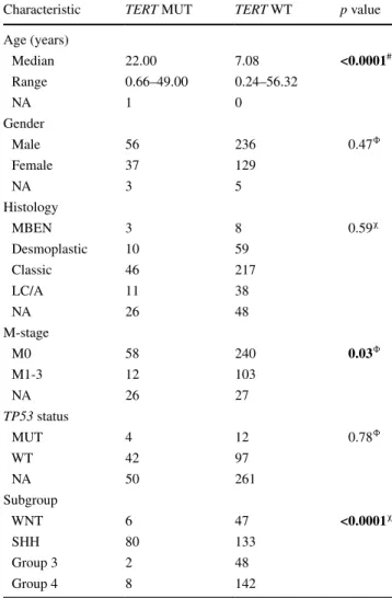
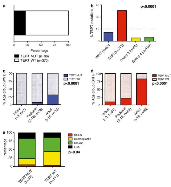
Characteristics of TERT-mutated medulloblastomas We performed Sanger sequencing on a clinically well- annotated medulloblastoma cohort (n = 466), reflecting the spectrum of demographics and histological subtypes of the disease (Table 1; Supplementary Figure 1A). Our results were verified using a Taqman-based genotyping assay that detects both of the most highly recurrent TERT promoter mutations (C228T and C250T). Since both mutational hotspots are located in highly homologous sequences, C228T and C250T mutations result in an iden- tical binding sequence for the mutation-specific probe (CCCggAAgggg; Supplementary Figure 1B). A total of 21 % of medulloblastomas harbored TERT mutations (Fig. 1a). In line with a previous report, these mutations were enriched in older patients (Table 1; p < 0.0001), all muta- tions were heterozygous, and none of the available matched germline controls displayed this mutation [13]. Interestingly, we found that TERT-mutated medulloblastomas present less
Table 1 Clinicopathological and molecular characteristics according to TERT mutational status
F female, LC/A large cell/anaplastic, M male, MB medulloblastomal, MBEN medulloblastoma with extensive nodularity, NA not available (data were excluded from statistical comparison)
Bold values indicate p < 0.05
# Mann–Whitney U test
Φ Fisher’s exact test
χ Chi-square test
Characteristic TERT MUT TERT WT p value
Age (years)
Median 22.00 7.08 <0.0001#
range 0.66–49.00 0.24–56.32
NA 1 0
gender
Male 56 236 0.47Φ
Female 37 129
NA 3 5
Histology
MBeN 3 8 0.59χ
Desmoplastic 10 59
Classic 46 217
lC/A 11 38
NA 26 48
M-stage
M0 58 240 0.03Φ
M1-3 12 103
NA 26 27
TP53 status
MUT 4 12 0.78Φ
WT 42 97
NA 50 261
Subgroup
WNT 6 47 <0.0001χ
SHH 80 133
group 3 2 48
group 4 8 142
frequently with metastatic dissemination at diagnosis com- pared to TERT wild-type tumors (p = 0.03).
TERT mutations are specifically enriched in SHH medulloblastomas
In a subgroup-specific analysis, we revealed that TERT mutations were significantly enriched in SHH tumors (80/213; 38 %; p < 0.0001) compared to WNT (6/53; 11 %) and group 3 (2/50; 4 %) or group 4 tumors (8/150; 5 %).
TERT mutations in both WNT and SHH medulloblasto- mas were positively correlated with age. TERT mutations were significantly enriched in adult patients (Fig. 1c, d, both p < 0.0001). Increasing age was not associated with increased mutational frequency in either group 3 or group 4 tumors (n.s.). While histopathological features were simi- lar between TERT-mutated and wild-type tumors across subgroups, we observed that classic histology was more commonly observed in TERT mutant SHH tumors, and
desmoplastic histology in wild-type SHH tumors (Fig. 1e;
Table 2; p = 0.04), respectively.
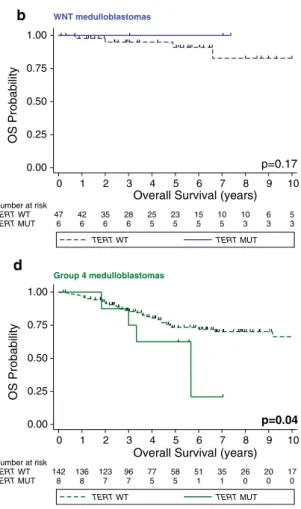
Prognostic implications of TERT mutations
When medulloblastoma patients across all subgroups were stratified by TERT mutational status, we observed no significant differences in survival (Fig. 2a; p = 0.45).
Further after normalizing the subgroup composition to reported subgroup ratios, a statistical difference was still not revealed (data not shown; p = 0.36) [1, 15, 26, 41].
However, when TERT mutational status is re-analyzed in a subgroup-specific manner, several important survival asso- ciations are observed. TERT mutations had no prognostic impact within WNT tumors (Fig. 2b; p = 0.17). However, a significant association between TERT promoter mutations and outcomes was noted in SHH and group 4 medullo- blastomas. Specifically, the 5-year overall survival of SHH tumors with and without TERT mutations was 77.6 ± 7 %
Fig. 1 TERT promoter-mutated medulloblastomas display dis- tinct demographics, histology, and subgroup affiliation. a Bar graph indicating the frequency of TERT mutations in 466 primary medulloblastomas. b Prevalence of TERT mutations according to medulloblastoma subgroups, and within, c WNT and d SHH subgroups accord- ing to age groups. Distribution of histological variants within SHH tumors according to TERT mutational status (e). MUT mutation, OS overall survival, WT wild-type
a b
0 25 50 75 100
TERT MUT (n=96) TERT WT (n=370) Percentage
WNT (n=53) SHH (n=213)
Group 3 (n=50) Group 4 (n=150) 0
15 30 45
c d
Infant (<3; n=2)
Pediatric (3-16; n=38)
Adult (>16; n=13) 0
25 50 75
100 TERT MUT
TERT WT
Infant (<3; n=64)
Pediatric (3-16; n=82)
Adult (>16; n=66) 0
25 50 75 100
TERT WT
% Age group (WNT) % Age group (SHH)% TERT mutations
TERT MUT
e
p<0.0001
p<0.0001 p<0.0001
TERT MUT (n=57)
TERT WT(n=111) 0
25 50 75
100 MBEN
Desmoplastic Classic LCA
Percentage
p=0.04
and 64.1 ± 5.1 %, respectively (Fig. 2c; p = 0.04). In contrast to the improved prognosis of TERT mutant SHH tumors, we observed the inverse pattern in group 4 tumors where the 5-year overall survival for patients with- out and with TERT mutations was 73.3 % ± 4.3 % and 62.5 % ± 17.1 % (Fig. 2d; p = 0.04). Similar to the unfa- vorable prognosis of TERT mutations in group 4 tumors, both of the patients with TERT-mutated group 3 tumors died after 7 and 45 months of follow-up, respectively (Sup- plementary Table 1). Thus, we conclude that TERT muta- tions define distinct prognostic patient cohorts in a sub- group-specific fashion with good prognosis in SHH and poor prognosis in group 4 medulloblastomas.
Survival analysis restricted to specific age groups
As TERT mutations are predominantly observed in non- infant medulloblastomas, we evaluated the prognostic
implications of these promoter mutations across all four medulloblastoma subgroups in an age-dependent manner.
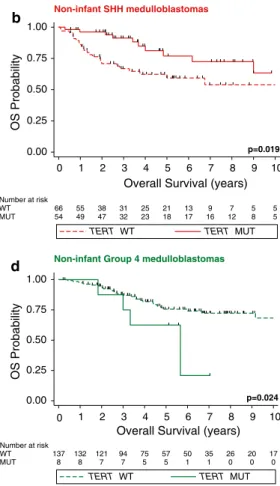
TERT mutational status across subgroups had no prog- nostic impact among patients older than 3 years of age at diagnosis (Fig. 3a; p = 0.59). Interestingly, the prognos- tic impact of TERT mutation was more pronounced in the non-infant SHH population with a 5-year overall survival of 76.9 % ± 7.6 % and 59.3 % ± 6.9 % of non-infants with and without TERT promoter mutations, respectively (Fig. 3b; p = 0.019). These prognostic implications were similar in adult medulloblastoma patients and in the adult SHH subgroup (Supplementary Figure 2). In a subset of 76 SHH cases with known TP53 mutational status [44], we revealed that TP53 mutations identify non-infant SHH tumors with a particularly poor prognosis, while in con- trast TERT mutations identify a subsets with good prog- nosis (Fig. 3c; p = 0.047). Mutations of both TERT and TP53 were observed in 4/12 SHH tumors (Supplementary Table 2). Non-infant group 4 showed an inverse prognos- tic association with poor outcome of TERT-mutated cases (Fig. 3d; p = 0.024). lastly, we analyzed the overall sur- vival of SHH patients under a multivariate Cox propor- tional hazards model comprising age at diagnosis, TERT mutational status, M-stage, and histology. In addition to the known prognostic significance of M-stage (p < 0.001) and histology (p = 0.02), we revealed that TERT status con- tinued to be associated with good prognosis (Hr 0.17, CI 0.04–0.69, p = 0.01), independent of other prognostic fac- tors including age at diagnosis (p = 0.35).
Distinct somatic copy number alterations of TERT-mutated medulloblastomas
To identify additional genetic features associated with these distinct demographic and clinical differences, we evaluated broad and focal copy number alterations according to sub- group affiliation and TERT promoter mutations. Notably, only 1/6 (17 %) of TERT-mutated WNT tumors harbored monosomy 6, while this alteration is observed in approxi- mately 80 % of TERT wild-type medulloblastomas of the WNT subgroup (Fig. 4a; p = 0.005). loss of chromo- some 2 and 10q loss were significantly enriched in TERT wild-type SHH tumors, while 3q loss was more frequently observed in their TERT mutant counterparts (Fig. 4b). Pre- viously described focal alterations characteristic for SHH tumors including amplification of MYCN/GLI2/CDK6/
YAP1/PPM1D, and deletions targeting PTCH1/CDKN2A/
CDKN2B/PTEN were largely confined to TERT wild-type SHH medulloblastomas, while TERT mutant SHH (Fig. 5) and group 4 (Supplementary Figure 3) showed very few focal SCNAs. Consistent with the higher frequency of TERT mutations in SHH tumors, we observed increased TERT expression in the SHH subgroup compared to group
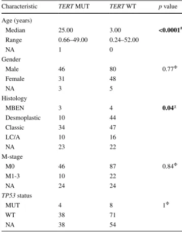
Table 2 Clinicopathological and molecular characteristics of SHH medulloblastoma according to TERT mutational status
F female, LC/A large cell/anaplastic, M male, MB medulloblastoma, MBEN medulloblastoma with extensive nodularity, NA not available (data were excluded from statistical comparison)
Bold values indicate p < 0.05
# Mann–Whitney U test
Φ Fisher’s exact test
χ Chi-square test
Characteristic TERT MUT TERT WT p value
Age (years)
Median 25.00 3.00 <0.0001#
range 0.66–49.00 0.24–52.00
NA 1 0
gender
Male 46 80 0.77Φ
Female 31 48
NA 3 5
Histology
MBeN 3 4 0.04χ
Desmoplastic 10 44
Classic 34 47
lC/A 10 16
NA 23 22
M-stage
M0 46 87 0.84Φ
M1-3 10 22
NA 24 24
TP53 status
MUT 4 8 1Φ
WT 38 71
NA 38 54
4 tumors in two independent gene expression profiling studies (p < 0.001; Supplementary Figure 4). Furthermore, we observed TERT amplification in two tumors included in the entire cohort of 1,088 previously studied tumors [30].
Both of these cases with TERT amplification were SHH- driven medulloblastomas with wild-type TERT status, which were derived from pediatric patients who were both alive after 15 and 83 months of follow-up (Supplementary Figure 5). Thus, broad and focal SCNAs underline that TERT mutations define a genetically distinct subset within SHH tumors and possibly within the WNT and group 4 tumors.
Discussion
The underlying biology of adult medulloblastomas remains poorly understood. Next-generation sequencing stud- ies have revealed a broad spectrum of novel, potentially tumorigenic mutations in the recent past, but none of these
studies focused on adult medulloblastomas [12, 25, 32, 34, 39]. In addition, the vast majority of these mutations are not recurrent enough to stratify patients into distinct clini- cal and prognostic subgroups.
In this study, we demonstrate that TERT promoter muta- tions, initially described in melanoma [10, 11], comprise the most recurrent mutation described so far across medul- loblastoma subgroups, with a particular enrichment in older patient cohorts. These somatic mutations are espe- cially common in older patients with SHH tumors (83 %) and to a lesser extent in adults with WNT medulloblasto- mas (11 %). Based on the transcriptional heterogeneity of SHH tumors in infant and adult patients, we suspect that the adult cluster mainly comprised TERT-mutated medul- loblastomas [24]. According to the initial classification of tumor types with TERT mutations at frequencies over 15 % (TERT-high) vs. below this threshold (TERT-low) [13], our report suggests distinct baseline telomerase activity of the cell of origin in each of the subgroups (group 3 ≥ group 4 > WNT >> SHH). Furthermore, the identification of
0.00 0.25 0.50 0.75 1.00
OS Probability
79 70 67 52 38 32 26 24 17 12 9
TERT MUT 354 312 262 206 164 133 106 73 58 42 37 TERT WT
Number at risk
0 1 2 3 4 5 6 7 8 9 10
Overall Survival (years)
TERT WT TERT MUT
0.00 0.25 0.50 0.75 1.00
OS Probability
6 6 6 6 5 5 5 5 3 3 3
TERT MUT 47 42 35 28 25 23 15 10 10 6 5 TERT WT
Number at risk
0 1 2 3 4 5 6 7 8 9 10
Overall Survival (years)
TERT WT TERT MUT
0.00 0.25 0.50 0.75 1.00
OS Probability
63 55 53 38 28 22 20 18 14 9 6
TERT MUT 118 97 70 55 42 37 26 17 14 11 10 TERT WT
Number at risk
0 1 2 3 4 5 6 7 8 9 10
Overall Survival (years)
TERT WT TERT MUT
0.00 0.25 0.50 0.75 1.00
OS Probability
8 8 7 7 5 5 1 1 0 0 0
TERT MUT 142 136 123 96 77 58 51 35 26 20 17 TERT WT
Number at risk
0 1 2 3 4 5 6 7 8 9 10
Overall Survival (years)
TERT WT TERT MUT
p=0.45 p=0.17
p=0.04 p=0.04
a b
c d
Medulloblastomas across subgroups WNT medulloblastomas
SHH medulloblastomas Group 4 medulloblastomas
Fig. 2 Prognostic impact of TERT promoter mutations varies accord- ing to medulloblastoma subgroups. Kaplan–Meier estimate display- ing overall survival (OS) according to TERT mutational status in pri-
mary medulloblastomas (a), within WNT (b), SHH (c), and group 4 (d) subgroups. Survival differences were calculated using continuous log-rank tests. MUT mutation, OS overall survival, WT wild-type
recurrent TERT promoter mutations makes a compelling argument that the increasing availability of whole-genome sequencing results may substantially add to a refined under- standing of the mutational landscape of different biological and age-driven medulloblastoma subgroups, since earlier
next-generation sequencing studies focusing on the pro- tein-coding regions had not encompassed gene-regulatory regions including promoter mutations.
In this study, we demonstrate that the mutational status of the TERT promoter can segregate individuals with SHH a
c
b
d
0.00 0.25 0.50 0.75 1.00
OS Probability
70 64 61 46 33 28 23 22 15 11 8
MUT 290 261 223 176 141 114 90 63 49 36 32
WT Number at risk
0 1 2 3 4 5 6 7 8 9 10
Overall survival (years)
TERT WT TERT MUT
0.00 0.25 0.50 0.75 1.00
OS Probability
54 49 47 32 23 18 17 16 12 8 5
MUT 66 55 38 31 25 21 13 9 7 5 5
WT Number at risk
0 1 2 3 4 5 6 7 8 9 10
Overall Survival (years)
TERT WT TERT MUT
0.00 0.25 0.50 0.75 1.00
OS Probability
8 8 7 7 5 5 1 1 0 0 0
MUT 137 132 121 94 75 57 50 35 26 20 17
WTNumber at risk
0 1 2 3 4 5 6 7 8 9 10
Overall Survival (years)
TERT WT TERT MUT
0.00 0.25 0.50 0.75 1.00
OS Probability
6 5 4 3 2 1 1 0 0 0 0
TP53 MUT 34 31 29 21 17 14 13 12 8 5 3
TERT MUT 36 32 23 20 15 13 9 7 5 3 3
WTNumber at risk
0 1 2 3 4 5 6 7 8 9 10
Overall Survival (years)
TERT /TP53 WT TERT MUT TP53 MUT p=0.047
p=0.59 p=0.019
p=0.024 Non-infant medulloblastomas across subgroups Non-infant SHH medulloblastomas
Non-infant Group 4 medulloblastomas Non-infant SHH medulloblastomas
Fig. 3 TERT promoter mutations delineate prognostic subsets within non-infant SHH and group 4 medulloblastomas. Kaplan–Meier esti- mate displaying overall survival (OS) in non-infant medulloblastomas (>3 years of age at diagnosis) according to TERT mutational status
across subgroups (a), in SHH tumors (b), in SHH tumors (TP53 mutated/wild-type) (c), and group 4 (d). Survival differences were calculated using continuous log-rank tests
Fig. 4 WNT and SHH medul- loblastoma harbor distinct broad genomic imbalances depending on the mutational status of TERT. Bar graphs indicating the frequency of broad cytogenetic alterations in WNT (a), and SHH (b) tumors.
★★ p < 0.01; ★ p < 0.05; MUT mutation, WT wild-type
a WNT tumors b
0.0 0.2 0.4 0.6 0.8 1.0
6q loss 6p loss
TERT MUT (n=6) TERT WT (n=43) Frequency
SHH tumors
0.0 0.1 0.2 0.3 0.4
10q loss 3q loss chr. 2 loss
TERT MUT (n=64) TERT WT (n=108) Frequency
and group 4 medulloblastomas with distinct prognostic outcomes, while a prognostic impact of this mutation was not observed in glioblastomas [23]. Molecular mecha- nisms converging on TERT up-regulation were recently reported to be associated with dismal prognosis in pediat- ric brain cancers [4]. Our findings in group 4 tumors with TERT mutations follow this pattern, while SHH tumors with TERT mutations comprise a prognostically favorable subgroup. Notably, survival curves of SHH tumors increas- ingly approximate with extended follow-up. We hypoth- esize that this pattern might be due to secondary malignan- cies and late relapses in older SHH tumors [36–38]. Since virtually all of the TERT promoter mutations encompass the mutational hotspots C228T and C250T, patient strati- fication can be carried out using a single PCr followed up with Sanger sequencing or with a single experiment using our newly designed Taqman-based genotyping assay.
The latter assay is particularly suitable for routine clini- cal applications as it is highly sensitive and specific (5 ng DNA input is sufficient). Furthermore, our Taqman-based genotyping assay can be used on DNA derived from fresh- frozen and formalin-fixed paraffin-embedded tissue, since it only amplifies a short DNA fragment.
Both hotspot mutations C228T and C250T create an e-twenty-six (eTS) binding motif [10, 11] resulting in up-regulation of TERT expression at the mrNA level [2], which was not observed at the protein level in glioblas- tomas [43]. We now demonstrate that SHH tumors with TERT mutations are mostly mutually exclusive with those harboring 10q loss (p = 0.017) Notably, the relatively favorable prognosis of TERT-mutated SHH medulloblas- tomas may be explained by the relative lack of high-risk biomarkers [17, 18, 24, 44].
In summary, we describe the demographic, clinico- pathological, and biological implications of TERT pro- moter mutations in a subgroup-specific fashion. This study underlines the dependence of adult WNT and SHH tumors to reacquire telomerase activity and suggests a potential prognostic utility of TERT mutational analysis in an era of individualized therapy.
Acknowledgments We thank Susan Archer for technical writing, and Nick Downey from Integrated DNA Technologies for support with probe/primer design. We acknowledge CrB HCl—Neurobiotec tumor bank (Hospices Civils de lyon, lyon, France). MDT is sup- ported by a CIHr Clinician Scientist Phase II award, funds from the garron Family Chair in Childhood Cancer research at The Hospital for Sick Children and the University of Toronto, and operating funds
from the Canadian Institutes of Health research, the National Insti- tutes of Health (r01CA159859 and r01CA148699) and the Pediatric Brain Tumor Foundation. Mr is supported by a fellowship from the Dr. Mildred Scheel Foundation for Cancer research/german Cancer Aid and funds from the Baden-Wurttemberg Foundation. Vr is sup- ported by a CIHr fellowship and an Alberta Innovates-Health Solu- tions Clinical Fellowship. KZ acknowledges research support from MH CZ-DrO FNBr 65269705. AK was supported by the TAMOP- 4.2.2A-11/1/KONV-2012-0025 project and the János Bolyai Scholar- ship of the Hungarian Academy of Sciences.
Conflict of interest The authors declare no conflicts of interest.
Open Access This article is distributed under the terms of the Crea- tive Commons Attribution license which permits any use, distribu- tion, and reproduction in any medium, provided the original author(s) and the source are credited.
References
1. Aaron rH, elion gB, Colvin OM, graham M, Keir S, Bigner DD, Friedman HS (1994) Busulfan therapy of central nervous system xenografts in athymic mice. Cancer Chemother Pharma- col 35(2):127–131
2. Arita H, Narita Y, Fukushima S, Tateishi K, Matsushita Y, Yoshida A, Miyakita Y, Ohno M, Collins VP, Kawahara N, Shibui S, Ichimura K (2013) Upregulating mutations in the TerT promoter commonly occur in adult malignant gliomas and are strongly associated with total 1p19q loss. Acta Neuropathol 126(2):267–
276. doi:10.1007/s00401-013-1141-6
3. Blasco MA (2005) Telomeres and human disease: ageing, cancer and beyond. Nat rev genet 6(8):611–622. doi:10.1038/nrg1656 4. Castelo-Branco P, Choufani S, Mack S, gallagher D, Zhang C,
lipman T, Zhukova N, Walker eJ, Martin D, Merino D, Wasser- man JD, elizabeth C, Alon N, Zhang l, Hovestadt V, Kool M, Jones DT, Zadeh g, Croul S, Hawkins C, Hitzler J, Wang JC, Baruchel S, Dirks PB, Malkin D, Pfister S, Taylor MD, Weksberg r, Tabori U (2013) Methylation of the TerT promoter and risk stratification of childhood brain tumours: an integrative genomic and molecular study. lancet Oncol 14(6):534–542. doi:10.1016/
S1470-2045(13)70110-4
5. Cho YJ, Tsherniak A, Tamayo P, Santagata S, ligon A, greulich H, Berhoukim r, Amani V, goumnerova l, eberhart Cg, lau CC, Olson JM, gilbertson rJ, gajjar A, Delattre O, Kool M, ligon K, Meyerson M, Mesirov JP, Pomeroy Sl (2011) Integra- tive genomic analysis of medulloblastoma identifies a molecu- lar subgroup that drives poor clinical outcome. J Clin Oncol 29(11):1424–1430. doi:10.1200/JCO.2010.28.5148
6. Dubuc AM, Northcott PA, Mack S, Witt H, Pfister S, Taylor MD (2010) The genetics of pediatric brain tumors. Curr Neurol Neu- rosci rep 10(3):215–223. doi:10.1007/s11910-010-0103-9 7. Forsyth Nr, Wright We, Shay JW (2002) Telomerase and dif-
ferentiation in multicellular organisms: turn it off, turn it on, and turn it off again. Differentiation 69(4–5):188–197.
doi:10.1046/j.1432-0436.2002.690412.x
8. Heaphy CM, Subhawong AP, Hong SM, goggins Mg, Mont- gomery eA, gabrielson e, Netto gJ, epstein JI, lotan Tl, Westra WH, Shih Ie M, Iacobuzio-Donahue CA, Maitra A, li QK, eber- hart Cg, Taube JM, rakheja D, Kurman rJ, Wu TC, roden rB, Argani P, De Marzo AM, Terracciano l, Torbenson M, Meeker AK (2011) Prevalence of the alternative lengthening of telomeres telomere maintenance mechanism in human cancer subtypes. Am J Pathol 179(4):1608–1615. doi:10.1016/j.ajpath.2011.06.018 Fig. 5 Focal somatic copy number alterations are largely confined
to TERT wild-type SHH medulloblastomas. gISTIC2 analysis indi- cating focal amplifications/deletions in 108 wild-type (a, c) and 64 mutant (b, d) SHH tumors, respectively. Star regions enriched for reported DNA copy number variations
◂
9. Hiyama e, Hiyama K (2007) Telomere and telomerase in stem cells. Br J Cancer 96(7):1020–1024. doi:10.1038/sj.bjc.6603671 10. Horn S, Figl A, rachakonda PS, Fischer C, Sucker A, gast
A, Kadel S, Moll I, Nagore e, Hemminki K, Schadendorf D, Kumar r (2013) TerT promoter mutations in familial and spo- radic melanoma. Science 339(6122):959–961. doi:10.1126/
science.1230062
11. Huang FW, Hodis e, Xu MJ, Kryukov gV, Chin l, garra- way lA (2013) Highly recurrent TerT promoter mutations in human melanoma. Science 339(6122):957–959. doi:10.1126/
science.1229259
12. Jones DT, Jager N, Kool M, Zichner T, Hutter B, Sultan M, Cho YJ, Pugh TJ, Hovestadt V, Stutz AM, rausch T, Warnatz HJ, ryzhova M, Bender S, Sturm D, Pleier S, Cin H, Pfaff e, Sieber l, Wittmann A, remke M, Witt H, Hutter S, Tzaridis T, Weischenfeldt J, raeder B, Avci M, Amstislavskiy V, Zapatka M, Weber UD, Wang Q, lasitschka B, Bartholomae CC, Schmidt M, von Kalle C, Ast V, lawerenz C, eils J, Kabbe r, Benes V, van Sluis P, Koster J, Volckmann r, Shih D, Betts MJ, russell rB, Coco S, Tonini gP, Schuller U, Hans V, graf N, Kim YJ, Monoranu C, roggendorf W, Unterberg A, Herold-Mende C, Milde T, Kulozik Ae, von Deimling A, Witt O, Maass e, rossler J, ebinger M, Schuhmann MU, Fruhwald MC, Hasselblatt M, Jabado N, rutkowski S, von Bueren AO, Williamson D, Clif- ford SC, McCabe Mg, Collins VP, Wolf S, Wiemann S, lehrach H, Brors B, Scheurlen W, Felsberg J, reifenberger g, Northcott PA, Taylor MD, Meyerson M, Pomeroy Sl, Yaspo Ml, Korbel JO, Korshunov A, eils r, Pfister SM, lichter P (2012) Dissect- ing the genomic complexity underlying medulloblastoma. Nature 488(7409):100–105. doi:10.1038/nature11284
13. Killela PJ, reitman ZJ, Jiao Y, Bettegowda C, Agrawal N, Diaz lA Jr, Friedman AH, Friedman H, gallia gl, giovanella BC, grollman AP, He TC, He Y, Hruban rH, Jallo gI, Mandahl N, Meeker AK, Mertens F, Netto gJ, rasheed BA, riggins gJ, rosenquist TA, Schiffman M, Shih Ie M, Theodorescu D, Tor- benson MS, Velculescu Ve, Wang Tl, Wentzensen N, Wood lD, Zhang M, Mclendon re, Bigner DD, Kinzler KW, Vogelstein B, Papadopoulos N, Yan H (2013) TerT promoter mutations occur frequently in gliomas and a subset of tumors derived from cells with low rates of self-renewal. Proc Natl Acad Sci USA 110(15):6021–6026. doi:10.1073/pnas.1303607110
14. Kim NW, Piatyszek MA, Prowse Kr, Harley CB, West MD, Ho Pl, Coviello gM, Wright We, Weinrich Sl, Shay JW (1994) Specific association of human telomerase activity with immortal cells and cancer. Science 266(5193):2011–2015
15. Kool M, Korshunov A, remke M, Jones DT, Schlanstein M, Northcott PA, Cho YJ, Koster J, Schouten-van Meeteren A, van Vuurden D, Clifford SC, Pietsch T, von Bueren AO, rutkowski S, McCabe M, Collins VP, Backlund Ml, Haberler C, Bourdeaut F, Delattre O, Doz F, ellison DW, gilbertson rJ, Pomeroy Sl, Taylor MD, lichter P, Pfister SM (2012) Molecular subgroups of medulloblastoma: an international meta-analysis of transcrip- tome, genetic aberrations, and clinical data of WNT, SHH, group 3, and group 4 medulloblastomas. Acta Neuropathol 123(4):473–
484. doi:10.1007/s00401-012-0958-8
16. Kool M, Koster J, Bunt J, Hasselt Ne, lakeman A, van Sluis P, Troost D, Meeteren NS, Caron HN, Cloos J, Mrsic A, Ylstra B, grajkowska W, Hartmann W, Pietsch T, ellison D, Clifford SC, Versteeg r (2008) Integrated genomics identifies five medul- loblastoma subtypes with distinct genetic profiles, pathway sig- natures and clinicopathological features. PloS One 3(8):e3088.
doi:10.1371/journal.pone.0003088
17. Korshunov A, remke M, Kool M, Hielscher T, Northcott PA, Williamson D, Pfaff e, Witt H, Jones DT, ryzhova M, Cho YJ, Wittmann A, Benner A, Weiss WA, von Deimling A, Scheurlen W, Kulozik Ae, Clifford SC, Peter Collins V, Westermann F,
Taylor MD, lichter P, Pfister SM (2012) Biological and clinical heterogeneity of MYCN-amplified medulloblastoma. Acta Neu- ropathol 123(4):515–527. doi:10.1007/s00401-011-0918-8 18. Korshunov A, remke M, Werft W, Benner A, ryzhova M, Witt
H, Sturm D, Wittmann A, Schottler A, Felsberg J, reifenberger g, rutkowski S, Scheurlen W, Kulozik Ae, von Deimling A, lichter P, Pfister SM (2010) Adult and pediatric medulloblasto- mas are genetically distinct and require different algorithms for molecular risk stratification. J Clin Oncol 28(18):3054–3060. doi:
10.1200/JCO.2009.25.7121
19. liu X, Bishop J, Shan Y, Pai S, liu D, Murugan AK, Sun H, el- Naggar AK, Xing M (2013) Highly prevalent TerT promoter mutations in aggressive thyroid cancers. endocr relat Cancer 20(4):603–610. doi:10.1530/erC-13-0210
20. louis DN, Ohgaki H, Wiestler OD, Cavenee WK, Burger PC, Jouvet A, Scheithauer BW, Kleihues P (2007) The 2007 WHO classification of tumours of the central nervous system. Acta Neuropathol 114(2):97–109. doi:10.1007/s00401-007-0243-4 21. Mermel CH, Schumacher Se, Hill B, Meyerson Ml, Ber-
oukhim r, getz g (2011) gISTIC2.0 facilitates sensitive and confident localization of the targets of focal somatic copy- number alteration in human cancers. genome Biol 12(4):r41.
doi:10.1186/gb-2011-12-4-r41
22. Morrison SJ, Prowse Kr, Ho P, Weissman Il (1996) Telomer- ase activity in hematopoietic cells is associated with self-renewal potential. Immunity 5(3):207–216
23. Nonoguchi N, Ohta T, Oh Je, Kim YH, Kleihues P, Ohgaki H (2013) TerT promoter mutations in primary and secondary glio- blastomas. Acta Neuropathol. doi:10.1007/s00401-013-1163-0 24. Northcott PA, Hielscher T, Dubuc A, Mack S, Shih D, remke M,
Al-Halabi H, Albrecht S, Jabado N, eberhart Cg, grajkowska W, Weiss WA, Clifford SC, Bouffet e, rutka JT, Korshunov A, Pfister S, Taylor MD (2011) Pediatric and adult sonic hedgehog medulloblastomas are clinically and molecularly distinct. Acta Neuropathol 122(2):231–240. doi:10.1007/s00401-011-0846-7 25. Northcott PA, Jones DT, Kool M, robinson gW, gilbertson rJ,
Cho YJ, Pomeroy Sl, Korshunov A, lichter P, Taylor MD, Pfister SM (2012) Medulloblastomics: the end of the beginning. Nat rev Cancer 12(12):818–834. doi:10.1038/nrc3410
26. Northcott PA, Korshunov A, Pfister SM, Taylor MD (2012) The clinical implications of medulloblastoma subgroups. Nat rev Neurol 8(6):340–351. doi:10.1038/nrneurol.2012.78
27. Northcott PA, Korshunov A, Witt H, Hielscher T, eberhart Cg, Mack S, Bouffet e, Clifford SC, Hawkins Ce, French P, rutka JT, Pfister S, Taylor MD (2011) Medulloblastoma comprises four distinct molecular variants. J Clin Oncol 29(11):1408–1414. doi:
10.1200/JCO.2009.27.4324
28. Northcott PA, Nakahara Y, Wu X, Feuk l, ellison DW, Croul S, Mack S, Kongkham PN, Peacock J, Dubuc A, ra YS, Zilber- berg K, Mcleod J, Scherer SW, Sunil rao J, eberhart Cg, gra- jkowska W, gillespie Y, lach B, grundy r, Pollack IF, Hamilton rl, Van Meter T, Carlotti Cg, Boop F, Bigner D, gilbertson rJ, rutka JT, Taylor MD (2009) Multiple recurrent genetic events converge on control of histone lysine methylation in medulloblas- toma. Nat genet 41(4):465–472. doi:10.1038/ng.336
29. Northcott PA, rutka JT, Taylor MD (2010) genomics of medul- loblastoma: from giemsa-banding to next-generation sequencing in 20 years. Neurosurg Focus 28(1):e6. doi:10.3171/2009.10.
FOCUS09218
30. Northcott PA, Shih DJ, Peacock J, garzia l, Morrissy AS, Zich- ner T, Stutz AM, Korshunov A, reimand J, Schumacher Se, Beroukhim r, ellison DW, Marshall Cr, lionel AC, Mack S, Dubuc A, Yao Y, ramaswamy V, luu B, rolider A, Cavalli FM, Wang X, remke M, Wu X, Chiu rY, Chu A, Chuah e, Corbett rD, Hoad gr, Jackman SD, li Y, lo A, Mungall Kl, Nip KM, Qian JQ, raymond Ag, Thiessen NT, Varhol rJ, Birol I, Moore
rA, Mungall AJ, Holt r, Kawauchi D, roussel MF, Kool M, Jones DT, Witt H, Fernandez lA, Kenney AM, Wechsler-reya rJ, Dirks P, Aviv T, grajkowska WA, Perek-Polnik M, Haberler CC, Delattre O, reynaud SS, Doz FF, Pernet-Fattet SS, Cho BK, Kim SK, Wang KC, Scheurlen W, eberhart Cg, Fevre-Montange M, Jouvet A, Pollack IF, Fan X, Muraszko KM, gillespie gY, Di rocco C, Massimi l, Michiels eM, Kloosterhof NK, French PJ, Kros JM, Olson JM, ellenbogen rg, Zitterbart K, Kren l, Thompson rC, Cooper MK, lach B, Mclendon re, Bigner DD, Fontebasso A, Albrecht S, Jabado N, lindsey JC, Bailey S, gupta N, Weiss WA, Bognar l, Klekner A, Van Meter Te, Kumabe T, Tominaga T, elbabaa SK, leonard Jr, rubin JB, liau lM, Van Meir eg, Fouladi M, Nakamura H, Cinalli g, garami M, Hauser P, Saad Ag, Iolascon A, Jung S, Carlotti Cg, Vibhakar r, ra YS, robinson S, Zollo M, Faria CC, Chan JA, levy Ml, Sorensen PH, Meyerson M, Pomeroy Sl, Cho YJ, Bader gD, Tabori U, Hawkins Ce, Bouffet e, Scherer SW, rutka JT, Malkin D, Clif- ford SC, Jones SJ, Korbel JO, Pfister SM, Marra MA, Taylor MD (2012) Subgroup-specific structural variation across 1,000 medul- loblastoma genomes. Nature 488(7409):49–56. doi:10.1038/
nature11327
31. Northcott PA, Shih DJ, remke M, Cho YJ, Kool M, Hawkins C, eberhart Cg, Dubuc A, guettouche T, Cardentey Y, Bouffet e, Pomeroy Sl, Marra M, Malkin D, rutka JT, Korshunov A, Pfis- ter S, Taylor MD (2011) rapid, reliable, and reproducible molec- ular sub-grouping of clinical medulloblastoma samples. Acta Neuropathol. doi:10.1007/s00401-011-0899-7
32. Parsons DW, li M, Zhang X, Jones S, leary rJ, lin JC, Boca SM, Carter H, Samayoa J, Bettegowda C, gallia gl, Jallo gI, Binder ZA, Nikolsky Y, Hartigan J, Smith Dr, gerhard DS, Fults DW, VandenBerg S, Berger MS, Marie SK, Shinjo SM, Clara C, Phillips PC, Minturn Je, Biegel JA, Judkins Ar, resnick AC, Storm PB, Curran T, He Y, rasheed BA, Friedman HS, Keir ST, Mclendon r, Northcott PA, Taylor MD, Burger PC, riggins gJ, Karchin r, Parmigiani g, Bigner DD, Yan H, Papadopoulos N, Vogelstein B, Kinzler KW, Velculescu Ve (2011) The genetic landscape of the childhood cancer medulloblastoma. Science 331(6016):435–439. doi:10.1126/science.1198056
33. Pfister SM, Korshunov A, Kool M, Hasselblatt M, eberhart C, Taylor MD (2010) Molecular diagnostics of CNS embryo- nal tumors. Acta Neuropathol 120(5):553–566. doi:10.1007/
s00401-010-0751-5
34. Pugh TJ, Weeraratne SD, Archer TC, Pomeranz Krummel DA, Auclair D, Bochicchio J, Carneiro MO, Carter Sl, Cibulskis K, erlich rl, greulich H, lawrence MS, lennon NJ, McK- enna A, Meldrim J, ramos AH, ross Mg, russ C, Shefler e, Sivachenko A, Sogoloff B, Stojanov P, Tamayo P, Mesirov JP, Amani V, Teider N, Sengupta S, Francois JP, Northcott PA, Tay- lor MD, Yu F, Crabtree gr, Kautzman Ag, gabriel SB, getz g, Jager N, Jones DT, lichter P, Pfister SM, roberts TM, Meyer- son M, Pomeroy Sl, Cho YJ (2012) Medulloblastoma exome sequencing uncovers subtype-specific somatic mutations. Nature 488(7409):106–110. doi:10.1038/nature11329
35. ramaswamy V, Northcott PA, Taylor MD (2011) FISH and chips:
the recipe for improved prognostication and outcomes for chil- dren with medulloblastoma. Cancer genet 204(11):577–588.
doi:10.1016/j.cancergen.2011.11.001
36. ramaswamy V, remke M, Bouffet e, Faria CC, Perreault S, Cho YJ, Shih DJ, luu B, Dubuc AM, Northcott PA, Schüller U, gururangan S, Mclendon r, Bigner D, Fouladi M, ligon Kl, Pomeroy Sl, Dunn S, Triscott J, Jabado N, Fontebasso A, Jones DT, Kool M, Karajannis MA, gardner Sl, Zagzag D, Nunes S, Pimentel J, Mora J, lipp e, Walter AW, ryzhova M, Zhelud- kova O, Kumirova e, Alshami J, Croul Se, rutka JT, Hawkins C, Tabori U, Codispoti Ke, Packer rJ, Pfister SM, Korshunov A, Taylor MD (2013) recurrence patterns across medulloblastoma
subgroups: an integrated clinical and molecular analysis. lancet Oncol. doi:10.1016/S1470-2045(13)70449-2
37. remke M, Hielscher T, Korshunov A, Northcott PA, Bender S, Kool M, Westermann F, Benner A, Cin H, ryzhova M, Sturm D, Witt H, Haag D, Toedt g, Wittmann A, Schottler A, von Bueren AO, von Deimling A, rutkowski S, Scheurlen W, Kulozik Ae, Taylor MD, lichter P, Pfister SM (2011) FSTl5 Is a marker of poor prognosis in Non-WNT/Non-SHH medulloblastoma. J Clin Oncol 29(29):3852–3861. doi:10.1200/JCO.2011.36.2798 38. remke M, Hielscher T, Northcott PA, Witt H, ryzhova M, Witt-
mann A, Benner A, von Deimling A, Scheurlen W, Perry A, Croul S, Kulozik Ae, lichter P, Taylor MD, Pfister SM, Kors- hunov A (2011) Adult medulloblastoma comprises three major molecular variants. J Clin Oncol 29(19):2717–2723. doi:10.1200/
JCO.2011.34.9373
39. robinson g, Parker M, Kranenburg TA, lu C, Chen X, Ding l, Phoenix TN, Hedlund e, Wei l, Zhu X, Chalhoub N, Baker SJ, Huether r, Kriwacki r, Curley N, Thiruvenkatam r, Wang J, Wu g, rusch M, Hong X, Becksfort J, gupta P, Ma J, easton J, Vadodaria B, Onar-Thomas A, lin T, li S, Pounds S, Paugh S, Zhao D, Kawauchi D, roussel MF, Finkelstein D, ellison DW, lau CC, Bouffet e, Hassall T, gururangan S, Cohn r, Ful- ton rS, Fulton ll, Dooling DJ, Ochoa K, gajjar A, Mardis er, Wilson rK, Downing Jr, Zhang J, gilbertson rJ (2012) Novel mutations target distinct subgroups of medulloblastoma. Nature 488(7409):43–48. doi:10.1038/nature11213
40. rutkowski S, von Hoff K, emser A, Zwiener I, Pietsch T, Fig- arella-Branger D, giangaspero F, ellison DW, garre Ml, Bias- soni V, grundy rg, Finlay Jl, Dhall g, raquin MA, grill J (2010) Survival and prognostic factors of early childhood medulloblastoma: an international meta-analysis. J Clin Oncol 28(33):4961–4968. doi:10.1200/JCO.2010.30.2299
41. Taylor MD, Northcott PA, Korshunov A, remke M, Cho YJ, Clif- ford SC, eberhart Cg, Parsons DW, rutkowski S, gajjar A, elli- son DW, lichter P, gilbertson rJ, Pomeroy Sl, Kool M, Pfister SM (2012) Molecular subgroups of medulloblastoma: the cur- rent consensus. Acta Neuropathol 123(4):465–472. doi:10.1007/
s00401-011-0922-z
42. Thompson MC, Fuller C, Hogg Tl, Dalton J, Finkelstein D, lau CC, Chintagumpala M, Adesina A, Ashley DM, Kellie SJ, Taylor MD, Curran T, gajjar A, gilbertson rJ (2006) genomics iden- tifies medulloblastoma subgroups that are enriched for specific genetic alterations. J Clin Oncol 24(12):1924–1931. doi:10.1200/
JCO.2005.04.4974
43. Vinagre J, Almeida A, Populo H, Batista r, lyra J, Pinto V, Coelho r, Celestino r, Prazeres H, lima l, Melo M, rocha Ag, Preto A, Castro P, Castro l, Pardal F, lopes JM, Santos ll, reis rM, Cameselle-Teijeiro J, Sobrinho-Simoes M, lima J, Maximo V, Soares P (2013) Frequency of TerT promoter muta- tions in human cancers. Nature Commun 4:2185. doi:10.1038/nc omms3185
44. Zhukova N, ramaswamy V, remke M, Pfaff e, Shih DJ, Mar- tin DC, Castelo-Branco P, Baskin B, ray PN, Bouffet e, von Bueren AO, Jones DT, Northcott PA, Kool M, Sturm D, Pugh TJ, Pomeroy Sl, Cho YJ, Pietsch T, gessi M, rutkowski S, Bognar l, Klekner A, Cho BK, Kim SK, Wang KC, eberhart Cg, Fevre- Montange M, Fouladi M, French PJ, Kros M, grajkowska WA, gupta N, Weiss WA, Hauser P, Jabado N, Jouvet A, Jung S, Kum- abe T, lach B, leonard Jr, rubin JB, liau lM, Massimi l, Pol- lack IF, Shin ra Y, Van Meir eg, Zitterbart K, Schuller U, Hill rM, lindsey JC, Schwalbe eC, Bailey S, ellison DW, Hawkins C, Malkin D, Clifford SC, Korshunov A, Pfister S, Taylor MD, Tabori U (2013) Subgroup-specific prognostic implications of TP53 mutation in medulloblastoma. J Clin Oncol 31(23):2927–
2935. doi:10.1200/JCO.2012.48.5052