R E V I E W Open Access
Molecular markers and potential
therapeutic targets in non-WNT/non-SHH (group 3 and group 4) medulloblastomas
Otília Menyhárt1,2, Felice Giangaspero3,4and Balázs Győrffy1,2*
Abstract
Childhood medulloblastomas (MB) are heterogeneous and are divided into four molecular subgroups. The provisional non-wingless-activated (WNT)/non-sonic hedgehog-activated (SHH) category combining group 3 and group 4 represents over two thirds of all MBs, coupled with the highest rates of metastases and least understood pathology. The molecular era expanded our knowledge about molecular aberrations involved in MB tumorigenesis, and here, we review processes leading to non-WNT/non-SHH MB formations.
The heterogeneous group 3 and group 4 MBs frequently harbor rare individual genetic alterations, yet the
emerging profiles suggest that infrequent events converge on common, potentially targetable signaling pathways.
A mutual theme is the altered epigenetic regulation, and in vitro approaches targeting epigenetic machinery are promising. Growing evidence indicates the presence of an intermediate, mixed signature group along group 3 and group 4, and future clarifications are imperative for concordant classification, as misidentifying patient samples has serious implications for therapy and clinical trials.
To subdue the high MB mortality, we need to discern mechanisms of disease spread and recurrence. Current preclinical models do not represent the full scale of group 3 and group 4 heterogeneity: all of existing group 3 cell lines are MYC-amplified and most mouse models resemble MYC-activated MBs. Clinical samples provide a wealth of information about the genetic divergence between primary tumors and metastatic clones, but recurrent MBs are rarely resected. Molecularly stratified treatment options are limited, and targeted therapies are still in preclinical development. Attacking these aggressive tumors at multiple frontiers will be needed to improve stagnant survival rates.
Keywords:Medulloblastoma, Prognostic biomarker, Risk stratification, Survival, Non-WNT/non-SHH, Group 3, Group 4
Introduction
Medulloblastoma (MB) is the most common pediatric brain tumor [1], with a culminating incidence among chil- dren before the age of five [2]. Unfortunately, disease dis- semination is an early event, and as many as 40% of patients carry metastases already at diagnosis [3], with a grim outlook for survival [4]. Metastatic disease and tumor recurrence are responsible for the stagnant survival
rates of the past decades [1,2], while survivors frequently face treatment-related adverse effects [1].
The current consensus agrees upon four distinct molecu- lar entities within MBs: wingless-activated (WNT), sonic hedgehog-activated (SHH), group 3, and group 4 MBs [5], each characterized by specific mutations, copy number al- terations, transcriptomic/methylomic profiles, and clinical outcomes [6–9]. Subgroup assignment is prognostic with markedly different survival rates; a 5-year overall survival (OS) is as high as 95% in WNT, while group 3 patients fea- ture the worst (45–60%), with the shortest survival among infants. An intermediate (75–80%) OS characterizes group 4 and SHH MBs, although it also depends on histology, presence of metastases, and molecular abnormalities such as mutations and oncogene amplifications [10–14].
© The Author(s). 2019Open AccessThis article is distributed under the terms of the Creative Commons Attribution 4.0 International License (http://creativecommons.org/licenses/by/4.0/), which permits unrestricted use, distribution, and reproduction in any medium, provided you give appropriate credit to the original author(s) and the source, provide a link to the Creative Commons license, and indicate if changes were made. The Creative Commons Public Domain Dedication waiver (http://creativecommons.org/publicdomain/zero/1.0/) applies to the data made available in this article, unless otherwise stated.
* Correspondence:gyorffy.balazs@ttk.mta.hu
12nd Department of Pediatrics, Semmelweis University, Tűzoltó u. 7-9, Budapest H-1094, Hungary
2MTA TTK Lendület Cancer Biomarker Research Group, Institute of Enzymology, Hungarian Academy of Sciences, Magyar tudósok körútja 2, Budapest H-1117, Hungary
Full list of author information is available at the end of the article
Group 3 and group 4 MBs are more related to each other than to WNT and SHH and appear as non-WNT/
non-SHH in the revised 2016 WHO classification [15], yet they are molecularly and clinically heterogeneous with diverse outcomes [16–18]. The provisional non-WNT/non-SHH category presents a complex chal- lenge as these tumors represent over two thirds of all MBs, coupled with the highest rates of disseminated dis- ease and least understood pathology.
Here we aim to summarize the present state of non-WNT/non-SHH MB research, with a particular focus on molecular similarities and differences between group 3 and group 4 MBs.
Clinical attributes of group 3 and group 4 MBs The demography of group 3 or group 4 MB patients overlaps although the subtypes are associated with radic- ally different prognosis and clinical outcome.
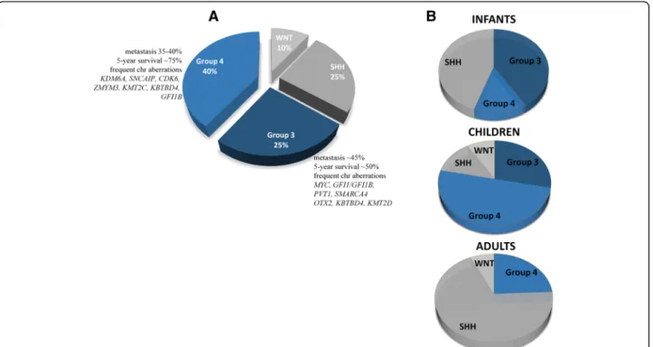
Group 3 MBs account for approximately 25% of all cases, predominantly among infants and children, with a peak diagnosis between ages 3 and 5 years and almost never in adults; hence, in adults, only three MB sub- groups can be differentiated [19, 20] (Fig. 1). The male-to-female ratio is approximately two to one [12].
Group 3 MBs are the deadliest of all molecular sub- groups, with a 58% 5-year OS in children and a 45%
5-year OS in infants [10, 16,21]. The grim outcome re- sults from the aggregation of adverse prognostic factors,
such as young age or presence of metastases (in up to 50% of patients) at diagnosis, large cell/anaplastic (LCA) histology, andMYCamplification. Group 3 is most likely to consist of multiple subcategories, out of which MYC-amplified tumors confer an especially short sur- vival; only 20% of these patients survive up to 5 years [18,22]. Group 3 MBs rarely recur at the original tumor site, but reappear as metastases [23]. The rate of metas- tasis does not necessarily reflect survival [12]; thus, chil- dren with group 3 disease without disease spread who are assigned to be standard-risk may face undertreat- ment [10]. Targeted treatments are not yet developed for group 3 patients due to our limited understanding of tumorigenesis.
Group 4 MB is the most prevalent biological subtype, comprising approximately 40% of all MB patients, pre- dominantly between ages 3 and 16 years, and yet, its pathogenesis is the least understood [5, 10]. Very few in- fants, approximately 45% of childhood and 25% of adult cases, belong to this subgroup (Fig.1), and it is three times more frequent in males than in females across all age groups [5,10]. The prognosis for group 4 patients is inter- mediate, and the 5-year survival reaches 80% when treated with standard therapy [13], although non-metastatic group 4 patients with chromosome 11 loss have an excel- lent prognosis, with > 90% survival [8]. Approximately 30–40% of group 4 MB patients have metastases at diag- nosis and are currently treated as high risk, including
Fig. 1Molecular subgroups of medulloblastoma. The current consensus divides medulloblastoma into four subgroups: WNT-activated (WNT), SHH-activated (SHH), group 3, and group 4. Only the most frequently altered genes are listed for group 3 and group 4 (a). Adult samples are extremely rare among group 3 patients, while the majority of group 4 tumors consist of children (b)
those with an LCA histology. The 5-year survival of high-risk patients is approximately 60% [8,10,14]. Adults with group 4 MBs have a significantly worse prognosis compared to the SHH- or WNT-activated subtypes [19].
Molecular identification of group 3 and group 4 MBs
The 2016 WHO classification refers to MB subgroups as genetically defined variants with prognostic value and treats group 3 and group 4 MBs as provisional entities. The rec- ommendation integrates histological and molecular classifi- cations, with different prognosis for classical or LCA histology, the latter usually associated with a high-risk dis- ease (although extremely rare in group 4) [15].
Initially, immunohistochemistry (IHC)-based markers were developed to allocate molecular subgroup identity to clinical samples. A diagnostic method involving a dis- tinct set of antibodies (GAB1, β-catenin, filamin A, and YAP1) distinguished WNT- and SHH-activated and non-WNT/non-SHH MB subgroups in FFPE samples [24]. Another four-antibody approach to identify sub- groups also from FFPE samples included DKK1 for WNT, SFRP1 for SHH, NPR3 for group 3, and KCNA1 for group 4 MBs, allocating 98% of samples into each subcategory [12]. Nevertheless, subgroup assignment solely based on IHC is not recommended any longer:
patchy nuclear β-catenin accumulation might be mis- leading [25, 26] and validation studies revealed KCNA1 expression in all subgroups, making it unsuitable for clas- sification [27]. Suboptimal reproducibility of IHC results arising from different protocols, institutional standards, and interpretations arrange for inconsistencies [25].
Identification of group 3 and group 4 MBs should be based on either methylation or transcriptional profiling to identifying samples clustering with other tumors of the same type [8,25]. Transcription may be assessed by either genome-wide transcriptomics or specific gene panels, for instance, the NanoString 22 gene signature. The assay evaluates group 3 identity utilizing the expression of IMPG2,GABRA5, EGFL11,NRL,MAB21 L2, andNPR3, while allocates group 4 tumors based onKCNA1,EOMES, KHDRBS2, RBM24, UNC5D, and OASI expression [28].
The methylation- or transcriptional profiling-based classi- fications are robust, although their implementation might be challenging in the daily practice.
A clinically applicable rapid approach classified non-WNT/non-SHH MBs with 92% accuracy based on highly specific epigenetic biomarkersfrom both fresh frozen and FFPE samples. The differentially methylated CpG probes were located within an intergenic region of chromo- some 12, the intronic regions of RPTORand RIMS2, and the 3′-UTR region of VPS37B genes. The method accur- ately classified unambiguous group 3 and group 4 cases,
however demonstrated limited discrimination capacity with tumors harboring intermediate methylation profiles[29].
MBs with ambiguous subgroup identity
A growing number of studies suggest that subgroups within non-WNT/non-SHH tumors should be explored further to capture patient diversity. A large-scale study utilizing methylomic data revealed a shared biological signature between group 3 and group 4 tumors, suggest- ing a likelihood of common origin. Combining the two subgroups, especially low-risk group 3 and group 4 sam- ples for clinical purposes, results in a categorization out- performing the current risk stratification models [30]
(Fig. 2a and 3a). Integration of methylomic and tran- scriptomic data found ambiguous subgroup identity in 3% of samples [31]. Gene expression-based clustering also identified non-WNT/non-SHH subtypes with mixed signatures [18, 32]. The ambiguity of categorization has been reflected in established MB cell lines: D283 cells have been categorized in the past as both group 3 [33]
and group 4 [34] and, along with the D721 cell line, ex- press high levels of bothMYC andOTX2mRNA. These cell lines were placed eventually to an intermediate cat- egory [35].
Three MB subgroups within non-WNT/non-SHH tumors were recently described: group 3, group 4, and intermediate group 3/4 MBs, the latter with re- markably good prognosis [36]. Although based on a limited sample size, the results imply that provisional group 3 and group 4 distinctions could misplace a portion of patients. The study extended the Nano- String 22 gene signature [28] further, including the expression of SNCAIP, MYCC, RCVRN, and PDC genes. Future clarifications ought to standardize the methods for diagnostic purposes as patient misclassifi- cation has serious implications for treatment and en- rollment into clinical trials.
Molecular biology of group 3 and group 4 MBs Genetic predispositions
Damaging germline mutations in known cancer predis- position genes is rare in non-WNT/non-SHH MB pediatric patients. In a sample of 1022 MBs, germline BRCA2 and PALB2 mutations were present in 1–2% of group 3/group 4 tumors, associated with mutational sig- natures typical of homologous recombination repair (HR) deficiency. Occasional heterozygous germline FANCA(n= 1, group 3) orFANCQ(n= 1, group 4) mu- tations were also identified and linked to an HR-deficiency mutation spectrum. Genetic testing for these patients is recommended in case of a familial his- tory of BRCA-associated cancers or if mutational signa- tures are suggestive of HR deficiency [37].
Recurrent somatic driver events
Group 3 and group 4 MBs are genetically heterogeneous and, unlike WNT and SHH-activated MBs, are not driven by well-defined, constitutively activated signaling pathways. Tetraploidy is a recurrent early genetic event in both group 3 and group 4 MBs, leading to an in- creased number of large-scale copy number gains [38].
A meta-analysis based on 550 samples identified a gain of 17q (in 58% of samples) and loss of 17p (55%) along with a loss of 16q(42%), 10q(43%), and 9q(21%) and gain of 7 (39%) and 1q (41%) as most recurrent struc- tural aberrations in Group 3 MBs [10] (Table 1). Tetra- ploidy also occurs early in approximately 40% of group 4 tumors [38], but its prognostic significance is yet un- clear. Isochromosome 17q (a chromosome with two 17q arms) is present in about 80% of all group 4 samples but is not predictive of outcome. Chromosome 7 gain (47%),8p loss (41%),10q loss (15%), and11p and18q aberrations are also regular events (Table 2). Approxi- mately 80% of females have a complete loss of one X
chromosome [10,12,18,39]. Both group 3 and group 4 MBs harbor frequent chromosomal aberrations although somatic mutations are relatively infrequent. In fact, more than half of group 3 samples are thought to be devoided of mutations; based on deep sequencing of 92 samples, none of the 12 most significantly mutated genes were al- tered in group 3 and group 4 tumors [21,40].
Somatic MYC (17% in group 3) and MYCN (6% in group 4) amplifications are the most frequently observed driver events [28]. The link between MYC and group 3 MB outcome is well established, and high MYC levels are associated with significantly reduced survival [18, 41].MYCactivation develops because of amplification at the MYC loci, genomic rearrangement of PVT1–MYC, or other yet-unknown mechanisms [22,28,42–44].
Recently, a study with a large sample size identified at least one potential driver events in 76% of group 3 and 82% of group 4 MBs, with an almost equal occurrence ofMYCN amplifications across group 3 (5%) and group 4 (6%), with MYC amplifications restricted to group 3
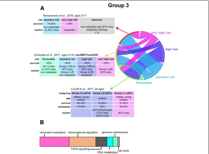
Fig. 2Risk stratification, proposed prognostic biomarkers, and major mechanisms of tumorigenesis in group 3 medulloblastomas (a). Schematic representation of major mechanisms most frequently affected by somatic alterations within group 3 tumors contributing to medulloblastoma development (b). LR, low risk; HR, high risk
tumors (17%) [6]. Activation of the mutually exclusive GFI1/GFI1Bwas identified as the most prevalent driver event through“enhancer hijacking”, by depositing them near active regulatory elements. Hotspot insertions tar- geting a novel potential oncogene, KBTBD4, were also frequent both in group 3 and group 4 samples [6, 38].
The prognostic significance of GFI1/GFI1B activation is not yet clear [45], although a large-scale integrative ana- lysis of gene expression and methylation data indicated the presence ofGFI1/GFI1B activations mainly within a particular subtype of group 3 tumors [31].
A single copy gain of the SNCAIPgene is present in over 10% of group 4 tumors and represents the most distinctly upregulated gene within the group 4 signature.
SNCAIP is involved in the development of Parkinson’s disease, and its tandem duplications in group 4 MBs are mutually exclusive with MYCN and CDK6 amplifica- tions, the latter present in 5–10% of all group 4 tumors [18, 28]. In group 4 MBs, PRDM6, an epigenetic
regulator of gene activity, is the probable target of SNCAIP-associated enhancer hijacking and is activated in about 17% of tumors [6].
SMARCA4 encoding subunits of the SWI/SNF-like chromatin-remodeling complex is among the most re- currently (~ 9%) mutated genes in group 3 tumors [6,38].
Network analysis of group 4 somatic copy number aberra- tions revealed the enrichment of genes responsible for chromatin modification and identified a novel homolo- gous deletion of a histone-lysine demethylase, KDM6A [28], that preferentially demethylates the H3K27 trimethyl mark (H3K27me3) [46]. Somatic mutations of the KDM6A gene are exclusively present in approximately 12% of group 4 tumors, along with frequent mutations of other 6 KDM family members (KDM1A, KDM3A, KDM4A,KDM5A,KDM5B, andKDM7A) [21,38,40,47]
(Table 2). EZH2 is also amplified or overexpressed in group 3 and 4 tumors, contributing to the inscription of H3K27me3, and is mutually exclusive with KDM6A
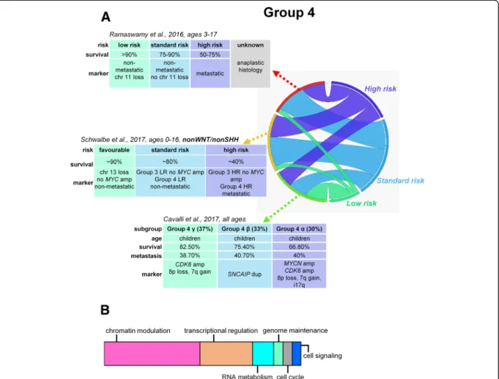
Fig. 3Risk stratification, proposed prognostic biomarkers, and major mechanisms of tumorigenesis in group 4 medulloblastomas (a). Schematic representation of major mechanisms most frequently affected by somatic alterations within group 4 MBs contributing to medulloblastoma development (b). LR, low risk; HR, high risk
Table 1Frequent genetic alterations in group 3 MBs according to [6,12,28,38,40,113]
Percentage of patients
Gene/
chromosome
Modification Gene name Gene
location
Gene function
58 17q Mainly gain – – –
55 17p Mainly loss – – –
55 8q Gain or loss – – –
51 8p Gain or loss – – –
48 7q Mainly gain – – –
43 10q Mainly loss – – –
42 16q Mainly loss – – –
41 1q Mainly gain – – –
39 7p Mainly gain – – –
38 13q Gain or loss – – –
34 11q Mainly loss – – –
32 11p Mainly loss – – –
31 5q Mainly gain – – –
30 5p Mainly gain – – –
21 X Loss – – –
17 MYC Amplification,
overexpression
MYC proto-oncogene, bHLH transcription factor
8q24.21 Transcriptional regulation
12 PVT1 Amplification Pvt1 oncogene (non-protein coding) 8q24.21 Oncogenic lncRNA
11 GFI1B overexpression,
amplification, deletion
Growth factor independent 1B transcriptional repressor
9q34.13 Transcriptional regulation
9 SMARCA4 Mutation SWI/SNF-related, matrix-associated, actin-dependent regulator of chromatin, subfamily a, member 4
19p13.2 Chromatin modulation, SWI/SNF Nucleosome-remodeling complex 6 KBTBD4 Mutation Kelch repeat and BTB domain containing 4 11p11.2 Ubiquitination of target
substrates
6 SHPRH Low level
amplification
SNF2 histone linker PHD RING helicase 6q24.3 Genome maintenance
5 CD109 Deletion CD109 molecule 6q13 TGF-βsignaling
5 CTDNEP1 Mutation CTD nuclear envelope phosphatase 1 17p13.1 Metabolism of fatty acids
5 KMT2D Mutation Lysine methyltransferase 2D 12q13.12 Chromatin modulation
5 KDM7A Mutation Lysine demethylase 7A 7q34 Chromatin modulation
5 CHD7 Mutation Chromodomain helicase DNA binding protein 7 8q12.2 Chromatin modulation
5 DDX3X Mutation DEAD-box helicase 3, X-linked Xp11.4 RNA metabolism
5 KDM3A Mutation Lysine demethylase 3A 2p11.2 Chromatin modulation
5 KDM4C Mutation Lysine demethylase 4C 9p24.1 Chromatin modulation
5 KDM5B Mutation Lysine demethylase 5B 1q32.1 Chromatin modulation
5 KDM6A Mutation Lysine demethylase 6A Xp11.3 Chromatin modulation
5 MYCN Amplification MYCN proto-oncogene, bHLH transcription factor 2p24.3 Transcriptional regulation
5 CREBBP Amplification CREB binding protein 16p13.3 Chromatin modulation,
transcription initiation
5 DDX31 Amplification DEAD-box helicase 31 9q34.13 RNA metabolism
4 ESRRG Low level
amplification
Estrogen-related receptor gamma 1q41 Transcriptional regulation, estrogen signaling
4 SNX6 Deletion Sorting nexin 6 14q13.1 TGF-βsignaling
4 GFI1 Overexpression,
amplification
Growth factor independent 1 transcriptional repressor
1p22.1 Transcriptional regulation
3 OTX2 Amplification, Orthodenticle homeobox 2 14q22.3 Transcriptional regulation
mutations. About 50% of tumors with KDM6A and KDM1Amutations also harbor ZMYM3 mutations, sug- gesting a cooperation between these two genes [47]. The relatively numerousCHD7orZMYM3mutations partake in the regulation of the H3K4me3 mark [6]. Inactivating mutations in MLL2 and MLL3 genes also participate in the reduction of H3K4me3 levels, promoting the deactiva- tion of prodifferentiation genes [38, 48]. TBR1 and EOMESexpression is significantly higher in group 3 and 4 tumors compared to other subgroups and strongly corre- lates with gene methylation [38]. These observations sug- gest that by preserving methylation marks, both group 3 and group 4 MBs retain a stem-like epigenetic state and their pattern of gene expression is more consistent with progenitor and undifferentiated cells than cells with SHH- and WNT-activated MBs [49]. Genes participating in chromatin remodeling, such asKDM6AandZMYM3, are located on the X chromosome, explaining the higher prevalence of group 3 and group 4 MBs in males [47].
The mutual theme of altered epigenetic regulation in tumorigenesis across group 3 and group 4 tumors (Fig.2b and3b) emphasizes the potential utility of drugs targeting dysregulated epigenetic modifiers, with promising in vitro results [50].
Another hallmark of non-WNT/non-SHH MBs is the elevated expression ofOTX2, a target of TGFβsignaling.
OTX2 amplification in group 3 MBs is mutually exclu- sive toMYCamplification and is also routinely found in group 4 MBs [6, 28]. OTX2 regulates cell cycle, drives
proliferation, inhibits cellular differentiation, and has been associated with MB development [51]. Overexpres- sion and knockdown of OTX2 are associated with al- tered expression levels of several polycomb genes (EED, SUZ12, and RBBP4) and genes encoding H3K27 demethylases (KDM6A, KDM6B, JARID2, andKDM7A) [52]. Additionally, OTX2 targets EZH2 that could be pharmacologically manipulated and is a potential target especially for patients with hematological malignancies [53]. Transcriptional profiling identified an elevated ex- pression of a photoreceptor program in Group 3 MBs, well characterized in the retina [32]. OTX2 transactiva- tion contributes to the regulation of transcription factors NRL and CRX, acting as master regulators of the photoreceptor-specific program. Both genes are required for tumor maintenance while the target ofNRL, the pro- tein BCL-XL, is necessary for tumor cell survival.
Anti-BCL therapy may serve as a rational therapeutic target in this subset of group 3 MBs [54].
Approximately 20% of group 3 cases involve copy number alterations in TGFβ pathway genes, including the deletion of pathway inhibitors (CD109, FKBP1A, SNX6) and amplification of regulators (ACVR2A, ACVR2B,TGFBR1); thus, TGFβsignaling may represent a rational target for personalized therapy [6, 28].
Notch-mediated signaling pathway plays a critical role in CNS development, stem cell maintenance, and differ- entiation of cerebellar granule neuron precursors; modu- lates epithelial-to-mesenchymal transition; and has been Table 1Frequent genetic alterations in group 3 MBs according to [6,12,28,38,40,113](Continued)
Percentage of patients
Gene/
chromosome
Modification Gene name Gene
location
Gene function
overexpression
3 FKBP1A Deletion FK506 binding protein 1A 20p13 TGF-βsignaling
3 CDK6 Amplification Cyclin-dependent kinase 6 7q21.2 Cell cycle
2 ACVR2A Amplification Activin A receptor type 2A 2q22.3-
q23.1
TGF-βsignaling
2 TGFBR1 Amplification Transforming growth factor beta receptor 1 9q22.33 TGF-βsignaling
2 BRCA2 Mutation BRCA2, DNA repair associated 13q13.1 Genome maintenance
1 ACVR2B Amplification Activin A receptor type 2B 3p22.2 TGF-βsignaling
1 E2F5 Amplification E2F transcription factor 5 8q21.2 Transcriptional regulation
– FOXG1 Overexpression Forkhead box G1 14q12 Transcriptional regulation
– IMPG2 Overexpression Interphotoreceptor matrix proteoglycan 2 3q12.3 Proteoglycan – GABRA5 Overexpression Gamma-aminobutyric acid type A receptor alpha5
subunit
15q12 Neurotransmission
– EGFL11 Overexpression Eyes shut homolog (Drosophila) 6q12 Cell signaling
– NRL Overexpression Neural retina leucine zipper 14q11.2-
q12
Transcriptional regulation
– MAB21L2 Overexpression Mab-21 like 2 4q31.3 TGF-βsignaling, neural
development
– NPR3 Overexpression Natriuretic peptide receptor 3 5p13.3 Natriuretic peptide metabolism
Table 2Frequent genetic alterations in group 4 MBs according to [6,12,28,38,40,113]
Percentage of patients
Gene/
chromosome
Modification Gene name Location Function
86 17q Mainly gain – – –
79 17p Mainly loss – – –
54 7q Mainly gain – – –
50 8p Loss – – –
43 7p Mainly gain – – –
43 8q Loss – – –
32 11p Loss – – –
28 11q Mainly loss – – –
21 X Loss – – –
17 PRDM6 Amplification,
overexpression
PR/SET domain 6 5q23.2 Chromatin modulation
10 SNCAIP Tandem duplication Synuclein alpha interacting protein 5q23.2 Chromatin modulation
9 GFI1B Amplification,
overexpression, deletion
Growth factor independent 1B transcriptional repressor
9q34.13 Transcriptional regulation
8 DDX31 Deletion DEAD-box helicase 31 9q34.13 RNA metabolism
8 MYC Amplification MYC proto-oncogene, bHLH
transcription factor
8q24.21 Transcriptional regulation
8 CHD7 Mutation Chromodomain helicase DNA binding
protein 7
8q12.2 Chromatin modulation
8 DDX31 Mutation DEAD-box helicase 31 9q34.13 RNA metabolism
7 KDM6A Mutation Lysine demethylase 6A Xp11.3 Chromatin modulation
6 KBTBD4 Mutation Kelch repeat and BTB domain
containing 4
11p11.2 Ubiquitination of target substrates
6 KMT2C Mutation Lysine methyltransferase 2C 7q36.1 Chromatin modulation
6 ZMYM3 Mutation Zinc finger MYM-type containing 3 Xq13.1 Chromatin modulation
6 OTX2 Amplification Orthodenticle homeobox 2 14q22.3 Transcriptional regulation
6 MYCN Amplification MYCN proto-oncogene, bHLH
transcription factor
2p24.3 Transcriptional regulation
5 KDM4C Mutation Lysine demethylase 4C 9p24.1 Chromatin modulation
4 ZIC1 Mutation Zic family member 1 3q24 Transcriptional regulation
4 CDK6 Amplification Cyclin-dependent kinase 6 7q21.2 Cell cycle
3 FLG Mutation Filaggrin 1q21.3 Matrix protein
3 KMT2D Mutation Lysine methyltransferase 2D 12q13.12 Chromatin modulation
3 TBR1 Mutation T-box, brain 1 2q24.2 Transcriptional regulation
3 TERT Mutation Telomerase reverse transcriptase 5p15.33 Genome maintenance
3 GFI1 Amplification,
overexpression
Growth factor independent 1 transcriptional repressor
1p22.1 Transcriptional regulation
3 CCND2 Amplification Cyclin D2 12p13.32 Cell cycle
3 CTNNB1 Low level amplification Catenin beta 1 3p22.1 Wingless signaling
3 CTDNEP1 Mutation CTD nuclear envelope phosphatase 1 17p13.1 Metabolism of fatty acids
3 KDM1A Mutation Lysine demethylase 1A 1p36.12 Chromatin modulation
3 KDM5A Mutation Lysine demethylase 5A 12p13.33 Chromatin modulation
3 PIK3CA Mutation Phosphatidylinositol-4,5-bisphosphate
3-kinase
catalytic subunit alpha
3q26.32 Cell signaling
2 ATM Mutation ATM serine/threonine kinase 11q22.3 Genome maintenance
2 BRCA2 Mutation BRCA2, DNA repair associated 13q13.1 Genome maintenance
implicated in MB disease etiology [55]. Mutations in Notch signaling genes have been described in group 3 MBs [6], with especially elevated expression of NOTCH1 in spinal metastases [56]. Somatic copy number varia- tions in group 4 MBs affect regulators of theNF-κB sig- nalingpathway, such as deletions ofNFKBIAand USP4, providing an opportunity for a rational targeted treat- ment [28].
We summarize the most frequent genetic aberrations of group 3 MBs in Table1and group 4 MBs in Table2.
Tumor proteome analysis defines novel potentially targetable signaling pathways
Both group 3 and group 4 MBs are characterized by abundant within-subgroup genetic heterogeneity. The low rate of recurrent lesions sets a challenge for success- ful therapy development. Moreover, it is difficult to infer phenotypes based on genomic data only; thus, global proteome and phosphoproteome profiles may uncover yet unknown subgroup-specific biological processes [43, 44, 57]. A recent phosphoproteomic comparison re- vealed profound divergence in post-transcriptional regu- lation and differential kinase activity between group 3 and group 4 samples: in group 3, the PDHK, CLK, and CK2 kinase families, while in group 4 MBs, the kinases downstream of the RTK-GPCR axis were primarily enriched. The study identified aberrant RTK signaling as a unifying feature of group 4, with a potentially pivotal role of ERBB4 and SRC signaling in MB development [44]. Another tumor proteome analysis underlies the limited number of potentially targetable pathways; differ- ent transcriptional patterns from untreated SHH, group 3, and group 4 MB samples converged into only two
protein-signaling profiles. The first profile resembled MYC-like signaling, encompassing all of the SHH-activated and majority of group 3 samples. The other protein profile consisted of the rest of group 3 and the bulk of group 4 tumors, displaying DNA damage/
apoptosis/neuronal signaling [58].
Elevated MYC-expression is a discriminatory feature of a subset of group 3 tumors. Some group 3 MBs are characterized with an increased post-translational activa- tion of MYC even in the absence of MYC amplification and are linked to the elevated expression of kinases, such as PRKDC, providing targets for future therapies [43]. HMGA1 is a stem cell phenotype regulator that targets MYC and is also targeted by MYC, and plays a role in cell growth and invasion in cancer. In a prote- omic analysis, HMGA1 isoforms a and b showed ele- vated expression in Group 3 MBs associated with poor outcome [57].
In summary, proteomic platforms complement cytogen- etic, transcriptomic, and mutation-based data and expand translational opportunities. Data integration on multiple levels yields a more complete understanding of cancer biology for the sake of novel therapeutic strategies.
Prognostic biomarkers of survival
Within each MB subgroup, additional subtypes can be identified with distinct biological backgrounds and clin- ical outcomes [5, 30, 31]. Subgroup-specific markers of prognosis may present the most beneficial route to avoid over- or undertreatment [14]. The proposed four cat- egories consist of low-, standard-, high- and very high-risk non-WNT/non-SHH MBs for non-infant (age 3–17 years) patients [25].
Table 2Frequent genetic alterations in group 4 MBs according to [6,12,28,38,40,113](Continued) Percentage
of patients Gene/
chromosome
Modification Gene name Location Function
2 FAT1 Mutation FAT atypical cadherin 1 4q35.2 Cell signaling
2 MED12 Mutation Mediator complex subunit 12 Xq13.1 Chromatin modulation
2 SMARCA4 Mutation SWI/SNF-related, matrix-associated, actin-dependent regulator of chromatin, subfamily a, member 4
19p13.2 Chromatin modulation, SWI/SNF nucleosome-remodeling complex
2 ACVR2B Amplification Activin A receptor type 2B 3p22.2 Cell signaling
2 SEMA3D Amplification Semaphorin 3D 7q21.11 Axon guidance during development
– FOXG1 Overexpression Forkhead box G1 14q12 Transcriptional regulation
– KCNA1 Overexpression Potassium voltage-gated channel subfamily A member 1
12p13.32 Voltage-gated potassium (K+) channel
– EOMES Overexpression Eomesodermin 3p24.1 Transcriptional regulation
– KHDRBS2 Overexpression KH RNA binding domain containing, signal transduction associated 2
6q11.1 RNA metabolism
– RBM24 Overexpression RNA binding motif protein 24 6p22.3 RNA metabolism
– UNC5D Overexpression Unc-5 netrin receptor D 8p12 Cell adhesion, axon guidance
– OAS1 Overexpression 2′-5′-Oligoadenylate synthetase 1 12q24.2 Cellular innate antiviral response
The low-risk (> 90% survival) group consists of non-metastatic group 4 patients with chromosome 11 loss (approximately one-third) and/or gain of whole chromosome 17 (approximately 5%). Thestandard-risk (75–90% survival) population includes patients with non-metastatic group 3 without MYC amplification and non-metastatic group 4 without chromosome 11 loss.
The high-risk (50–75% survival) cohort consists of metastatic group 4 patients, and very high-risk (< 50%
survival) refers to metastatic group 3 patients withMYC amplification [8,14,59].
Risk evaluation of non-metastatic but MYC-amplified group 3 tumors with an LCA histology or isochromo- some i17q or group 4 MBs with anaplastic histology re- quires further clarifications [8] (Fig. 2a, 3a). The Medulloblastoma Advanced Genomics International Consortium identified good outcome regardless of the presence of metastases in a noteworthy portion of group 4 MB patients with loss of chromosome 11 (15%) and/or gain of whole chromosome 17 (5%) [14]. Therapy de-escalation in these subtypes requires prospective clin- ical investigations.
Emerging risk stratification models
Based on the utilized patient populations (children vs.
children and adults) and statistical methods, divergent new stratification schemes started to emerge. A recent methylation pattern-based stratification split Group 3 and Group 4 children into high-risk (HR) and low-risk (LR) categories with dramatically different survival rates (group 3, 10-year OS of 22% in HR vs. 69% in LR; group 4, 36% in HR vs. 72% in LR). Group 4 HR was character- ized by frequent metastatic disease, residual disease after surgery, frequent GFI1 mutations, and high rates of i17p, compared to group 4 LR which was characterized by MYCN amplifications. Group 3 HR was associated with frequent MYC amplification, GFI1 mutations, pre- dominance in males, and LCA histology, while the oc- currence of group 3 LR was most frequent in infants and was associated with metastases. Shared biological signature between group 3 and group 4 tumors prompted their combination in the stratification algo- rithm that outperformed the current risk stratification models. In addition, a novel biomarker, loss of chromo- some 13, was identified as an independent risk factor in non-WNT/non-SHH cohorts [30] (Fig.2aand3a).
Another methylation-based study divided group3/
group4 MBs into eight subtypes, assigning MYC-driven samples to subtype II [6]. Clustering group 3 MBs based on post-translational modifications resulted in two sub- types, out of which G3a corresponded to the earlier identified subtype II [6], representing theMYC-activated group 3 MBs.
Expression- and methylation-based integrated cluster- ing divided group 3 and group 4 tumors into six sub- types altogether; group 3αand group 3β yielded equal survival outcomes. Group 3αpatients were younger with frequent metastases, while group 3βwas represented by usually slightly older, non-metastatic patients with a high frequency of GFI1 and GFI1B oncogene activation, OTX2 amplification, and loss of DDX31. Group 3γhad the worst prognosis, with repeated MYC amplification and i17p enrichment [31] (Fig. 2a). Group 4α was enriched forMYCNamplification, group4βfor SNCAIP duplications, and group 4γ mainly forCDK6 amplifica- tions; nevertheless, the rate of metastatic spread or sur- vival was not different across group 4 subtypes [31]
(Fig.3a).
Well-planned collaborative prospective studies will be necessary to reach a consensus among emerging risk stratification algorithms.
Preclinical models of group 3 MBs reveal potential therapeutic targets
Group 3 MBs mostly develop in the fourth ventricle as small primary tumors with early dissemination [60] and appear to originate from at least two different cell types;
tumors resembling human MYC-enriched group 3 de- velop from cerebellar progenitors with stem-like proper- ties after an enforced expression of MYC [61, 62] or from GABAergic neuronal progenitors [63].MYCfamily genes encode transcription factors that form heterodi- mers to activate or repress downstream signaling. The Myc-Miz1 (a Pox virus and zinc finger (POZ) domain transcription factor) complex represses the transcription of negative cell cycle regulators [64] and activates a gene repression program responsible for maintaining a stem-like phenotype. Target genes of Myc-Miz1 are re- pressed in murine models of group 3 MBs, and the dis- ruption of Myc-Miz1 inhibits group 3 tumor formation;
thus, the critical interaction between Myc and Miz1 rep- resents a defining hallmark of group 3 MB development [65]. In the same cerebellar progenitor cells, MycN forms complexes with Miz1 less efficiently and induces instead sonic hedgehog-activated (SHH) MBs [65].
MYC is a poor target of small molecule inhibition;
therefore, alternative strategies are necessary to target MYC transcription or MYC target genes. Spontaneous animal models recapitulating group 3 MB development are lacking. Several orthotopic murine models of MYC-driven group 3 oncogenesis have attempted to clarify MYC involvement in MB tumor initiation, main- tenance, and progression and provide models for new therapeutic strategies [61, 62, 66]. Conditional expres- sion of MYC and loss of TRP53 in a murine model in- duced different tumor types in situ from various multipotent embryonic cerebellar progenitor cells [63].
MYC overexpression coupled with TRP53 inactivation resulted in tumors that resemble human MB exhibiting an LCA histology with similarity in gene expression sig- natures. The generated tumors were enriched for genes targeted by PI3K and mTOR inhibitors, indicating the importance of PI3K/mTOR signaling in MYC-driven MBs [61]. Drug screening within this model identified histone deacetylase inhibitors (HDACIs, such as LBH-589) demonstrating synergistic activity with phos- phatidylinositol 3-kinase inhibitors (PI3KI) via activating the expression of the FOXO1 tumor suppressor [67].
Another murine model utilizing human neural stem and progenitor cells harboring transformed c-MYC, dominant-negative p53, and constitutively active AKT and hTERT revealed tumor sensitivity to cyclin-dependent kinase (CDK) inhibitors, such as pal- bociclib [66]. Based on proteomics, a subset of group 3 MBs was identified with increased post-translational ac- tivation ofMYCeven in the absence of MYC-amplifica- tions, with the potential role of the PRKDC kinase in promoting MYC stability. PRKDC assists DNA double-strand breaks repair through non-homologous end-joining and in MYC-amplified group 3 cell lines;
both MYC and PRKDC protein were highly enriched.
ThePRKDCinhibitor NU7441 preferentially sensitized theMYC-amplified cell line D458 to radiation [43].
Bromodomain and extraterminal (BET)-containing proteins facilitate gene transcription by recognizing side chain acetylated lysine on open chromatin and have been identified as novel potential targets of MYC or MYCN transcription [68]. BET bromodomain inhibi- torsof MYC-amplified MBs, such as compound JQ1, re- duced in vitro cell proliferation and prolonged survival in MYC-amplified MB xenografts, possibly through the inhibition of BRD4 [69], a cofactor of MYC-dependent transcription [68].
Based on gene set enrichment analyses, group 3 MBs are enriched in the folate and purine metabolism pathways compared to group 4 MBs. The combined application of the folate synthesis inhibitor peme- trexed and nucleoside analog gemcitabine inhibited cellular growth in vitro and increased the survival of mice bearing cortical group 3 implants overexpressing MYC-protein. Nonetheless, resistance developed in all cases [70].
The expression of GABAA receptor α5 subunit gene (GABRA5) is elevated in MYC-driven group 3 MBs [40].
Benzodiazepines function as receptor ligands of GABAA
receptorα5 subunit, but they also have undesirable toxic side effects, such as respiratory depression in mouse xenograft models [33]. High-throughput localized intra- tumor drug delivery of a new benzodiazepine deriva- tive, KRM-II-08, demonstrated higher in vivo activity compared to cisplatin in nude mouse xenografts [71].
A model investigating angiogenesis found significantly elevatedVEGFAmRNA expression in Group 3 compared to the other subgroups, strongly associated with reduced overall survival. Gene enrichment analysis using the xeno- graft mouse models of group 3 MB identified five potential driver genes linked to angiogenesis, of which RNH1, SCG2, and AGGF1 expression were associated with de- creased survival. The clinical significance of these genes requires further analysis, while VEGFA already provides a druggable target, suggesting that anti-vascularization therapiesmay be a potential route to treat group 3 MBs.
Finally, dynamic susceptibility-weighted (DSC) MRI and susceptibility-weighted imaging (SWI) were able to iden- tify three distinct organization patterns in the tumor vas- cular architecture associated with survival, thus presenting a probable clinically relevant biomarker of survival [72].
CD47 is a membrane protein that functions as an anti-phagocytic cell surface ligand that blocks macro- phages from destroying tumor cells [73]. CD47 is expressed on the cell surface of malignant pediatric brain tumors [74]. CD47 binds and activates the inhibi- tory signal regulatory protein-a (SIRPα) on the cell sur- face. Humanized anti-CD47 antibody, Hu5F9-G4, blocked CD47-SIRPαinteractions efficiently and demon- strated high therapeutic efficacy in vitro and in patient-derived xenograft models of group 3 MBs. Sys- temic treatment reduced the growth of both primary tu- mors and leptomeningeal metastases. Intraventricular administration of Hu5F9-G4 was associated with in- creased survival in xenograft models with metastases, al- though this type of drug administration was ineffective on primary tumors [74].
In summary, most preclinical in vitro and murine models resembleMYC-activated MBs, and the field lacks adequate representation of heterogeneity within group 3 tumors. In fact, all of existing group 3 MB cell lines are MYC amplified [35] compared to the presence of MYC amplifications in 17% of group 3 patients [28]. Model systems focusing on mechanisms of non-MYC-amplified group 3 tumorigenesis are in great demand.
Preclinical models of group 4 MBs are limited
Group 3 and group 4 MBs generally develop in similar locations [63], although differences of expression pat- terns imply distinct cellular compartment of origin [13, 28]. A study investigating the regulatory role of pre- dicted super-enhancers localized the expression of a master regulator exclusive to group 4 MBs (the tran- scription factorLMX1A)in neurons of the nuclear tran- sitory zone, possibly originating from the upper rhombic lip of the cerebellum [75]. Proteogenomic studies impli- cated aberrantERBB4andSRCsignaling as hallmarks of group 4 MBs [44]. Constitutive activation ofSRC along with a forced expression of a dominant negative form of
p53in a murine model resulted in tumors in the poster- ior cerebellum and dorsal hindbrain, a typical location of group 4 MBs, with a gene expression pattern similar to group 4 tumors [44]. Consistently, ERBB4 and phos- phorylated SRC were detectable in the nuclear transitory zone of the murine cerebellum at embryonic day 13, but absent from granule neuron progenitors on postnatal day 7 [44]. In another murine model, the enforced ex- pression ofMYCNunder the GLT1promoter or glial fi- brillary acidic protein-positive (GFAP+) neonatal cells induced MB development expressing KCNA1, a known marker of group 4 tumors[76].
In summary, preclinical models recapitulating group 4 MB development and progression are mostly lacking.
There is only a single pair of cell lines unambiguously classified as group 4, derived from the same patient:
CHLA-01-MED and CHLA-01R-MED [35]. Separate models of the mutually exclusive MYCN-, SNCAIP-, or CDK6-driven tumorigenesis are greatly needed. Preclin- ical systems modeling the effects of PRDM6 activation, present in 17% of group 4 patients, would promote our understanding of group 4 tumorigenesis. Given the large portion of patients (~ 40%) diagnosed with group 4 MBs, it is of utmost importance to identify common molecu- lar mechanisms and therapy targets, especially for pa- tients with high-risk disease. Integrative proteogenomic approaches might provide promising means to unravel novel targetable pathways [44].
Risk-specific treatment strategies of non-WNT/
non-SHH MBs
Medulloblastoma treatment strategy is multimodal, in- cluding maximal safe resection, radiotherapy, and chemotherapy. The treatment type and intensity are de- fined by age at diagnosis, metastatic status, and extent of surgical resection [77, 78]. The extent of disease deter- mines the risk of recurrence, while patient age restricts the treatment options, as young children (< 3 years of age) are particularly vulnerable to radiation therapy.
Patients with minimal tumor residue have a better long-term outcome, especially when metastases are ab- sent [78, 79]. With the help of modern imaging tech- niques during surgery, gross total (no remaining tumor residue) or near-total (diameter of residue is less than 1.5 cm) resection is achieved in the majority of patients.
When accounting for molecular subgroups, a study based on 787 patients identified a progression-free sur- vival benefit for gross total resection over subtotal resec- tion (tumor residue larger than 1.5 cm), but no benefits in the overall survival. Improvement was most notice- able for group 4 patients, for whom gross total resection increased the progression-free survival compared to that of subtotal resection, especially in the case of metastatic disease [16]. Thus, maximum safe resection provides the
best outcome without being overly aggressive by pre- serving the neurologic integrity, especially when the risk of neurologic morbidity is high.
Based on these factors, patients can be divided into two different treatment groups. Children older than 3 years with total or near-total resection and no metastatic dissemination are classified as average or standard risk, while patients with suboptimal tumor resection, dissem- ination, or metastasis and/or LCA histology are treated as having high-risk disease [77]. The LCA histology is enriched in SHHTP53mutant and high-risk group 3 tu- mors and is associated with a poor outcome across all age groups, with a 5-year overall survival (OS) as low as 22% in infants [10]. Risk stratification also determines the intensity of craniospinal irradiation [80]. The average risk, non-infant patients are treated with 23.4 Gy cra- niospinal irradiation with a boost of 55 Gy to the tumor bed in the posterior fossa, followed by adjuvant chemo- therapy [81]. High-risk patients receive a dose of 36–39 Gy, a boost of 55 Gy to the tumor bed, and adjuvant chemotherapy [82]. Typical chemotherapy regimens consist of cisplatin/carboplatin-vincristine-cyclophospha- mide combinations. A prospective study of average-risk group 4 patients aged 3–17 years treated with surgery, irradiation, and chemotherapy found excellent 5-year progression-free survival (95.9% and 88.7%) for patients treated by two different protocols [17].
Infants under the age of 3 years require delayed radi- ation therapy and are preferably treated by multiagent chemotherapy. The tested chemotherapy regimens in- clude vincristine, cyclophosphamide, etoposide, and cis- platin followed by autologous hematopoietic cell rescue (CCG-99703) and methotrexate (intravenous and intra- ventricular), vincristine, cyclophosphamide, and carbo- platin (HIT-SKK’92) [83, 84]. This approach provides a better outcome for children with gross total resection with an absence of metastatic dissemination compared to patients with residual or metastatic disease [84–86].
Delay of radiation therapy may be particularly favorable in young children with an MB of desmoplastic/extensive nodular histology; thus, the advantage of deferred radio- therapy is histological subtype-specific as well [87]. Fur- thermore, radiation avoidance in infants reduces treatment-related neurocognitive deficits [88].
In adults, due to the relatively low incidence of MBs (< 1% of all adult CNS tumors), there is no accepted standard of care. The current treatment strategy involves craniospinal irradiation given mostly post-resection as well as occasional chemotherapy mainly for high-risk disease, both with unknown outcomes [89,90].
The clinicopathologic feature-based risk stratification fails to consider heterogeneity within standard- and high-risk patients. Nonetheless, an exciting transform- ation is ongoing with the integration of molecular data
into MB classification [15]. Ongoing clinical trials inves- tigate the optimal clinical and molecular risk-directed therapy in a subtype-specific manner in non-WNT/
non-SHH MBs, although rational targeted approaches are still absent in existing trials. A phase II trial NCT01878617 with a primary completion date of 2023 contains a treatment arm that investigates the value of new chemotherapy agents (pemetrexed and gemcitabine) supplemented to standard treatment in intermediate- and high-risk patients and the effects of reduced-dose cyclophosphamide as first line in standard risk of non-WNT/non-SHH MBs.
Therapy optimization awaits solutions for a number of ongoing challenges. High-risk MBs have been a neglected entity in international clinical trials. It is of top priority especially for very high-risk patients (such as group 3 with MYCamplifications) to clinically evalu- ate substances previously determined as effective in pre- clinical studies, such as histone deacetylases and PI3K inhibitors. Therapies are also in demand for metastatic patients. Moreover, prospective studies are required to validate the clinical utility of low-risk biomarkers, par- ticularly in metastatic tumors, and clinical trials are needed to test therapy de-escalation in low-risk populations.
Metastatic non-WNT/non-SHH medulloblastomas MBs have the tendency to disseminate early via the cere- brospinal fluid (CSF) in the leptomeningeal space in three biologically distinct forms: free-floating tumor cells in the CSF, nodular and laminar metastases, and the last with the shortest survival [91]. About 45% of group 3 and 40% of group 4 patients have disease dissemination at the time of diagnosis, frequently at distant locations, and dissemination is independent of the type of therapy [23]. Group 3 metastases are mostly laminar compared to the more nodular pattern in metastatic group 4 pa- tients, and suprasellar metastases are highly specific to group 4 MBs, suggesting different molecular mecha- nisms of disease spread across subtypes [92]. Disease dissemination occurs in the central nervous system in half of the patients, and extraneural metastases (ENMs) are located frequently in the bone (84%), bone marrow (27%), lymph nodes (15%), and liver and lung (6–6%) [93]. Metastatic patients are treated for a high-risk dis- ease, but most patients experience relapse and disease spread regardless of therapy. The prognosis is particu- larly poor for group 3 patients withMYCorMYCNam- plifications; nevertheless, not all group 3 metastatic patients have a uniformly poor outcome [94].
The outlook for previously irradiated patients with MB recurrence is grim in spite of the multitude of treatment options including surgery, radiation, high-dose chemo- therapy, and participation in clinical trials [95–97].
Overall, relapse is responsible for 95% of MB-associated deaths, emphasizing the need for more competent ther- apies [3]. To prevent disease spread and recurrence, we must understand the molecular mechanisms regulating migration and invasion better.
Targetable somatic mutations, assessed by multire- gional biopsies, are spatially heterogeneous even within primary tumors [98]. Even though metastases maintain the subgroup identity of their corresponding primary le- sions, primary tumors and metastatic clones are substan- tially different as a consequence of clonal selection.
Nevertheless, the preserved subgroup identity suggests a different cellular origin across group 3 and group 4 MBs [99–101].
Molecular pathways involved in self-renewal and metas- tases are starting to emerge. Notch signaling has been linked to medulloblastoma development [55], with a par- ticular focus onNOTCH1driving group 3 MB metastases [56]. Spinal metastases expressed higher levels of NOTCH1 and Notch1 pathway-regulated genes (including genes responsible for motility, migration, and adhesion, such asTWIST1) compared to primary tumor sites, sug- gesting a distinct population of MB cells that are able to metastasize. NOTCH1+ cells also represent a population of stem cells implicated in self-renewal and maintenance of the primary tumors. Mice bearing group 3 MBs devel- oped lower rates of spinal metastases after treatment with a NOTCH1-blocking antibody anti-NRR1, supporting the importance of the Notch1 pathway as a therapy target [56].BMI1has been implicated in MB pathogenesis and poor outcome [102] and is a direct downstream target of NOTCH1andTWIST.NOTCH1silencing downregulated MYC expression, while silencing TWIST1 resulted in MYC levels comparable with controls, suggesting different regulatory models of NOTCH1-MYC and NOTCH1-TWIST1-BMI1axes [56].
Overexpression of PRUNE1 promotes motility and metastatic processes in solid tumors and is associated with poor survival [103, 104]. Protein products of PRUNE1 and NME1are preferentially expressed during brain development [105] and form a protein complex [106]. In metastatic group 3, MBs PRUNE1 enhanced TGFβ signalingthrough the upregulation ofOTX2and SNAIL and suppression of PTEN, and induced epithelial-to-mesenchymal transition [107]. Disrupting the interaction between PRUNE1 and NME1 with a competitive permeable peptide in orthotropic xenografts inhibited primary tumor growth and cancer spread;
moreover, a small molecule PRUNE1 inhibitor, AA7.1, impaired MB progression and dissemination in xeno- grafts [107]. MBs and leptomeningeal metastases contain abundant and activated IGF1R, IGF1, and IGF2 com- pared to normal cerebellar tissue [108], promoting sur- vival and proliferation of granule neuron precursors
[109]. In MYC-amplified MB cells,IGF1 induces migra- tion; thus, the bioavailability ofIGF1from the leptomen- ingeal surface may promote migration and metastatic growth. Targeting IGF1R may represent a feasible ap- proach to prevent spread within high-risk MBs [110].
UpregulatedPDGFRA and downstream members of the RAS/MAPK signaling pathways have also been identi- fied in metastases, associated with in vitro migratory be- havior [111].
Preclinical models of anti-metastatic treatment are scarce. In a recent study, humanized anti-CD47 anti- body, Hu5F9-G4, blocked CD47-SIRPαinteractions that halt macrophages from destroying tumor cells. Systemic Hu5F9-G4 administration reduced the growth of both primary tumors and leptomeningeal metastases in Group 3 MB xenografts [74]. Intraventricular drug ad- ministration increased survival in xenografts with metas- tases, although it was ineffective on primary tumors.
Additionally, Hu5F9-G4 eliminated CD15+
tumor-initiating cells significantly, suggesting to be a po- tential treatment against stem cells to prevent relapses [74].
Collection of clinical samples from primary lesions and metastases would facilitate the exploration of func- tional heterogeneity within primary tumors and target- able signaling pathways in metastases, albeit group 3 and group 4 MBs usually relapse as metastases, making the resampling difficult. Despite emerging molecular mecha- nisms of self-renewal and disease spread, clinically rele- vant substances targeting metastases are just starting to emerge. Eliminating treatment-resistant stem-like cells could provide a feasible approach to treat high-risk MBs in the future [112], although cell populations responsible for treatment resistance are not fully explored.
Conclusions
Molecular synthesis suggests that despite tumor hetero- geneity, rare molecular events converge on a limited number of potentially targetable signaling pathways, and the dysregulated epigenetic machinery offers rational tar- gets for drug development across subgroups.
Current preclinical models explore only a thin layer of phenotypes in high-risk tumors (MYC- orMYCN-ampli- fied group 3 MBs), but additional models are needed to analyze mechanisms of tumorigenesis. Samples from re- lapses compared to primary tumors would also provide a wealth of information, but recurrent MBs are rarely resected.
Nonetheless, unknown territories are still abundant, especially within non-WNT/non-SHH tumors. Mo- lecular stratification is not conclusive, as intermediate subgroups are emerging. Reliable methods, accessible for daily clinical application, are sought after to assess subgroup (and subtype) affiliation, as the correct
classification of patients is needed to bring a revolu- tion in systemic treatment. Molecularly stratified treatment options are limited, and targeted therapies are only in preclinical development. The development of rational treatment approaches especially for high-risk and metastatic non-WNT/non-SHH patients is of first priority to suppress stagnant survival rates of the past decades.
Acknowledgements N/A
Funding
The study was supported by the NVKP_16-1-2016-0037, 2018-1.3.1-VKE-2018- 00032 and KH-129581 grants of the National Research, Development and Innovation Office, Hungary.
Availability of data and materials
Data sharing is not applicable to this article as no datasets were generated or analysed during the current study.
Authors’contributions
OM studied the concept and design and drafted and reviewed the manuscript for intellectual content. FG studied the concept and design and reviewed the manuscript. BG studied the concept and design and drafted and reviewed the manuscript for intellectual content. All authors read and approved the final manuscript.
Ethics approval and consent to participate N/A
Consent for publication
All coauthors have reviewed and approved the contents of the manuscript.
Competing interests
The authors declare that they have no competing of interest.
Publisher’s Note
Springer Nature remains neutral with regard to jurisdictional claims in published maps and institutional affiliations.
Author details
12nd Department of Pediatrics, Semmelweis University, Tűzoltó u. 7-9, Budapest H-1094, Hungary.2MTA TTK Lendület Cancer Biomarker Research Group, Institute of Enzymology, Hungarian Academy of Sciences, Magyar tudósok körútja 2, Budapest H-1117, Hungary.3Department of Radiological, Oncological, and Anatomo-Pathological Sciences, University Sapienza of Rome, Rome, Italy.4IRCCS Neuromed, Pozzilli (Is), Italy.
Received: 30 November 2018 Accepted: 26 February 2019
References
1. Smoll NR. Relative survival of childhood and adult medulloblastomas and primitive neuroectodermal tumors (PNETs). Cancer. 2012;118(5):1313–22.
2. Ward E, DeSantis C, Robbins A, Kohler B, Jemal A. Childhood and adolescent cancer statistics, 2014. CA Cancer J Clin. 2014;64(2):83–103.
3. Pizer BL, Clifford SC. The potential impact of tumour biology on improved clinical practice for medulloblastoma: progress towards biologically driven clinical trials. Br J Neurosurg. 2009;23(4):364–75.
4. Pui CH, Gajjar AJ, Kane JR, Qaddoumi IA, Pappo AS. Challenging issues in pediatric oncology. Nat Rev Clin Oncol. 2011;8(9):540–9.
5. Northcott PA, Korshunov A, Pfister SM, Taylor MD. The clinical implications of medulloblastoma subgroups. Nat Rev Neurol. 2012;8(6):340–51.
6. Northcott PA, Buchhalter I, Morrissy AS, Hovestadt V, Weischenfeldt J, Ehrenberger T, et al. The whole-genome landscape of medulloblastoma subtypes. Nature. 2017;547(7663):311–7.
![Table 1 Frequent genetic alterations in group 3 MBs according to [6, 12, 28, 38, 40, 113]](https://thumb-eu.123doks.com/thumbv2/9dokorg/1332950.107963/6.892.85.808.150.1102/table-frequent-genetic-alterations-group-mbs-according.webp)
![Table 2 Frequent genetic alterations in group 4 MBs according to [6, 12, 28, 38, 40, 113]](https://thumb-eu.123doks.com/thumbv2/9dokorg/1332950.107963/8.892.84.792.140.1105/table-frequent-genetic-alterations-group-mbs-according.webp)
![Table 2 Frequent genetic alterations in group 4 MBs according to [6, 12, 28, 38, 40, 113] (Continued) Percentage](https://thumb-eu.123doks.com/thumbv2/9dokorg/1332950.107963/9.892.84.807.144.487/table-frequent-genetic-alterations-group-according-continued-percentage.webp)
