SULFUR DOPED FLUORESCENT CARBON DOTS AS NANOSENSORS FOR RAPID AND SENSITIVE MONITORING OF CALCIUM IN HARD WATER
Alexandre Loukanov
1,2, Polina Mladenova
2, Hibiki Udono
1, Zsombor Miskolczy
3, Anatoly Angelov
1, László Biczók
3, Seiichiro Nakabayashi
1ABSTRACT
Herein, we have developed carbon nanodots (C-dots) doped with sulfur as fluorescent nanosensor for rapid and sensitive determination of dissolved calcium ion concentrations and water hardness in natural aqueous media.
The nanoparticles were synthesized via green microwave assisted pyrolysis of citric acid and cysteamine mixture.
They exhibited highly intensive blue fluorescence under irradiation with UV light (365 nm excitation) with high quantum yield (~ 49 %). The specific sensor selectivity to dissolved Ca2+ arose from the variety of organic functional groups on the nanoparticle surface and the presence of sulfur. The detection process was based on quenching of C-dots photoluminescence and linear relationship between concentration of Ca2+ ions and fluorescence intensity.
On the other hand, the fluorescence of the amino-passivated C-dots without sulfur was sensitive to the total amount of bicarbonate in the solution. In addition, the presence of polar organic groups at the surface facilitated the good dispersibility of C-dots in natural aqueous media, particularly in groundwater with diversity of ionic strength and salts. The nanoparticles could be regenerated and multiply re-used by rinsing with hydrochloric acid solution and purification with desalting column. Furthermore, the quantity of calcium ions in mineral water determined by C-dots agreed well with the Ca2+ content of the sample, suggesting the potential application of the developed carbon nanodots.
Keywords: carbon nanodots, calcium detection, water hardness monitoring.
Received 10 January 2018 Accepted 28 February 2018
1 Graduate School of Science and Engineering, Saitama University Shimo–Ohkubo 255, Sakura Ku, Saitama 338-8570, Japan E-mail: loukanov@mail.saitama-u.ac.jp
2 Department of Engineering Geoecology
University of Mining and Geology “St. Ivan Rilski”, Sofia, Bulgaria
3 Institute of Materials and Environmental Chemistry
Research Centre for Natural Sciences, Hungarian Academy of Sciences P.O. Box 17, 1525 Budapest, Hungary
INTRODUCTION
The rapid and sensitive monitoring of the water hardness is of particular importance in numerous envi- ronmental and industrial fields. The hardness is caused generally by dissolved calcium and magnesium salts, whose concentration is usually higher than that of the other alkaline ions [1]. A minor contribution to the to- tal hardness of water is also made by other polyvalent ions. However, the water hardness is most commonly expressed as milligrams of calcium carbonate equivalent
per liter. Therefore, the amount of dissolved calcium is an indicator for the total hardness value and for the water quality. According to the United States Geology Survey there are four types of water hardness, based on the dissolved amount of calcium salts: soft (0 - 0.60 mmol/L), moderately hard (0.61 - 1.20 mmol/L), hard (1.21 - 1.80 mmol/L) and very hard (> 1.81 mmol/L) [2, 3]. The concentration of calcium up to and exceed- ing 100 mg/L is common in natural sources of water, particularly groundwater. The dissolved bicarbonates have contribution to the so called temporary hardness.
They can be reduced by water boiling. Nevertheless, the permanent hardness is caused generally by calcium and magnesium chlorides and it strongly influences the state of the environment and biodiversity [4]. Magnesium is present in natural groundwater usually at lower concen- trations (from negligible to about 50 mg/L and rarely above 100 mg/L), so calcium-based hardness usually predominates [5]. The standard analytical methods for determination of Ca2+ are based on the atomic absorption spectrometry technique or complexometric titrations [6]. Although each analytical method for detection of calcium possesses certain advantages over others, each also suffers drawbacks. The commercially available sensors and analytical devices that can detect water hardness and calcium concentration are bulky, expensive and require additional configuration to use [7]. In addi- tion, their sample preparation often involves the use of complicate chemical protocols and approaches. Another disadvantage of the sensors is that they are unable to pervade and analyze the amount of Ca2+ at cellular and subcellular level [8]. Because of the importance of Ca2+
in biology and environmental science an universal and simple sensor approach could be useful and convenient for the monitoring of the dissolved ion content in both natural waters and unicellular bioindicator organisms.
Recently, the carbon nanodots (C-dots) were devel- oped as promising fluorescent probes for biosensing and metal ion detection [9]. For example, Zhang and Chen reported economical and green approach for preparation of C-dots and their sensor application for detection of Hg2+ [10]. In another study the fluorescent C-dots were utilized for the detection of ferric ions in biosystems [11] and additionally for sensing of other metal ions [12, 13]. Now, we developed sulfur doted carbon nanodots (C-dots-SH) as effective fluorescent nanosensors for rapid detection of dissolved calcium in aqueous media and water hardness. Ca2+ ions can be captured by the sulfur-containing groups located at the surface to form a complex, resulting in a quenching of the nanoparticle photoluminescence. We investigated the effect of the interaction between thiol-functionalized C-dots and dis- solved Ca2+ on the fluorescence and found that the sulfur- containing groups at the nanoparticle surface can bind the ions, leading to selective and significant quenching of the emission. Millimolar concentrations of calcium in hard water could be detected, suggesting promising applications of C-dots-SH in the analytical chemistry.
EXPERIMENTAL
Preparation of sulfur-doped highly fluorescent car- bon nanodots by microwave-assisted pyrolysis
All chemicals were used as received without further purification. Citric acid, cysteamine, ethylenediamine and all metal salts (CaCl2, MgCl2, NaHCO3, Ca(HCO3)2, KCl, NaCl, NiCl2, FeCl2, etc.) were purchased from Wako. Milli-Q ultrapure water was used throughout all control experiments for detection and monitoring.
Amino-passivated C-dots as control experiment were prepared as described previously [14]. In a typical prepa- ration, first 1 g of citric acid was dissolved in 10 ml of Mili-Q water. Then, 0.2 g of cysteamine was added into the reaction solution under vigorous stirring (for syn- thesis of C-dots-SH). For preparation of non-modified C-dots as control experiment, 0.2 ml ethylenediamine was used instead of cysteamine. The obtained transpar- ent solutions were subject to microwave irradiation (3 min at 600 W). During the microwave heating, the solution color was changed from colorless to yellow for C-dots-SH and from colorless to brown for C-dots. The formed yellow gums were purified by dissolving and centrifugation in acetone. In third control experiment, sulfur-contained C-dots with –S–S– bond were synthe- sized as described above but 0.2 g cysteine amino acid was used instead of cysteamine.
Sensing of metal ions and bicarbonates. Determina- tion of Ca2+ concentration in real sample of conven- tional drinking mineral water
Stock solutions of above mentioned metal ions (Conc. = 100 mmol/L), sodium bicarbonate (Conc. = 100 mmol/L), C-dots and C-dots-SH (Conc. = 50 mg/
ml) were prepared in MilliQ water with pH 6. The fluo- rescence titration assays were carried out by adding 50 µL metal ion (or bicarbonate) solutions into 3 ml C-dots solution of 80 µg/ml concentration in a quartz cuvette.
After adding of the metal ion, the cuvette was shaken and the fluorescence of the mixed solution was measured immediately. In all experiments the pH of the reaction sensing solution was near to neutral. A real water sample was taken from mineral water (Ikoma-Natural mineral water, pH = 6.9 and with known concentrations of Ca2+
= 14 mg/L, Mg2+ = 5.2 mg/L, Na+ = 6.2 mg/L and K+
= 2.4 mg/L). The calcium quantity was measured as described above and the obtained value was compared
with the concentration data written on the bottle label.
Finally, the sulfur-contained C-dots with –S–S– bond were tested in assay experiment only for detection of Ca2+ ions with various concentrations.
Instrumentation
The optical spectra of purified nanoparticles were measured by UV-VIS Jasco analytical spectrophotometer (model № V-570) and Jasco analytical photoluminescence spectrophotometer (model № FP-6300). The quantum yield (QY) of control and sulfur-doped C-dots was meas- ured by an absolute photoluminescence quantum yield measurement system (model № C9920-03G, Hamamatsu Photonics). FT-IR spectra were obtained by Bruker FT-IT spectrometer TENSOR II equipped with Platinum ATR.
RESULTS AND DISCUSSION Optical characterization
The carbon nanodots were synthesized by bottom-up approach via microwave-assisted pyrolysis using citric acid as a carbon source and cysteamine (or 1,2-ethyl- enediamine) as passivation agent. These components enabled to obtain functionalized C-dots with carboxy, amino and thiol groups on their surface. The FTIR spec- tra revealed non-significant changes of the functional groups on the surface for both type nanoparticles. The presence of stretches for hydroxyl (–OH) and amino (–NH2) groups were confirmed in the region of about 3400 - 3100 cm–1. The thiol (S–H) stretch appears at 2400 cm–1. Intensive stretching vibration of C=O and asym- metric stretching vibrations of N–H and C–NH–C groups are located at 1697, 1617, and 1126 cm–1, respectively.
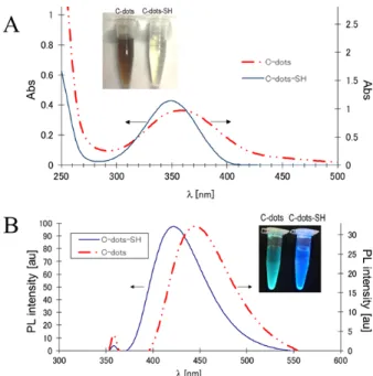
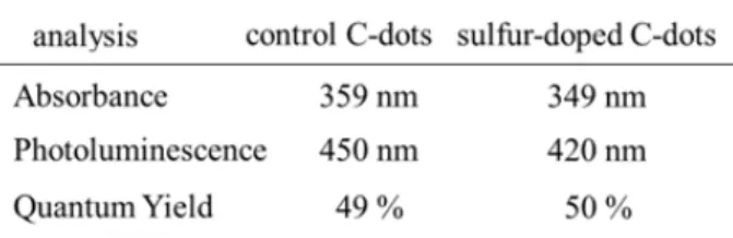
The bands around 1300-1410 cm–1 were assigned to aro- matic groups [15]. UV-VIS and fluorescence spectra of aqueous solutions (1 mg/ml) of sulfur-doped C-dots and non-modified C-dots as control experiment are shown in Fig. 1. The absorption maximum of non-modified C-dots (prepared by passivation with 1,2-ethylenediamine) was observed at 359 nm and the absorption peak of sulfur doped C-dots (passivated with cysteamine) was at 349 nm (Fig. 1A). These peaks are attributed to n-π* transi- tions of C=O bonds and the connected nitrogen- and sulfur- containing groups [16]. The band shift is due to the different passivation precursors in the synthesis of these two type nanoparticles. On the upper photo- graphic image in Fig. 1A are shown both the solution of C-dots-SH and non-modified C-dots. The solution
of the non-modified C-dots possesses dark visible color (left sample), which is due to the higher amount of amino passivating groups. The exchange with sulfur causes light yellow color. There is also a blue-shift in the fluorescence spectra of C-dots-SH compared to that of C-dots (Fig. 1B). The PL spectra of both nanoparticles are roughly symmetrical on the wavelength scale, wide with large Stokes shift. Under the excitation at 365 nm, C-dots-SH solution exhibits strong blue emission with a maximum around 420 nm. For comparison, the fluorescence maximum of C-dots is around 450 nm.
In addition, the emission peak position of C-dots has excitation wavelength-dependent behavior in the range 350 - 500 nm (as reported elsewhere [14]). However, the same effect is not observed for the C-dots-SH, whose emission peak position remains always at 420 nm. The explanation of this phenomenon is beyond the scope of the present report.
The fluorescence quantum yield (QY) is an impor- tant parameter for the detection sensitivity in the sensing
Fig. 1. Optical properties of the non-modified nano- particles (C-dots) and sulfur doped (C-dots-SH). (A) Absorption spectra of C-dots-SH (solid line) and C- dots (dash line). The upper photographic image shows the original colors of the solution under visible light.
(B) Fluorescence spectra of C-dots-SH (solid line) and C-dots (dash line). The upper photo presents the blue and bluish-green emission of the C-dots-SH and C-dots solutions upon excitation at 365 nm.
reaction. Generally, without surface passivation, the carbon nanodots possess rather low QY (lower than 1 %).
After passivation with amino and thiol groups their QY increases dramatically to ~ 50 % owing to the strongly PL centers on the nanoparticle surface. The obtained values are close to those of organic fluorescent dyes. The optical spectroscopic data for the synthesized C-dots and C-dots-SH are summarized in Table 1.
Additionally, there was a good linear relationship between the emission intensity and C-dots-SH concen- tration in the range of 0 - 0.100 mg/ml.
Sensitivity and detection of Ca2+ ions
The highly-fluorescent carbon nanodots have been widely used for sensitive detection of various metal ions. In most cases the detection is based on the induced fluorescence quenching, but there is also exception for PL enhancement [17]. Fig. 2 shows the influence of the metal ions and bicarbonate on the fluorescence intensity of the carbon nanodots in MilliQ water. The alkali cati- ons have no effect on the PL intensity of both C-dots and
C-dots-SH in solutions from slightly acidic to neutral pH.
The obtained data also confirm the negligible effect of Mg2+, which is a common component of the hard water.
The strong blue emission of C-dots-SH is significantly quenched when Ca2+ ions are added.
The addition of dissolved calcium decreases the intensity of nanoparticle fluorescence, but has no effect on the location of the maximum, as shown in Fig. 3.
Thus, the quenching of C-dots-SH fluorescence inten- sity by Ca2+ ions might be attributed to the formation of complexes between Ca2+ and thiol groups at the surface of the nanoparticle.
The pH value of the analyte is another factor that af- fects the sensing ability of the nanoparticles. In strongly acidic solution (pH ≤ 2.5) the addition of Ca2+ has no effect on the nanoparticles fluorescence intensity. The reason can be attributed to the protonation of the surface groups, which precludes the formation of stable Ca2+
complex and/or the quenching of the excited carbon nanodots by H3O+ ions. The quenching efficiency is much higher in weakly acidic media with pH 4 - 6 [14, 15]. Unexpectedly, the sample containing bicarbonate (NaHCO3) enhanced the fluorescence emission of the non-modified C-dots, however HCO3– did not influenced the intensity of C-dots-SH. The bicarbonates are the negative ions, which balance the dissolved Ca2+ and Mg2+
ions and the detection sensitivity of C-dots to HCO3– is important for their practical application as nanosensors.
It should be noted that Mg2+ might have similar reactiv- ity to C-dots-SH as Ca2+. Nevertheless, the experimental Table 1. Optical spectroscopic data of the carbon nano-
dots prepared by microwave-assisted pyrolysis.
Fig. 2. Fluorescence spectra of (A) non-modified C-dots, and (B) sulfur doped C-dots in the presence of different ions with concentration = 10–2 mol/L. λex = 365 nm, F and F0 denote the measured fluorescence intensity of the analyzed sample and the initial fluorescence intensity of the control sample, respectively.
data from Fig. 3 show that Mg2+ did not quench PL of either type of nanoparticles.
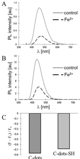
At neutral pH Ni2+ and Fe2+ might interfere seri- ously with the detection of Ca2+ because of their strong quenching effect. In all cases the fluorescence intensity of C-dots is drastically quenched by Fe2+ or Fe3+ (Fig.
4). It is known that the iron ions have the greatest effect on the emission among the other tested metal ions. The reason is the special coordination interaction between the phenolic hydroxyl groups of the carbon nanodots and d orbitals of the iron ions [18]. The fluorescence quenching is due to the nonradiative electron-transfer occurring within the excited iron complex. So, this effect must be taken into account when the carbon nanodots are used in practice as nanosensors.
The applicability of C-dots-SH to detect Ca2+ in a real sample of drinking mineral water was further evaluated. For that purpose the developed approach was applied to the famous brand mineral water with a known chemical composition. The concentration of Ca2+ ions in the sample was measured to be 11.8 ppb, which was consistent with the value written on the package.
The presence of thiol group on nanoparticle surface An important question which arose during the investigation, was in which oxidation state existed the reactive sulfur on the nanoparticle surface. The FTIR spectra showed the presence of thiol group around 2400 cm–1 as described above. However, an independent experiment was
Fig. 3. Fluorescence spectra of sulfur-doped carbon nanodots (C-dots-SH, conc. = 80 µg/ml) in the presence of differ- ent concentrations of Ca2+ (0 - 100 mmol/L). The right upper window displays the relation between the concentration of Ca2+ ions and fluorescence intensity.
Fig. 4. Fluorescence quenching of (A) non-modified C-dots and (B) sulfur-doped C-dots-SH by iron(II) chloride, (C) Comparison of the emission quenching of both nanoparticles.
conducted in which the sulfur was introduced on the nanoparticle surface in the state of –S–S– group (by use of precursor cysteine). Calcium ions did not quench the fluorescence of the carbon nanodots prepared in such a way as shown in Fig. 5.
CONCLUSIONS
Highly-luminescent sulfur-doped carbon nanodots were successfully synthesized from citric acid and cysteamine by microwave-assisted method. The re- sults confirm the validity of sulfur-doped C-dots and their fluorescence to detect Ca2+ ions in real sample of drinking mineral water. More important, the quenched fluorescence intensity exhibited a good linear correlation with the concentration Ca2+ ions in the sample solutions.
The method has many advantages as rapid detection, good reproducibility, selectivity and low performance cost. The method has also potential application for de- termination of calcium ions in biological specimen by fluorescence microscopic techniques.
Acknowledgements
The authors are grateful to Saitama University and to JSPS Bilateral Joint Research Projects, titled “Multi- electron transfer in supramolecular nanosystems”
between Saitama University and Hungarian Academy of Sciences as well as to NKFIH Grant K123995 for the financial support.
REFERENCES
1. J. Saurina, E. López-Aviles, A. Le Moal, S. Hernán- dez-Cassou, Determination of calcium and total hardness in natural waters using a potentiometric sensor array, Anal. Chim. Acta, 464, 2004, 89-98.
2. T. Bhattacharjee, H. Jiang, N. Behdad, A fluidic col- orimetric sensor design for water hardness detection, IEEE sensors Journal, 15, 2015, 819-826.
3. USGS – U.S. Geological Survey Office of Water Quality. “USGS Water-Quality Information: Water Hardness and Alkalinity. usgs.gov
4. P. Sengupta, Potential health impacts of hard water, Int. J. Prev. Med., 4, 2013, 866-875.
5. National Research Council, Drinking water and health, Washington, DC, National Academy of Sci- ence, 1977.
6. A.D. Eaton, L.A. Clesceri, A.E. Greenberg (Eds.), APHA-AWWA-WEF, Standard methods for the examination of water and wastewater, 19th Ed., APHA, Washington, 1995.
7. S. Karita, T. Kaneta, Chelate titrations of Ca2+ and Mg2+ using paper based analytical devices, Anal.
Chim. Acta, 924, 2016, 60-67.
8. A. Takahashi, P. Camacho, J.D. Lechleiter, B. Her- man, Measurement of intracellular calcium, Physi- ological Reviews, 79, 1999, 1089-1125.
9. X. Gao, Y. Lu, R. Zhang, S. He, J. Ju, M. Liu, L.
Li, W. Chen, One-pot synthesis of carbon nanodots for fluorescence turn-on detection of Ag+ based on the Ag+-induced enhancement of fluorescence, J.
Mater. Chem. C, 3, 2015, 2302-2309.
10. R. Zhang, W. Chen, Nitrogen-doped carbon quantum dots: facile synthesis and application as a “turn-off”
fluorescent probe for detection of Hg2+ ions, Bio- sens. Bioelectron., 55, 2014, 83-90.
11. S.J. Zhu, Q.N. Meng, L. Wang, J.H. Zhang, Y.B.
Song, H. Jin, K. Zhang, H.C. Sun, H.Y. Wang, B.
Yang, Highly photoluminescent carbon dots for multicolor patterning, sensors, and bioimaging, Angew. Chem., Int. Ed., 52, 2013, 3953-3957.
12. Y.Q. Dong, R.X. Wang, G.L. Li, C.Q. Chen, Y.W.
Chi, G.N. Chen, Polyamine-functionalized carbon quantum dots as fluorescent probes for selective and sensitive detection of copper ions, Anal. Chem., 84, Fig. 5. Fluorescence spectra of sulfur-doped C-dots
(obtained from cysteine) with different concentration of Ca2+ (0-100 mmol/L).
2012, 6220-6224.
13. H.M.R. Goncalves, A.J. Duarte, J.C.G.E. da Silva, Optical fiber sensor for Hg(II) based on carbon dots, Biosens. Bioelectron., 26, 2010, 1302-1306.
14. A. Loukanov, R. Sekiya, M. Yoshikawa, N. Kob- ayashi, Y. Moriyasu, S. Nakabayashi, Photosen- sitizer-conjugated ultrasmall carbon nanodots as multifunctional fluorescent probes for bioimaging, J. Phys. Chem. C, 120, 2016, 15867-15874.
15. A. Loukanov, H. Udono, R. Takakura, P. Mladenova, N. Kobayashi, R. Kawamura, S. Nakabayashi, Monitoring and extraction of uranium in polluted acid mine drainage by super-paramagnetic nano- particles coated with carbon nanodots, J. Radioanal.
Nucl. Chem., 314, 2017, 1149-1159.
16. Y. Wang, S. Kalytchuk, Y. Zhang, H. Shi, S.V. Ker- shaw, A.L. Rogach, Thickness-dependent full-color emission tenability in a flexible carbon dot ionogel, J. Phys. Chem. Lett., 5, 2014, 1412-1420.
17. N. Dhenadhayalan, K.C. Lin, Chemically induced fluorescence swithing of carbon-dots and its multiple logic gate implementation, Scientific Reports, 5, 2015, 10012.
18. S. Zhu, Q. Meng, L. Wang, J. Zhang, Y. Song, H.
Jin, K. Zhang, H. Sun, H. Wang, B. Yang, Highly photoluminescent carbon dots for multicolor pat- terning, sensors, and bioimaging, Angew. Chem.
Int. Ed. 52, 2013, 3953-3957.
![fi edintothreeseparatetechniques:DNA, – 3].Dependingontargetbiomolecules,microarraycanbeclassi fl uentcombinationofscienti fi cationofmicrobialcausativeagentsofinfectiousdiseases,microarraytechnologyisthelatestproductionofaharmonicmultidisciplinaryorchestra;](data:image/gif;base64,R0lGODlhAQABAIAAAP///wAAACH5BAEAAAAALAAAAAABAAEAAAICRAEAOw==)