This is an Accepted Manuscript, which has been through the Royal Society of Chemistry peer review process and has been accepted for publication.
Accepted Manuscripts are published online shortly after
acceptance, before technical editing, formatting and proof reading.
Using this free service, authors can make their results available to the community, in citable form, before we publish the edited article. We will replace this Accepted Manuscript with the edited and formatted Advance Article as soon as it is available.
You can find more information about Accepted Manuscripts in the
author guidelines.Please note that technical editing may introduce minor changes to the text and/or graphics, which may alter content. The journal’s standard Terms & Conditions and the ethical guidelines, outlined in our author and reviewer resource centre, still apply. In no event shall the Royal Society of Chemistry be held responsible for any errors or omissions in this Accepted Manuscript or any consequences arising from the use of any information it contains.
Accepted Manuscript
Dalton
Transactions
An international journal of inorganic chemistry www.rsc.org/dalton
ISSN 1477-9226
PAPER Joseph T. Hupp, Omar K. Farha et al.
Effi cient extraction of sulfate from water using a Zr-metal–organic framework
Volume 45 Number 1 7 January 2016 Pages 1–398
Dalton
Transactions
An international journal of inorganic chemistry
This article can be cited before page numbers have been issued, to do this please use: L. I. Szekeres, S.
Bálint, G. Galbács, I. Kálomista, T. Kiss, F. H. H. Larsen, L. Hemmingsen and A. Jancsó, Dalton Trans., 2019, DOI: 10.1039/C9DT01141B.
Journal Name ARTICLE
Received 00th January 20xx, Accepted 00th January 20xx DOI: 10.1039/x0xx00000x www.rsc.org/
Hg
2+and Cd
2+binding of a bioinspired hexapeptide with two cysteine units constructed as a minimalistic metal ion sensing fluorescent probe
Levente I. Szekeres,a Sára Bálint,a Gábor Galbács,a Ildikó Kálomista,a Tamás Kiss,a Flemming H.
Larsen,b Lars Hemmingsen,c Attila Jancsó*a
Hg2+ and Cd2+ complexation of a short hexapeptide, Ac-DCSSCY-NH2 (DY), was studied by pH-potentiometry, UV and NMR spectroscopies and fluorimetry in aqueous solutions and the Hg2+-binding ability of the ligand was also described in immobilized form, where the peptides were anchored to a hydrophilic resin. Hg2+ was demonstrated to form a 1:1 complex with the ligand even at pH = 2.0 while Cd2+ coordination by the peptide takes place only above pH ~ 3.5. Both metal ions form bis-ligand complexes by the coordination of four Cys-thiolates at ligand excess above pH ~ 5.5 (Cd2+) and 7.0 (Hg2+).
Fluorescence studies demonstrated a Hg2+ induced concentration-dependent quenching of the Tyr fluorescence until a 1:1 Hg2+:DY ratio. The fluorescence emission intensity decreases linearly with the increasing Hg2+ concentration in a range of over two orders of magnitude. The fact that this occurs even in the presence of 1.0 eq. of Cd2+ per ligand reflects a complete displacement of the latter metal ion by Hg2+ from its peptide-bound form. The immobilized peptide was also shown to bind Hg2+ very efficiently even from samples at pH = 2.0. However, the existence of lower affinity binding sites was also demonstrated by binding of more than 1.0 eq. of Hg2+ per immobilized DY molecules under Hg2+-excess conditions.
Experiments performed with a mixture of four metal ions, Hg2+, Cd2+, Zn2+ and Ni2+, indicate that this molecular probe may potentially be used in Hg2+-sensing systems under acidic conditions for the measurement of µM range concentrations.
Introduction
Hg2+ and Cd2+ ions can induce serious cytotoxic effects and cancer development in living organisms. Among others, Cd2+
can interfere with DNA repair mechanisms and causes osteoporosis by mimicking Zn2+ and Ca2+ ions.1,2 The high affinity of Hg2+ to the sulfhydryl groups of thiols has long been accepted to play a role in Hg2+ toxicity. Nevertheless, it has also been suggested that the major mechanism of toxicity is related to its interaction with the selenocysteine residues of glutathione peroxidase and thioredoxin reductase, leading to the inhibition of gap junction-mediated intercellular communication and proinflammatory cytokine production.3,4 Cellular defence mechanisms remove these toxic metal ions from the cytosol. In many bacteria the removal of Cd2+ ions, owing to their similarity to Zn2+, can be implemented by multication resistance- mediating CzcABC efflux pumps5 and by the P-type ATPase ZntA or the more specific CadA systems.6 In contrast, the detoxification of Hg2+ takes place in the form of Hg0 via the
specific mercury resistance mer system.7,8 Cys-abundant sequence motifs can provide efficient Hg2+ and Cd2+ binding for proteins. The CXXC motif often constitutes the metal ion binding site in proteins, like in rubredoxins,9 P-type ATPases,10 alcohol dehydrogenases,11 zinc fingers,12 metallochaperones,13,14 metallothioneins,15 as well as in the members of the Hg2+ and Cd2+ selective resistance systems (MerP,16 MerC,17 CadA,6 ZntA6 NMerA18).
Hg2+ can form extremely stable complexes with linear, {HgS2} type coordination mode with thiol ligands, e.g. Cys (log2 = 39.4 – 43.6)19–21, BAL (log1 = 44.8),22 etc. The formation of this binding mode was observed during the interaction of Hg2+ with many CXXC-type proteins, like MerP,16 NMerA,23 Atx1,24 and with cyclic25,26 or linear27,28 metalloprotein model peptides, etc.
The presence of amino acids with high rigidity and/or helical penalty (e.g Pro, Gly) in the positions of X1 and X2 increases the Hg2+ binding efficiency by preorganising the peptide structure.
Moreover, the Hg2+ complex of the rigid CDPPC peptide displays an exceptionally high stability constant (log = 40.0) even compared to the effective CPPC sequence.29 There are also examples for the formation of bis complexes with a {HgS4} binding mode among proteins bearing a CXXC motif (HAH1,24 Rubredoxins,30 etc).
In its complexes, Cd2+, similarly to Zn2+, prefers to possess a tetrahedral coordination sphere with two or four thiolate moieties but Cd2+ has a higher thiophilic character. When the coordination sphere of Cd2+ is not saturated by thiolate groups,
a.Department of Inorganic and Analytical Chemistry, University of Szeged, Dóm tér 7, Szeged, H-6720, Hungary; E-mail: jancso@chem.u-szeged.hu.
b.Department of Food Science, University of Copenhagen, Rolighedsvej 30, 1958 Frederiksberg C, Denmark. – Should we add his present affiliation?
c.Department of Chemistry, University of Copenhagen, Universitetsparken 5, 2100 Copenhagen, Denmark
Electronic Supplementary Information (ESI) available: [details of any supplementary information available should be included here]. See DOI: 10.1039/x0xx00000x
Dalton Transactions Accepted Manuscript
Open Access Article. Published on 02 May 2019. Downloaded on 5/2/2019 1:39:34 PM. This article is licensed under a Creative Commons Attribution-NonCommercial 3.0 Unported Licence.
View Article Online DOI: 10.1039/C9DT01141B
the complexes are stabilized mostly by complementary binding of other functional groups. Interactions with the carboxylate groups of Glu and Asp units probably play a role in the selectivity of CadA and ZntA.31 Besides monomeric bis complexes with a pure {CdS4} coordination mode, the presence of dimeric species with a mixed Cys-Glu ({CdS3O}) coordination was also observed in the system of Cd2+ and a 71 amino acid long polypeptide, derived from CadA, at pH = 6.0.31
CXXC moieties are potentially applicable as metal ion binding units in Hg2+ and Cd2+ sensing systems, since their interaction is typically characterized by a well-defined stoichiometry, high efficiency and selectivity. Since these metal ions can accumulate in the food chain and therefore ingested by humans, they pose significant health risks, and therefore their fast, selective sensing and remediation is highly prioritised.8 Sensing schemes for these metal ions, based on fluorimetry,32 potentiometry,33–
35 impedance measurement,36 protein-functionalized microcantilevers,37 or differential surface plasmon resonance38 etc., were already proposed. Peptide ionophores can be designed with an ability for signalling by introducing fluorescing residues or amino acids bearing fluorescing side chains into their sequence.39–41 Metal ion binding to peptidic probes mostly induce fluorescence quenching, especially with Cu2+ 42–53 and Hg2+.42,48,54 However, chelation can also enhance the fluorescence (CHEF) via electronic interactions between the fluorophore and the altered coordination sphere. This is very frequent with Zn2+ 43,46–49,51,53 and Cd2+ 43,46–49 but examples are also known with Ag+,42,46–48 Pb2+46,47 and Hg2+.48 In addition, the metal ion promoted conformational change of the peptide may lead to a “close contact” and thus to a Förster resonance energy transfer (FRET) effect between different fluorophore residues.46,50,52,54–56 Aggregation induced emission (AIE) enhancement can also be utilized for the analysis of metal ions.57,58 The response mechanism and the sensitivity and selectivity of a peptide sensor designed for a specific metal ion is highly influenced by the quality and quantity of the binding groups, as well as the fluorophores themselves, in addition to the amino acid sequence and the higher order peptide structure. Although many fluorescent peptides have already been tested as potential sensor candidates for Hg2+, Cd2+ or other metal ions, examples where the studied ionophore bears a CXXC motif are rather scarce. In the unique case of the short Dansyl-CPGCW-NH2 peptide, the intervening amino acids between the two Cys residues provide high backbone rigidity for the ionophore. Upon excitation of either of the two fluorophore units, Cd2+ ions induced a significant enhancement in the light emission of dansyl. Rather interestingly, Hg2+ and Zn2+ were shown to have no effect on the fluorescent response of the molecule.56 Another notable, previously studied dicysteinyl peptidic probe possesses a CXXHC binding motif combined with dansyl and Trp fluorophores. This probe displayed a ratiometric turn on response for Hg2+, Cd2+, Pb2+, Zn2+, and Ag+ ions upon the excitation of either of the two fluorophore units.46 In addition, Joshi and co-workers also observed a turn-on response for Zn2+ with two fluorescent peptides containing a Cys and a His residue in a 1,4-arrangement.47,49
Optochemical sensors, constructed by the covalent attachment of fluorescent peptides to the surface of solid carriers (e.g.
optical fibers) may allow in-situ trace analysis of toxic metal ions with low sample quantity requirements. Such systems may present simple alternatives to currently used reliable, but expensive and robust laboratory-based instrumental methods.
Despite the fact that several studies have already reported the successful design of such sensors,32,59–61 there have only been a very few attempts to characterize the metal capturing peptide elements both in their dissolved and immobilized forms.47,49 With an aim of exploiting the metal ion binding properties of the CXXC motif, we designed and synthesized the short, DCSSCY hexapeptide in an acetylated and amidated free form (Ac- DCSSCY-NH2 (DY)) and in an immobilized, resin-attached form (DY-NTG). Compared to other aromatic amino acids, Tyr is less hydrophobic and still exhibits a reasonable quantum yield (0.14).62 The fluorescence of Tyr has proved to be applicable in determining the Cu2+ complex stabilities of different peptides,63–65 in spite of the relatively close excitation (EX = 275 nm) and emission (EM = 304 nm) maxima of the fluorophore.62 Accordingly, this simple ligand and the obtained data may be relevant in the design of peptide-based metal ion sensing systems.
Results and discussion
UV absorption experiments
UV absorption spectra of DY, as a function of pH, were recorded in the absence and presence of metal ions with the purpose of monitoring the effect of Hg2+ and Cd2+ on the deprotonation processes of the ligand. As it is expected, the proton dissociation of both the Cys –SH groups and the phenolic –OH group markedly influence the recorded spectra, even in the absence of metal ions (Figure S1). Deprotonated thiol groups are characterized by a UV-band centred around = 235-236 nm66,67 while the proton release from the Tyr–OH function is known to result in a shift of two characteristic absorption peaks from 223 to 240 nm and from 277 to 295 nm.68 The dissociation of the Asp sidechain carboxyl group does not have a noticeable spectral influence in the = 220 – 400 nm range. Since the Cys and Tyr residues all deprotonate above neutral pH, a continuous evolution of the absorbance is observed between pH ~ 6.5 and 11.0 around 240 nm while the absorbance at 295 nm starts increasing from a notably higher pH, above pH ~ 8.5 (Figure S1), indicating the exclusive effect of the deprotonation of the Tyr residue. One may estimate the pKa value of the Tyr –OH function from the A295nm vs. pH plot (Figure S1) to be around 10.0 which is in line with the data determined from pH-metric titrations (see later).
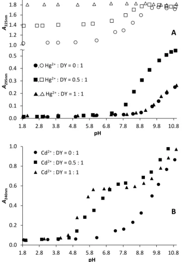
UV/pH titrations were conducted in the presence of 0.5 and 1.0 equivalent of Hg2+ ions within the range of pH ~ 1.8 – 11.0 (Figure S2) and representative absorbance traces from selected wavelength values are depicted in Figure 1.A. The significantly larger absorbances observed at pH = 1.8 at ~ 210 – 230 nm in the presence of Hg2+ relative to the metal ion free sample is originated from S−–Hg2+ charge transfer bands and clearly
Dalton Transactions Accepted Manuscript
Open Access Article. Published on 02 May 2019. Downloaded on 5/2/2019 1:39:34 PM. This article is licensed under a Creative Commons Attribution-NonCommercial 3.0 Unported Licence.
View Article Online DOI: 10.1039/C9DT01141B
indicate peptide binding to the metal ion, as was also observed previously for other systems of Hg2+ and cysteine containing peptides under acidic conditions.29,69–71 Furthermore, the absorbance increase, induced by Hg2+-binding, seems to be proportional to the applied Hg2+:DY ratio (Figures 1.A and S2.B) between pH ~ 1.8 – 7.0. At the same time, absorbances in the higher wavelength regime, i.e. around 295 nm, are almost identical independently of the Hg2+ concentration. These findings suggest an unvarying speciation up to neutral pH at any Hg2+:DY ratio (and even up to pH 11.0 at one equivalent of Hg2+
per peptide) and the high energy charge transfer band, characterized by an absorption maximum below 220 nm (see the calculated difference spectra in Figure S3.), indicates a {HgS2} type coordination mode in the complexes formed.26,29,69,70,72,73
1.0 1.2 1.4 1.6 1.8
A225nm
A
~~
0.0 0.1 0.2 0.3 0.4 0.5
1.8 2.8 3.8 4.8 5.8 6.8 7.8 8.8 9.8 10.8
A295nm
pH
,Hg2+: DY = 0 : 1
,Hg2+: DY = 0.5 : 1
,Hg2+: DY = 1 : 1
0.0 0.2 0.4 0.6 0.8 1.0
1.8 2.8 3.8 4.8 5.8 6.8 7.8 8.8 9.8 10.8
A240nm
pH
B
Cd2+: DY = 0 : 1 Cd2+: DY = 0.5 : 1 Cd2+: DY = 1 : 1
Figure 1. Absorbances at selected wavelength values as a function of pH, recorded for Hg2+:DY (A) and Cd2+:DY (B). (cDY = 110−4 M (A) or 510−5 M (B), T = 298 K).
A remarkable difference between the speciation in Hg2+:DY 0.5:1 and 1:1 above pH ~ 7.5 is reflected by the A vs. pH profiles at 295 nm. The absorbances observed for Hg2+:DY 1:1 up to pH
= 11.0 are nearly identical with those obtained for the free ligand suggesting that the bound Hg2+ has only a minor influence, if any, on the deprotonation of the Tyr sidechain group. The remarkably different A295nm vs. pH trace of Hg2+:DY 0.5:1 (Figure 1.A) suggests a fundamental rearrangement in the
coordination sphere of Hg2+ above neutral pH, most probably induced by the coordination of a second ligand and the formation of additional S−–Hg2+ bonds. LMCT bands with maxima around 240-250 nm or 280-300 nm in the UV spectra of Hg2+-thiolate complexes were previously assigned as signatures of trigonal {HgS3}74,75 or (pseudo)tetrahedral {HgS4} species, respectively,30,75,76 and this suggests that the two DY ligands binds Hg2+ with both of their two thiolates under basic conditions (see Figure S3). The slight batochromic shift of the absorption maximum, observed above pH ~ 9.5 (Figure S2.A), is presumably the effect of the deprotonation of the Tyr residues of the bound ligands taking place without metal ion assistance.
This interpretation is further supported by the fact that isosbestic points are observed above pH 10 at ~ 264 nm and ~ 282 nm (Figure S2.A), indicting a simple transition between two species.
The pH-dependent series of UV spectra recorded for Cd2+:DY 0.5:1 and 1:1 (Figures 1.B and S4) indicate metal ion coordination via the formation of thiolate–Cd2+ bonds from pH
~ 4.5. The A240nm vs. pH profiles, representing the evolution of S−–Cd2+ charge transfer bands,77–83 suggests that metal ion binding is essentially complete by pH ~ 6 at 1 eq. of Cd2+ per ligand, however, the curve for Cd2+:DY 0.5:1 is characterized by a narrow plateau near pH 6 and a second step levelling off above pH ~ 7.5 (Figure 1.B). This indicates a different complexation pathway when the peptide is in excess over Cd2+. One may presume the formation of bis-ligand species, as was also reported for the Cd2+ binding of other short, two Cys-containing peptides.79–81,84 This is also supported by the systematically higher absorbances observed for Cd2+:DY 0.5 relative to those observed both for Cd2+:DY 1:1 and for the free ligand (Figure 1.B). Indeed, the molar extinction coefficients for = 240 nm that one may estimate for the species present at pH ~ 8.0 at 0.5 and 1 eq. of Cd2+ per ligand (25 100 M−1 cm−1 and 11 700 M−1 cm−1) correspond well to four and two thiolates coordinated Cd2+-centres, respectively.77,78 The further increase of absorbance above pH 8 (at any Cd2+:DY ratio) is most likely the consequence of the deprotonation of the Tyr sidechain phenols, nevertheless, other processes that may affect the coordination sphere of the metal ion, e.g. proton release(s) from Cd2+- coordinated water molecules, may also contribute to the observed spectral changes under alkaline conditions.
1H NMR studies
1H NMR spectra of the free peptide were recorded as a function of pH (Figure S6) and resonances of DY were assigned as described in the Experimental part and are shown in Figure S5.
We performed NMR titrations in the Hg2+:DY (Figure 2) and Cd2+:DY (Figure S9) systems by changing the metal ion to ligand ratio at selected pH values or at constant Hg2+:DY ratios (0.5:1 and 1:1) as a function of pH (Figures S7-S8). However, pH- dependent 1H NMR experiments were not executed for Cd2+:DY because of severe line broadening and poorly resolved resonances observed for all of the signals already at 0.25 eq. of Cd2+ per ligand (see Figure S9). We had previously observed a similarly strong influence of Cd2+ on the 1H NMR signals of other,
Dalton Transactions Accepted Manuscript
Open Access Article. Published on 02 May 2019. Downloaded on 5/2/2019 1:39:34 PM. This article is licensed under a Creative Commons Attribution-NonCommercial 3.0 Unported Licence.
View Article Online DOI: 10.1039/C9DT01141B
relatively short, Cys-containing peptides81,85 and as in those reported cases, the line broadening seen in the spectra of Cd2+:DY indicates ligand exchange and/or conformational dynamics occurring in an intermediate time regime, relative to the NMR time scale.
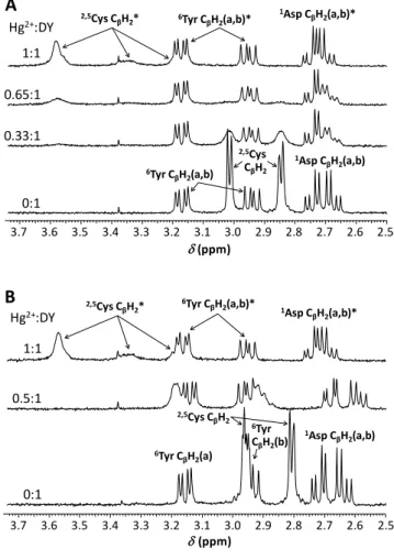
The deprotonation processes of the carboxyl group of the Asp residue, the two Cys-thiols and the sidechain phenolic function of Tyr in the free DY ligand can be clearly followed by the gradual shifting of the corresponding CβH2 resonances or the CδH and CεH signals of Tyr towards lower chemical shifts in the relevant pH-ranges (Figure S6). The presence of Hg2+ has only minor effect on the shape and chemical shift of these resonances (with the exception of the slightly more influenced CβH2 signals of Asp – see later), suggesting that Hg2+ does not bind to these moieties. As opposed to this, below neutral pH Hg2+ binding of the two thiolates are represented by the collapse of the Cys CβH2 signals of the unbound ligand and the emerging new resonances attributed to the bound molecule (see the effect of the increasing Hg2+:DY ratio at pH = 5.7, Figure 2.A). This indicates that the ligand exchange dynamics is moderately slow at this pH, relative to the NMR timescale.
3.7 3.6 3.5 3.4 3.3 3.2 3.1 3.0 2.9 2.8 2.7 2.6 2.5
(ppm) Hg2+:DY
1:1 0.65:1
0:1 0.33:1
A
1Asp CβH2(a,b)
1Asp CβH2(a,b)*
6Tyr CβH2(a,b)*
2,5Cys CβH2*
2,5Cys CβH2 6Tyr CβH2(a,b)
3.7 3.6 3.5 3.4 3.3 3.2 3.1 3.0 2.9 2.8 2.7 2.6 2.5 Hg2+:DY
1:1
0.5:1
0:1
(ppm)
1Asp CβH2(a,b)
1Asp CβH2(a,b)*
6Tyr CβH2(a,b)*
2,5Cys CβH2*
2,5Cys CβH2
6Tyr CβH2(a)
6Tyr CβH2(b)
B
Figure 2. 1H NMR spectra of DY obtained with varying equivalents of Hg2+ per peptide at pH = 5.7 (A) and 8.5 (B) (H2O:D2O = 90:10 % v/v, cDY = 1.010−3 M, T = 298 K). The symbol
‘*‘ denotes 1H resonances of the ligand in its metal bound state.
The pH-dependence of resonances observed with 0.5 eq. of Hg2+ per peptide agrees excellently with the UV data and with
the formation of a bis-ligand complex with a {HgS4} coordination geometry. The fact that the chemical shifts of the Cys protons lie in between those of the free peptide and the resonances of the mono-complex (see the broad peaks around 2.9 and 3.2 ppm on the middle spectrum in Figure 2.B) may simply reflect that Hg2+ quite efficiently attracts electrons in the {HgS2} type species, but that this effect (per cysteine) is less pronounced in the {HgS4} structure. Consequently, the deshielding effect on the Cys protons caused by Hg2+ coordination is weaker for the {HgS4} than for {HgS2} structures. It is also interesting to note that the signals of the Cys CβH2 protons are hardly observable around neutral pH at 0.5:1 Hg2+:DY ratio (Figure S7.A), possibly as a consequence of severe line broadening. This might imply that the increase of pH and approaching the range where the thiol groups of the unbound ligands deprotonate has an influence on the ligand exchange rate and that ligand exchange dynamics also affects the observed Cys CβH2 resonances.
Complex speciation in the metal ion – peptide systems, unravelled by pH-potentiometric titrations
pH-potentiometry is, in general, a powerful method for the characterization of complex formation processes that lead to the release of protons from the metal ion binding moieties of the ligand, and pKa values of the free ligand and its Cd2+
complexes are presented in Table 1. However, Hg2+ ions are bound so efficiently to ligands with thiol groups, including the studied DY peptide, that the free metal ion fraction is negligible even at the very acidic end of the pH-range coverable by a glass electrode.29,69–71 As a consequence, the relevant metal-ion induced deprotonation processes are completed at the initial pH of the titrations and thus the method is not directly applicable to the determination of stability constants in such systems. (An indirect pH-metric approach for the characterization of Hg2+-peptide complex speciation using a competing Hg2+-binding ligand has been previously reported by Iranzo et al.29). In spite of the limitations of this method for the present system, we executed pH-potentiometric titrations aiming to follow the various deprotonation processes occurring at the Hg2+-bound peptide(s) and to determine the proton dissociation constants characterizing the interconversion of the differently protonated complex species. These data, collected in Table S1 in the ESI, allowed of the calculation of species distribution curves for Hg2+:DY 0.5:1 and 1:1 (Figure 3) based on the plausible assumption that the species present at pH = 2.0 is HgH2L with two Hg2+-coordinated thiolate groups and protonated Asp and Tyr residues of the ligand. Indeed, the calculation of such a speciation scheme required the overall formation constant (log) of one of the species to be fixed (conveniently the log of HgH2L). Since we did not have an experimentally measured value for logHgH2L we assumed that the Hg2+ + [H2L]2− ⇌ [HgH2L] process of DY could be characterized by the same stability constant as that of the parent mono complex (HgL) of the terminally protected CDPPC peptide, determined by Iranzo et al. (logK = 40.0).29 By this assumption we could set a fixed, “arbitrary” logHgH2L value (see more details in the Supplementary information) required for
Dalton Transactions Accepted Manuscript
Open Access Article. Published on 02 May 2019. Downloaded on 5/2/2019 1:39:34 PM. This article is licensed under a Creative Commons Attribution-NonCommercial 3.0 Unported Licence.
View Article Online DOI: 10.1039/C9DT01141B
data evaluation. Note that it does not affect the conclusions of this work how closely this arbitrary stability data approaches its real value, as long as it is high enough to ensure a complete binding of Hg2+ at the lowest utilized pH. This is also reflected by model calculations where in spite of the different applied logHgH2L values the resulting pKa data for the deprotonation processes of the complexes turned out to be identical (Table S2). The calculated distribution of species (Figure 3) well supports all of our previous conclusions drawn from the spectroscopic data. The pKa values associated with the release of the two protons, leading to [HgHL]− (pKa = 4.1, Table S1) and then to [HgL]2− (pKa = 9.6, Table S1), are close to the values determined for the deprotonations of the Asp and Tyr units of the free ligand, respectively (pKa,1 = 3.80 and pKa,4 = 10.05, see Table 1). This indicates that the deprotonation of these groups are essentially not affected by the metal ion in the Hg2+-bound peptide. At Hg2+:DY = 0.5:1 the bis-ligand complexes dominate above pH 8. Considering the characteristic pH-range for the presence of the differently protonated bis complexes and the observed position of their S−–Hg2+ charge transfer bands, it is straightforward to suggest three coordinating thiolate groups in [HgH3L2]3− and thus a {HgS4} coordination mode in [HgH2L2]4−. Similar to the deprotonation step of the [HgL]2− parent complex, the two H+-dissociation processes of the [HgH2L2]4− species, occurring in the alkaline pH range (pKa = 10.0 and 11.2 - Table S1), are assigned to the deprotonation of non-coordinating Tyr sidechains. Proposed schematic structures for the Hg2+:DY complexes are collected in Scheme S1.
0.0 0.2 0.4 0.6 0.8 1.0
2 3 4 5 6 7 8 9 10 11
XHg2+
pH
HgH2L [HgHL]−
[HgL]2−
[HgH3L2]3−
[HgH2L2]4−
[HgHL2]5−
[HgL2]6−
Figure 3. Distribution of species in the Hg2+:DY 0.5:1 (continuous lines) and 1:1 (dashed lines) systems (cDY = 1.010−3 M, T = 298 K).
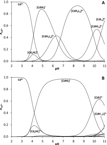
Evaluation of pH-potentiometric data obtained for Cd2+:DY reveal that Cd2+-binding occurs above pH ~ 3.5 and the first dominant species, formed in parallel with a small amount of a dinuclear species, is a mono-protonated mono complex independently of the metal ion to ligand ratio (Figure 4).
The composition ‘[CdHL]−‘ corresponds well with the coordination of both thiolates to Cd2+ and with a protonated Tyr phenol group in this species. Obviously, the Asp carboxyl function is deprotonated in [CdHL]−, however, since the formation of this complex spans the pH-range where the Asp sidechain of the free ligand also deprotonates, pH-metric data
are not conclusive with regard to the Cd2+-binding of this group.
Previous studies on short oligopeptides containing both Cys and Asp residues reflected, however, that participation of the available carboxylate groups in Cd2+-binding is possible,80,81,83 even if two thiolate donors are also coordinated.80,81 Binding of the Asp sidechain of DY to Cd2+ is supported by the stability constant calculated for the Cd2+ + [HL]3− = [CdHL]− process (logK1+H = 12.56). This value is nearly identical to the stability that may be calculated for the relevant, {CdS2O2} type complex of a short phytochelatin peptide (( -Glu-Cys)2-Gly - PC2, logK1+H
= 12.57)80 and significantly higher than the stability of the protonated mono complex of ACSSACS-NH2 with only two coordinating thiolate groups (logK1+H = 10.22).84 The remarkable extra stability observed for the mono complex of DY is presumably only partly related to the additional binding of the Asp carboxylate group, and may also be a consequence of the more favoured CXXC amino acid pattern vs. the CXXXC sequence in the ACSSACS-NH2 heptapeptide.
0.0 0.2 0.4 0.6 0.8 1.0
2 3 4 5 6 7 8 9 10 11
XCd2+
pH Cd2+
[CdH2L2]4−
[CdH3L2]3−
[CdHL2]5−
[CdL2]6−
A
[Cd2HL]+ [CdHL]−
0.0 0.2 0.4 0.6 0.8 1.0
2 3 4 5 6 7 8 9 10 11
XCd2+
pH
Cd2+ [CdHL]−
[CdL]2−
[Cd2HL]+
[CdH−1L]3−
B
Figure 4. Distribution of species in the Cd2+:DY 0.5:1 (A) and 1:1 (B) systems (cDY = 1.010−3 M, T = 298 K).
Furthermore, the additional coordination of one of the internal peptide carbonyl groups, as suggested for PC2, is also possible in the [CdHL]− complex of DY resulting in a {S−,S−,COO–,CO}
donor group environment around the metal ion. The pKa value determined for the [CdHL]− = [CdL]2− + H+ process (pKa = 9.83) is very close to the pKa of the Tyr residue in the free ligand indicating that the sidechain phenolate is not bound to Cd2+,
Dalton Transactions Accepted Manuscript
Open Access Article. Published on 02 May 2019. Downloaded on 5/2/2019 1:39:34 PM. This article is licensed under a Creative Commons Attribution-NonCommercial 3.0 Unported Licence.
View Article Online DOI: 10.1039/C9DT01141B
similarly to what was concluded for the Hg2+ complexes of DY.
(Suggested schematic structures for the Cd2+:DY complexes are shown in Scheme S2.) Calculation of the apparent dissociation constant for the Cd2+:DY mono complexes at pH = 7.0, according to the equation Kd = (Cd2+]free [Ligand]free) / [Cd2+]complexed, may allow to compare the Cd2+-binding affinity of DY to other relevant cysteine containing peptides or proteins. The calculated Kd = 9.110−10 M value indicates a slightly larger Cd2+- binding affinity of DY compared to that of the PC2 phytochelatin peptide (Kd = 1.710−9 M)80 but a substantially stronger Cd2+- binding relative to the reduced glutathione (Kd = 1.410−5 M).86 However, the average dissociation constants determined for the and domains of MT-2 metallothioneins (Kd = 1.010−15 M () and 0.1-1.010−12 M ())87 clearly reflect a more efficient Cd2+-coordination by these proteins, provided by the thiolate- clusters.
At twofold DY excess over Cd2+ the bis-ligand complexes start to form from ca. pH 5 and become fully dominant by pH ~ 8 (Figure 4.A), as also suggested by the pH dependent evolution of the S−–Cd2+ charge transfer band and the calculated molar extinction coefficient for = 240 nm at Cd2+:DY 0.5:1, vide supra. The two proton release processes, leading from the four- thiolate coordinated [CdH2L2]4− complex to [CdHL2]5− and then to [CdL2]6−, are assigned to dissociations of the non- coordinating Tyr phenol sidechains (see Table 1). The stability constant calculated for the protonated bis complex ([CdHL]− + [HL]3− = [CdH2L2]4−, logK2CdH2L2 = 8.21) and the relative stability of the protonated mono and bis-ligand species (log(K1+H/K2+2H)
= 4.35) indicate that binding of the second ligand is substantially less favoured than that of the first one. Very similar data have been reported for the bis complex formation of the phytochelatin pentapeptide PC2 (log(K1+H/K2+2H) = 4.39)80 leading to a four thiolate coordinated species. The relative positions of the two Cys residues in DY and PC2 are different (CXXC, CXC) just as the overall charges of their relevant mono and bis complexes (DY: −1/−4, PC2: −2/−6), nevertheless, the two peptides are of similar sizes. This suggests that the relative binding strength of the second DY or PC2 ligand is determined by similar factors, i.e. the notably high stability of their mono complex and steric hindrance (bulkiness of the peptides) but the log(K1+H/K2+2H) values are probably not large enough to assume a major change in the coordination geometry of Cd2+.
Logarithmic protonation constants and formation constants (log) of the Cd2+ complexes of DY (with the estimated errors in parentheses, last digit) together with some calculated data (I = 0.1 M NaClO4, T = 298 K)
Species log pKa
[HL]3 10.05(1) 10.05
[H2L]2 19.29(1) 9.24
[H3L] 27.56(2) 8.27
[H4L] 31.36(2) 3.80
pKaCdxHyLz a
[CdHL] 22.61(4) 9.83
[CdL]2 12.78(8) 11.48
[CdH–1L]3 1.3(2) −
[CdH3L2]3 47.6(2) 6.7
[CdH2L2]4 40.9(1) 9.9
[CdHL2]5 31.0(2) 10.6
[CdL2]6 20.4(1) −
[Cd2HL]+ 25.4(2) −
logK1+H b 12.56
logK2+2H c 8.21
log(K1+H/K2+2H) 4.35
NP. d 279
FP (cm3) e 0.004
a pKaCdxHyLz = logCdxHyLz – logCdxHy−1Lz; b stability constant calculated for Cd2+ + [HL]3−
= [CdHL]−, logK1+H = log[CdHL]− – log[HL]3−; c stability constant calculated for [CdHL]− + [HL]3− = [CdH2L2]4−, logK2+2H= log[CdH2L2]4− – log[CdHL]− – log[HL]3− ; d NP = number of points; e FP = fitting parameter
Influence of metal ions on the fluorescence of the ligand The effect of the metal ions on the fluorescence of the Tyr residue, as a function of the metal ion to DY ratio, was monitored at selected pH values, applying excitation at = 278 nm. Gradual addition of Cd2+ ions to a solution of the peptide at pH = 7.1 (Figure S10) showed a partial quenching (~35 % drop) of the original emission intensities, levelling off at a ca. 0.6-0.7:1 Cd2+:DY ratio. This I308nm vs. Cd2+:DY ratio profile perfectly correlates with the disappearance of the free ligand, occurring slightly above a 0.5:1 Cd2+:peptide ratio at pH = 7.1 (see Figure 4.A), especially under the small concentrations used for the fluorimetric studies. Since further increase of the Cd2+:DY ratio was found to have no influence on the observed emission intensity, Cd2+ seems to exert very similar quenching effect on the fluorescence of the Tyr residue, independently of the number of coordinated DY molecules. In a double (Trp and Dansyl) labelled peptide containing two Cys residues, Cd2+ was found to induce a turn-on response, nevertheless, the chelation enhanced fluorescence also reached a maximum around a 0.5:1 Cd2+:peptide ratio, and accordingly, it was attributed to the presence of a bis complex.56
For the experiments in the Hg2+:DY system pH = 6.0 was chosen to apply a close-to-neutral condition while avoiding the occurrence of bis complexes and thus maintaining a relatively simple speciation that should be desirable for a molecular probe. In view of the outstanding affinity of DY towards Hg2+
that, in contrast to Cd2+, results in a complete binding of this metal ion even at rather low pH values, we recorded Hg2+:DY ratio dependent emission spectra at pH = 2.0, as well.
Dalton Transactions Accepted Manuscript
Open Access Article. Published on 02 May 2019. Downloaded on 5/2/2019 1:39:34 PM. This article is licensed under a Creative Commons Attribution-NonCommercial 3.0 Unported Licence.
View Article Online DOI: 10.1039/C9DT01141B
700 1000 1300 1600 1900 2200
0.0 0.5 1.0 1.5 2.0 2.5 3.0
I308 nm
cHg2+/ cDY pH = 2.0
pH = 6.0
pH = 2.0, 1 eq. Cd2+
pH = 6.0, 1 eq. Cd2+
0 500 1000 1500 2000
295 325 355
I
(nm) cHg2+
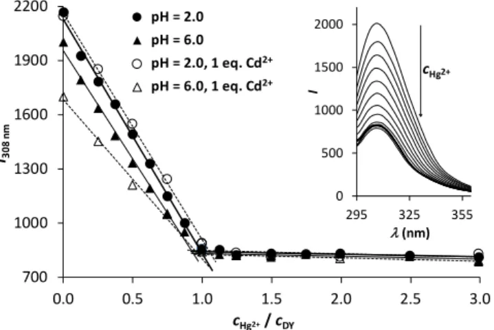
Figure 5. Fluorescence titration of DY by Hg2+ at pH = 2.0 (circles) and pH = 6.0 (triangles) in the absence (filled symbols) and presence (open symbols) of Cd2+ ions. Fitted lines represent the trends in the decrease of intensity and the breakpoint at 1.0 eq. of Hg2+
(cDY = 3.010−5 M, EM = 308 nm, EX = 278 nm). The inset shows the recorded spectra at pH = 6.0 in the absence of Cd2+ ions.
As Figure 5 clearly shows, the increase of the concentration of Hg2+ leads to a significant drop of emission intensities, following a linear trend until reaching a 1:1 Hg2+:DY ratio both at pH = 6.0 and 2.0. This correlates well with the calculated complex speciation (Figure 3) and indicates a strong and complete binding of 1 eq. Hg2+ per DY. However, it also suggests that the ligand cannot capture a second Hg2+ ion in solution, which is also a desirable property for a Hg2+ sensor. Although different species dominate at the two selected pH values (HgH2L at pH = 2.0 and [HgHL]− at pH = 6.0), the observed trends in the decreasing fluorescence emission intensities are nearly identical in the two cases. This suggests that the protonation state of the uncoordinated Asp carboxyl group has no impact on the Hg2+-induced quenching of the Tyr fluorescence.
Different possibilities may be considered for the mechanism of the metal ion induced quenching of the fluorescence of DY, as suggested by Chen’s in-depth overview of the interaction of Hg2+ with proteins containing aromatic amino acids.88 Indeed, Hg2+ was shown to quench the fluorescence of the vast majority of tested proteins, especially of those that contained Cys residues near the aromatic amino acids.88 Although direct interaction of both of the studied metal ions with the Tyr residue of DY has been excluded in our systems, an overlap between the thiolate-related absorption bands and the Tyr emission might cause quenching via energy transfer. Besides, metal ion promoted conformational changes in the peptide structure may also play a role in the altered fluorescence.
We also tested the Hg2+-induced quenching of the fluorescence of DY in the presence of 1.0 eq. of Cd2+. Since the latter metal ion is not bound to the peptide at pH = 2.0 (Figure 4) it has no influence on the emission intensities at any Hg2+:DY ratio.
Nevertheless, 1.0 eq. Cd2+ per DY at pH = 6.0 induces a notable fluorescence quenching, as compared to the Cd2+-free sample, reflecting that Cd2+-DY complexes are present at the start of the titration. When this sample is titrated by Hg2+ the observed fluorescence is gradually quenched down to the same level as
seen in the absence of Cd2+, suggesting that the DY-bound Cd2+
is completely displaced by Hg2+.
In view of the potential analytical applicability of such a molecular probe the most promising findings of the above experiments are the significant fluorescence response of the ligand, proportional to Hg2+-concentration up to a 1:1 Hg2+:DY ratio, as well as the remarkable metal ion binding selectivity that is also manifested in the Hg2+-mediated changes in emission intensities. In this context, the data series recorded at pH 2 (Figure 5) demonstrates selective binding of Hg2+ at low pH, which may be used to eliminate interferences from other metal ions, such as Cd2+, in a fluorophore based Hg2+ assay. A quick assessment of the potential analytical figures of merit for Hg2+ sensing under our measurement conditions yields an estimated limit of detection of ca. 210−7 M and a linear dynamic range exceeding 2 orders of magnitude.
Hg2+ binding of the DY peptide immobilized to a resin
Next we aimed to explore Hg2+ binding to the DY ligand in an immobilized form. To achieve this, we synthesized a construct carrying the peptide anchored to a hydrophilic resin (DY-NTG).
Hg2+ binding to DY-NTG was studied as a function of contact time, pH and metal ion concentration at pH = 2.0. As a reference system, the peptide-free resin with acetylated terminal amino groups (Ac-NTG) was also tested at pH = 2.0 by adding 1.5 eq. of Hg2+ per the available blocked peptide anchoring sites. The determined 0.0354 mmole Hg2+ per g Ac-NTG metal ion binding capacity represents ca. 13% of the theoretical loading of the resin (quantity of available functionalization sites on the resin beads, L = 0.267 mmole/g). It seems that a small but non- negligible amount of Hg2+ may be bound by Ac-NTG, probably at the polyethylene-glycol chains. However, such binding sites in the peptide functionalized resin, DY-NTG, may play a role in metal ion binding only under metal ion excess conditions.
Since metal ion complexation processes could be slower at the resin beads-supported binding sites as compared to the solution phase, first we determined the contact time necessary for a complete equilibration in the reaction of Hg2+ and DY-NTG.
Samples were prepared at pH = 2.0 by adding Hg2+ ions in concentrations corresponding to 1.5, 2.0 and 3.0 eq. of Hg2+
relative to the theoretical loading of the resin, i.e. to the available immobilized peptide chains (0.227 mmole/g, see the sample preparation protocol in the Methods section). The excess of metal ion ensured that the concentration of Hg2+ could be conveniently and accurately measured back by ICP-MS after the termination of reaction. The studied samples were shaken for 30, 45, 60, 120, 180 and 300 minutes. Three additional samples, prepared with 0.33, 0.66 and 1.0 eq. of Hg2+ per peptide, were also tested by using 60 and 120 min contact times. Analysis of the remaining Hg2+ concentrations in all these samples showed that up to 1.0 eq. of Hg2+ per ligand the concentration of the metal ion was stable after 60 min (or even less, as suggested by other preliminary tests), however, 120 min equilibration time seemed to be necessary for samples containing metal ion excess. Accordingly, a contact time of 120
Dalton Transactions Accepted Manuscript
Open Access Article. Published on 02 May 2019. Downloaded on 5/2/2019 1:39:34 PM. This article is licensed under a Creative Commons Attribution-NonCommercial 3.0 Unported Licence.
View Article Online DOI: 10.1039/C9DT01141B
min was used for all further experiments, including those performed with the reference compound, Ac-NTG.
0.0 0.5 1.0 1.5 2.0 2.5 3.0 3.5 4.0
0.0 1.0 2.0 3.0 4.0 5.0 6.0 7.0
nHg2+,bound(µmol)
nHg2+,0 (µmol)
Figure 6. Hg2+ binding (mol) to the immobilized ligand at pH = 2.0 plotted as a function of the original Hg2+ content (mol) of samples (Vsample = 10.0 mL; mDY-NTG = 10.0 mg). The dashed line is a linear fit of data up to 1.0 eq. of added Hg2+ relative to the theoretical Hg2+ binding capacity of DY-NTG (L = 2.27 mol/10 mg). The intercept of the narrow dotted lines represents an expected data point if 1.0 eq. of added Hg2+ is fully captured.
The effect of pH on the Hg2+ binding ability of DY-NTG was monitored between pH = 1.0 and 6.0 by preparing samples of
~1.0 eq. Hg2+ per DY ligand. In a good correlation with the complex speciation determined for the Hg2+:DY system in solution (Figure 3), no variation in the bound metal ion quantity was observed. Consequently, the immobilized peptide resembles well the Hg2+ binding ability of DY in solution up to a 1:1 Hg2+:DY ratio, as indicated by the binding of ca. 95 % of the added Hg2+ ions to DY-NTG independently of the applied pH (Figure S11).
Hg2+ binding to DY-NTG was investigated at pH = 2.0 as a function of Hg2+ concentration in a range of 0.774 – 6.49 µmole/10.0 mL sample representing ca. 0.34 – 2.86 eq. of metal ion per available immobilized peptide (Figure 6). The bound quantity of Hg2+, calculated from the remaining metal ion concentrations of the samples contacted with DY-NTG, increases linearly up to ca. 1.0 eq. Hg2+ per peptide. At this specific point the bound quantity of Hg2+, as measured by ICP- MS, indicates that nearly 95 % (2.07 µmole) of the added Hg2+
ions (2.20 µmole) are bound. As seen in Figure 6, the bound Hg2+
quantity further increases in parallel with the increasing Hg2+
concentration of the samples and the obtained curve follows a saturation-like trend converging towards what appears to be
~2.0 eq. bound Hg2+ per peptide. This suggests that other, weaker binding sites also participate in metal ion binding and the quantity of Hg2+ ions, captured at these sites, cannot be explained by the metal ion binding ability of the non-peptidic parts of the resin, considering the value measured for the reference Ac-NTG (0.0354 mmole/g). In some of the previously published studies on the metal ion binding abilities of cysteine containing peptides,89–91 immobilized to various matrices, the authors also reported about the presence of binding sites with different affinities in their constructs.91 Since solution phase experiments with the DY peptide showed that it was able to bind only one Hg2+ ion per molecule, coordination of the second
metal ion must be attributed to binding positions originating from the immobilization of the ligands. These positions are probably less accessible than the primary binding positions and might require a slow structural change of the solid supported ligands, as suggested by the longer contact times needed for equilibration when metal ion excess is used.
These results demonstrate that DY-NTG efficiently binds Hg2+
even at low pH (pH = 2.0) and the bound quantity linearly increases with the increasing metal ion concentration in samples where the Hg2+ content does not exceed the theoretical Hg2+-binding capacity of the resin.
nM2+,0= 4.54 mol Zn2+ Cd2+
Hg2+
Ni2+
B
0.0 0.5 1.0 1.5 2.0 2.5 3.0
nM2+,bound(µmol)
nM2+,0= 2.27 mol Zn2+
Cd2+
Hg2+
Ni2+
A
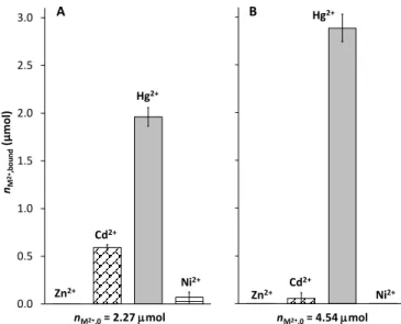
Figure 7. Binding of different M2+ ions to the immobilized ligand (mol) at pH = 6.0 (cMES
= 0.02 M) from two sample solutions containing 1.0 eq. (2.27 mol) (A) or 2.0 eq. (4.54
mol) (B) of all the four metal ions relative to the theoretical M2+ binding capacity of DY- NTG. (Vsample = 10.0 mL; mDY-NTG = 10.0 mg)
In order to test whether DY-NTG displays a selectivity in the binding of Hg2+, two types of samples containing the resin and four selected metal ions, Hg2+, Cd2+, Zn2+ and Ni2+, were prepared. Since data for Cd2+:DY showed that this metal ion cannot be bound by the ligand at pH = 2.0, which most probably also holds for Zn2+ or Ni2+, the metal ion selectivity experiments were performed at pH 6.0 by applying 1.0 or 2.0 equivalents of all the four metal ions, relative to the available immobilized peptide, in both types of samples. The results obtained reveal a dominance of Hg2+ in occupying the available binding sites of DY-NTG, especially at twofold excess of the metal ions relative to the peptide where only Hg2+ is bound by DY-NTG. However, a significant amount of Cd2+ (0.59 µmole), besides 1.96 µmole Hg2+, was also captured from the sample containing 1.0 eq. of all the four metal ions (Figure 7). These data represent ca. 90 % and 24 % binding of the added Hg2+ and Cd2+ ions, respectively.
Consequently, at pH = 6.0 Cd2+ can successfully occupy presumably mainly some of the lower affinity sites of the resin, even in the presence of 1.0 eq. of Hg2+ per peptide. Since Cd2+
induces a notable quenching of the fluorescence of DY in solution it may potentially act as an interference in the Hg2+- binding of this molecular probe at pH = 6.0 and in samples with sub-stoichiometric M2+:peptide ratios. However, Zn2+ and Ni2+
Dalton Transactions Accepted Manuscript
Open Access Article. Published on 02 May 2019. Downloaded on 5/2/2019 1:39:34 PM. This article is licensed under a Creative Commons Attribution-NonCommercial 3.0 Unported Licence.
View Article Online DOI: 10.1039/C9DT01141B