T O N A R C O T I C S MAURICE H. SEEVERS
Department of Pharmacology, University of Michigan, Ann Arbor, Michigan
I. Introduction 244 II. Criteria for the Establishment of Physical Dependence and Elicitation of
the Signs of Abstinence 246 III. The Development of Physical Dependence (and Tolerance) 248
A. Optimol Conditions for Development to Depressants of the Central
Nervous System 248 B. Failure of Development to Stimulants of the Central Nervous System. 249
IV. Locus of Origin of Abstinence and Species Specificity 249
V. Possible Mechanisms of Action 251 A. Factors that Might Alter the Cell Environment by Remote Actions on
Other Tissues 251 1. By an Action of the Drug on Other Body Tissues 251
2. By an Action of Tissues on the Drug (Detoxication) 253 B. Factors that Might Establish a "Barrier" Between the Cell and Its
Environment ("Surface" Forces) 255 1. Alterations in Membrane Permeability 255
2. Preferential Occupation of Surface Receptors by These Com
pounds 255 C. Factors that Might Modify the Capacity of the Cell to Respond to Its
Environment ("Intracellular" Effects) 256 1. Formation of Addition Products with Cell Constituents 256
2. Substitution of the Drug for a Cellular Constituent 256 3. Modifications of the Quantity or Activity of Enzymes 256 4. Morphological Alterations in Cytoarchitecture 258
VI. Some General Correlations and Newer Concepts 260
References 262 I. Introduction
The term dependence is used commonly by microbiologists to desig
nate a type of adaptation in bacteria exposed constantly to an environ
ment containing certain inhibitors of growth, notably streptomycin. In this situation the criterion for the establishment of dependence is a reduction in the rate, or the complete inhibition, of growth after drug withdrawal.
In searching for an explanation of this phenomenon it is natural to seek analogy in other biological systems. Such a search will not reveal
244
many examples of real dependence to alien chemical compounds. There has been a recent tendency to expand the usage of the term dependence to include components of the normal cell. For example, a report has just appeared in which the term dependence is applied to a situation in which an abnormally large quantity of pyridoxine is required regularly to prevent convulsions in an infant whose mother was treated during the early months of pregnancy with large quantities of this vitamin (Hunt et at, 1954). It is postulated that the infant may have acquired in utero an increased capacity to degrade pyridoxine enzymatically in view of the enormous dose required. The proper terminology for this condition would appear to be tolerance rather than dependence. Be that as it may, the author believes firmly that the term dependence should be reserved exclusively for those phenomena that occur after the ex
posure of tissue cells to an alien chemical environment, even if it is conceded that similar mechanisms could operate with abnormal quan
tities of normal metabolites. Otherwise, the term has no specific scientific value, since it is obvious that the biochemical and physiological integrity of the cell is "dependent" upon every one of its multitude of biochemical and structural components.
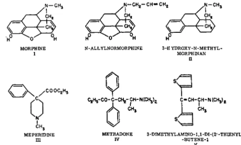
One of the relatively few examples of dependence as so defined is that which occurs in certain higher organisms after exposure to five different chemical types of narcotic analgesics (Fig. 1), the barbiturates, alcohol, and certain hydrocarbons.
It will be my purpose to review briefly our present knowledge regard
ing physical dependence to morphine as a prototype of the analgesic
Π
<~X / °Ο Ο° Λ V
A,
CH,1
CH,0
4M,S>
MEPERIDINE METHADONE 3-ΟΙΜΕΤΗΥίΑΜΙΝΟ-1,1-ΟΙ-(2·-ΤΗΙΕΝΥί) ΠΙ IV -BUTENE-1
V
FIG. 1. Five chemical types of analgesic drugs and their antagonist N-allylnor- morphine.
class of narcotics. Special attention will be directed toward an evaluation of the evidence that might suggest a common denominator for depend
ence as a general biological phenomenon.
The psychological and psychiatric aspects of the narcotic problem involve a type of dependence that includes many factors relating to the prior emotional state, motivations, environmental influences, and be- havioristic patterns of the individual. These aspects of the problem are categorized by such terms as craving, habituation, and addiction; they will not be considered here since they can be dissociated scientifically from the phenomena of tolerance and physical dependence (Krueger et al, 1941; Wikler and Rasor, 1953).
Physical dependence to narcotics, as distinguished from psychological or emotional dependence, may be defined as follows: a state of poten
tial hyperexcitability of the cerebrospinal axis which remains latent during the continuous administration of the narcotic and is manifest overtly during withdrawal of the chemical from the cellular environ
ment. The signs and symptoms of hyperactivity resulting from the unmasking of this latent state are termed the abstinence syndrome.
The term, physical dependence, as applied in this specific sense is used to designate only one of the two primary processes of adaptation (Seevers, 1954), which have been demonstrated in several species of vertebrates of higher cerebral development following the chronic admin
istration of these drugs. Dependence is intimately associated, and de
velops simultaneously, with the second adaptive process, tolerance. The latter represents an acquired resistance to the depressant effects of these substances on certain portions of the central nervous system. Although they are considered separately in this symposium, these two phenomena can hardly be dealt with as separate entities as they relate to the central nervous system. Whereas tolerance development can be demonstrated in types of cells other than those of the nervous system, for example with morphine in the smooth muscle of the blood vessels, physical dependence has not been demonstrated to occur outside of the nervous system. Neither has it been possible thus far to prove the existence of physical dependence in the nervous system without the concurrent development of tolerance.
II. Criteria for the establishment of physical dependence a n d elicitation of the signs of abstinence
The signs of physical dependence to morphine are quite typical and generally similar to those which occur with the synthetic analgesics.
Those that occur in the rhesus monkey are shown in Table 1 (Seevers,
TABLE 1. Classification of the Signs of Abstinence in the Monkey MILD (may be considered of no significance by the untrained observer): Appre
hension, continual yawning, rhinorrhea, lacrimation, hiccup, shivering, perspiration on face, chattering, quarreling and fighting.
MODERATE: Intention tremor, anorexia, pilomotor activity, muscle twitchings and rigidity, holding the abdomen (cramps).
SEVERE: Extreme restlessness, assumption of peculiar attitudes, vomiting, severe diarrhea, erection and continued masturbation, inflammation of the eyelids and con
junctiva (insomnia), continual calling and crying, lying on the side with eyes closed, marked spasticity.
VERY SEVERE: Docility in the normally excitable animal, dyspnea, pallor, strabis
mus, dehydration, weight loss, prostration, circulatory collapse, convulsions and death.
1936). The actual differences in individual signs in each species are related to the anatomic limitations of the animal.
With these five types of drugs the signs of abstinence may be elicited by either one of two procedures: (a) abrupt withdrawal, which is the classic method and involves stopping administration of the drug, or (b) injection of N-allylnormorphine, a specific antagonist of this class of compounds, which "neutralizes" most of the depressant effects of the drug at their locus of action on or in the cell.
With abrupt withdrawal, the signs of abstinence appear first within a matter of 6 to 10 hours after the last dose, depending on the drug and its rate of detoxication in the individual species. They increase in inten
sity with morphine up to the 32nd or 48th hour (Isbell and Fraser, 1950; Isbell and White, 1953). With shorter-acting compounds this interval is diminished, and with longer-acting compounds the peak is delayed. With morphine the intensity of the signs of abstinence is inversely proportional to the quantity of morphine remaining in the body. In a general way, loss of tolerance follows morphine detoxication;
curves representing residual tolerance and residual morphine parallel each other rather closely, although in human addicts who have had a very long and heavy drug experience the rate of tolerance loss is delayed materially. Possibly some tolerance is retained permanently. Once the abstinence syndrome has reached its peak intensity, it then remains at this level for 24 to 48 hours longer, subsiding slowly over a period of 7 to 10 days.
With nalorphine-induced abstinence the situation is markedly differ
ent, representing an explosive type of response, which is precipitated within a few moments after administration of the antagonist. It reaches a peak of intensity within 5 to 10 minutes and then subsides within one to two hours as the N-allylnormorphine is detoxified. Although the
manifestations of abstinence in the latter instance may appear to be more intense because of their explosive nature, the actual level of hyper- excitability of the central nervous system is not elevated appreciably above that which one might expect at the peak of abrupt withdrawal.
Certain signs appear more commonly, particularly those related to the gastrointestinal tract, at least in the monkey. There is one outstanding difference between the two types of withdrawal. The slow onset and prolonged course of abrupt withdrawal results in a severe stressing situation associated with marked fatigue from the insomnia and exces
sive muscle activity. The lowered food intake and water imbalance from anorexia contribute to the abnormal situation. These and other factors modify the total picture but do not in themselves appear to increase the level of nervous excitability per se. In our experience with the monkey, for example, we have never observed convulsive seizures in nalorphine- induced withdrawal, although this is commonly experienced following abrupt withdrawal in this species.
III. The development of physical dependence
A. OPTIMAL CONDITIONS FOR DEVELOPMENT TO DEPRESSANTS OF THE CENTRAL NERVOUS SYSTEM
The development of maximal tolerance and maximal dependence with this class of compounds requires exposure of the affected cells to a continuous and uninterrupted optimal concentration of the drugs at all times. This means that the quantity administered will be increased as tolerance is developed and that the frequency of administration is such that no period of abstinence is permitted. Under optimal conditions the quantity and frequency should be such that the pharmacological effects are continuous, the contribution of each new dose overlapping the effects of the previous administration in order to maintain maximal receptor occupation continuously. This not only creates the most rapid and maximum tolerance development, but also builds up the "hyper- excitatory" state in the neuron to a maximum, creating the greatest possi
ble degree of physical dependence. Under these circumstances tolerance and dependence can be detected within a relatively short time. With morphine, maximal dependence can be produced within 30 days or earlier in the monkey judging by the appearance of convulsions follow
ing abrupt withdrawal (Deneau, Kissel, and Seevers, 1954a).
In contrast to the situation that occurs with tolerance, further in
creases of dosage beyond a certain point do not engender a greater in-
crease in the latent hyperexcitability of the nervous system. In other words, a ceiling is reached beyond which further increments of dosage do not appear to enhance the intensity of signs of abstinence. Using the appearance of grand mal type seizures as an end point, we have been able to produce as great effects in the monkey with doses of 3 mg per kilogram 4 times a day (12 mg per kilogram per day) as with 75 mg per kilogram 2 times a day (150 mg per kilogram per day). Andrews and Himmelsbach (1944) believe that in man total daily doses beyond 500 mg (up to 4400 mg) do not produce any significant increment in the level of excitability over that occurring with the smaller doses.
B . FAILURE OF DEVELOPMENT OF DEPENDENCE (OR TOLERANCE) TO STIMULANTS OF THE CENTRAL NERVOUS SYSTEM
One of the fundamental principles, which cannot be disregarded in any consideration of this problem, relates to the failure of development of either tolerance or physical dependence in the cells of the nervous system to drugs that are primarily excitant and that lack primary depressant qualities. These include substances such as sympathomimetic amines (amphetamine, mescaline, etc.), cocaine, strychnine, caffeine, and agents of this type. It is of equal importance to recognize that vascular tolerance or tachyphylaxis can be developed to many agents of this general type and that increased detoxication rates are also probable with continued administration. The examples of tolerance that are ordin
arily cited to refute the basic principle stated above probably belong to the latter types of tolerance, which may be reflected indirectly in the central nervous system.
Following continuous exposure to CNS excitants the nervous system becomes "sensitized" or "intolerant," requiring a reduction rather than an increase in dosage. No abstinence syndrome can be demonstrated with cocaine (Tatum and Seevers, 1929), amphetamine, mescaline, or with other drugs having this general type of pharmacologic action, unless weakness and fatigue are so categorized.
IV. Locus of origin of abstinence a n d species specificity η
Physical dependence can be demonstrated only in vertebrates with highly developed cerebral hemispheres. These include man, the chim
panzee, the monkey, and the dog. Many reports have been made that certain other species, such as the cat, the rabbit, the guinea pig, the rat, etc., show signs of physical dependence. These reports are largely
unreliable since the effects in these animals do not meet the criterion of physical dependence already outlined (Seevers, 1948). It should be pointed out that virtually all experimental studies on animals have in
volved drug administration on a 24-hour five-day week basis (Deneau, Kissel and Seevers, 1954b). Further studies of small animals are needed, utilizing six-hour seven-day week administration, although the author does not believe this will alter the conclusions drawn here concerning qualitative species differences. Since tolerance development is also ex
ceedingly poor or nonexistent in these same species, a rather close nega
tive parallelism seems to exist between the two phenomena. Neither tolerance nor physical dependence has been demonstrated in cold
blooded animals. Attempts have been made to establish evidence of physical dependence and tolerance to these same drugs in cultures of embryonal tissue. Several positive reports (Krueger et al., review, 1941) have been made based upon the same criteria as used for drug resist
ance and dependence in microorganisms. The author is at a loss to interpret these results as they might apply to the problems we are dis
cussing here. If it is true that there is a common denominator between this type of dependence and the dependence to opiates, then transfer of these results may have validity. It seems clear that further work along these lines is indicated, even though microorganisms are known to be very resistant to the action of morphine.
The foregoing facts, which relate to species differences, suggest that animals of higher cerebral development may possess some specifically sensitive neurological unit that is entirely absent or present in only small numbers in the lower species. An alternative explanation might conceive of the specialized distribution of a drug-sensitive enzyme or tissue component limited to the more highly developed species.
The evidence available at the present time, culminating largely from the neurophysiologic observations of Wikler (1950, 1953), indicates that the phenomenon of physical dependence resides exclusively in the central nervous system and involves probably all of the major divisions, including the cerebral hemispheres, the subcortical areas, and the cord.
The comparatively small amount of neurophysiologic evidence now available suggests that the internuncial neuron is a principal, if not an exclusive, point of action. Peripheral nerve does not appear to be involved. Such inferences are based largely on the studies of spinal reflexes in cord-transected dogs and man. An acute "abstinence syn
drome," as evinced by enhancement or inhibition of multisynaptic reflexes, can be elicited either by nalorphine or by abrupt withdrawal.
Since this extends only to multineuronal arcs (crossed extensor reflex, etc.) and not to two-neuron arcs such as the knee jerk, it is concluded that the internuncial neuron is involved primarily.
It has been stated previously that physical dependence has not been demonstrated to occur in structures outside the central nervous system.
Shideman and Seevers (1942) demonstrated that the oxygen uptake of minced skeletal muscle from chronically morphinized rats, killed at various intervals after the last dose, showed a time-activity curve paral
leling rather closely the known curves of abstinence intensity in other species. These results have been confirmed in the mouse and, to a lesser extent, in the dog. An interpretation of these observations as it might relate to physical dependence in general is difficult, especially in view of the recent observations of Irwin and Shideman (1953), which indicate that the oxygen uptake of skeletal muscle of the rhesus monkey is de
creased rather than increased during abstinence. This decrease coincides with a marked fall in body temperature, which occurs at the peak of abstinence intensity in the latter species.
Complete local tolerance (tachyphylaxis) to the depressor actions of morphine can be developed in vascular smooth muscle within a few hours (Schmidt and Livingston, 1933). It might be expected that if dependence is a universal cellular response in those species in which it can be produced satisfactorily in the central nervous system, that a local vasoconstriction should occur when morphine is withdrawn. This actually does occur in man, as is evidenced by skin temperature changes and by a reduction in the oxygen saturation of venous blood.
It has been generally assumed that this response is neurogenic in origin, although this has not been established. It would be of considerable theoretical importance to ascertain whether this is a primary cellular response or whether it is secondary to the effect on the central nervous system.
V . Possible mechanisms of action
A. FACTORS THAT MIGHT ALTER THE CELL ENVIRONMENT BY REMOTE EFFECTS ON OTHER TISSUES
1. By an Action of the Drug on Other Body Tissues
a. Blood-Borne Factors. Under this general heading come a variety of possible mechanisms that have been suggested, such as antitoxins (Hirschlaff, 1902; Valenti, 1914) or substances with allergenic properties that might cause the appearance of antibodies (Sioli and Wrinkel, 1933).
For example, Ostromislensky (1935) postulated a water-soluble protein- morphine combination that could be hydrolyzed easily; the morphine could be removed by oxidation, leaving behind the protein that was to serve as antigen. All these concepts are purely speculative and there is no evidence to support the view that tolerance or dependence has any relation to immunity or sensitization in the immunological connotation of these terms.
Many attempts have been made to relate dependence to the actions of morphine on the endocrine glands, notably the thyroid and the adrenal, in view of some similarities between the abstinence syndrome and the toxic actions of these hormones. The pituitary gland has also been invoked, in view of the well-known antidiuretic action of morphine.
Since this effect is blocked by nalorphine it is believed to be neurogenic in origin.
There is sufficient ancillary evidence to keep such hypotheses alive.
For example, morphine exerts a calorigenic action in the dog, and thyroid feeding increases markedly the toxicity of morphine. It has now been established that in chronic morphinism there is a suppression of the activity of the adrenal cortex with a reduction in excretion of 17-ketosteroids (Eisenman et al, 1954).
Recently, efforts have been devoted toward an examination of epinephrine and histamine as possible mediators of these phenomena.
It is presumed that they would be liberated in peripheral tissues and exert their effects after transport to the nerve centers themselves. It is a well-established fact that a single dose of morphine liberates epinephrine (Gross et al, 1948) and histamine (Feldberg and Paton, 1951). This occurs in those species that develop marked physical dependence, as well as in others, like the cat and rabbit, that do not develop any significant tolerance or physical dependence. Whereas it has been established that a certain grade of central tolerance develops to epinephrine (Essex et al, 1952), this does not appear to be the case with histamine. The main objection to the acceptance of explanations based upon the activity of these substances lies in the fact that as toler
ance develops liberation of these two substances no longer occurs.
Furthermore, neither one is capable of suppressing the abstinence syn
drome. This would be expected theoretically if they were responsible for the development of tolerance or dependence.
The concept that morphine may modify the type or extent of circu
lating substrates has been suggested, in view of the fact that morphine is known to have a glycogenolytic action. Since this operates largely
through the medium of epinephrine release and disappears with toler
ance, this view is very difficult to harmonize with the facts.
b. Neurogenic Influences. Several hypotheses involving various modi
fications of the principles of homeostasis have been invoked to explain tolerance and dependence, the latter being conceived to represent a physiologic rebound after release from drug suppression. These have generally been conceived to operate via neurogenic influences from the more primitive autonomic regulating centers. The concepts and findings of Magoun (1950) as they relate to the action of chemical substances on the inhibiting-activating centers in the reticular substance give greater substance to such views. In general, however, such suggestions suffer from exactly the same deficiencies and must bear the same type of criticism as that which may be directed against most current views, i.e., they are descriptive of events which occur rather than explanations of subtle mechanism.
2. By an Action of Tissues on the Drug (Detoxication)
a. To Less Toxic Substances. One of the more attractive hypotheses presented to account for tolerance is that which postulates that tissues acquire the ability to degrade the drug more rapidly.
Gross and Thompson (1940) demonstrated that a large fraction of administered morphine is excreted as a conjugate that they termed
"bound" morphine. They also reported that morphine-tolerant animals possessed a reduced capacity to excrete the bound form in comparison with nontolerant animals. Recent studies (Woods et al, 1954), utilizing newly developed methods that are both specific and sensitive indicate that approximately 40% of an administered dose appears as morphine monoglucuronide (Woods, 1954) in bile within a few hours after administration. This conjugate is ultimately reabsorbed from the intes
tine and excreted in the urine, so that 75 to 80% of an administered dose, 15% free and 60 to 65% conjugated, is recovered within 48 to 72 hours.
Of the remaining fraction, 5 to 10% is recovered in feces, leaving approx
imately 10% not accounted for (Woods et al., 1953; Cochin et al., 1954).
Preliminary pharmacological observations indicate the morphine mon
oglucuronide to be inert (Woods, 1954a). In contrast to the observations of Gross and Thompson noted above, these workers were unable to find any significant difference between tolerant and nontolerant dogs after tolerance had been well developed after several months of drug admin
istration. It is quite clear that, with this drug at least, tolerance cannot be explained on this basis.
Analysis of tissues of tolerant and nontolerant rats, dogs, and monkeys
fails to reveal any significant differential accumulation of morphine in any single tissue except possibly the thyroid and the spleen. The amount of morphine present in the brain or in the spinal fluid of tolerant or non- tolerant animals is so low that it can rarely be detected by a method that is sensitive to 1 ^g of morphine per gram of tissue (Woods, 1954b).
These results suggest that the concentration in brain, even at the time of peak plasma levels, does not exceed 10'6 M.
These studies also prove that morphine has been virtually eliminated from the body 48 hours after the last dose in tolerant as well as non- tolerant animals.
b. To More Toxic Substances. The possibility that some residue, such as an oxidation product of morphine, might remain in tissues, cre
ating the excitatory state, was postulated many years ago by Marme (1883). No such substance has ever been found in tissues, although it is well known that morphine is easily susceptible to oxidation and that a definite compound, pseudomorphine, can be readily produced in vitro.
The only product about which there is indirect evidence to presume its occurrence in the body is normorphine. Recent studies (Elliott et al., 1954) of morphine tagged with C1 4 in the methyl group suggest N-demethylation, since C14-tagged carbon dioxide is found in the expired air. Normorphine has not been identified in tissues chemically.
Although the hypothesis of formation of a toxic product is not supported by evidence, it is a fact that approximately 10% of any dose of morphine has not yet been accounted for.
A great difficulty is encountered when an explanation based upon toxic degradation products is applied to tolerance, cross-tolerance, de
pendence, and mutual dependence satisfaction of the five types of narcotics of different chemical structure. These all have widely differing chemical characteristics. Some are relatively easily detoxified in vivo.
It does not appear to be fruitful to search for a common degradation product in these five different substances. The evidence available at present lends no support for the view that the phenomena of tolerance or dependence are related primarily to changes in the nature of the compound itself.
Recent studies (Deneau et al., 1954a) indicate that physical de
pendence can be detected easily with nalorphine in monkeys given 100 μg per kilogram four times daily for 50 days (shorter periods were not tried) and in man (Wikler et al., 1953), using large doses of nalor
phine, following 15 mg four times daily for two or three days.
These observations and the data of Woods point out very strikingly
the marked potency of morphine as a pharmacological agent. Assuming the value previously mentioned, 10"6M, to apply at the peak plasma con
centration following 30 mg per kilogram in the dog, it seems improbable that a single dose of 15 mg in man would produce a brain concentration exceeding 10"9 M. If these values are correct, then a mechanism based on competition for sites of action of hormones, vitamins, or chemical mediators could be more easily visualized.
c. To Utilizable Substrates. This seems somewhat remote for the class of drugs considered here, although it is a possibility. It must be considered with agents like alcohol, streptomycin on bacteria, etc. With the latter, adaptation could occur by cellular utilization of the pentose moiety, much like the adaptation of yeasts to galactose.
B . FACTORS THAT MIGHT ESTABLISH A "BARRIER" BETWEEN THE CELL AND ITS ENVIRONMENT ("SURFACE" FORCES)
Such factors might in a general way be designated as surface influ
ences, although the author recognizes that this is an exceedingly tenuous line of demarcation, since it is not possible to make a sharp distinction between the surface and the internal environment of the cell. Nonethe
less, it seems to be worthwhile to consider such effects as might be produced largely by physiochemical forces at the surface of the cell or by an action on receptors (enzymes) oriented to the surface.
1. Alterations in Membrane Permeability
a. To the Drug or to Its Degradation Products. We have no good evidence upon which to base a hypothesis along these lines. It would appear that morphine and compounds of this type do not find easy access to the central nervous system, in view of the low concentration found there. Since there is no evidence that an increased concentration of these drugs is present in brains of tolerant or dependent animals, the possibility that this type of an effect occurs appears to be unlikely. Nor is there any evidence concerning possible changes in electrolyte or ion transfer. Little work has been done along these lines. If it is correct that the phenomenon of physical dependence can be demonstrated only in certain types of interneurons, such types of evidence will be difficult to obtain.
b. To Substrates and Other Chemical Requirements. The fact that the physiologic response of the cell appears to be quite normal during drug administration makes it difficult to conceive of major variations resulting from limited substrate or other metabolite concentration.
2. Preferential Occupation of Surface Receptors by These Com-
pounds. It is theoretically possible for a large molecule of the morphine type, which has several centers of high electron density, to umbrella the surface and prevent access of other potent chemical influences, such as hormones, vitamins, chemical mediators, etc. It seems logical that if such an effect did occur it would be short-lived, in view of the fact that once tolerance has developed and a high concentration of morphine is maintained in the cellular environment, it is difficult to obtain any evidence of altered physiological activity of the cell.
Whereas the author believes that morphine and this class of com
pounds probably exerts surface effects (in the case of the neuron), such as the partial blockade of nervous conduction, it must be admitted that no direct evidence for such a view is available.
C. FACTORS THAT MIGHT MODIFY THE CAPACITY OF THE CELL TO RESPOND TO ITS ENVIRONMENT ( "INTRACELLULAR" EFFECTS )
1. Formation of Addition Products with Cell Constituents. The chem
ical reactivity of morphine renders it possible to conceive of morphine as the prosthetic group of an enzyme or serving as a carrier with the capacity to accept or transfer groups as "active acetyl," possibly even to compete with choline in this regard for the acetyl group at the synapse.
Its redox potential puts it in the category of substances capable of acting in hydrogen transfer mechanisms, such as phosphorylation, etc.
2. Substitution of the Compound for Some Cellular Constituent.
With morphine and the other narcotic analgesics, the essential basicity of the compounds suggests that they might compete for cell constituents such as choline and actually become incorporated into a larger molecule, for example, lecithin. Loofs (1922) presented such a view. Whereas his idea was purely speculative, it inspired several investigators to seek evidence along this line. It has been claimed by Klee and Grossman (1925) that choline infusion during withdrawal reduces the intensity of the abstinence syndrome. Ma (1931) claimed that lecithin feeding diminished the intensity of the abstinence syndrome. No modern work supports the theory, nor have these latter claims been substantiated, although the concept is not easy to discard even as it might relate to some particular substance such as lecithin.
3. Modification of the Quantity or the Activity of Enzymes. A sum
mary of the older literature is found in Kreuger et al. (1941). Most of the older studies involved the proteolytic and hydrolytic enzymes.
The recent work has dealt principally with the oxidative enzymes, al
though a considerable amount of attention has been paid to esterases,
particularly Cholinesterase. The more recent studies really began with the work of Quastel and Wheatley (1932), who noted that several depressant drugs were capable of inhibiting oxygen uptake in various types of cells, including those of the nervous system. Current studies of the effects of these substances on the cytochrome enzymes have been made by Wang and Bain (1953a), who examined this problem with the prime objective of determining minimal effective concentrations.
In general, it may be stated that morphine and the synthetic anal
gesics are not very potent enzyme inhibitors. Bernheim and Bernheim (1936) demonstrated that morphine in a concentration 8 Χ 10~5 Μ produced a 50% reduction in the hydrolysis of acetylcholine by brain esterase. The most sensitive enzyme found by Wang and Bain was DPN cytochrome c reductase, the minimal effective concentration in this instance being 10"3 M.
Reasoning by analogy, and accepting the data of Woods that the concentrations in brain appear to be no more than 10"6 M, it seems logical to state that at this time there is very little evidence to support the view that the action of these drugs is related to a direct effect on enzymes. It must be recognized, of course, that there is a possibility of serious error in this type of argument. If, as appears to be the case, morphine and these other compounds have difficulty in gaining access to the intracellular environment, such argument does not rule out the possibility that surface-orientated enzymes may be placed in contact with relatively large quantities of morphine, especially during chronic administration. With a single therapeutic dose in man, however, it seems probable that even the surface concentrations must be much below those shown to be effective in vitro. This is borne out by studies made on brain tissues studied in vivo after chronic administration. Wang and Bain (1953b) have been unable to find any significant difference in either the quantity or activity of enzymes in brains removed from chronically morphinized rats compared with controls.
The author would like to point out that the essentially negative evidence obtained thus far does not preclude the possibility of an explanation based upon changes in enzyme activity. Very little work has been done on species in which dependence can be demonstrated to be of significance. The author is of the opinion, however, that the limita
tions of the phenomenon to what appear to be specialized types of inter- neurons suggest that if enzymes are involved, the pattern of distribution is unique and in all likelihood the type of enzyme involved might not be distributed universally in tissues.
As mentioned previously, Shideman and Seevers (1942) believed that they had some evidence for the development of physical dependence in rat skeletal muscle, which showed an increased oxygen uptake during withdrawal. This excessive oxygen uptake was azide-sensitive, the latter reducing the level to, but not below, normal. This seemed to fit in with the general concept of activity versus resting metabolism in this par
ticular type of tissue. Since the reverse situation is now known to occur in the monkey during withdrawal, the data as a whole do not furnish much information concerning dependence in the broader sense.
It seems fair to summarize the work on enzymes at this time by concluding that, although no clear-cut evidence is available to relate physical dependence to enzyme activity, it remains an attractive hypoth
esis, and the author believes that further study should be made to deter
mine factors related to energy-release mechanisms in the central nervous system in dependent animals. The possibility that alternative metabolic pathways may take over the function of an inhibited component in a multienzyme system is neither proved nor disproved at this time. Fur
thermore, until an exact cellular distribution of these drugs as well as
"sensitive" enzymes can be ascertained, it does not seem logical to con
clude that an inhibiting concentration of morphine might not be present at the site of enzyme activity.
4. Morphological Alterations in Cytoarchitecture of the Cell. The concept that these drugs might produce alterations in the colloidal concept that these drugs might produce alterations in the collodial state of nervous tissue or produce actual changes in the types of protein or lipoproteins has been considered many times. Bancroft and Richter (1931) revived the concept of Claude Bernard that anesthesia and narcosis could be explained on a physicochemical basis. Bancroft's idea engendered a considerable amount of work with sodium rhodanate, based upon the theory that this compound should reverse the agglom
erated brain colloids and return them to the normal critical size. There is no substantiating evidence whatsoever for this concept in terms of physical dependence in animals or man.
Nonetheless, marked structural changes can be produced by this class of substances in the brain of the rhesus monkey, by acute or chronic administration.
Irwin and Seevers (1954) reported recently that single large sub
lethal doses of members of the morphine, meperidine, methadone and morphinan series of drugs are capable of producing profound neuro
physiologic and pathologic changes in this species. These effects are
graded to some extent with dose and are completely preventable by the administration of nalorphine shortly after the drug has been admin
istered. Tolerant animals do not show the effects of doses several times those required to initiate the effects in nontolerant animals.
The administration of a large single sublethal dose produces an immediate neurologic syndrome in this species, which is characterized by an increase in the deep reflexes, hypertonicity of the flexors and abductors of the lower limbs, hypertonicity of the extensors of the upper limbs and, occasionally, the flexors. These changes are associated with disturbances in gait, overreaching of the forelimbs, increase in motor activity, asymmetric pupils, apprehension, convulsions, etc. The animal may be completely paralyzed and strikingly resemble a decorticate preparation.
The effects occur within a short time after administration. This con
dition may remain temporarily and disappear with complete recovery within a matter of 24 hours, or recovery may occur with large but temporary neurological residues, with ultimate, partial, or complete functional recovery. If survival is effected for several weeks in the most severe cases, by artificial feeding, neuropathological examination reveals damage affecting principally the white matter of all portions of the cerebral hemispheres (Bebin et al., 1954). Cortex, basal ganglia, and cerebellar hemispheres are also affected, but to a lesser extent. No pathological changes were observed in the diencephalon, brain stem, or spinal cord. All gradations of demyelinization were observed, including complete breakdown of myelin sheaths and axis cylinders.
Chronic administration of morphine and certain members of the other classes of compounds in large daily doses results in the appear
ance of abnormal neurologic changes, which are detectable only fol
lowing careful examination, but which are associated with well-defined neuropathology with the same distribution in the nervous system out
lined above. With methadone, a persistent deformity of the lower limb is noted, consisting of marked adduction of the thighs, abduction below the knees and turning in at the feet. These permanent changes are associated with neuropathology in the hemispheres, but no cord lesions were noted.
The exact significance of these findings is not clear at this time.
Although they have not been demonstrated in other species by using similar techniques, the author believes that they represent quantitative rather than qualitative species differences. The monkey is notably sus
ceptible to the pathology-inducing properties of drugs that affect the
central nervous system. It is quite clear that these results indicate the widespread distribution and action of morphine in the central nervous system. It is difficult to escape the view that it is a direct action on the neuronal structures, in view of the complete antagonism by N-allylnor- morphine. It appears also that these neurologic signs and neuropathology are probably related to the depressant actions of the drug, rather than to the excitant effects, since both nalorphine antagonism and tolerance development extend primarily to the depressant properties of these drugs. It is of considerable interest to note that lesions of strikingly similar character may be produced in the rhesus monkey by the chronic administration of cyanide or azide (Hurst, 1944). This might suggest that the cytochrome enzymes may be involved in this phenomenon. The author feels that further extensions of work along these lines is urgently needed and that it should be correlated directly with chemical studies on the intimate tissue distribution of these compounds.
V I . Some general correlations a n d concepts
It has long been known that morphine acts both as a depressant and as an excitant, the effects varying with the type of cell involved, the species of animal that serves as a source of these cells, the dose, the route of administration, and a multitude of other factors. Various areas of the central nervous system respond in a different manner in the same species and with the same dose. It is important also to recognize that acquired tolerance is developed only to the depressant functions of this drug, not to its excitant actions. The latter appear to be fundamental pharmacological phenomena in the nervous system, since there is no description of tolerance but only increased sensitivity to drugs that augment the total functional activity of the central nervous system.
In recognition of these facts, Tatum, et al. (1929) proposed a hypoth
esis, which has become to be known as the "dual action" hypothesis:
tolerance being described as an acquired resistance to the narcotic effects; dependence to the cumulative effects of a longer-lasting direct excitation, the effected cells become sensitized rather than tolerant to this excitant effect.
Seevers and Woods (1953), accepting the basic tenets of this hypoth
esis as representative of the facts, have presented an interpretation that postulates a biphasic action of this and similar drugs on the same cell, the internuncial neuron. This new concept was engendered primarily from the work of Wikler; our observations that the axon appears to be a vulnerable point of attack in the monkey, judging from the ease with
TABLE 2. Hypothetical Concept of "Dual Action" of Morphine, Involving Two Types of Receptors on Certain Internuncial Neurons
Depression Excitation Single Drug Administration
Locus of receptors Nature of bond
Number of bonds Pharmacologic
effects
Nalorphine action
Pharmacological resultant Relation to total
daily dose
Specific cross-toler
ance to chemical and pharmaco
logical analogues.
Nonspecific cross- tolerance to bar
biturates, alcohol, etc.
Abrupt withdrawal
Nalorphine withdrawal
Cell surface
Weak (physicochemical)
Many (high extracellular morphine concentration) Partial blockade of conduction
in sensory and motor neurons producing anal
gesia, motor weakness, etc.
Antagonizes this action of morphine by "neutraliz
ing" drug effect at receptor site
Intracellular
Strong chemical bonding (incorporation into macromolecule?)
Relatively few (low intracellu
lar morphine concentration) Facilitates impulse formation or
propagation (convulsions)
No significant effect on large convulsant doses of mor
phine; probable summation with large doses of both drugs
Repeated (Chronic) Administration Almost complete tolerance
to this effect
Rate and degree of tolerance development logarithmic function of receptor occu
pation—never complete High grade mutual capacity
to occupy receptors and produce similar pharmaco
logical effects with small doses
Low-grade "smothering"
effect on CNS with very large doses only Rapid loss of tolerance
paralleling desaturation of receptors and loss of body morphine
Acts to abolish depression by "displacing" morphine from surface receptors.
This "unmasks" the latent
"hyperexcitatory" state
No tolerance to this effect but development of latent
"hyperexcitatory" state Degree of excitation increases
with dose up to ceiling- further increases with dose do not occur
Suppression of excitability as with morphine by "surface"
effects
Suppression of excitability only with very large doses
Gradual emergence of state of CNS hyperexcitability as
"extracellular" morphine is excreted
Gains access to this site only with difficulty and in large dosage and then summates with morphine to induce excitation
which demyelination occurs; and the low concentrations of morphine demonstrable in brain tissue. This concept, which is subject to experi
mental verification or rejection, makes a fair fit with the multitude of known facts.
It is conceived that morphine and its congeners and other synthetic analgesics combine with receptors (more likely of two types) located (a) one at or near the surface of the neuron (extracellular phase) and the other within the neuron (intracellular phase).
The essential features of this concept are presented here in tabular form (Table 2) to conserve space. A more detailed description will be found elsewhere (Seevers, 1954).
The importance of the phenomena of physical dependence to nar
cotics as a fundamental process of biological adaptation is sufficient to warrant a concentrated attack on the problem by the chemist, the biochemist, the neurophysiologist, and the pharmacologist. Its real nature is not likely to be disclosed until we know the histochemical distribution of the drugs in the interneuron; whether the effect is produced by the original drug or by its detoxication products; its effects on energy- storing and release mechanisms and on the total biochemical architec
ture of the cell. Its intimate relation to the phenomena of tolerance sug
gests that the solution of either mechanism will hasten greatly an under
standing of the other. Even though marked differences would appear to exist between the overt manifestations of physical dependence to narcotics in primates and dependence to antibiotics in microorganisms some similar biochemical mechanism, such as enzyme adaptation, mav be involved in both instances.
References
Andrews, H. L., and Himmelsbach, C. K. (1944). /. Pharmacol. Exptl. Med. 81, 288.
Bancroft, W. D., and Richter, G. H. (1931). /. Phys. Chem. 35, 215.
Bebin, J . , Scharenberg, Κ., Irwin, S., and Seevers, Μ. H. (1954). /. Pharmacol.
Exptl. Therap. 110, 4.
Bernheim, F., and Bernheim, Μ. L. C. (1936). J. Pharmacol. Exptl. Therap. 57, 427.
Cochin, J . , Haggart, J . , Woods, L. Α., and Seevers, Μ. H. (1954). /. Pharmacol.
Exptl. Therap. In press.
Deneau, G., Kissel, J., and Seevers, Μ. H. (1954a). Unpublished experiments.
Deneau, G , Kissel, J., and Seevers, Μ. H. (1954b). Federation Proc. 13, 347.
Eisenman, A. J., Fräser, H. F., and Isbell, H. (1954). Federation Proc. 13, 203.
Elliott, H. W., Tolbert, Β. M., Adler, Τ. Κ., and Anderson, Η. Η. (1954). Proc. Soc.
Exptl Biol Med. 85, 77.
Essex, Η. E . , Meeker, W. Α., and Brown, B. (1952). Am. ] . Physiol. 171, 78.
Feldberg, W., and Paton, W. D. M. (1951). /. Physiol. 114, 490.
Gross, E . G., and Thompson, V. (1940). J. Pharmacol Exptl Therap. 68, 413.
Gross, E . G., Holland, H., Carter, H. R., and Christensen, Ε . Μ. (1948). Anesthesiol
ogy 9, 459.
Hirschlaff, L. (1902). Klin. Wochschr. 39, 1149, 1174.
Hunt, A. D., Jr., Stokes, J . , Jr., McCrory, W. W., and Strand, Η. Η. (1954). Pedi
atrics 13, 140.
Hurst, Ε . W. (1944). Brain 67, 103.
Irwin, S., and Seevers, Μ. H. (1954). /. Pharmacol. Exptl. Therap. 110, 27.
Irwin, S., and Shideman, F. E . (1953). Unpublished experiments.
Isbell, H. and Fräser, H. F. (1950). Pharmacol Revs. 2, 355.
Isbell, H., and White, W. W. (1953). Am. J. Med. 14, 558.
Klee, P. H., and Grossman, O. (1925). Münch, med. Wochschr. 72, 251.
Krueger, H., Eddy, Ν. B., and Sumwalt, M. (1941). Public Health Repts. (U.S.) Suppl. 165.
Loofs, F. A. (1922). Z. ges. Neurol. Psychiat. 79, 433.
Ma, W.-C. (1931). Chinese J. Physiol. 5, 251.
Magoun, H. W. (1950). Physiol Revs. 30, 459.
Marme, W. (1883). Beut. med. Wochschr. 9, 33.
Ostromislensky, I. (1935). Med. Record 141, 556.
Quastel, J. H., and Wheatley, Α. Η. M. (1932). Proc. Roy. Soc. (London) B112, 60.
Schmidt, C. F., and Livingston, A. E . (1933). /. Pharmacol Exptl Therap. 47, 473.
Seevers, Μ. H. (1948). Ann. Ν. Y. Acad. Set. 57, 99.
Seevers, Μ. H. (1954). Federation Proc. June. In press.
Seevers, Μ. H. (1936). J. Pharmacol. Exptl. Therap. 56, 157.
Seevers, Μ. H., and Woods, L. A. (1953). Am. J. Med. 14, 546.
Shideman, F. E., and Seevers, Μ. H. (1942). J. Pharmacol. Exptl. Therap. 74, 88.
Sioli, F., and Rinkel, M. (1933). Deut. med. Wochschr. 59, 323.
Tatum, A. L., and Seevers, Μ. H. (1929). J. Pharmacol. Exptl. Therap. 36, 401.
Tatum, A. L., Seevers, Μ. H., and Collins, Κ. H. (1929). J. Pharmacol Exptl.
Therap. 36, 447.
Valenti, A. (1914). Arch, exptl. Pathol Pharmacol. 75, 437.
Wang, R. I. H., and Bain, J. A. (1953a). J. Pharmacol. Exptl. Therap. 108, 349.
Wang, R. I. H., and Bain, J. A. (1953b). J. Pharmacol. Exptl. Therap. 108, 354.
Wikler, Α., Fräser, Η. F., and Isbell, Η. (1953). /. Pharmacol Exptl Therap. 109, 8.
Wikler, Α., and Rasor, R. W. (1953). Am. J. Med. 14, 566.
Wikler, A. (1950). Pharmacol. Revs. 2, 435.
Wikler, A. "Opiate Addiction." C. C. Thomas, Springfield, 1953.
Woods, L. A. (1954a). Federation Proc. 13, 419.
Woods, L. A. (1954b). J. Pharmacol Exptl Therap. In press.
Woods, L. Α., Cochin, J., Fornefeld, E. G., and Seevers, Μ. H. (1954). J. Pharmacol Exptl Therap. In press.
Woods, L Α., Cochin, J . , Mellett, L. B., and Seevers, Μ. H. (1953). Abstract XIX Intern. Physiol. Cong. Montreal, p. 901.