Transradial access for renal artery intervention
ZOLTÁN RUZSA1,2,*, KÁROLY TÓTH2, ZOLTÁN JAMBRIK1,3, NÁNDOR KOVÁCS2, SÁNDOR NARDAI1, BALÁZS NEMES1, KÁLMÁN HÜTTL1,2, BÉLA MERKELY1
1Cardiac and Vascular Center, Semmelweis University, Budapest, Hungary
2Cardiology Division, Invasive Cardiology, Bács-Kiskun County Hospital, Kecskemét, Hungary
3Cardiology Division, Pándy Kálmán County Hospital, Gyula, Hungary
*Corresponding author: Zoltán Ruzsa MD, PhD; Cardiac and Vascular Center, Semmelweis University, Városmajor u. 68, H-1122 Budapest, Hungary; E-mail: zruzsa25@gmail.com
(Received: May 11, 2014; Revised manuscript received: June 2, 2014; Accepted: June 2, 2014)
Abstract: Introduction: Percutaneous interventional procedures in the renal arteries are usually performed using a femoral or brachial vascular access. The transradial approach is becoming more popular for peripheral interventions, but limited data exists for renal artery angioplasty and stenting. Methods: We have analyzed the clinical, angiographic and technical results of renal artery stenting performed from radial artery access between 2012 and 2013. The radial artery anatomy was identifi ed with aortography using 100 cm pig tail catheter. After engagement of the renal artery ostium with a 6F Multipurpose or 6F JR5 guiding catheter, the stenosis was passed with a 0.014” guidewire followed by angioplasty and stent implantation. Results: In 27 patients (mean age: 65.4 ± 9.17) with hemodynamically relevant renal artery stenosis (mean diameter stenosis:
77.7 ± 10.6%; right, n = 7; left, n = 20), interventional treatment with angioplasty and stenting was performed using a left (n = 3) or right (n = 24) radial artery access. Direct stenting was successfully performed in 13 (48%) cases, and predilatations were required in ten cases 10 (37%). Primary technical success (residual stenosis <30%) could be achieved in all cases. The mean contrast consumption was 119 ± 65 ml and the mean procedure time was 30 ± 8.2 min. There were no major periprocedural vascular complications and in one patient transient creatinine level elevation was observed (3.7%). In one patient asymptomatic radial artery occlusion was detected (3.7%). Conclusion: Transradial renal artery angioplasty and stenting is technically feasible and safe procedure.
Keywords: carotid stenosis, carotid stenting, radial approach
Introduction
The radial artery (RA) is getting to be the preferred access site for coronary interventions worldwide and the access site is very popular for many peripheral an- giographies and interventions as well [1, 2]. Percutane- ous renal intervention (PRI) is usually performed using the femoral or brachial vascular access [3] but limited data exists for renal artery angioplasty and stenting per- formed from RA [4–5]. The major advantage of the RA access is the extreme low rate of vascular complication rate [6]. Several vascular complications were reported after transradial (TR) angioplasty, but many of them are asymptomatic due to dual blood supply of the hand [6–9]. The aim of the study was to analyze the angio- graphic and clinical results of the renal artery stenting from our database; we describe our technique and pro- vide a review of the literature.
Methods
Study population
We have analyzed the clinical, angiographic and techni- cal results of renal artery stenting performed from radial artery access between 2012 and 2013. Several param- eters were applied to evaluate the potential advantages or drawbacks of TR access: access site cross over, rate of access site and renal complications, major adverse events (MAE) at 1-month and consumption of angioplasty equipment. The access site was an operator decision.
Medication
All patients were pretreated with the traditional TR cocktail (5000 U Heparin and 2.5 mg Verapamil) and
Aspirin plus Clopidogrel. A bolus of Heparin was given to reach the dose of 100 U/kg Heparin.
Angioplasty technique
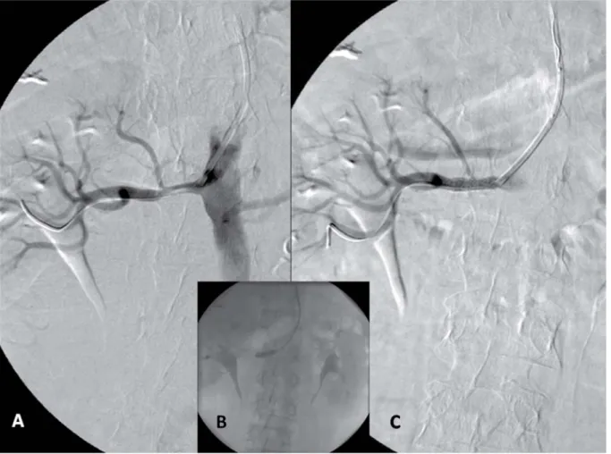
After local anesthesia, the RA was punctured with a dedicated TR needle and 6F sheath (Terumo). The RA anatomy was identifi ed in AP or left anterior oblique 20 view with aortography using a 5F 100 cm long pig tail catheter. The cannulation of the renal artery was performed with a 100 cm long 6F JR5 or a 125 cm long 6F Multipurpose (Cordis) guiding catheter (the available guiding length was estimated after the an- giography with the 100 cm long pig tail catheter) (Fig. 1A–B). In some cases the “no-touch technique”
was used when the ostium was severely diseased [10]
(Fig. 2A–D). The renal artery lesion was passed with a conventional coronary guidewire (0.014 BMW or Whisper Extra support, 180 cm) or when the support was insuffi cient a 0.018 Steelcore (Abbot) was used for buddy wire during the intervention. After performing a road map imaging the lesion was stented directly with a dedicated renal artery stent Express SD (Boston Sci,
150 cm long shaft) or Herculink (Abbot Co, 135 cm long shaft). In calcifi ed arteries or when the stent did not pass the lesion and predilatation was performed with a non-compliant monorail balloon (135–150 cm long) (Fig. 1C–D). In the case of fi bro muscular dys- plasia or stent restenosis balloon angioplasty was per- formed primarily.
Postoperative treatment
After the procedure the sheath was removed immedi- ately and hemostasis was achieved with a tourniquet for 6 hours. We did not apply a dedicated hemostatic device.
All patients were mobilized immediately.
Post procedural follow-up
The RA was investigated after the procedure with palpa- tion and in the case of impalpable RA, the artery was investigated with Doppler Ultrasound. The creatinine level and the blood pressure was controlled 3 days after the intervention.
Fig. 1. A: High grade stenosis of the left renal artery and selective cannulation with a Multipurpose 6F guiding catheter. B: Direct stenting with a 6 × 18 mm Herculink stent. C: Final result
Quantitative angiography
Angioplasty was performed according to the standard clinical practice. The vessels and the lesions were ana- lyzed by using a computerized quantitative analysis sys- tem (General Electric, Innova 3100). Measurements were obtained with digital caliper method.
Statistical analysis
All statistical analysis was performed with a commercial- ly available software Statistica 8.0 (StatSoft Inc., Tulsa, OK, USA). Continue variables were expressed as mean ± standard deviation and range, and were compared using
unpaired t-tests. Probability values lower than 0.05 were considered to be signifi cant.
Results
Demographic and clinical data are summarized on Ta- ble I. The indication of the intervention was resistant hypertension in 18 patients (66.7%) and worsening re- nal failure in 9 patients (33.3%).
Procedural data are summarized in Table II. In 27 patients (mean age: 65.4 ± 9.17) with hemodynamically relevant renal artery stenosis (mean diameter stenosis:
77.7 ± 10.6%, right; n = 7; left, n = 20), interventional treatment with angioplasty and stenting was performed
Fig. 2. A–B: Calcifi ed high grade ostial left renal stenosis and cannulation with a Multipurpose 125 cm diagnostic catheter over a 6F 120 cm sheathless guiding using the telescoping technique. C–D: Predilatation with a Quantum 4 × 20 mm balloon and stenting with a 6.5 × 18 mm Herculink stent. E: Final result
using a left (n = 3) or right (n = 24) radial artery access.
The cross over rate was 0%. Direct stenting was success- fully performed in 13 (48%) cases, and predilatations was required in ten cases 10 (37%). Primary technical success (residual stenosis <30%) could be achieved in all cases.
The mean contrast consumption was 119 ± 65 ml.
Complications are summarized in Table III. Proce- dural complications: In one patient the stent was dis- lodged from the ostium during balloon removal, but it was successfully mobilized and parked in the exter- nal iliac artery. Renal complications: In the investigated population renal failure was not detected, however, in one patient the creatinine and carbamide nitrogen level was increased after the intervention (3.7%). Temporary hematuria was observed in one patient (3.7%). Major ad- verse event was not detected in the investigated popula- tion. Vascular complications: There were no major peri- procedural major vascular complications. Minor vascu- lar complication was detected in three patients (11.1%) (1 asymptomatic radial artery occlusion and 2 transient spasm).
Clinical results
Clinically systolic blood pressure was improved from 162.5 ± 22.2 to 151 ± 21.9 (p = ns). Serum creatinine
Table III Complications
Complications n (%)
Procedural complications
Stent dislodgement 0 (0)
Other 0 (0)
Renal complications
Renal failure 0 (0)
Transient creatinine elevation 1 (3.7) Access complications
Major
Symptomatic RAO 0 (0)
Compartment syndrome 0 (0)
Other 0 (0)
Minor
Spasm 2 (7.4)
Asymptomatic RAO 1 (3.7)
Perforation without compartment sy 0 (0) MAE
Death 0 (0)
Urgent bypass operation or PTA 0 (0)
AMI 0 (0)
Table I Demographic and clinical data
Demographic and clinical data n (%)
Age (years) 65.4 ± 9.1
Male 22 (81.5)
Hypertension 27 (100)
Renal insuffi ciency 9 (33.3)
– Hemodialysis 1 (3.7)
Dyslipidaemia 26 (96.3)
Active smokers 10 (37)
Diabetes mellitus 8 (29.6)
Severe obesity 6 (22.2)
Coronary artery disease 7 (25.9)
– Previous PCI 4 (14.8)
Peripheral artery disease 17 (62.9)
– Previous PTA 10 (37)
Table II Angiographic and procedural data
Angiographic and procedural data n (%) Lesion site
Right renal artery 8 (29.6)
Left renal artery 19 (70.4)
Lesion location
Isolated ostial 7 (25.9)
Proximal shaft 19 (70.4)
Middle shaft 1 (3.7)
Distal shaft 0 (0)
Quantitative measurements and angiography
Lesion length (mm) 14.36 ± 3.25
Reference diameter (mm) 5.89 ± 0.80 Stenosis diameter (%) 76.68 ± 10.56
Severe calcifi cation 7 (25.9)
Severe tortuosity 0 (0)
Visible thrombus 0 (0)
Angiographic result of the intervention
Residual stenosis <30% with good fl ow 27 (100) Equipments
Diagnostic catheter / procedure (%) 27 (107.4) Guide catheter / procedure (%) 25 (92.6)
Sheathless guiding (%) 2 (7.4)
Guide wire 0.35 (%) 28 (103.7)
Guide wire 0.14 (%) 29 (107.4)
Balloon / procedure (%) 13 (48.1)
Stent used / procedure (%) 24 (88.8)
values dropped from 170.3 ± 124.4 to 167.7 ± 119 (p = ns).
Discussion
Atherosclerotic renal artery stenosis (RAS) can be asso- ciated with three main clinical syndromes such as reno- vascular hypertension; ischemic nephropathy; and cardiac syndromes (refractory heart failure, unstable angina, and sudden-onset pulmonary edema). The treatment of the RAS can be optimal medical or interventional treatment.
Many studies has confi rmed the potential of the PRI to decrease the hypertension [11] and to reverse the pro- gression of renal failure [12], but some studies show no benefi t in patients presented with RAS [13] for this rea- son patient selection has a paramount importance due to the high number of non-responsive patients and because the procedure is associated with frequent vascular access complications and worsening renal failure [11–14].
Vascular complications
Femoral access complications occur more frequently in patients with peripheral artery disease [15] and renal dysfunction [16] and both diseases are associated with RAS and make these patients more vulnerable for femo- ral complications [17]. Due to high number of the ac- cess complications of the PRI, many eff orts were made to decrease the incidence of their frequency, such as in- troducing new alternative access sites (brachial, radial), small new low profi le 6F compatible devices, routine use of closure devices [4–5]. BA access is associated with high risk of vascular and nerve complications [18], but despite the disadvantages of the BA access this is the sec- ond access site for many interventionalist for PRI, due to the large size of the brachial artery, which makes the puncture easy, while it is not spasmogenic and allows the use of 6–7F large sheaths and the traditional catheter length allows the use of nearly all devices used for PRI.
Another potential access site for PRI is the radial and ulnar way which has shown in many coronary and pe- ripheral studies an extremely low rate of access site com- plications [1, 2, 6].
Renal complications
The most severe complication associated after PRI is the worsening renal failure and the potential cause can be macro and microembolisation during the intervention and contrast nephropathy. Distal embolization can be prevented with careful guiding catheter engagement of the ostium with “no-touch technique” [10] and with using distal embolic protection devices [14]. TR and
TB access allows relatively atraumatic cannulation of the renal artery ostia especially in cases when the renal ar- tery has a downward origin and potentially decrease the chance of distal embolization.
Disadvantages
On the low rate of early (spasm, perforation, brachial artery dissection, distal embolization) and late [pseudo aneurysm formation, radial artery occlusion (RAO)] re- ported vascular complications of radial approach it is im- portant to note that most of them are asymptomatic due to dual blood supply of the hand [6–9]. Symptomatic complications causing critical hand ischemia can occur when the forearm circulation is not complete and the radial artery occludes or when the brachial or subcla- vian artery is damaged [19]. RAO is reported in 3–5% in diff erent studies [6–9], and as the radial artery must be preserved for further interventions and Cimino fi stulae, all preventive measures must be taken to prevent RAO including fast and atraumatic puncture, intra-arterial administration of Heparin and verapamil, and the use of non-occlusive bandage [20]. Additionally in patients with renal insuffi ciency, in whom the below-the-knee arteries are frequently aff ected by atherosclerosis and calciphylaxis symptomatic RAO might occur more easily than in normal population [21].
Advantages
The potential of immediate mobilization of the patients increase the patient comfort and shorten the hospitaliza- tion, but the most important advantage of the technique is the extreme low rate of vascular complications [1].
Technical aspects
Puncture and traversing the radial and brachial artery is the same as during coronary procedures and is important that the operator be aware of the radial artery anomalies especially radial artery loops to avoid complications. The catheter advancement in the ascending aorta is some- times diffi cult, especially from the right radial artery, but the pig tail catheter can be advanced in the ascending aorta with Terumo hydrophilic wire in LAO60 projec- tion easily. There are two techniques for guide-catheter advancement: direct and indirect engagement. We prefer the direct catheter advancement, when the ostium is not diseased, but in the case of high-grade ostial lesions or several adjacent aortic disease, there is a risk of guide catheter dissection, distal embolization therefore in these patients we prefer the “no-touch technique”. A long 5F diagnostic catheter (125 cm) is telescoped through a
shorter 6F guide catheter the long 300 cm 0.014–0.018 GW is placed over the stenosis, and after removing the diagnostic catheter a long monorail balloon is used for predilatation. We are following the trends which sug- gest a movement forward 0.014 GWs and long coronary monorail balloons for PRI. The one caveat to keep in mind is that radial access requires the use of balloons and stents with long shaft lengths (135 or 150 cm). We prefer 135–150 cm long balloon expandable stents for PRI, but sometimes these 135 cm long stents has not suffi cient length in 125 long guiding catheter, therefore the Y connector must be removed and the stent must be implanted under roadmap image. Another option is to use a long 120 cm sheathless guiding (Asahi) and 125 long diagnostic catheter for no-touch technique (Fig. 2). The left radial artery is generally preferred for renal angiography and interventions due to shorter dis- tance and the avoidance of crossing the carotid vessels, but we prefer the right radial approach because the right radial approach is more comfortable for the operator.
Study limitations
The main limitation of the study is the retrospective study design and the lack of femoral control group. An- other limitation of the study that the right and left radial approach was not investigated in the study.
Conclusion
Transradial renal artery angioplasty and stenting is tech- nically feasible and safe. Further randomized studies are needed to confi rm the superiority over the traditional femoral artery access.
Abbreviations
RA: radial artery; FA: femoral; BA: brachial artery; PRI: percutane- ous renal intervention; RAO: radial artery occlusion; RA: radial artery;
TR: transradial
* * *
Funding sources: None.
Authors’ contribution: ZR, KT and ZJ introduced the study idea.
ZR and BN performed the interventions. KT performed the off -line analysis and acquired the angiography images. NK and SN helped in the interpretation of the results and statistical analysis. ZR wrote the manuscript. KH and BM added clinical discussion to the manuscript.
ZR and BN reviewed the manuscript. Finally, all authors have read and approved the manuscript.
Confl ict of interest: The authors have no real or perceived confl icts of interest to disclose.
References
1. Caputo RP, Tremmel JA, Rao S, Gilchrist IC, Pyne C, Pancholy S, Frasier D, Gulati R, Skelding K, Bertrand O, Patel T: Transradial arterial access for coronary and peripheral procedures: Executive summary by the Transradial Committee of the SCAI. Catheter Cardiovasc Interv 78(6), 823–839 (2011)
2. Staniloae CS, Korabathina R, Coppola JT: Transradial access for peripheral vascular interventions. Catheter Cardiovasc Interv 81(7), 1194–1203 (2013)
3. Kaukanen ET, Manninen HI, Matsi PJ, Söder HK: Brachial artery access for percutaneous renal artery interventions. Cardiovasc In- tervent Radiol 20(5), 353–358 (1997)
4. Scheinert D, Bräunlich S, Nonnast-Daniel B, Schroeder M, Schmidt A, Biamino G, Daniel WG, Ludwig J: Transradial ap- proach for renal artery stenting. Catheter Cardiovasc Interv 54(4), 442–447 (2001)
5. Sanghvi K, Coppola J, Patel T: Cranio-caudal (transradial) ap- proach for renal artery intervention. J Interv Cardiol 26(5), 530–
535 (2013)
6. Kolluri R, Fowler B, Nandish S: Vascular access complications:
Diagnosis and management. Curr Treat Options Cardiovasc Med 15(2), 173–187 (2013)
7. Kanei Y, Kwan T, Nakra NC, Liou M, Huang Y, Vales LL, Fox JT, Chen JP, Saito S: Transradial cardiac catheterization: A review of access site complications. Catheter Cardiovasc Interv 78(6), 840–846 (2011)
8. Stella PR, Kiemeneij F, Laarman GJ, Odekerken D, Slagboom T, van der Wieken R: Incidence and outcome of radial artery occlu- sion following transradial coronary angioplasty. Cathet Cardiovasc Diagn 40(2), 156–158 (1997)
9. Uhlemann M, Möbius-Winkler S, Mende M, Eitel I, Fuernau G, Sandri M, Adams V, Thiele H, Linke A, Schuler G, Gielen S: The Leipzig prospective vascular ultrasound registry in radial artery catheterization: Impact of sheath size on vascular complications.
JACC Cardiovasc Interv 5(1), 36–43 (2012)
10. Feldman RL, Wargovich TJ, Bittl JA: No-touch technique for re- ducing aortic wall trauma during renal artery stenting. Catheter Cardiovasc Interv 46(2), 245–248 (1999)
11. Dorros G, Jaff M, Mathiak L, He T; Multicenter Registry Par- ticipants: Multicenter Palmaz stent renal artery stenosis revascu- larization registry report: Four-year follow-up of 1,058 successful patients. Catheter Cardiovasc Interv 55(2), 182–188 (2002) 12. Ramos F, Kotliar C, Alvarez D, Baglivo H, Rafaelle P, Londero H,
Sánchez R, Wilcox CS: Renal function and outcome of PTRA and stenting for atherosclerotic renal artery stenosis. Kidney Int 63(1), 276–282 (2003)
13. ASTRAL Investigators, Wheatley K, Ives N, Gray R, Kalra PA, Moss JG, Baigent C, Carr S, Chalmers N, Eadington D, Hamil- ton G, Lipkin G, Nicholson A, Scoble J: Revascularization versus medical therapy for renal-artery stenosis. N Engl J Med 361(20), 1953–1962 (2009)
14. Cooper CJ, Haller ST, Colyer W, Steff es M, Burket MW, Thomas WJ, Safi an R, Reddy B, Brewster P, Ankenbrandt MA, Virmani R, Dippel E, Rocha-Singh K, Murphy TP, Kennedy DJ, Shapiro JI, D’Agostino RD, Pencina MJ, Khuder S: Embolic protection and platelet inhibition during renal artery stenting. Circulation 117(21), 2752–2760 (2008)
15. Jeremias A, Gruberg L, Patel J, Connors G, Brown DL: Eff ect of peripheral arterial disease on in-hospital outcomes after primary percutaneous coronary intervention for acute myocardial infarc- tion. Am J Cardiol 105(9), 1268–1271 (2010)
16. Aziz EF, Pulimi S, Coleman C, Musat D, Florita C, Platzman Z, Fawzy A, Herzog E, Tamis-Holland J, Hong MK: Role of renal function in vascular access complication rates in patients undergo- ing percutaneous coronary procedures using arteriotomy closure devices. J Invasive Cardiol 22(1), 8–13 (2010)
17. Wilms G, Marchal G: The angiographic incidence of renal artery stenosis in the atherosclerotic population. Eur J Radiol 10, 195–
197 (1990)
18. Alvarez-Tostado JA, Moise MA, Bena JF, Pavkov ML, Greenberg RK, Clair DG, Kashyap VS: The brachial artery: A critical access for endovascular procedures. J Vasc Surg 49(2), 378–385 (2009) 19. Ruzsa Z, Molnár L, Szabó G, Merkely B: Catheter-induced bra-
chial artery dissection during transradial angioplasty. J Vasc Access 14(4), 392–393 (2013)
20. Sanmartin M, Gomez M, Rumoroso JR, Sadaba M, Martinez M, Baz JA, Iniguez A: Interruption of blood fl ow during compres- sion and radial artery occlusion after transradial catheterization.
Catheter Cardiovasc Interv 70(2), 185–189 (2007)
21. Ruzsa Z, Tóth K, Berta B, Koncz I, Szabó Gy, Jambrik Z, Varga I, Hüttl K, Merkely B, Nemes A: Allen’s test in patients with periph- eral artery disease. Central European Journal of Medicine 9(1), 34–39 (2014)