O R I G I N A L S T U D I E S
Short- and long-term results with a percutaneous treatment in critical hand ischaemia
Zoltán Ruzsa MD, PhD
1,2| Balázs Berta MD, PhD
1| Júlia Tóth MD
2| Balázs Nemes MD, PhD
1| András Katona MD
3| Arthúr Hüttl MD
1|
Imre Ungi MD, PhD
3| Olivier F. Bertrand MD, PhD
4| Béla Merkely MD, DSc
11Heart and Vascular Center, Semmelweis University, Budapest, Hungary
2Invasive Cardiology, Bács-Kiskun County Hospital, Kecskemét, Hungary
3Albert Szent-Györgyi Clinical Center, 2nd Department of Medicine and Cardiology Center, Medical Faculty, University of Szeged, Szeged, Hungary
4Cardiology Department, University Laval, Quebec, Canada
Correspondence
Zoltán Ruzsa, Heart and Vascular Center, Semmelweis University, 68 Városmajor Street, Budapest 1122, Hungary.
Email: zruzsa25@gmail.com
Abstract
Background:The aim of this prospective registry was to determine the feasibility, safety, and outcomes of percutaneous transluminal angioplasty and thrombolysis in the treatment of critical hand ischemia (CHI).
Methods:One-hundred one patients (aged 60.6 ± 15.3 years) were treated for CHI between 2012 and 2016 in three cardiovascular centers. Anatomically, the upper arm was divided into three seg- ments (I-subclavian, II-brachial, and III-forearm). We examined the rates of technical and clinical suc- cess, major adverse events (MAEs), and vascular complications at 1 year and at long-term follow-up.
Results:Nineteen patients (18.8%) were treated for acute CHI, and 82 (81.2%) for chronic CHI.
Median follow-up was 36.9 (19.6–68.3) months. Clinical symptoms were isolated rest pain in 91 patients (90.1%) and digital ulcer or gangrene in 10 patients (9.9%). The technical and clinical success rate of intervention was 96.0% (97/101) and 84.2% (85/101) at 1 year. Angioplasty was performed in Segments I, II, and III in 28 (27.7%), in 29 (28.7%), and 44 (43.5%) patients. Stent implantation was necessary in 47 patients (46.8%). Vascular access site complications were found in 2.1% of the sample. After 1 year, MAEs occurred in 27 patients (26.9%), and the target lesion revascularization rate was 11.9%. In two patients (1.9%), thoracic sympatectomy was nec- essary, and two patients (1.9%) underwent minor finger amputations.
Conclusions:Angioplasty of hand vessels for CHI is a feasible and safe procedure with accept- able rates of technical success and hand healing. MAEs are frequent because the rate of severe comorbidities is high.
K E Y W O R D S
balloon angioplasty, brachial/radial/ulnar, catheterization, complications, vascular access
1 | I N T R O D U C T I O N
Upper extremity arterial disease (UEAD) is a rare disease because ather- erosclerosis is approximately 20 times less common in the arms than in the legs and is usually limited to the proximal or inflow arteries.
Clinically, it can be manifested in upper extremity claudication or criti- cal hand ischemia (CHI) with rest pain, ulceration, and gangrene. CHI is either acute (aCHI) or chronic (cCHI), determined by characteris- tics of disease onset. In the literature, only case reports1,2and a lim- ited number of studies about interventional treatment can be found.3–6The aim of this prospective study was to assess the long- term feasibility, safety, and outcomes of percutaneous transluminal angioplasty (PTA) and catheter-directed thrombolysis in the treat- ment of CHI.
Abbreviations:BA, balloon angioplasty; CHI, critical hand ischemia; FA, femoral access; GW, guidewire; RA, radial artery; RAO, radial artery occlusion; TR, transra- dial; UA, ulnar artery; UEAD, upperextremityarterialdisease; US, duplex ultrasound.
Received: 7 October 2018 Revised: 27 December 2018 Accepted: 9 February 2019 DOI: 10.1002/ccd.28166
This is an open access article under the terms of the Creative Commons Attribution-NonCommercial-NoDerivs License, which permits use and distribution in any medium, provided the original work is properly cited, the use is non-commercial and no modifications or adaptations are made.
© 2019 The Authors.Catheterization and Cardiovascular Interventionspublished by Wiley Periodicals, Inc.
Catheter Cardiovasc Interv.2019;93:1301–1310. wileyonlinelibrary.com/journal/ccd 1301
2 | M E T H O D S
Patients with CHI were evaluated in a prospective study spanning three cardio-vascular centers, and their clinical and angiographic data had been analyzed. All patients underwent careful angiological examination, vascular team consultation, diagnostic angiography, and vascular ultra- sound before intervention. Patients who were candidates for surgical revascularization were excluded from the study. The Institutional Ethi- cal Review Committees approved the study in all three centers, and all patients provided written informed consent prior to study inclusion.
Primary endpoints were the rates of technical success and major adverse events (MAEs).
Secondary endpoints were clinical success, fluoroscopy time, X-ray dose, procedural time, contrast consumption, duration of hospitaliza- tion in days, and the rate of access-site complications.
Inclusion criteria were aCHI or cCHI associated with pain at rest and/or ischemic hand lesions (finger gangrene or ulceration) and the presence of hemodynamic significant lesions. A significant lesion was defined as one with a diameter at least 50% that of the stenosis or occlusion involving the subclavian, axillo-brachial, radial, ulnar, or interosseous artery (the distal arches originating from the radial or ulnar arteries were considered part of these arteries) or CHI after transradial or transbrachial angioplasty.
Exclusion criteria were forearm claudication and subclavian steel syndrome from subclavian artery stenosis, known thoracic outlet syn- drome, Cimino Brescia fistulae at the ipsilateral site, and surgical revascularization candidacy.
2.1 | Antithrombotic and thrombolytic regimen
After a loading dose of 325 mg aspirin and 300 mg clopidogrel, patients underwent aspirin or clopidogrel monotherapy (100 mg aspi- rin or 75 mg clopidogrel daily) in patients without stent implantation
and dual antiplatelet therapy for 2 months if a stent implantation was present. Additional Na-heparin was given until reaching 100 IU/kg for the procedure. Routine activated clotting time as not measured during intervention.
Catheter directed thrombolysis was done with intra-arterial recombinant tissue plasminogen activator (r-tPA; 10 mg bolus and 1 mg/h maintenance dose) and additional intravenous NaHeparin was given to prevent catheter thrombosis (bolus 60 IU/kg, maintenance dose 1,000 IU/h corrected by activated plasmin thromboplastin time).
2.2 | Access site selection
In aCHI, the access site for diagnostic angiography and for thromboly- sis was the conventional femoral artery (FA).
In cCHI, primary access for angioplasty was via the FA method when the lesion was proximal (Segments I–II), multiplex, or complex.
Where isolated, short lesions were below the elbow (BTE) (Segment III), anterograde brachial artery access was preferred. Dual access site was obtained in failed anterograde cases, using the distal radial (snuff- box segment) or ulnar artery (UA) when the anterograde recanaliza- tion failed. In some cases, the palmar arch technique was used as a retrograde technique.1
2.3 | Angioplasty technique in cCHI
When FA access was used, the FA was punctured the conventional way, and the subclavian artery was cannulated with a vertebral catheter. After a selective angiography, a long hydrophilic sheath (90 cm, 6F, Cook Co., USA) was advanced in the subclavian artery proximal to the stenosis over a long (260 cm) J-tip guidewire (GW). Subclavian and anonym artery interventions were performed in routine clinical practice. When brachial or BTE lesions were found, the stenosis was dilated with dedicated below-the-knee balloons using a long inflation time, and stent implan- tation was performed only when suboptimal results were reached
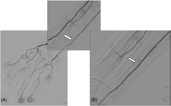
FIGURE 1 Angioplasty in chronic critical hand ischemia. A, High grade stenosis of the right ulnar artery (white arrow) in a patient with a chronic renal failure and diabetes mellitus. B, The ulnar artery lesion after balloon angioplasty (white arrow)
(Figure 1). Balloon expandable stents were used in short lesions, while long lesions were stented with long self-expandable stents.
2.4 | Chronic total occlusion recanalization
Subclavian artery chronic total occlusion (CTO) recanalization was performed using femoral access in the conventional way (Figure 2).
Brachial artery CTO recanalization was performed using a 0.018 guidewire and the penetration or subintimal technique (Figure 3).
Occlusions of the BTE artery were recanalized using 0.014 coronary or with below-the-knee GWs 300 cm in length, applying the penetra- tion or subintimal technique. Prolonged inflations were always used with long, low-profile below-the-knee or coronary balloons, and stent implantation was performed only when the lesions were flow limiting.
The anterograde recanalization was preferred for CTO, but after failed anterograde attempts, recanalization was performed using a retro- grade technique via the radial or UA. In some cases, the palmar arch was used as a conduit (palmar arch technique, Figure 4).
2.5 | Angioplasty technique in aCHI
After selective angiography, the treatment strategy was different depending on whether the thrombus burden was small or large. If it
was small, the patient underwent mechanical thrombectomy with a thrombectomy catheter. If large, the patient underwent a local throm- bolysis for 24 hr with local administration of r-tPA, and the angiogra- phy was repeated after 24 hr. Additional thrombus aspiration was performed if the thrombus remained flow limiting or occlusive after thrombolysis, and additional balloon angioplasty was performed when the lesion was significantly stenosed or dissected.
2.6 | Treatment of restenosis
Restenosis was treated with paclitaxel-eluting balloons using pro- longed balloon dilatation. When the recoil was significant or the lesions were flow-limiting, commercially available drug-eluting stents were used (Figure 5).
2.7 | Postoperative treatment
When the FA approach was used in cCHI, the sheath was removed immediately, and an Angio-Seal closure device (St Jude Medical) was used. A mechanical compression bandage was used for 4 hr, and patients were mobilized afterward. In patients with aCHI, the sheath was removed only after discontinuation of r-tPA, and the FA site was closed with Angioseal.
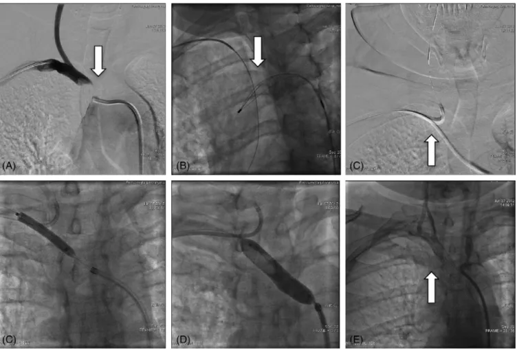
FIGURE 2 Subclavian artery CTO recanalization. A, Subclavian artery angiography from right radial artery access showing anonym artery CTO (white arrow). B, The anonym artery CTO was passed with a Terumo wire, but balloon passage was unsuccessful, therefore the guidewire was captured from femoral access and externalized (white arrow). C, The angioplasty was done from femoral approach, but the radial carotid artery was protected from right radial access with a filter wire (white arrow). D, Balloon dilalation with a 6×40 mm balloon. E, Stent implantation with a 10×29 mm Omnilink balloon expandable stent. E, Final angiography show good angiographic result (white arrow)
RUZSAET AL. 1303
When brachial artery access was used in cCHI, the sheath was removed immediately, and hemostasis was achieved using a tourni- quet for 6 hr. All patients were mobilized immediately.
When dual access was used, the distal ulnar access site was com- pressed with dedicated compression device (RadiStop, Terumo, Japan) obtaining patent hemostasis. The distal radial artery (RA) in the snuff- box area was compressed manually for 5 min and thereafter tourni- quet was used for 2 hr.
2.8 | Duplex ultrasound protocol
Duplex ultrasound was performed for all patients by a trained physi- cian after the intervention and after 12 months. The diameter and the peak systolic velocity of the RA and UA were measured at the wrist.
On the first postoperative day, the patency of the UA and RA was evaluated by palpation, and vascular ultrasound as performed for symptomatic patients or those with unpalpable RAs or UAs.
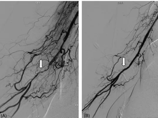
FIGURE 3 Brachial artery CTO recanalization. A, Brachial artery angiography shows a brachial artery CTO with highly developed collateral system (white arrow). B, Final angiography after ballon dilatation (white arrow)
FIGURE 4 Local thrombolysis in acute hand ischemia. A, Brachial artery angiography of a patient with acute hand ischemia shows a total occlusion of the axillary artery (white arrow). B, Control angiography after 24 hr local thrombolysis shoes good angiographic result (white arrow)
FIGURE 5 Treatment of restenosis. A, Brachial artery repeat angiography after brachial artery angioplasty shows a critical restenosis (white arrow). B, Control angiography after drug eluting balloon dilatation (Impact, Medtronic) shows no residual stenosis (white arrow)
FIGURE 6 The pathomechanism of CHI after transradial catheterization. A, Figure shows radial artery occlusion and incomplett or weak palmar arch. B, Figure shows a radial artery occlusion beside ulnar artery chronic total occlusion. C, Figure shows a radial artery occlusion with distal embolization or thrombus propagation [Color figure can be viewed at wileyonlinelibrary.com]
RUZSAET AL. 1305
2.9 | Quantitative angiography
Quantitative angiography and measurements were performed according to standard clinical practice. Vessels and lesions were analyzed using a computerized quantification system (Innova 3100; General Electric). The arteries were localized as Segment I (anonym and subclavian artery), Segment II (axillary and brachial artery), or Segment III (below-the-elbow arteries, that is, ulnar, radial, and interosseal arteries and the palmar arch).
2.10 | Postprocedural follow-up
Access arteries were investigated physically and using duplex ultra- sound after the procedure. All patients underwent detailed clinical follow-up examinations at 2 and 12 months and on a long term after the procedure.
2.11 | Definitions
2.11.1 | Major adverse event
An MAE was assessed as the composite of death, myocardial infarc- tion, stroke, major upper arm amputation, and repeated revasculariza- tion of the target vessel by PTA or by arterial bypass graft surgery at 2 months, 12 months, and on a long term.
2.11.2 | Definition of vascular complication
A major vascular complication was defined as diminished or lost arte- rial pulse or the presence of any pseudo-aneurysm or arteriovenous fistula during clinical follow-up. A minor complication was defined as a hematoma requiring no further treatment, measuring≤2 cm in diame- ter over the radial or ulnar puncture area, or measuring≤5 cm in diam- eter over the femoral puncture site. Major bleeding was defined as a fall in hemoglobin to more than 3 g/dL or any bleeding requiring a blood transfusion.
2.11.3 | Technical success
A technical success was when PTA resulted in less than 30% residual stenosis with sufficient anterograde flow. A suboptimal result was characterized by sluggish flow and/or a residual stenosis between 30 and 50% after repeated dilatations.
2.11.4 | Clinical success
Primary clinical success was when an improvement of at least one clinical category in the Rutherford-Becker classification7 was seen.
Primary patency was when persistent patency was seen without re- intervention (angioplasty, surgery, or amputation) on or at the margins of the treated lesion. Limb salvage was when the prevention of major amputation was achieved. A finger amputation was classified minor, whereas any amputation above that level was considered major.
2.11.5 | Contrast induced nepropathy
Contrast induced nepropathy was defined as an absolute≥0.5 mg/dL or a relative≥25% increase in creatinine level at 24–72 hr after the procedure without another clear cause for the acute kidney injury.
2.12 | Statistical analysis
Statistical analysis was performed using commercially available Graph Pad Prism 6.0 software. Each continuous variable was expressed as the mean ± standard deviation or as the median (inter-quartile range), as appropriate. Each categorical variable was expressed as a percent- age. The various patient cohorts were compared using either the Mann–Whitney test or the Kruskal–Wallis test. Probability values not exceeding 0.05 were considered significant.
3 | R E S U L T S
Nineteen patients (18.8%) with aCHI and 82 patients (81.2%) with cCHI were treated. Clinical symptoms were isolated rest pain in 91 patients (90.1%) and digital ulcer or gangrene in 10 (9.9%). Demo- graphic and clinical data are summarized in Table 1, while disease eti- ology is summarized in Table 2.
3.1 | Anatomic and procedural data
Anatomic and procedural data are summarized in Tables 3 and 4.
Primary technical success (residual stenosis <30%) could be achieved in 96 patients (95.0%). Clinical success at 1 year was reached for 85 patients (84.2%). Single and dual access was obtained in 78 and 23 patients, respectively. The primary lesion was found in Segment I in 28 patients (27.7%), in Segment II in 29 (28.7%), and in Segment III in TABLE 1 Demographic and clinical data of the study group patients
Acute CHI n= 19, (%)
Chronic CHI n= 82, (%)
All patients n= 101 (%) Age (years) 54.9 ± 21.3 62.4 ± 13.2 60.6 ± 15.3
Female 13 (68.4) 47 (57.3) 60 (59.4)
Hypertension 16 (84.2) 73 (89) 89 (88.1)
Hyperlipidemia 6 (31.5) 65 (79.2)* 71 (70.3)
Diabetes 3 (15.7) 30 (36.6)* 33 (32.7)
Smokers 9 (47.3) 54 (65.8)* 63 (62.4)
Renal failure 7 (36.8) 24 (29.2) 31 (30.7) Coronary artery disease 5 (26.3) 37 (45.1)* 42 (41.6) Atrial fibrillation (all type) 6 (31.5)* 15 (18.3) 21 (20.8) Chronic obstructive
pulmonary disease
3 (15.8) 11 (13.4) 14 (13.8)
Raynaud syndrome 2 (10.5)* 2 (2.4) 4 (3.9) Peripheral artery disease
(lower extremity)
11 (57.9) 57 (69.5) 68 (67.3)
*P < 0.05
TABLE 2 Etiology
Acute CHI n= 19, (%)
Chronic CHI n= 82, (%)
All patients n= 101, (%) Etiology
Atherosclerosis/thrombosis 7 (36.8) 59 (71.9) 66 (65.3)
Embolism 9 (47.3) 1 (1.2) 10 (9.9)
Trauma 2 (10.5) 1 (1.2) 3 (2.9)
Iatrogenic 0 (0) 18 (21.9) 18 (17.8)
Vasculitis 1 (0.5) 3 (3.7) 4 (3.9)
44 (43.5%). In our sample of 101 patients, 133 lesions were treated, and they were located in the anonym artery in 2 patients, the subcla- vian artery in 28, the axillary artery in 15, the brachial artery in 27, the RA in 35, the UA in 22, the interosseal artery in 1, the palmar arch in 4, and the digital artery in 1 patient. Stent implantation was performed for 47 patients (46.5%). Singular (n= 61, 60.4%), multilevel (n= 31, 30.7%), and parallel (n= 7, 6.9%) dilatations were performed. Throm- bolysis and mechanical thrombectomy was performed for 55.5%
(11/19) and 47.4% (9/19), respectively in the 19 patients with aCHI.
Complete and partial thrombus resolution has been achieved in 3/11 (27.3%) and in 6/11 (54.5%) patients with large thrombus burden with
initial thrombolysis and additional thrombectomy and angioplasty was necessary in 6/11 (54.5%) and 7/11 (63.6%) patients with 81.8% final technical success. Complete thrombus resolution has been achieved in 8/8 (100%) patients with small thrombus burden with initial mechanical thrombectomy (100%) and balloon angioplasty (100%) with 100% final technical success.
Chronic total occlusion recanalization was performed for 49 patients, and a 95.9% success rate was achieved; however, dual arterial access (femoral/brachial and distal radial/palmar arch) was necessary in eight individuals (16.3%) because re-entry was unsuc- cessful. Retrograde technique was obtained in 31 patients with a 93.5% successful result. Limb salvage was achieved in all patients (100.0%). Major amputation was not necessary in any patient, but additional distal finger amputations were performed in six patients (5.9%) and thoracal sympetectomy in two patients (2%).
3.2 | Procedural complications
As indicated in Table 5, intraprocedural complications were not observed, but one asymptomatic radial artery occlusion (RAO) occurred after intervention. Contrast induced nepropathy was detected in one patient, however the creatinine and carbamide nitro- gen levels were normalized in 2 weeks. The rates of major and minor vascular access complications were 2.9% (three brachial pseudoaneur- ysm) and 2.0% (two femoral hematoma), respectively.
3.3 | One year and long-term follow-up
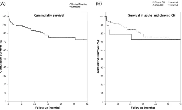
As shown in Table 5 and Figures 7 and 8, median follow-up was 36.9 (19.6–68.3) months. The MAE rate at 1 year and after long-term follow-up was 26.9 and 45.0%, respectively. Similarly, the mortality rate was 10.9 and 21.8%, respectively. Major amputation was not observed in the investigated population, but minor amputations were necessary in two patients (2.0%). The reintervention rate at 1 year and 3 years was 11.9 and 18.3%, respectively.
3.4 | Repeat angiography
Angiography was repeated for 21 (20.8%) patients at long-term follow-up, revealing restenosis or reocclusion in all cases. The clinically driven restenosis rate was 14.3% in Segment I, 24.1% in Segment II, and 27.3% in Segment III, while the restenosis rate after balloon angioplasty or stent implantation was 12.8 versus 9.3% (p = ns) at 1 year follow-up. Stent or balloon restenosis rate was 9 versus 16.6%
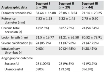
in Segment I (p= ns), 23.1 versus 12.5% in Segment II (p< 0.05), and TABLE 3 Angiographic data
Angiographic data
Segment I (n= 28)
Segment II (n= 29)
Segment III (n= 44) Diameter stenosis (%) 84.64 ± 16.88 95.86 ± 8.24 91.14 ± 23.25 Reference
diameter (mm)
7.53 ± 1.23 5.32 ± 1.45 2.75 ± 0.60
Chronic total occlusion (n)
4 (12.5%) 8 (27.75%) 24 (54.54%)
Lesion length (mm) 31.5 ± 16.77 81.21 ± 63.58 80.52 ± 78.91 Severe calcification (n) 24 (85.7%) 11 (37.93%) 21 (47.72%) Intraluminary
thrombus (n)
0 (0%) 10 (34.48%) 9 (20.45%)
Angiographic outcome
Successful 28 (100%) 28 (96.5%) 41 (93.2%)
Unsuccessful 0 (0%) 1 (3.5%) 3 (6.8%)
TABLE 4 Procedural data
Access n(%)
Single access (n= 78, 77.2)
Femoral 41 (52.6)
Anterograde brachial/ axillary 16 (20.5)
Radial or ulnar 21 (26.9)
Dual access (n= 23, 22.8)
Femoral + radial or ulnar 13 (56.5)
Femoral + brachial 4 (17.4)
Brachial + radial or ulnar 3 (13)
Radial + contralateral radial 3 (13) Intervention
Balloon angioplasty 93 (100)
Stent implantation by patients 47 (46.5) Stent implantation by segments
Segment I 22 (78.6%)
Segment II 13 (44.8%)
Segment III 14 (31.8%)
Aspiration thrombectomy 12 (11.9)
Thrombolysis 11 (10.9)
Equipments per procedure
Guidewire 2.2
Balloon 1.6
Stent 0.58
Procedural factors
Contrast consumption (mL) 142.7 [125.8–159.6]
X-ray dose (Gy/cm2) 331.2 [86.0–576.4]
TABLE 5 Complications
1 year FU n(%)
3 years FU n(%)
Myocardial infarction 2 (2.1) 8 (10.0)
Stroke 5 (5.2) 7 (8.0)
Major unplaned amputation 0 (0) 0 (0)
Re-angioplasty or bypass 11 (11.9) 15 (18.3)
Death 11 (10.9) 20 (21.8)
Major adverse event 27 (26.9) 42 (45.0)
RUZSAET AL. 1307
14.3 versus 13.3% in Segment III (p= ns) at 1 year follow-up, but the long-term results were statistically significant in Segment I only (13.6 vs. 50%; p< 0.05). Reintervention achieved good angiographic and clinical results for all patients (n= 21, 100.0%). Re-restenosis after a successful procedure was achieved in 2 of 20 patients (10.0%).
4 | D I S C U S S I O N
Pathophysiologically, UEADs can be divided into large artery (proximal to the wrist) and small artery (distal to the wrist) diseases, and the primary cause can be classified into thromboembolic (embolism and thrombosis), atherosclerotic (large and small artery disease), or vasospastic disease.
Independently of pathophysiology, the end-stage of UEAD is CHI with rest pain, ulceration, and gangrene.
The etiology of aCHI is mostly thromboembolic, and the emboli of the digits can originate from the heart or from the upper arm arter- ies. Additional radial an UA trauma, usually from intrarterial lines or after transradial catheterization, leads to thrombosis and/or embolism to distal digital arteries. Acute ischemia requires emergency surgical intervention, but embolectomy of the distal arteries has many limita- tions or can be impossible. Immediate administration of Na-heparin to prevent thrombus propagation and urgent angiography is necessary in these cases. Local thrombolytic therapy was published in some case reports and original papers with good angiographic and clinical results6,8–10with using streptokinase, urokinase, and r-tPA in different FIGURE 7 Major adverse events. A, Major adverse events. B, Myocardial infarction. C, Stroke. D, Target lesion revascularization
studies. In a recent publication, A. M. Schrijver et al. achieved >95%
clot lysis in 61% of patients with the need of 35% additional angio- plasty, with final 98% technical, 68% clinical success and an amputa- tion rate of 7%,8while in our study population the success depended by initial thrombus burden. In patients with large thrombus burden thrombolysis was fully or partially effective 9/11 patients (81.8%) and in patients with small thrombus burden, thrombectomy was effective in 8/8 patients (100%); however, the need of additional angioplasty was high in both patient groups (63.6 vs 100%). Final clinical success has been achieved in all patients and amputation was not necessary in any patient.
The etiology of cCHI is quite variable, and the causes can be ath- erosclerosis, thoracic outlet syndrome, vasculitis, autoimmune disease, hypothenar hammer syndrome, iatrogenic calciphylaxis, or vasospasm.
Large artery occlusions in upper extremity circulation requires treat- ment, either open or endovascular, because they cause hand claudica- tion or CHI. The proximal inflow lesions (subclavian or anonym artery) can be treated with angioplasty and stenting with good long-term results,11 but the endovascular treatment of brachial or below-the- elbow lesions has limited long-term evidence. Ferraresi et al. showed that isolated below-the-elbow lesions can be treated with an 82%
technical success rate and hand healing up to 65%.4The clinical reste- nosis rate was found to be 18% after a half year, and 36% of the patients died during follow-up. Our results show a 96.0% technical success rate, and mortality and TLR rates after 1 year were 10.9 and 11.9%, respectively.
4.1 | RAO after transradial angioplasty
RA access for percutaneous intervention is associated with very low major vascular complication rates in general, but the frequency of RAO remains relatively high, though it has no clinical importance in most cases because there is collateral formation from the interosseal artery or the palmar arch.7,12Possible mechanisms involved in occlusion
of the RA after transradial catheterization can be thrombus formation after vessel injury (dissection, prolonged spasm, and hematoma), occlusive compressure bandage, and distal embolization. When symp- tomatic RAO was accompanied by CHI, conservative treatment is not adequate, and invasive treatment with angioplasty should be consid- ered. The data in the literature are very limited regarding long-term results in this patient subgroup, and only case reports exist.1,2Our patient group consisted of 15 symptomatic patients after transradial angioplasty; all had rest pain and one patient also had a digital ulcer.
This group was treated successfully with BA (15/15) and additional stenting (3/15) with good clinical improvement (15/15), but the rein- tervention rate at 1 year was 40.0% (two with stent thrombosis and four with restenosis). The cause of the hand ischemia after transradial intervention was radial artery occlusion beside week palmar arch (5/15 pts.), radial artery occlusion and ulnar artery stenosis (9/15 pts.) and radial artery occlusion with distal embolisation (1/15 pts.). Figure 6 rep- resents the possible cause of the hand ischemia after transradial cathe- terization. Balloon angioplasty can be performed anterogradely via the brachial or femoral approach, and retrogradely via the palmar arch or distally from the occlusion site.
CTO recanalization can be luminal or subintimal. The luminal recanalization technique is generally successful in short occlusions, but is rarely effective in long occlusions, where subintimal angioplasty is mandatory. The rationale of using subintimal angioplasty is to create a subintimal lumen with the GW, and after proper BA, the new subin- timal space is maintained open by blood pressure. The technique has promising long-term results in the femoro-popliteal and infra-inguinal tract,10,11 but only limited data for the below-the-elbow segment is available.
4.2 | Study limitation
The primary limitation of this study is the lack of angiographic control in all patients.
FIGURE 8 Survival. A, Cumulative survival. B, Survival in chronic and acute critical limb ischemia
RUZSAET AL. 1309
5 | C O N C L U S I O N S
Angioplasty of the hand vessels for CHI is a feasible and safe proce- dure with acceptable rates of technical success and hand healing.
MAEs are frequent because of a high rate of severe comorbidities.
C O N F L I C T O F I N T E R E S T No conflict of interest for any author.
O R C I D
Zoltán Ruzsa https://orcid.org/0000-0002-2474-5723 Olivier F. Bertrand https://orcid.org/0000-0001-6335-2888
R E F E R E N C E S
1. Ruzsa Z, Kovács N, Merkely B. Retrograde subintimal recanalization of a radial artery occlusion after coronary angiography using the palmar loop technique. Cardiovasc Revasc Med. 2015;16(4):259-261.
2. Tasal A, Bacaksiz A, Erdogan E, Goktekin O. Successful angioplasty for radial artery chronic total occlusion in a patient with digital gangrene.
Postepy Kardiol Interwencyjnej. 2013;9(3):304-306.
3. Ferraresi R, Palloshi A, Aprigliano G, et al. Angioplasty of below- the-elbow arteries in critical hand ischemia. Eur J Vasc Endovasc Surg.
2012;43(1):73-80.
4. Tomoi Y, Soga Y, Fujihara M, et al. Outcomes of endovascular therapy for upper extremity peripheral artery disease with critical hand ische- mia. J Endovasc Ther. 2016;23(5):717-722.
5.Gandini R, Fabiano S, Morosetti D, Merolla S, Mauri G, Ferraresi R.
Endovascular treatment of below-the-elbow arteries in criticalhan- dischemia. Minerva Cardioangiol. 2016;64(6):662-671.
6.Coulon M, Goffette P, Dondelinger RF. Local thrombolytic infusion in arterial ischemia of the upper limb: Mid-term results. Cardiovasc Inter- vent Radiol. 1994;17(2):81-86.
7.Ruzsa Z, Tóth K, Nemes B, et al. Transradial and transulnar access for iliac artery interventions using sheathless guiding systems: A feasibility study. Catheter Cardiovasc Interv. 2016;88(6):923-931.
8.Schrijver AM, De Borst GJ, Van Herwaarden JA, et al. J Cardiovasc Surg (Torino). 2015;56(3):433-439.
9.Miyayama S, Yamashiro M, Shibata Y, et al. Thrombolysis and throm- boaspiration for acute thromboembolic occlusion in the upper extrem- ity. Jpn J Radiol. 2012;30(2):180.
10. Cejna M, Salomonowitz E, Wohlschlager H, Zwrtek K, Böck R, Zwrtek R. rt-PA thrombolysis in acute thromboembolic upper- extremity arterial occlusion. Cardiovasc Intervent Radiol. 2001;24(4):
218-223.
11. Kanei Y, Kwan T, Nakra NC, et al. Transradial cardiac catheterization:
A review of access site complications. Catheter Cardiovasc Interv.
2011 Nov 15;78(6):840-846.
12. Stella PR, Odekerken D, Kiemeneij F, Laarman GJ, Slagboom T, van der Wieken R. Incidence and outcome of radial artery occlusion fol- lowing transradial coronary angioplasty. Cathet Cardiovasc Diagn.
1997;40:156-158.
How to cite this article: Ruzsa Z, Berta B, Tóth J, et al. Short- and long-term results with a percutaneous treatment in critical hand ischaemia. Catheter Cardiovasc Interv. 2019;93:
1301–1310.https://doi.org/10.1002/ccd.28166