INVESTIGATION OF NOVEL THERAPEUTIC OPTIONS FOR THE TREATMENT OF ISCHEMIC AND METABOLIC CARDIOVASCULAR DISEASES
Ph.D thesis
Csilla Terézia Nagy
Semmelweis University
Doctoral School of Pharmaceutical Sciences
Supervisor: Zoltán Giricz, PharmD, Ph.D Official reviewers: Péter Sántha, MD, Ph.D
Attila Szijártó, MD, Ph.D Head of the Final Examination Committee:
Barna Vásárhelyi, MD, Ph.D Members of the Final Examination Committee:
Erika Pintér, MD, Ph.D Balázs Szalay, MD, Ph.D
Budapest
2018
1
1 Introduction
1.1 Obesity and therapies of obesity
In recent decades, obesity and its metabolic complications have become one of the biggest public health issues worldwide. Obesity is most commonly caused by a combination of excessive food intake, lack of physical activity, and genetic susceptibility. Occasionally it can be triggered by medications, endocrine disorders or mental disorder. Obesity is a condition in which excess fat has accumulated in the body.
Excess adiposity, particularly visceral adiposity leads to adipocyte dysfunction which results in dysregulation of adipokines, which may contribute to impaired glucose and lipid metabolism, as well as inflammatory responses. Visceral obesity greatly increases the risk of several diseases, such as Type 2 diabetes, non-alcoholic fatty liver disease, dyslipidaemia, cardiovascular disease, certain types of cancers, and depression.
The purpose of treatment of overweight and obesity is to reduce body weight and prevention of weight regain and thus to reduce the consequent health risks. Weight reduction can be achieved by a combination of the following: lifestyle interventions (diet, physical activity), surgery (bariatric surgery) and pharmacotherapy (appetite suppressants, weight loss drugs). At present there are only few available anti-obesity drugs on the market. Despite all the significant achievements, the current pharmacological treatments for obesity are only modestly effective and have several side effects. Therefore, further research is needed to develop a novel strategy to enhance the effectiveness of obesity treatment and to reduce side effects, including repurposing of drugs already in use or development of novel agents.
1.1.3 Monoamine oxidase (MAO) inhibitors
Monoamines (epinephrine, norepinephrine (NE), dopamine (DA) and 5-HT) participate in many physiological activities of the body. In mammals there are two subtypes of MAO: MAO-A and MAO-B. MAO inhibitors have two main groups:
selective and non-selective, within these we distinguish reversible and irreversible inhibitors. At present, MAO inhibitors are mostly used for psychiatric and neurological disorders. However, previous studies have shown that semicarbazide sensitive amine
2
oxidase inhibitors administered in combination with certain non-selective and/or irreversible MAO inhibitors can reduce body weight and fat deposition in animal models of diet-induced obesity. Selegiline is a clinically widely used, irreversible and selective inhibitor of MAO-B, which is primarily used to treat Parkinson's disease, Alzheimer's disease and depression. Békési et al. showed that selegiline (5–10 mg kg-1) significantly decreased liver fat but not body weight in rats fed with a lipid-rich diet (cholesterol 1%, olive oil 10%). Therefore, we hypothesized that MAO inhibitors may have favourable metabolic effects in obesity.
1.2 Cardiovascular diseases (CVDs) and therapies of CVDs
CVDs such as heart failure, hypertensive heart disease, cardiomyopathy, cardiac arrhythmia and coronary artery diseases (angina and myocardial infarction) are leading causes of morbidity and mortality worldwide. Nearly 300 risk factors play a role in the development of CVDs. The most common type of CVD is myocardial infarction (MI).
MI occurs when the blood flow reduces or stops for a part of the heart that causes an oxygen and nutrient depletion in the heart muscle, which leads to myocardial necrosis.
Most myocardial infarction in the world is due to atherosclerosis, when a coronary artery becomes occluded following the rupture of an atherosclerotic plaque, which leads to the formation of a blood clot.
The purpose of pharmacological therapies is to prevent CVDs and to improve patient outcomes. The use of current cardiovascular drugs such as antiplatelet drugs, anticoagulants, nitrates, beta-blockers, renin-angiotensin (ACE) inhibitors, and statins depends on the heart condition and symptoms. However, despite all the achievements in this field, there are still certain types of CVDs that do not have appropriate therapies.
Numerous studies demonstrated that the efficacy of cardioprotective therapies is significantly affected by comorbidities such as obesity, diabetes or dyslipidaemia. A growing amount of evidence suggests that increased autophagy plays a critical role in cardioprotective interventions and that besides MAO inhibition, autophagy could be a potential therapeutic target for the treatment of metabolic- and cardiovascular diseases.
3 1.2.2 Autophagy
Autophagy is an intracellular degradation process which eliminates unnecessary or dysfunctional organelles and long-lived proteins through lysosomal breakdown for recycling intracellular components. In mammalian cells, three main types of autophagy are described: microautophagy, chaperone-mediated autophagy and macroautophagy.
Macroautophagy, (herein referred to as autophagy) eradicate damaged cell organelles or unused proteins. Autophagy consists of sequential steps: the initiation phase, sequestration phase and degradation phase. Under normal circumstances autophagy has a low activity in cells but metabolic stress such as nutrient starvation or hypoxia can increase the activity of autophagy. Furthermore, the role of autophagy in the development of metabolic disorders (insulin resistance, obesity, atherosclerosis) has been studied extensively using different genetic animal models. Therefore, the use of autophagy-inducing agents could be an effective therapy in CVDs and metabolic diseases.
1.2.3 Compounds influencing autophagy
Previous studies reported that several therapeutic agents which are already in clinical use, for example hydrophobic statins, sevoflurane, sulfaphenazole and certain antibiotics, such as chloramphenicol (CAP), may also induce autophagy in addition to their primary effects. Since previous studies reported that CAP protects the heart against ischemia/reperfusion injury and upregulates autophagy markers. It has been shown that the induction of autophagy is required for cardioprotective mechanisms but no detailed investigation has been performed on which stage of autophagy is necessary for cardioprotection. Therefore, here we hypothesized that CAP induces cardioprotection, via increasing the certain step of autophagy in cardiomyocytes.
4
2 Objectives
To investigate the effect of selegiline treatment on metabolic parameters in high- fat, high-sucrose (HFS) diet-induced moderate obesity.
To investigate whether CAP-induced autophagy is necessary for cardioprotection.
To assess whether sequestration and/or degradation phases of autophagy are necessary for the cardioprotective effect of CAP.
3 Methods
This investigation conforms to the Guide for the Care and Use of Laboratory Animals published by the US National Institutes of Health and was approved by the animal ethics committee of the San Diego State University, San Diego, California and Semmelweis University, Budapest, Hungary.
3.1. Study design: diet-induced experimental obesity
Male Long-Evans rats were randomly divided into two groups: control diet (CON) and high-fat, high-sucrose (HFS) diet group. The CON group was fed control rat chow, whereas the HFS group was fed a chow supplemented with 20% lard and 15%
sucrose as a HFS diet. From week 16, groups of animals were further randomly divided and received subcutaneous injections of 0.25 mg kg-1 selegiline (CON+S and HFS+S) or vehicle (CON, HFS) once daily.
Body weights were measured monthly. Blood was taken, and fasting blood glucose levels were measured from the saphenous vein every month. At week 24, oral glucose tolerance test (OGTT) and insulin tolerance test (ITT) was performed. At week 24, food intake was observed for 24 hours in a metabolic cage. At week 25 animals were anesthetized with pentobarbital (60 mg kg-1, intraperitoneally). After hemodynamic measurements, animals were sacrificed. Epididymal and interscapular brown fat tissue - markers of adiposity - were isolated, and their weights were measured. Blood and tissue samples were collected and stored at −80 °C (Figure 1).
5 Figure 1. Experimental protocol.
Male Long-Evans rats were fed with control (CON, n=10) diet or with high-fat, high sucrose (HFS, n=10) diet for 25 weeks; CON+S (n=10) and HFS+S (n=10) groups were treated with 0.25 mg kg-1 selegiline (S) from week 16 to 24. Body weights and blood glucose were measured monthly. Neuropathy, behavioural tests were measured at weeks 22-23. Oral glucose tolerance test (OGTT), insulin tolerance test (ITT), metabolic cage and computed tomography (CT) were performed at week 24. Hemodynamic analysis was performed at week 25. Tissue sampling was performed after terminal procedures.
3.2 Study design: effect of CAP on autophagy in different cardiac cells
To identify the most suitable model system, in a pilot study we examined the effect of CAP on autophagy in neonatal rat cardiomyocytes (NRCMs), in H9c2 cardiac myoblast cells and in isolated hearts. We found that CAP induced autophagy in isolated hearts but not in NRCMs or in H9c2 cells. The efficacy of TAT-HAAtg5K130R was also assessed in a pilot experiment where we observed that in the left atrium LC3-II/I ratio was decreased after 15 min of administration of 200 nM TAT-HA-Atg5K130R to isolated hearts as compared to vehicle controls.
Therefore, in the main series of experiments, we used an ex vivo model.
3.3 Evaluation of whole body fat
To assess obesity, computed tomography (CT) measurements were performed on NanoSPECT/CT PLUS at week 24. Adiposity index was calculated by the following
6
formula: (whole body adipose tissue volume/body weight) ×100. Subcutaneous and total visceral fat volumes were evaluated on CT images by CTan software.
3.4 Immunohistochemistry
For immunohistochemistry, deparaffinized sections underwent antigen retrieval.
After blocking endogenous peroxidase activity, the sections were blocked in appropriate sera (2.5% goat serum in PBS). The sections were incubated with primary antibody then secondary antibody. The specific staining was visualized by Leica DM3000 LED microscope. After routine formalin-fixed paraffin-embedded specimen processing, 4 µm thick tissue sections were prepared and stained with hematoxylin and eosin (H&E).
3.5 Adipocyte cross sectional area
Adipocyte cross sectional area was measured with Adiposoft software.
Adipocyte tissue areas in µm² were then further used for analysis.
3.6 Total RNA isolation
Total RNA from white adipose tissue (epididymal) was extracted with a precipitation method. Briefly, RNAzol® RT was added to each sample and homogenized with TissueLyser. Homogenates were centrifuged, and DNA and protein were precipitated. RNA concentrations were measured with NanoDrop.
3.7 cDNA synthesis and qRT-PCR
Total RNA was used as a template for cDNA synthesis, using Sensifast cDNA synthesis kit according to the manufacturer’s protocol. qRT-PCR reactions were performed with a LightCycler® 480 Real-Time PCR System in the presence LightCycler® RNA Master SYBR Green, according to the manufacturers protocol.
Results were calculated with 2log-ΔΔCp evaluation method.
3.8 Measurement of plasma lipid, insulin and leptin levels
Low density lipoprotein, high density lipoprotein and triglyceride were measured from plasma samples according to the manufacturer’s protocol. Plasma samples and pulverized pancreas samples were used to determine pancreatic insulin content [(I-125) IRMA Kit]. Plasma leptin was measured by ELISA according to manufacturer's instructions.
7 3.9 Measurement of liver lipid content
Total cholesterol and triglyceride were measured from homogenized liver samples according to the manufacturer’s protocol by Beckman Coulter AU 5800 Clinical Chemistry System. Hepatic cholesterol and triglyceride levels were normalized to protein content, measured with BCA method.
3.10 Hemodynamic measurements
Arterial blood pressure measurements were performed using a 2F microtip pressure microcatheter. Arterial blood pressure curve was recorded by the PowerLab data acquisition system and analyzed by the LabChart Software System.
3.11 Behaviour and nociceptive tests
To test if obesity may influence motor activity, motility was measured. The time spent with four mutually exclusive movement types were analyzed: rearing, immobility, ambulation and local movements. Novel object recognition assay (NOR) is a model for the investigation of visual recognition memory in rodents. To investigate sensory changes in prediabetes and obesity related to potential neuropathic complications, we performed two mechano-nociceptive tests: dynamic plantar aesthesiometer (DPA) and Randall-Selitto test. The Randall-Selitto test was performed after the DPA measurement to avoid the possible influence of the compression on touch sensitivity.
3.12 Isolation and treatment of NRCMs, treatment of H9c2 cells
Neonatal rats were sacrificed and hearts were removed. Cardiomyocytes from the left ventricles were isolated by digestion method. After 24 h, cells were placed into differentiation medium containing 1% fetal bovine serum (FBS). On the 3rd day of culturing, cell medium was supplemented with vehicle (saline, 5% v/v) or 300 μM CAP for 1 h. Then cells were homogenized, centrifuged and the supernatant was collected.
H9c2 cells were treated with 300 μM CAP or vehicle (CON). Cells were incubated for 1 h, then scraped in 200 μL RIPA lysis buffer. Then cells were homogenized, centrifuged and the supernatant was collected.
3.13 Preparation of K130R
TAT-HA-Atg5K130R protein was purified from BL21(DE3)pLysS E. coli bacteria transformed with pTAT-HA-Atg5K130R plasmid as described elsewhere.
8
Briefly, crude cellular extract was purified on a Ni-NTA column followed by desalting on a PD-10 column into PBS. Purified protein was used immediately after assessing its concentration by the BCA method.
3.14 Ex vivo heart perfusion
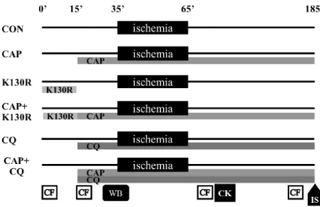
Sprague-Dawley rats (250–300 g) were anesthetized with i.p. pentobarbital (30 mg/kg) and anticoagulated with i.v. heparin (100 U/kg). Hearts were excised and perfused in Langendorff mode with Krebs-Henseleit solution (KH) for 15 min. From the 15th min, a group of heart received KH containing 300 μM CAP. To inhibit autophagosome formation a group of hearts were perfused with KH containing 300 μM CAP and 200 nM cell-permeable recombinant TAT-HA- Atg5K130R (CAP + K130R) for 15 min at the beginning of the protocol, then received CAP alone. Another group of hearts received 300 μM CAP and 10 μM CQ (CAP + CQ) throughout the protocol to inhibit lysosomal degradation of CAP-induced autophagosomes. Further groups of hearts were perfused with 10 μM CQ or 200 nM TAT-HA-Atg5K130R alone (CQ and K130R, respectively; see Figure 2).
Figure 2. Experimental protocol.
CAP- Chloramphenicol, CON- Control, K130R- TAT-HA-Atg5K130R, CK- Creatine kinase, CF- Coronary flow measurement, WB- Western blot, IS- Infarct size.
9
3.15 Measurement of infarct size, coronary flow and creatine kinase release
At the end of perfusion, hearts were sliced into 2 mm-thick slices, and right ventricles were removed. Slices were immersed in 1% triphenyltetrazolium- chloride, then in 4% formalin for 24 h and scanned. Coronary flow rate was measured throughout the protocol by timed collection of coronary effluent. Creatine kinase (CK) release was measured in the coronary effluent collected from 10 min to 20 min after the onset of the reperfusion by a colorimetric assay.
3.16 Western blotting
Equal amount of protein (left atria or whole hearts samples) was loaded and separated in a Tris-glycine-SDS polyacrylamide gel. Proteins were transferred onto a polyvinylidene difluoride membrane. Membranes were blocked with bovine serum albumin or non-fat dry milk. Membranes were probed with primary antibodies, and with corresponding horseradish peroxidase-conjugated secondary antibodies. Signals were detected with an enhanced chemiluminescence kit.
3.17 Data and statistical analysis
Data are expressed as mean ± standard error of mean (SEM). Statistical analysis was performed by two-way ANOVA using Fisher’s LSD as post hoc test or One-way ANOVA using Fisher's LSD as post-hoc test or Student's t-test or non-parametric Kruskal-Wallis test (coronary flow data, 14–20 min timepoint) by Prism 6. For motor activity multivariate analysis of variance was performed as statistical evaluation.
Differences were deemed significant in case p < 0.05.
4 Results
4.1 Effect of MAO-B inhibition in mild obesity
In this experiment, body weight increased slightly but significantly from the 16th week in HFS and HFS + S groups, with a 14% increase at the end of the study, which shows moderate obesity. Caloric intake was similar in all groups, as food intake was reduced in HFS and HFS + S groups. Selegiline had no effect on body weight or food intake.
10
We found, that total visceral and subcutaneous fat volumes increased significantly due to HFS diet and that selegiline treatment reduced these elevations in total visceral and subcutaneous fat depots. Weight of epididymal adipose tissue but not perirenal or brown adipose tissue was elevated by HFS diet compared to the control group. Furthermore, selegiline treatment significantly decreased epididymal adipose tissue weight in HFS. We found no significant differences in organ weights, plasma leptin and lipid levels. We assessed the blood level of thyroid hormones and we found no difference in their levels due to diet or selegiline treatment. Furthermore, we found that hepatic total cholesterol and triglyceride levels were significantly higher in HFS groups. However, selegiline had no effect on hepatic lipid levels in our model of obesity.
In our study, we aimed to characterize the changes in glucose homeostasis induced by chronic HFS diet. OGTT and ITT tests showed that insulin tolerance developed due to HFS diet on week 24, although fasting blood glucose level, or plasma and pancreas insulin levels were not affected by diet or selegiline treatment. These results suggest that chronic HFS diet led to the development of prediabetic state.
To examine the glucose uptake in visceral white adipose tissue, we measured Glut1, Glut4 expression. In our study, HFS diet significantly increased gene expression of Glut1 but not of Glut4. HFS diet induced the expression of Gapdh in adipose tissue, but selegiline did not affect the expression of Gapdh. Furthermore, we found that HFS diet induced the expression of Dgat in adipose tissue, but selegiline treatment had no effect on this parameter. We also found that neither diet nor selegiline had any influence on gene expression of Acc, Pnpla2, and Cd36 in white adipose tissue. However, we found that HFS diet induced Srebp-1c expression and selegiline treatment tended to reduce Srebp-1c gene expression in white adipose tissue in HFS diet. Furthermore, selegiline may have prevented the HFS diet-induced elevation in Ndufa1 in white adipose tissue. Furthermore, we measured gene expression of markers of adipose tissue inflammation. We found no difference in expression of Ccl2 and in the number of macrophages in the epididymal and inguinal white adipose tissue. However, HFS diet induced gene expression of Ccl3 which was reduced by selegiline treatment.
To examine whether HFS diet or selegiline influences hemodynamic parameters, we measured heart rate, arterial systolic and diastolic pressures. We found that
11
selegiline significantly decreased systolic pressure in rats fed control diet, however this effect was abolished upon HFS diet feeding.
We aimed to assess the effect of selegiline on movement pattern and recognition memory in our model. To investigate the, we performed spontaneous motor activity test and novel object recognition test at the end of our study. Neither HFS diet nor selegiline treatment significantly affected the motor activity or recognition memory. For detecting the presence of neuropathy and neuropathic pain in HFS diet we performed two nociceptive tests: Randall-Selitto paw pressure test and DPA. Results showed no difference in pain thresholds between groups neither for DPA nor Randall-Selitto experiments.
4.2 CAP reduces infarct size via induction of autophagy sequestration
In a pilot study we aimed to compare the effect of CAP on autophagy in neonatal cardiomyocytes, in H9c2 cells and in ex vivo-treated hearts. Therefore, we used the most common autophagy marker LC3 to measure changes in autophagy. In isolated rat neonatal cardiomyocytes and in H9c2 cells there was no significant difference in LC3-II to I ratio between corresponding CON and CAP groups. In the left atrium of hearts perfused with CAP, the ratio of LC3-II to LC3-I was significantly increased after 35 min compared to CON samples. Since CAP failed to induce autophagy in NRCMs and H9c2 cells, in our further experiments we used the isolated rat heart model. We also tested the effect of CAP and TAT-HA-Atg5K130R on autophagy-related protein LC3 levels in whole heart tissue.
In order to assess whether the sequestration and/or degradation phases of autophagy are necessary for the cardioprotective effect of CAP, we used ex vivo Langendorff heart perfusion experiments. CAP treatment significantly reduced infarct size and CK release as compared to CON hearts. Pretreatment with TAT-HA- Atg5K130R abolished the infarct size limiting effect of CAP, while pretreatment with CQ did not interfere with CAP-induced cardioprotection. We also measured LC3 expression in isolated heart samples and the western blot results showed that CAP increased LC3-II/I ratio. Meanwhile, administration of TAT-HA-Atg5K130R but not CQ reduced the increase of LC3-II due to CAP administration.
Since cardioprotection, as assessed by infarct size, and autophagosome formation, as assessed by LC3-II to LC3-I ratio, was lower in CAP +K130R group than
12
in CAP group, these results indicate that CAP-induced cardioprotection requires the process of autophagosome formation but not autophagosomal clearance. To characterize cardioprotective signaling mechanisms modulated by CAP, we assessed activation of proteins involved in the RISK/SAFE pathway. We found that the phosphorylation of Erk1/2 but not of Akt increased significantly due to CAP treatment.
5 Conclusions
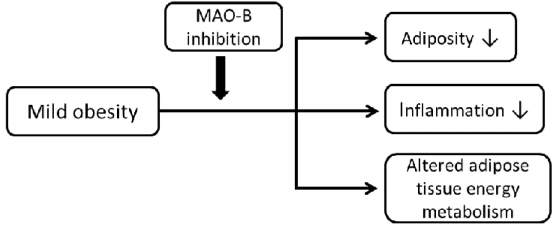
5.1 Selegiline moderates HFS diet-induced adiposity
This is the first demonstration that selegiline reduces adiposity, modulates adipose tissue energy metabolism and alleviates adipose inflammation induced by HFS diet (Figure 3). Here we have shown that HFS-induced expression of Srebp-1c, Glut1, and Ccl3 in adipose tissue was alleviated by selegiline treatment. However, in our experiment selegiline treatment did not affect the increase in body weight, prevent impairment of glucose homeostasis, or affect behaviour. Our results demonstrate that selegiline may influence glucose and lipid metabolism of white adipose tissue and may also alleviate inflammation in white adipose tissue.
These results suggest that specific inhibition of MAO-B by selegiline may mitigate harmful effects of obesity, and reduce the risk of cardiovascular diseases, thereby selegiline may serve as an adjuvant to anti-obesity therapy.
Figure 3. Effect of MAO-B inhibition in mild obesity.
13
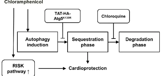
5.2 CAP reduces infarct size via induction of autophagy sequestration
Here we have shown for the first time in the literature that the sequestration, but not the clearance phase of autophagy is necessary for the cardioprotective effect of CAP (Figure 4). In our study, CAP administration reduced myocardial infarction in ex vivo rat hearts. Due to CAP treatment an increase in the LC3-II/I ratio and in the phosphorylation of Erk1/2 could be observed in the heart. The CAP-induced cardioprotection was abolished K130R treatment, an inhibitor of the initial phase of autophagy.
Therefore, therapeutic tools developed on the basis of induction of autophagic sequestration might lead to novel therapeutic options against acute myocardial ischemia/reperfusion injury.
Figure 4. Chloramphenicol-induced cardioprotection requires the sequestration phase of autophagy, not the degradation phase.
14
7
Bibliography of the candidate’s publications
7.1 Candidate’s publications involved in the current thesis
1. Nagy CT, Koncsos G, Varga ZV, Baranyai T, Tuza S, Kassai F, Ernyey AJ, Gyertyán I, Király K, Oláh A, Radovits T, Merkely B, Bukosza N, Szénási G, Hamar P, Mathé D, Szigeti K, Pelyhe C, Jelemenský M, Onódi Z, Helyes Z, Schulz R, Giricz Z, Ferdinandy P.
Selegiline reduces adiposity induced by high-fat, high-sucrose diet in male rats BRITISH JOURNAL OF PHARMACOLOGY 2018 Sep;175(18):3713-3726.
IF:6.81 (2017)
2. Giricz Z, Varga ZV, Koncsos G, Nagy CT, Gorbe A, Mentzer RM Jr, Gottlieb RA, Ferdinandy P
Autophagosome formation is required for cardioprotection by chloramphenicol LIFE SCIENCES 2017 Oct 1;186:11-16.
IF: 3.234
7.2 Candidate’s publications not involved in the current thesis
1. Onódi Z, Pelyhe C, Nagy CT, Brenner GB, Almási L, Kittel A, Manček-Keber M, Ferdinandy P, Buzás EI, Giricz Z
Isolation of high-purity extracellular vesicles by the combination of iodixanol density gradient ultracentrifugation and bind-elute chromatography from blood plasma FRONTIERS IN PHYSIOLOGY2018 Oct 23;9:1479.
IF: 3.394 (2017)
2. Kiscsatari L, Varga Z, Schally AV, Gaspar R, Nagy CT, Giricz Z, Ferdinandy P, Fabian G, Kahan Z, Gorbe A
Protection of neonatal rat cardiac myocytes against radiation-induced damage with agonists of growth hormone-releasing hormone
PHARMACOLOGICAL RESEARCH 2016 Sep;111:859-866.
IF: 4.48
15
3. Baranyai T, Nagy CT, Koncsos G, Onodi Z, Karolyi-Szabo M, Makkos A, Varga ZV, Ferdinandy P, Giricz Z
Acute hyperglycemia abolishes cardioprotection by remote ischemic perconditioning CARDIOVASCULAR DIABETOLOGY 2015 Nov 18;14:151.
IF: 4.534
4. Emri T, Tóth V, Nagy CT, Nagy G, Pócsi I, Gyémánt G, Antal K, Balla J, Balla G, Román G, Kovács I, Pócsi I.
Towards high-siderophore-content foods: optimisation of coprogen production in submerged cultures of Penicillium nalgiovense
JOURNAL OF THE SCIENCE OF FOOD AND AGRICULTURE 2013 Jul;93(9):2221-8.
IF: 1.879
5. Tóth V, Nagy CT, Pócsi I, Emri T
The echinocandin B producer fungus Aspergillus nidulans var. roseus ATCC 58397 does not possess innate resistance against its lipopeptide antimycotic
APPLIED MICROBIOLOGY AND BIOTECHNOLOGY 2012 Jul;95(1):113-22.
IF: 3.689
6. Tóth V, Nagy T C, Miskei M, Pócsi I, Emri T
Polyphasic characterization of “Aspergillus nidulans var. roseus” ATCC 58397.
FOLIA MICROBIOLOGICA 2011 Sep;56(5):381-8.
IF: 0.677