INVESTIGATION OF NOVEL THERAPEUTIC OPTIONS FOR THE TREATMENT OF ISCHEMIC AND METABOLIC CARDIOVASCULAR DISEASES
Ph.D thesis
Csilla Terézia Nagy
Semmelweis University
Doctoral School of Pharmaceutical Sciences
Supervisor: Zoltán Giricz, PharmD, Ph.D.
Official reviewers:
Péter Sántha, MD, Ph.D.
Attila Szijártó, MD, Ph.D.
Head of the Final Examination Committee:
Barna Vásárhelyi, MD, Ph.D.
Members of the Final Examination Committee:
Erika Pintér, MD, Ph.D.
Balázs Szalay, MD, Ph.D.
Budapest
2018
1 Table of contents
List of Abbreviations ... 4
1 Introduction ... 8
1.1 Obesity: diagnosis and treatment ... 8
1.1.1 Obesity and its metabolic complications ... 8
1.1.2 Therapies for treatment of obesity ... 12
1.1.3 Monoamine oxidase (MAO) inhibitors ... 16
1.2 Cardiovascular diseases (CVDs) ... 17
1.2.1 Therapies of CVDs ... 18
1.2.2 Autophagy ... 21
1.2.3 Compounds influencing autophagy ... 23
2 Objectives ... 25
3 Methods ... 26
3.1 Study designs ... 26
3.1.1 Study design: diet-induced experimental obesity ... 26
3.1.2 Study design: effect of CAP on autophagy in different cardiac cells ... 29
3.2 Evaluation of whole body fat ... 29
3.3 Immunohistochemistry ... 30
3.4 Adipocyte cross sectional area ... 31
3.5 Total RNA isolation ... 31
3.6 cDNA synthesis and qRT-PCR ... 31
3.7 Measurement of plasma lipid, insulin and leptin levels ... 32
3.8 Measurement of liver lipid content ... 33
3.9 Hemodynamic measurements ... 33
3.10 Behaviour and nociceptive tests ... 34
2
3.11 Isolation and treatment of NRCMs ... 35
3.12 Treatment of H9c2 cells ... 36
3.13 Preparation of K130R ... 36
3.14 Ex vivo heart perfusion ... 36
3.15 Measurement of infarct size, coronary flow and creatine kinase release ... 38
3.16 Western blotting ... 38
3.17 Data and statistical analysis ... 39
4 Results ... 40
4.1 HFS diet elevated body weight despite a similar caloric intake ... 40
4.2 Selegiline reduced HFS diet-induced adiposity ... 41
4.3 Selegiline had no effect on HFS diet-impaired glucose homeostasis ... 44
4.4 Selegiline interferes with glucose uptake and lipid metabolism via modulating expression of Glut1, Srebp-1c and Ndufa1 in HFS diet ... 46
4.5 Selegiline reduces HFS diet-induced inflammation in white adipose tissue by modulating expression of Ccl3 ... 48
4.6 Selegiline decreased arterial systolic pressure in control diet ... 49
4.7 HFS diet and selegiline did not induce behavioural alterations or sensory neuropathy ... 50
4.8 CAP induces autophagy in ex vivo perfused hearts but not in NRCMs and H9c2 cells ... 51
4.9 TAT-HA-Atg5K130R blocks CAP-induced cardioprotection in isolated hearts ... 53
5 Discussion ... 56
5. 1 Selegiline moderates adiposity induced by HFS diet ... 56
5.1.1 Selegiline reduced subcutaneous and visceral fat depots ... 56
5.1.2 Hypothesized mechanisms of the adiposity-lowering effect of selegiline ... 57
5.1.3 Selegiline alleviates WAT inflammation induced by HFS diet ... 58
5.1.4 Selegiline had no influence on HFS diet-induced prediabetes ... 59
3
5.1.5 Behavioural alterations or sensory neuropathy was not observed due to HFS
diet or selegiline ... 60
5.2 The process of autophagosome formation is necessary for the infarct size limiting effect of CAP ... 61
5.2.1 CAP induces cardioprotection via autophagy in ex vivo hearts ... 61
6 Conclusions ... 64
6.1 Selegiline moderates HFS diet-induced adiposity ... 64
6.2 CAP reduces infarct size via induction of autophagy sequestration ... 64
7 Summary ... 65
8 Összefoglalás ... 66
9 Bibliography ... 67
10 Bibliography of the candidate’s publications ... 88
10.1 Candidate’s publications involved in the current thesis ... 88
10.2 Candidate’s publications not involved in the current thesis ... 88
11 Acknowledgements ... 90
4
List of Abbreviations
ACC - acetyl-CoA carboxylase
ACE - angiotensin-converting-enzyme ADSCs - adipose-derived stem cells AKT - protein kinase B
AMPK - AMP-activated Protein Kinase ARBS - angiotensin II Receptor Blockers ATG - autophagy related
AUC - area under curve
BAT - brown adipose tissue
BCA - bicinchoninic Acid Protein Assay Kit
BMI - body mass index
BSA - bovine serum albumin
cAMP - cyclic adenosine monophosphate
CAP - chloramphenicol
CAPS - chloramphenicol succinate CB1 receptor - cannabinoid receptor type 1
CCL2 - monocyte chemoattractant protein 1 CCL3 - macrophage inflammatory protein 1-alpha CD36 - fatty acid translocase
5
CK - creatine kinase
CMA - chaperone-mediated autophagy CNS - central nervous system
CON - control diet
CQ - chloroquine
CT - computed tomography
CVD - cardiovascular disease
DA - dopamine
DGAT - diglyceride acyltransferase DI - discrimination index
DMEM - Dulbecco's Modified Eagle Medium DPA - dynamic plantar aesthesiometer EMA - European Medicines Agency
ERK1/2 - extracellular signal–regulated kinases 1/2 FAD - flavin-adenin-dinukleotid
FBS - fetal bovine serum
FDA - Food and Drug Administration
GAPDH - glyceraldehyde 3-phosphate dehydrogenase GLP-1 - glucagon-like peptide-1
GLUT1 - glucose transporter 1 GLUT4 - glucose transporter 4
GSK3b - glycogen synthase kinase 3 beta
6
5-HT - serotonin
HDL - low density lipoprotein H&E - hematoxylin and eosin HFS - high-fat, high-sucrose diet
HPRT1 - hypoxanthine phosphoribosyltransferase 1 ITT - insulin tolerance test
K130R - TAT-HA- Atg5K130R
KH - Krebs-Henseleit solution
LAD - left anterior descending coronary artery LC3 - microtubule-associated protein light chain 3 LDL - low density lipoprotein
MAO - monoamine oxidases
MAO-A - monoamine oxidase A MAO-B - monoamine oxidase B MI - myocardial infarction
mTORC1 - mammalian target of rapamycin complex 1 NDUFA1 - NADH dehydrogenase 1 alpha
NE - norepinephrine
NIH - National Institutes of Health NOR - novel objection recognition assay NRCMs - neonatal rat cardiomyocytes OGTT - oral glucose tolerance test
7 PBS - phosphate buffered saline PI3K - phosphoinositide 3-kinase PNPLA2 - adipose triglyceride lipase
RIPA - radio-Immunoprecipitation assay buffer
S - selegiline
SREBP-1c - sterol regulatory element-binding protein 1c TBST - tris-buffered saline with 0.05% Tween 20 T2DM - type 2 diabetes mellitus
t2familiar - exploration time of familiar object t2novel - exploration time of novel object
ULK1 - unc-51 Like Autophagy Activating Kinase 1 VAT - visceral adipose tissue
WAT - white adipose tissue
WHO - World Health Organization
8
1 Introduction
1.1 Obesity: diagnosis and treatment
In recent decades, obesity and its metabolic complications have become one of the biggest public health issues worldwide. According to the data of World Health Organization (WHO), the prevalence of obesity has nearly tripled between 1975 and 2016. In 2016, more than 1.9 billion adults were overweight, of whom over 650 million were obese (WHO 2018). Furthermore, obesity is a serious problem not just for adults but children also. The prevalence of obesity in children has dramatically increased in the last few years (Sahoo, Sahoo et al. 2015). In 2016, 41 million children under the age of 5 were overweight or obese in 2016 and over 340 million children and adolescents aged 5-19 were overweight or obese. Furthermore, obesity increases the risk of developing several comorbidities such as Type 2 diabetes mellitus (T2DM), high blood pressure, high blood cholesterol and high triglyceride levels, thereby reduces life quality and expectancy. Overweight and obesity are most common in developed and developing countries (Hruby and Hu 2015, Hales, Carroll et al. 2017). The cost of medical treatment of obesity and of the obesity-related diseases put a large economic burden on the individual and nations also (Levy, Levy et al. 1995, Birmingham, Muller et al. 1999, Tremmel, Gerdtham et al. 2017). Besides increased health care expenditure, obesity also imposes further costs in the form reduced ability to work, increased morbidity and mortality.
1.1.1 Obesity and its metabolic complications
Obesity is most commonly caused by a combination of excessive food intake, lack of physical activity, and genetic susceptibility (Yazdi, Clee et al. 2015). Occasionally it can be triggered by medications (Bernstein 1987, Ness-Abramof and Apovian 2005), endocrine disorders (Pujanek, Bronisz et al. 2013) or mental disorder (Bleich, Cutler et al. 2008). Obesity is a condition in which excess fat has accumulated in the body, such that it might have adverse effect on health (Van Itallie 1979). Usually it is defined and
9
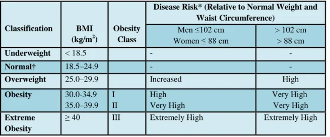
classified by body mass index (BMI, calculated by a person's weight in kilograms divided by the square of his height in meters), and further evaluated in terms of fat distribution via the waist circumference and body fat percentage. A person with a BMI of 18.5-24.9 kg/m² has a normal weight. A BMI of 25 to <30 kg/m² is defined as overweight and BMI ≥ 30 kg/m² is classified as obese, BMI ≥40 kg/m² defined as extreme obese (NIH 1998) (Table 1).
Table 1 Classification of overweight and obesity by BMI, waist circumference and associated disease risk.
Table was adopted from reference (NIH 1998).
*Disease risk for Type 2 diabetes, hypertension, and cardiovascular disease.
†Increased waist circumference can also be a marker for increased risk even in persons of normal weight.
Classification BMI (kg/m2)
Obesity Class
Disease Risk* (Relative to Normal Weight and Waist Circumference)
Men ≤102 cm Women ≤ 88 cm
> 102 cm
> 88 cm
Underweight < 18.5 - -
Normal† 18.5–24.9 - -
Overweight 25.0–29.9 Increased High
Obesity 30.0-34.9 35.0–39.9
I II
High Very High
Very High Very High Extreme
Obesity
≥ 40 III Extremely High Extremely High
Adipose tissue is one of the main types of a connective tissue which mainly comprises adipocytes. Besides adipocytes, adipose tissue contains the stromal vascular fraction (preadipocytes, fibroblasts, vascular endothelial cells) and a variety of immune cells (Frayn, Karpe et al. 2003). Adipose tissue is a complex, essential and highly active metabolic and endocrine organ and a main energy store of the body (Kershaw and Flier 2004). Beside the two main function of white adipose tissue (WAT) which are lipogenesis(fatty acid synthesis and storage) and lipolysis (mobilization or hydrolysis of triglycerides), WAT secretes also biologically active substances known as adipokines
10
(Proença, Sertié et al. 2014). Adipokines play a critical role in many biological functions, for example regulation of carbohydrate and lipid metabolism, regulation of food intake and insulin sensitivity (Fasshauer and Bluher 2015). Besides adipokines, monoamines (epinephrine, norepinephrine (NE), dopamine (DA) and serotonin (5-HT)) via dopaminergic and noradrenergic pathways play a key role in regulating carbohydrate and lipid metabolism (D’Souza and Abraham 2016).
Excess adiposity and adipocyte dysfunction result in dysregulation of adipokines, which may contribute to impaired glucose (Antuna-Puente, Feve et al.
2008) and lipid metabolism (Cao 2014, Tang 2016) as well as inflammatory responses (Balistreri, Caruso et al. 2010). Previous studies suggest that chronic inflammation in adipose tissue may play a significant role in the development of obesity-related metabolic dysfunction (Hotamisligil 2006, Lumeng and Saltiel 2011). Adipocyte dysfunction is also commonly associated with vascular diseases, like hypertension (Zhou and Qin 2012) and atherosclerotic vascular disease (Hajer, van Haeften et al.
2008, Lee, Wu et al. 2010).
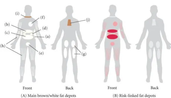
The detrimental effects of obesity on health are due to the enlargement of adipose tissue, particularly visceral adipose tissue. In mammals, there are two main types of adipose tissue, the WAT, which stores energy, and brown adipose tissue (BAT), which generates body heat. Moreover, researchers recently discovered another type of adipose tissue. After thermogenic stimuli brown adipocytes may appear in WAT, derived from precursor cells which are different from the classical BAT and are closer to the white adipocyte cell lineage. This process is called “browning”. These adipocytes are often referred as "inducible, beige, or brite adipocytes" (Giralt and Villarroya 2013). The adipose tissue divided into different regional depots with variation in biological function, structural organization and cellular size (Bjørndal, Burri et al. 2011). The main WATs are subcutaneous adipose tissue and visceral adipose tissue. Visceral fat, also known as organ fat or intra-abdominal fat is located between internal organs and torso, inside the peritoneal cavity. Visceral fat is composed of several adipose depots including mesenteric, epididymal white adipose tissue (omental) and perirenal fat (Figure 1.). The change in the volume of fat depots alters a number of physiological processes.
11
Figure 1. (A) The main white adipose tissues (WATs) are abdominal subcutaneous adipose tissue (SAT, (a)), and visceral adipose tissue (VAT). VAT surrounds the inner organs and can be divided in omental (b), mesenteric (c), retroperitoneal ((d): surrounding the kidney), gonadal ((e): attached to the uterus and ovaries in females and epididymis and testis in men), and pericardial (f). The omental depot stars near the stomach and spleen and can expand into the ventral abdomen, while the deeper mesenteric depot is attached in a web-form to the intestine.
The gluteofemoral adipose tissue (g) is the SAT located to the lower-body parts and is measured by hip, thigh, and leg circumference. WAT can also be found intramuscularly (h). Brown adipose tissue is found above the clavicle ((i): supraclavicular) and in the subscapular region (j). Although the mentioned subcutaneous and visceral adipose tissues are found in humans, depots (d) and (e) are mostly studied in rodents. (B) The adipose tissue depots that have been linked to risk of developing obesity-related diseases are indicated in red. The best-documented link to risk is found for the omental and mesenteric VAT. Figure was adopted from reference (Frayn, Karpe et al. 2003).
Researchers first started to focus on visceral obesity in the 1980s when they realized that the distribution between the fat depots is more important than the total adipose tissue mass for the risk of developing obesity-associated diseases. Visceral obesity greatly increases the risk of several diseases, such as T2DM (Golay and Ybarra 2005, Wells 2017), non-alcoholic fatty liver disease (Lopez-Velazquez, Silva-Vidal et al. 2014), dyslipidemia (Jung and Choi 2014), cardiovascular disease (Eckel 1997, Poirier, Giles et al. 2006), certain types of cancers (Gallagher and LeRoith 2015), and depression (Luppino, de Wit et al. 2010). Obesity and its metabolic complications are linked to more deaths worldwide than underweight. Therefore, pharmacological tools,
12
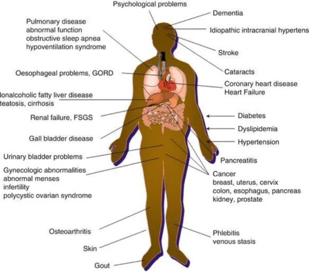
which can prevent or treat obesity and the co-morbidities associated with advanced obesity are intensively studied (Ferdinandy, Hausenloy et al. 2014) (Figure 2).
Figure 2. Medical complications of obesity.
GORD- gastro-oesophageal reflux disease, FSGS- Focal Segmental Glomerulosclerosis.
Figure was adopted from: Emmanuel J.J., Coppack S.W. (2016) Health Consequences–Obesity Associated Comorbidities. In Agrawal S. (eds) Obesity, Bariatric and Metabolic Surgery.
Springer, Cham).
1.1.2 Therapies for treatment of obesity
The purpose of treatment of overweight and obesity is to reduce body weight and prevention of weight regain and thus to reduce the consequent health risks. Weight reduction can be achieved by a combination of the following: lifestyle interventions (diet, physical activity), surgery (bariatric surgery) and pharmacotherapy (appetite
13
suppressants, metabolism inducers, absorption inhibitors) (Cannon and Kumar 2009).
All of these interventions are very important in the treatment of obesity but in this thesis we only focus on pharmacological therapy.
Nowadays, beside the pharmacotherapeutic treatment of obesity, cell-based therapies are getting more attention. Numerous studies suggest that, therapies using adipose-derived stem cells (ADSCs) (Illouz, Sterodimas et al. 2011, Payab, Goodarzi et al. 2018) or ADSCs-derived exosomes (Zhao, Shang et al. 2018) seems to be a potential treatment strategy to manage obesity and related metabolic disorders in the near future.
At the beginning of anti-obesity pharmacotherapy investigation, in the 70s, centrally-targeted and sympathetic-like agents showed very promising effects on decreasing body weight. These drugs activated the sympathetic nervous system inducing the release of catecholamines, meanwhile promoted satiety and reduced appetite (Motycka, St Onge et al. 2011). However, despite of these encouraging findings, numerous serious side effects occurred during the application of anti-obesity drugs. Due to the lack of sufficient efficacy and/or due to safety issues several drugs have been withdrawn from the market in the last 10 years. In 1973, US Food and Drug Administration (FDA) approved the combined pharmacological therapy fenfluramine- phentermine, and it was pulled out from the market in 1997 following life-threatening side effects such as pulmonary hypertension, and heart diseases (Connolly, Crary et al.
1997). Furthermore, rimonabant, a synthetic CB1 receptor inverse agonist was withdrawn in 2009 because patients exhibited increased depression and suicide risks (Moreira and Crippa 2009). Sibutramine, a serotonin (5-HT) or 5-hydroxytryptamine- norepinephrine reuptake inhibitor, was withdrawn from the market in 2010 due to increased risk of cardiovascular events (Krentz, Fujioka et al. 2016).
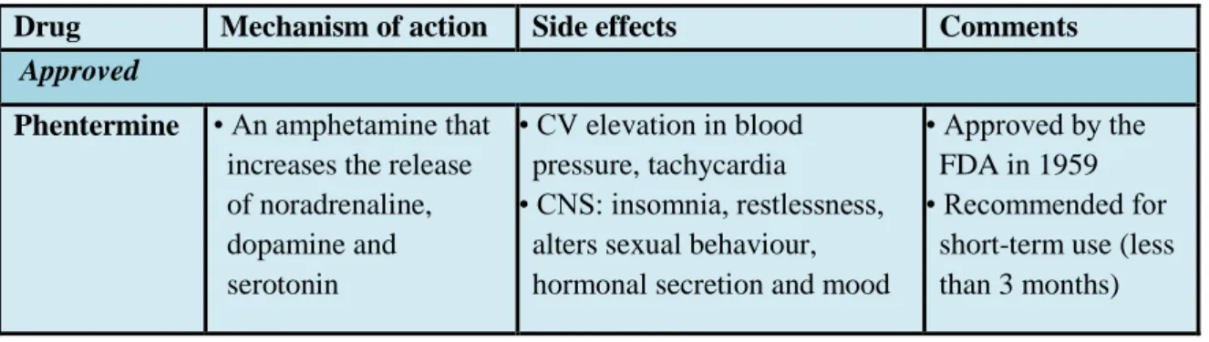
At present there are only few available anti-obesity drugs on the market (Srivastava and Apovian 2018) (Table 2). One of them is lorcaserin, with higher selectivity for the 5-HT2C receptors and less side effects than previous 5-HT agonists (fenfluramine and dexfenfluramine). In 2013, Fleming et al. showed that chronic injections of lorcaserin significantly decreasing food intake and body weight in high-fat diet obese-induced rats (Fleming, McClendon et al. 2013). Furthermore, Phase III clinical trial data show (BLOOM, BLOSSOM and BLOOM-DM) that lorcaserin is safe
14
and well tolerated in Type 2 diabetic patients and in non-diabetic patients, respectively (Greenway, Shanahan et al. 2016). Another anti-obesity drug in the market is Qnexa, which is the combination of phentermine and topiramate. Qnexa significantly decreases body weight (Garvey, Ryan et al. 2012) and improves obesity-related metabolic dysfunctions such as waist circumference, blood pressure, triglycerides and cholesterol levels,however, topiramate has teratogenic potential and increases the heart rate (Shin and Gadde 2013). Orlistat is the only anti-obesity drug which is driven by a peripheral approach and it has been on the market for more than 10 years. Orlistat reduces dietary fat absorption in the small intestines by inhibiting gastrointestinal lipase, however, may occur as a side effect the malabsorption of fat-soluble vitamins (Cahill and Lean 1999), severe liver injury (Sall, Wang et al. 2014) and kidney injury (Dietrich and Horvath 2012).
However, despite all the significant achievements, the current pharmacological treatments for obesity are only modestly effective and have several side effects.
Therefore, further research is needed to develop a novel strategy to enhance the effectiveness of obesity treatment and to reduce side effects, including repurposing of drugs already in use or development of novel agents.
Table 2 Anti-obesity drugs.
Table was adopted from reference (Srivastava and Apovian 2018) and (Dietrich and Horvath 2012). CNS- central nervous system, CV- cardiovascular; EMA- European Medicines Agency, FDA- US Food and Drug Administration, GLP-1- Glucagon-like peptide-1
*If applicable. ‡Never approved by the FDA owing to concerns related to adverse psychiatric side effects.
Drug Mechanism of action Side effects Comments Approved
Phentermine • An amphetamine that increases the release of noradrenaline, dopamine and serotonin
• CV elevation in blood pressure, tachycardia
• CNS: insomnia, restlessness, alters sexual behaviour, hormonal secretion and mood
• Approved by the FDA in 1959
• Recommended for short-term use (less than 3 months)
15 Table 2 (continued)
Drug Mechanism of action Side effects Comments
Orlistat • Pancreatic lipase inhibitor
• malabsorption of fat-soluble vitamins, steatorrhoea, fecal incontinence, flatulence
• Rare cases of severe liver injury
• Approved by the FDA in 1999
Lorcaserin • 5-HT receptor agonist that is more specific than previous compounds on the market, for example, fenfluramine
• Headache, dizziness, nausea, valvulopathy
• Possible carcinogenic effects in rodents
• Approved by the FDA in June 2012
• Under evaluation by the EMA
• Post-marketing, long-term CV outcomes trial required Phentermine
+ topiramate (Qnexa)
• Phentermine:
mechanism of action as above
• Topiramate:
anticonvulsant
• Possible teratogenic effects with topiramate
• Can increase heart rate
• Approved by the FDA in July 2012
• Post-marketing, long-term CV outcomes trial required Naltrexone/
bupropion sustained- release
• Bupropion: inhibitor of dopamine and noradrenalin uptake
• Naltrexone: μ-opioid receptor antagonist
• Nausea, constipation,
vomiting, dizziness, insomnia, dry mouth, and diarrhea
•Approved by the FDA in 2014
•Approved by the EMA in 2015
Liraglutide 3.0 mg
• GLP-1 analogue, binding to the same receptors as does the endogenous hormone GLP-1 that stimulates insulin secretion
•Nausea, hypoglycemia, diarrhea, constipation,
vomiting, headache, decreased appetite, dyspepsia, fatigue, dizziness, abdominal pain, and increased lipase
• Approved by the FDA in 2014
Withdrawn
Fenfluramine • Increases the release of serotonin
• Serotonin re-uptake inhibitor
• Hallucinations, valvulopathy, pulmonary hypertension
• Approved by the FDA in 1973
• Withdrawn in 1997 Sibutramine • Noradrenalin and
serotonin re-uptake inhibitor
• Increased risk of heart attack and stroke in patients with high risk of CV disorders
• Approved by the FDA in 1997
• Withdrawn in 2010 Rimonabant • Cannabinoid 1
receptor antagonist
• Risk of suicide • Approved by the EMA in 2006‡
• Withdrawn in 2009
16 1.1.3 Monoamine oxidase (MAO) inhibitors
Monoamines (epinephrine, NE, DA and 5-HT) participate in many physiological activities of the body. Monoamines are released from the synapse and exert their actions by activating dopaminergic, serotonergic and noradrenergic pathways. Monoaminergic signaling plays a critical role in regulating cognitive processes, as well as involved in the regulation of carbohydrate and lipid metabolism. MAOs are ubiquitous enzymes and they are responsible for the oxidative deamination of monoamines and they are found bound to the outer membrane of mitochondria in most cell types of the body. MAOs contain the covalently bound cofactor FAD, therefore, they are classified as flavoproteins. In mammals there are two subtypes of MAO: MAO-A and MAO-B. Both enzymes found in neurons and astroglia. Nevertheless, there are differences between their tissue distribution outside the central nervous system and the substrate selectivity of the two enzymes (Adeghate and Parvez 2004). MAO-B is mostly found in platelets and generally metabolizes DA, benzylamine and phenylethylamine. In contrast, MAO- A is found in the liver, pulmonary vascular endothelium, gastrointestinal tract and shows greater affinity for NE, 5-HT, DA and tyramine.
MAO inhibitors have two main groups: selective and non-selective, within these we distinguish reversible and irreversible inhibitors. At present, MAO inhibitors are mostly used for psychiatric and neurological disorders (Fiedorowicz and Swartz 2004, Finberg and Rabey 2016). MAO-B inhibitors (selegiline, rasagiline) are primarily used alone or in combination to treat Alzheimer’s and Parkinson’s diseases (Cai 2014, Dezsi and Vecsei 2017). MAO-A inhibitors (clorgyline, moclobemide) generally used as antidepressant (Meyer, Ginovart et al. 2006, Corbineau, Breton et al. 2017) and antianxiety drugs (Lader 1976).
However, in recent decades the use of MAO inhibitors by psychiatrists is significantly decreased; this decline is in context with the increase in the number of available novel antidepressants and with the concern about food- (Berlin and Lecrubier 1996, Flockhart 2012) and drug interactions (Sjöqvist 1965, Aboukarr and Giudice 2018). Nevertheless, since researchers showed that MAOs are highly expressed in human WAT, likely being involved in noradrenaline clearance and catecholamine-
17
dependent regulation of lipid metabolism in adipocytes (Pizzinat, Marti et al. 1999), thus, they may play an important role in obesity and lipid metabolism disorders and it may be an useful drug target in metabolic diseases.
Previous studies have shown that semicarbazide sensitive amine oxidase inhibitors administered in combination with certain non-selective and/or irreversible MAO inhibitors can reduce body weight and fat deposition in animal models of diet- induced obesity (Carpene, Iffiu-Soltesz et al. 2007, Carpene, Abello et al. 2008). Mattila et al. showed that administration of pargyline (30 mg kg-1) increased lipolysis and levels of plasma free fatty acid in rats (Mattila and Torsti 1966). Furthermore, diet-induced obesity increased MAO activity in the enlarged omental WAT of dogs (Wanecq, Bour et al. 2006). Selegiline is a clinically widely used, irreversible and selective inhibitor of MAO-B, which is primarily used to treat Parkinson's disease (Jankovic and Poewe 2012, Zhao, Cai et al. 2013, Chiu, Carlsson et al. 2014, Cereda, Cilia et al. 2017), Alzheimer's disease (Riederer, Lachenmayer et al. 2004) and depression (Fiedorowicz and Swartz 2004, Pae, Patkar et al. 2014, Thomas, Shin et al. 2015). However, it has also been shown to have pleiotropic effects not related to the MAO-B inhibition (Tatton, Ju et al. 1994, Tatton, Chalmers-Redman et al. 2002). Békési et al. showed that selegiline (5–10 mg kg-1) significantly decreased liver fat but not body weight in rats fed with a lipid-rich diet (Bekesi, Tulassay et al. 2012) (cholesterol 1%, olive oil 10%).
Therefore, we hypothesized that MAO inhibitors may have favourable metabolic effects in obesity.
1.2 Cardiovascular diseases (CVDs)
CVDs such as heart failure, hypertensive heart disease, cardiomyopathy, cardiac arrhythmia and coronary artery diseases (angina and myocardial infarction) are leading causes of morbidity and mortality worldwide (Pagidipati and Gaziano 2013, Roth, Johnson et al. 2017). Only in 2015, 17.7 million people died from CVDs in the world.
Most of these deaths occurred in low-income and middle-income countries (WHO 2018). Nearly 300 risk factors play a role in the development of CVDs. These include high blood pressure, high cholesterol, diabetes, obesity, smoking, excessive alcohol
18
consumption, inherited factors, age and psychosocial stress, etc. (Balakumar, Maung-U et al. 2016).
The most common type of CVD is myocardial infarction (MI). Only in 2015, about 15.9 million MI occurred, worldwide (Vos, Allen et al. 2016). MI occurs when the blood flow reduces or stops for a part of the heart that causes an oxygen and nutrient depletion in the heart muscle, which leads to myocardial necrosis (Boateng and Sanborn 2013). Most myocardial infarction in the world is due to atherosclerosis, when a coronary artery becomes occluded following the rupture of an atherosclerotic plaque, which leads to the formation of a blood clot. Besides atherosclerosis, many other factors may also cause myocardial infarction such as hyperthyroidism, low red blood cell count, or low blood pressure.
1.2.1 Therapies of CVDs
In the management of patients with heart disease, besides pharmacotherapy, cardioprotective interventions (Bousselmi, Lebbi et al. 2014, Thuret, Saint Yves et al.
2014, Aimo, Borrelli et al. 2015) and cell therapies (Reed, Foldes et al. 2013, Goradel, Hour et al. 2018) are becoming more and more important. In this thesis we focus on pharmacotherapy which is a major treatment modality in CVDs. The purpose of pharmacological therapies is to prevent CVDs and to improve patient outcomes. The use of current cardiovascular drugs such as antiplatelet drugs, anticoagulants, nitrates, beta-blockers, renin-angiotensin inhibitors, and statins depends on the heart condition and symptoms (Table 3). However, despite all the achievements in this field, there are still certain types of CVDs that do not have appropriate therapies. Furthermore, a major problem is that there are no adequate targets for treating several diseases, such as ischemic heart disease. Therefore, a further major objective of drug development is finding new cardiovascular agents (Stern and Lebowitz 2010, Huffman and Bhatnagar 2012) and searching for new pharmacological targets (Olson 2014).
19 Table 3 Classes of drug used in cardiovascular diseases
ACE- angiotensin-converting-enzyme; ARBS- Angiotensin II Receptor Blockers. Table was adopted from: http://www.secondscount.org/treatments/treatments-detail-2/medications- cardiovascular-disease#.W7MQhHszbDf.
Medication type Purpose Possible Side effects, Interactions, and Special Instructions
ACE Inhibitors and ARBS
To lower blood pressure and allow blood flow more easily from the heart
Dizziness, cough, low blood pressure. Kidneys and potassium levels should be monitored with blood tests.
Antiarrythmics To control irregular heartbeat
Depends on the class of drugs. Channel blockers can cause headaches, ankle swelling.
Amiodarone can increase sensitivity to sunlight and affect eyesight.
Antiplatelet Medications
To thin the blood and help prevent and dissolve clots in arteries and stents
Stomach pain, headache, dizziness, and breathing difficulties. Side effects more severe in patients with asthma and allergies. Take with food.
Aspirin To prevent and dissolve clots in the arteries
Stomach upset, headaches, and drowsiness. An allergic reaction could cause breathing difficulties. Other severe side effects include blood in the stool or coughing blood. Take with food to reduce risk of upset stomach.
Beta-Blockers To lower blood pressure and heart rate
Dizziness, fatigue, dry mouth, slow heart rate, weight gain, cold hands and feet. May reduce side effects if taken with food.
Thrombolytics To restore blood flow during a heart attack or stroke and to break up blood clots in the legs (deep vein thrombosis)
Bleeding, abnormal heart beat, new clotting.
Anticoagulants To prevent blood clots from forming in the arteries and heart
Bleeding, vomiting or coughing blood, blood in stool, headaches, and dizziness. Do not take with aspirin unless directed by doctor.
Digoxin To improve your heart's ability to pump blood and helps to slow down an irregular heartbeat
Side effects are more common if too much is taken and may include nausea, vomiting, diarrhea, stomach pain, loss of appetite, unusual tiredness, and slow heartbeat. Take on an empty stomach, high-fiber foods can decrease its absorption.
Smoking Cessation Medication
To make it easier to stop smoking
See "SecondsCount Guide to Medications That Help you Quit Smoking"
20 Table 3 (continued)
Medication type Purpose Possible Side effects, Interactions, and Special Instructions
Statins To lower your cholesterol level, reduce the risk of heart attacks, strokes
Muscle pain, liver damage, memory loss, nausea, gas, diarrhea, constipation, rash.
Diuretics To lower blood pressure
Frequent urination, dehydration, blurred vision, fatigue, rash, loss of appetite. Monitor blood pressure and kidney function.
Vasodilators To widen the blood vessels to increase the flow of blood and lower blood pressure
Headaches, nausea, and dizziness (especially older people). Do not drink grapefruit juice.
May interact negatively with cold medicine.
A number of cardioprotective interventions have been shown in the last decades, which reduced ischemic injury in animal models but failed to induce cardioprotection in human studies (Bolli, Becker et al. 2004). Then, numerous studies demonstrated that the efficacy of cardioprotective therapies is significantly affected by comorbidities such as obesity, diabetes or dyslipidaemia (Paulson 1997, Yi, Sun et al. 2011). For example, patients with T2DM have increased sensitivity to ischemia-reperfusion (IR) injury and that endogenous cardioprotective mechanisms are also inhibited (Ferdinandy, Schulz et al. 2007). Moreover, it has been shown that not only chronic diseases but also acute changes, such as acute hyperglycemia, can negatively affect cardioprotective interventions (Baranyai, Nagy et al. 2015). Furthermore, several publications reported that hypercholesterolemia changes cellular signalling and metabolism, thereby attenuates pharmacological and non-pharmacological cardioprotective interventions (Andreadou, Iliodromitis et al. 2017). In 2016, Ma et al. reported that remote ischemic preconditioning-induced cardioprotection was abolished in hypercholesterolemic rats by alteration of PI3K-Akt-GSK3b signaling pathway. In another study, Koncsos et al.
showed that hypercholesterolemia-induced decrease in basal autophagy and increase in apoptosis leads to loss of cardioprotection (Giricz, Koncsos et al. 2017).
Therefore, a growing amount of evidence suggests that increased autophagy plays a critical role in cardioprotective interventions (Huang, Yitzhaki et al. 2010, Giricz, Mentzer et al. 2012) and that besides MAO inhibition, autophagy could be a potential therapeutic target for the treatment of metabolic- and cardiovascular diseases.
21 1.2.2 Autophagy
Autophagy is an intracellular degradation process which eliminates unnecessary or dysfunctional organelles and long-lived proteins through lysosomal breakdown for recycling intracellular components (Gustafsson and Gottlieb 2008). Accordingly, autophagy is primarily considered as a cytoprotective process. However, excessive self- degradation has detrimental effects. Therefore, autophagic dysfunction is related with different human pathologies, such as liver, and heart disease, neurodegeneration, cancer and metabolic diseases (Rubinsztein, Codogno et al. 2012). In mammalian cells, three main types of autophagy are commonly described: microautophagy, chaperone- mediated autophagy (CMA) and macroautophagy.
Microautophagy is a process which involves the direct engulfment of cytoplasmic material into the lysosome. The cytoplasmic contents enter the lysosome through an invagination or deformation of the lysosomal membrane (Li, Li et al. 2012).
The second type of autophagy, CMA is a very complex and extremely selective for a subset of cytosolic soluble proteins (Kaushik and Cuervo 2008). Macroautophagy, (herein referred to as autophagy) eradicate damaged cell organelles or unused proteins.
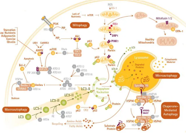
Autophagy consists of sequential steps: the initiation phase, which includes formation of the autophagosomal membrane also known as a phagophore that is usually derived from lipid bilayer contributed by the endoplasmic reticulum or the trans-Golgi. During sequestration phase, the phagophore expands and engulf protein aggregates or dysfunctional organelles and finally constitute a spherical double-membraned autophagosome. This is regulated by the autophagy proteins Atg4, Atg7, LC3, and the complex of Atg12-Atg5-Atg16L. In the degradation phase, autophagosome matures via fusion with lysosome, this structure is called autophagolysosome. This step initiates the degradation of the inner membrane of the autophagosome and cytoplasm-derived materials by lysosomal hydrolases. Lysosomal permeases and transporters export amino acids and other products of degradation to the cytosol for use by the cell in biosynthetic processes or to generate energy (Mizushima 2007, He and Klionsky 2009, Parzych and Klionsky 2014). Mitophagy is a specific type of autophagy, which means the selective degradation of injured or dysfunctional mitochondria (Ding and Yin 2012) (Figure 3).
22
Under normal circumstances autophagy has a low activity in cells but metabolic stress such as nutrient starvation or hypoxia can increase the activity of autophagy.
During metabolic stress degradation of intracellular components promotes cell survival by maintaining cellular energy levels. Furthermore, the role of autophagy in the development of metabolic disorders (insulin resistance, diabetes mellitus, obesity, atherosclerosis) has been studied extensively using different genetic animal models (Kim and Lee 2014). Therefore, the use of autophagy-inducing agents could be an effective therapy in cardiovascular diseases and metabolic diseases.
Figure 3. Autophagy mechanisms and signaling pathways. (macroautophagy, mitophagy and microautophagy).
Figure was adopted from: https://www.sigmaaldrich.com/technical-documents/
articles/biology/cell-culture/cellular-assays/autophagy-assays.html
23 1.2.3 Compounds influencing autophagy
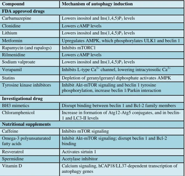
Previous studies reported that several therapeutic agents which are already in clinical use, for example hydrophobic statins (Andres, Hernandez et al. 2014), sevoflurane (Shiomi, Miyamae et al. 2014), sulfaphenazole (Huang, Liu et al. 2010) and certain antibiotics, such as chloramphenicol (CAP) (Prigione and Cortopassi 2007), may also induce autophagy in addition to their primary effects (Levine, Packer et al. 2015, Schiattarella and Hill 2016) (Table 4).
Table 4 Compounds that induce autophagy.
AMPK- AMP-activated Protein Kinase, cAMP- Cyclic adenosine monophosphate, mTORC1- mammalian target of rapamycin complex 1, ULK1- Unc-51 like autophagy activating kinase.
Table was adopted from reference (Levine, Packer et al. 2015).
Compound Mechanism of autophagy induction FDA approved drugs
Carbamazepine Lowers inositol and Ins(1,4,5)P3 levels
Clonidine Lowers cAMP levels
Lithium Lowers inositol and Ins(1,4,5)P3 levels
Metformin Upregulates AMPK, which phosphorylates ULK1 and beclin 1 Rapamycin (and rapalogs) Inhibits mTORC1
Rilmenidine Lowers cAMP levels
Sodium valproate Lowers inositol and Ins(1,4,5)P3 levels
Verapamil Inhibits L-type Ca2+ channel, lowering intracytosolic Ca2+
Statins Depletion of geranylgeranyl diphosphate activates AMPK Tyrosine kinase inhibitors Inhibit Akt-mTOR signaling and beclin 1 tyrosine
phosphorylation, increase beclin 1/Parkin interaction Investigational drug
BH3 mimetics Disrupt binding between beclin 1 and Bcl-2 family members Chloramphenicol Increase in formation of Atg12-Atg5 conjugates, and in beclin-
1 and LC3-II levels Nutritional supplements
Caffeine Inhibits mTOR signaling
Omega-3 polyunsaturated fatty acids
Inhibit Akt-mTOR signaling; disrupt beclin 1 and Bcl-2 binding
Resveratrol Activates sirtuin 1 Spermidine Acetylase inhibitor
Vitamin D Calcium signaling, hCAP18/LL37-dependent transcription of autophagy genes
24
Since previous studies reported that CAP protects the heart against ischemia/reperfusion injury and upregulates autophagy markers (He, Chen et al. 2001, Granville, Tashakkor et al. 2004, Sala-Mercado, Wider et al. 2010). It has been shown that the induction of autophagy is required for cardioprotective mechanisms but no detailed investigation has been performed on which stage of autophagy is necessary for cardioprotection. Since autophagy is a multi-step process, it can be inhibited at different steps. Previous studies have shown that inhibition of early-stage autophagy can be achieved by 3-methyladenine (Li, Liu et al. 2015) and wortmannin which are inhibit the class III phosphatidylinositol-3 kinase, or TAT-HA-Atg5K130R, a dominant negative mutant fusion protein of a key mediator of autophagy, Atg5 (Pyo, Jang et al. 2005, Hamacher-Brady, Brady et al. 2006, Hamacher-Brady, Brady et al. 2007). The late phase of autophagy, lysosomal fusion and degradation, can be arrested by elevating lysosomal pH, e.g., by the use of an antimalarial drug, chloroquine (CQ, see for review:
(Kimura, Takabatake et al. 2013)). Although a few studies utilized these substances to investigate the mechanism of cardioprotective stimuli, it is still unknown which phase of CAP-induced autophagy is necessary for cardioprotection.
Therefore, here we hypothesized that CAP induces cardioprotection, via increasing the level of autophagy in cardiomyocytes.
25
2 Objectives
Therefore, we had the following aims:
To investigate the effect of selegiline treatment on metabolic parameters in high- fat, high-sucrose diet-induced moderate obesity, together with alterations in hemodynamic parameters and markers of neuropathy and behavioural patterns.
To investigate whether CAP-induced autophagy is necessary for cardioprotection.
To assess whether sequestration and/or degradation phases of autophagy are necessary for the cardioprotective effect of CAP.
26
3 Methods
This investigation conforms to the Guide for the Care and Use of Laboratory Animals published by the US National Institutes of Health (NIH publication No. 85-23, revised 1996) and was approved by the animal ethics committee of the San Diego State University, San Diego, California and Semmelweis University, Budapest, Hungary.
Furthermore, conforms to Directive 2010/63/EU and was authorized by the regional animal health authority in Hungary (registration numbers: XIV-I-001/450-6/2012).
Animal studies are reported in compliance with the ARRIVE guidelines (Kilkenny, Browne et al. 2010, McGrath and Lilley 2015).
3.1 Study designs
3.1.1 Study design: diet-induced experimental obesity
5-7 weeks old male Long-Evans rats (RRID: RGD_2308852) were purchased from Charles River Laboratories (Wilmington, MA, USA). We chose Long-Evans rats for our study which are commonly used for experiments on diet-induced obesity (Li, Zhang et al. 2008, Estridge, Dey et al. 2017). Furthermore, this strain is leaner as compared to Sprague-Dawley and Wistar rats, therefore, the effects of mild obesity could be studied with higher reliability in this strain. Furthermore, our laboratory has already experience with this strain in metabolic studies (Koncsos, Varga et al. 2016).
Animals were housed in conventional animal house, 4 animals were housed in a polyethylene EUROSTANDARD TYPE IV S cages (480 x 375 x 210 mm) containing corncob bedding from J. Rettenmaier & Söhne GmbH & Co and housed in a room maintained in a 12/12 h light/dark cycle and constant temperature of 21 °C. Animals were allowed access to food and water ad libitum. After 1 week of acclimatization, rats were randomly divided into two groups: control (CON; n=20) and high-fat, high- sucrose group (HFS; n=20). The CON group was fed control rat chow, whereas the HFS group was fed a chow supplemented with 20% lard and 15% sucrose as a HFS diet (Table 5).
27 Table 5 Composition and nutritional data of chows.
Data are expressed as g/100g or otherwise noted. CON- control, HFS- high-fat, high-sucrose.
CON HFS
Corn 58 18
Gluten/corn 25 30
Sugar 0 15
Lard 0 20
Fasermix (straw pellet) 6 6
Sodium chloride 0.4 0.4
Zeolite, universal 6.7 6.7
Mono calcium phosphate (MCP) 1.5 1.5
Feed lime 1.3 1.3
Adhesive 0.6 0.6
Vitamin and trace element supplement
0.5 0.5 Gross energy (Mj/kg) 15.52 20.31
From week 16, groups of animals were further randomly divided and received subcutaneous injections of 0.25 mg kg-1 selegiline (CON+S and HFS+S) or vehicle (CON, HFS) once daily (n=10 in each group). Previous articles showed that rats treated with daily subcutaneous dose of 0.25 mg kg-1 selegiline selectively inhibited the B form of the enzyme, leaving the activity of the A form practically unchanged. With a higher (1 mg kg-1) dose the selectivity was nearly lost (Knoll 1978, Heinonen, Anttila et al.
1994).
Body weights were measured monthly. Blood was taken, and fasting blood glucose levels were measured from the saphenous vein every month (Accu-Check;
Roche, Basel, Switzerland). At week 24, oral glucose tolerance test (OGTT) was performed on conscious overnight fasted rats with per os administration of 1.5 g kg-1 glucose and measurements of plasma glucose levels at 15, 30, 60, and 120 min. Insulin tolerance test (ITT) was also performed at week 24 in overnight fasted rats. Insulin (0.5 IU/kg, Humulin R; Ely Lilly, Utrecht, The Netherlands) was injected intraperitoneally (i.p.), and plasma glucose levels were checked at 15, 30, 45, 60, 90, and 120 min. At week 24 after 1-day acclimatization, food intake was observed for 24 hours in a
28
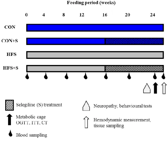
metabolic cage. At week 25 of the diet animals were sacrificed, anesthetized with pentobarbital (60 mg kg-1, intraperitoneally; Euthasol; Produlab Pharma, Raamsdonksveer, The Netherlands). After hemodynamic measurements, epididymal and interscapular brown fat tissue, markers of adiposity, were isolated, and their weights were measured. Blood and tissue samples were collected and stored at −80 °C (Figure 4.). To exclude the natural variability between weights, organ weights were normalized to tibia length (Tsujita, Muraski et al. 2006, Nemeth, Matyas et al. 2016).
Figure 4. Experimental protocol.
Male Long-Evans rats were fed with control (CON, n=10) diet or with high-fat, high sucrose (HFS, n=10) diet for 25 weeks; CON+S (n=10) and HFS+S (n=10) groups were treated with 0.25 mg kg-1 selegiline (S) from week 16 to 24. Body weights and blood glucose were measured monthly. Neuropathy, behavioural tests were measured at weeks 22-23. Oral glucose tolerance test (OGTT), insulin tolerance test (ITT), metabolic cage and CT (computed tomography) were performed at week 24. Hemodynamic analysis was performed at week 25. Tissue sampling was performed after terminal procedures.
29
3.1.2 Study design: effect of CAP on autophagy in different cardiac cells
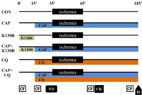
To identify the most suitable model system, in a pilot study we examined the effect of CAP on autophagy in neonatal rat cardiomyocytes (NRCMs), in H9c2 cardiac myoblast cells and in isolated hearts. We found that CAP induced autophagy in isolated hearts but not in NRCMs or in H9c2 cells (see Figure 14A–C). The efficacy of CAP and TAT-HAAtg5K130R was also assessed in a pilot experiment where we observed that LC3-II/I ratio was decreased after 15 min of administration of 200 nM TAT-HA- Atg5K130R in isolated hearts as compared to vehicle controls (Figure 14D).
Therefore, in the main series of experiments, we used an ex vivo model of acute cardiac ischemia/reperfusion injury to assess the effects of autophagy inhibitors (TAT- HA-Atg5K130R and CQ) on CAP-induced cardioprotection. Since the availability of TAT-HA-Atg5K130R was limited due to technical limitations in production and purification of the protein in quantities necessary for ex vivo heart perfusion experiments, we had to reduce the number of isolated hearts treated with TAT-HA- Atg5K130R.
3.2 Evaluation of whole body fat
To assess obesity, computed tomography (CT) measurements were performed on NanoSPECT/CT PLUS (Mediso, Budapest, Hungary) at week 24. The semicircular CT scanning was acquired with a 55 kV tube voltage, 500 msec exposure time, 1:4 binning, and 360 projections in 18 min. During the acquisitions, rats were placed in a prone position in a dedicated rat bed and were anesthetized with 2% isoflurane in oxygen. Temperature of the animals was kept at 37.2 ± 0.3 °C during imaging. In the reconstruction, 0.24 mm in-plane resolution and slice thickness were set, and Butterworth filter was applied (volume size: 76.8×76.8×190 mm). Images were further analysed with VivoQuant (inviCRO, Boston, MA) dedicated image analysis software products by placing appropriate volumes of interest on the whole body fat of animals.
The aim of segmentation was to separate the fat from other tissues. The connected threshold method helped to choose the adequate attenuated pixels for fat tissue analysis,
30
and then the isolated points were detected by erode 4 voxel and dilate 4 voxel steps.
After the measurements, animals recovered from anaesthesia. Adiposity index was calculated by the following formula: (CT whole body adipose tissue volume/body weight) ×100. Subcutaneous and total visceral fat volumes were evaluated on CT images by CTan software (Bruker microCT, Kontich, Belgium).
3.3 Immunohistochemistry
For immunohistochemistry, deparaffinized sections underwent antigen retrieval (pH=6 citrate buffer, at 95 °C for 15 min). After blocking endogenous peroxidase activity (Bloxall, Vector Laboratories, Burlingame, CA, USA), the sections were blocked in appropriate sera (2.5% goat serum in PBS). The primary antibody recognizing the monocyte/macrophage-specific protein Iba-1 (Wako Pure Chemical Industries, Chuo-Ku, Osaka, Japan), was incubated with the sections overnight in blocking solution at 4°C. After the primary antibody incubation, the sections were washed three times in PBS and incubated for an hour with an anti-rabbit IgG peroxidase polymer (ImmPress reagents, Vector Laboratories, Burlingame, CA, USA). Secondary antibodies were washed 3 times for 10 min and the specific signal was developed with diaminobenzidine (ImmPACT DAB EqV Peroxidase (HRP) Substrate, Vector Laboratories, Burlingame, CA, USA). The specific staining was visualized and images were acquired using Leica DM3000 LED microscope and MC 190 HD camera (Leica Microsystems, Wetzlar, Germany).
After routine formalin-fixed paraffin-embedded specimen processing, 4 µm thick tissue sections were prepared and stained with hematoxylin and eosin (H&E), for histological evaluation of adipocyte morphology. Adipocyte cross sectional area was measured with the ImageJ based software, Adiposoft (Galarraga, Campion et al. 2012).
31 3.4 Adipocyte cross sectional area
Adipocyte cross sectional area was measured with the ImageJ-based software, Adiposoft (Galarraga, Campion et al. 2012). Images of adipocyte cross sectional area were subtracted using ImageJ and then analyzed with Adiposoft without using automated method. Cells which were smaller than 25 µm in diameter were excluded.
Analyzed images were edited using manual method in Adiposoft.
Cells on the borders of image were excluded, separated sections that clearly belonged to one cell were merged together, and cells which were not recognized by software were added and analyzed manually. Adipocyte tissue areas in µm² were then further used for analysis.
3.5 Total RNA isolation
Total RNA from white adipose tissue (epididymal) was extracted with a precipitation method. Briefly, RNAzol® RT (Sigma, USA) was added to each sample and homogenized with TissueLyser (Qiagen, Germany). Homogenates were centrifuged, and DNA and protein were precipitated with nuclease-free water.
Furthermore, 4-bromoanisole (Sigma), phase separation step was incorporated to maximize the DNA elimination. Total RNA was precipitated with isopropanol (vWR, USA), and pellet was washed twice with ethanol (vWR, USA). Finally, total RNA was resuspended in nuclease-free water. RNA concentrations were measured with NanoDrop (Thermo Scientific, Waltham, USA).
3.6 cDNA synthesis and qRT-PCR
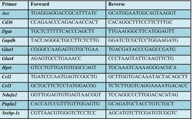
Total RNA was used as a template for cDNA synthesis, using Sensifast cDNA synthesis kit (Bioline; BLI 65053) according to the manufacturer’s protocol. qRT-PCR reactions were performed with a LightCycler® 480 Real-Time PCR System (Roche Diagnostics, Germany) in the presence LightCycler® RNA Master SYBR Green I (Roche; 03064760001) according to the manufacturers protocol. Forward and reverse primers for glucose transporter 1 (Glut1), glucose transporter 4 (Glut4), acetyl-CoA
32
carboxylase (Acc), adipose triglyceride lipase (Pnpla2), diglyceride acyltransferase (Dgat), fatty acid translocase (Cd36), sterol regulatory element-binding protein 1c (Srebp-1c), NADH dehydrogenase 1 alpha (Ndufa1), monocyte chemoattractant protein 1 (Ccl2) and macrophage inflammatory protein 1-alpha (Ccl3), glyceraldehyde 3- phosphate dehydrogenase (Gapdh) were used for analysis. Hprt was used as reference gene. Results were calculated with 2log-ΔΔCp evaluation method. Sequences of primers are indicated in Table 6.
Table 6 Sequences of primers
Primer Forward Reverse
Acc TGAGGAGGACCGCATTTATC GCATGGAATGGCAGTAAGGT
Cd36 CCAGAACCCAGACAACCACT CACAGGCTTTCCTTCTTTGC
Dgat TGCTCTTTTTCACCCAGCTT TTGAAGGGCTTCATGGAGTT
Gapdh TACCAGGGCTGCCTTCTCTTG GGATCTCGCTCCTGGAAGATG
Glut1 CGGGCCAAGAGTGTGCTGAA TGACGATACCCGAGCCGATG
Glut4 AGAGTGCCTGAAACC CCCTAAGTATTCAAGTTCTG
Hprt GTCCTGTTGATGTGGCCAGT TGCAAATCAAAAGGGACGCA
Ccl2 TGATCCCAATGAGTCGGCTG GCTTGGTGACAAATACTACAGCTT
Ccl3 GCTGCTTCTCCTATGGACGG TCTCTTGGTCAGGAAAATGACACC
Ndufa1 GGTTGGAGTGTGAGTAACGGT TCCAGGCCCTTGGACACATAG
Pnpla2 CACCATCCGTTTGTTGGAGTG GCAGATGCTACCTGTCTGCT
Srebp-1c CGTTAACGTGGGTCTCCTCC AGCATGTCTTCGATGTCGGTC
3.7 Measurement of plasma lipid, insulin and leptin levels
Low density lipoprotein (Beckman Coulter LDL-Cholesterol, Ref.: OSR6183), high density lipoprotein (Beckman Coulter HDL-Cholesterol, Ref.: OSR6187) and triglyceride (Beckman Coulter Triglyceride, Ref.: OSR60118) were measured from plasma samples according to the manufacturers protocol by Beckman Coulter AU 5800 Clinical Chemistry System.
33
Plasma samples and pulverized pancreas samples were used to determine pancreatic insulin content. Analysis was performed with insulin (I-125) IRMA Kit (Izotop Kft, Budapest, Hungary) according to the manufacturer's instructions.
Plasma leptin was measured by ELISA (KRC2281, Invitrogen, Camarillo, CA, USA) according to manufacturer's instructions. Technical triplicates were used to ensure the reliability of single values.
3.8 Measurement of liver lipid content
Approximately 50 mg of frozen liver tissue was homogenized with Tissuelyser LT (Quiagen, Germany) (50 s-1; 2×3 min) in a closed tube with 1.0-mm metal beads and 1.0 mL SET buffer (sucrose 250 mM, EDTA 2 mM, and Tris 10 mM). Complete cell destruction was done by 2 freeze-thaw cycles and 3 times passing through a 26- gauge syringe needle and a final freeze-thaw cycle. Total cholesterol (Beckman Coulter Cholesterol, Ref.: OSR6116), and triglyceride (Beckman Coulter Triglyceride, Ref.:
OSR60118) were measured from homogenized liver samples according to the manufacturers protocol by Beckman Coulter AU 5800 Clinical Chemistry System.
Hepatic cholesterol and triglyceride levels were normalized to protein content, measured with the bicinchoninic acid (BCA) method (Thermo Fisher Scientific, Waltham, USA) as did in previous article (Houben, Oligschlaeger et al. 2017).
3.9 Hemodynamic measurements
Arterial blood pressure measurements were performed using a 2F microtip pressure microcatheter (SPR-838, Millar Instruments, Houston, USA). Rats were anesthetized with pentobarbital (60 mg kg-1, intraperitoneally, Produlab Pharma, Raamsdonksveer, The Netherlands), tracheotomized, intubated and artificially ventilated, while core temperature was maintained at 37 °C. Thereafter the catheter was inserted into the right carotid artery and advanced into the ascending aorta. After stabilization for 5 min, arterial blood pressure curve was recorded by the PowerLab data
34
acquisition system (AD Instruments, Colorado Springs, USA), stored, displayed and analyzed on a personal computer by the LabChart Software System (AD Instruments) to calculate heart rate, systolic and diastolic blood pressure values.
3.10 Behaviour and nociceptive tests
To test if altered caloric intake and obesity may influence motor activity, motility was measured with a 3 channel CONDUCTA system (EXPERIMETRIA Ltd.
Hungary). Animals were placed to one of the 48×48×40 cm observation boxes for 30 min. Their movements were detected with infrared sensors. The time spent with four mutually exclusive movement types were analyzed: rearing (vertical activity), immobility (complete motionlessness), ambulation (horizontal activity) and local movements (non-locomotor activity).
Novel object recognition assay (NOR) was performed to assess the effects of selegiline on HFS diet-induced cognitive dysfunctions. This assay is a model for the investigation of visual recognition memory in rodents. The task procedure consists of three phases: Day 1: habituation to the test box for 3 min; Day 2: familiarization with two identical objects, (trial 1, t1); Day 3: test phase: after 24 h intertrial delay one of the familiar objects was replaced by a novel object and the exploration time of each object was measured for 3 min (trial 2, t2). The animals were observed through a video camera system. Recognition memory was characterised by the discrimination index (DI):
(t2novel-t2fam)/ (t2novel+t2fam) ×100, where t2novel and t2_fam are exploration time of novel and familiar object, respectively, in the test phase in seconds. Therefore, higher DI indicates better recognition memory.
To investigate sensory changes in prediabetes and obesity related to potential neuropathic complications, we performed two mechano-nociceptive tests. The dynamic plantar aesthesiometer (DPA; Ugo Basile Model No. 37450, Comerio, Italy) is a modified method of the classic Von Frey assay to assess touch sensitivity. The animals were put in mesh bottom plastic boxes. A blunt-end thin metal filament was targeted to the middle region of the plantar surface of the hind paws with an increasing force up to