TITLE PAGE
ADENOSINE PROMOTES HUMAN HAIR GROWTH AND INHIBITS CATAGEN TRANSITION IN VITRO – ROLE OF THE OUTER ROOT SHEATH
KERATINOCYTES
Erika Lisztes1,2*(ORCID: 0000-0002-8517-6536), Balázs István Tóth3*(ORCID: 0000-0002- 4103-4333), Marta Bertolini,4,5(ORCID: 0000-0002-5927-6998.), Imre Lőrinc Szabó3(ORCID: 0000-0002-9628-4372), Nóra Zákány3(ORCID: 0000-0003-4239-6106), Attila Oláh3 (ORCID: 0000-0003-4122-5639), Attila Gábor Szöllősi1,3 (ORCID: 0000-0001-
6046-8236), Ralf Paus6,7(ORCID: 0000-0002-3492-9358), Tamás Bíró1,8(ORCID: 0000- 0002-3770-6221)
1DE-MTA ‘Lendület’ Cellular Physiology Research Group, Department of Immunology, Faculty of Medicine, University of Debrecen, Debrecen, Hungary
2Doctoral School of Molecular Medicine, Faculty of Medicine, University of Debrecen, Debrecen, Hungary
3Department of Physiology, Faculty of Medicine, University of Debrecen, Debrecen, Hungary
4Department of Dermatology, University of Münster, Münster, Germany
5Monasterium Laboratory - Skin and Hair Research Solutions GmbH, Münster, Germany
6Department of Dermatology & Cutaneous Surgery, University of Miami Miller School of Medicine, Miami, FL, USA
7Centre for Dermatology Research, University of Manchester, Manchester, UK
8Hungarian Center of Excellence for Molecular Medicine, Szeged, Hungary
*these authors contributed equally to the work Correspondence to:
Tamás Bíró, MD, PhD, DSc
Department of Immunology, Faculty of Medicine University of Debrecen
H-4032 Debrecen, Egyetem tér 1.
Office Phone: +36 52 411-717/65242 FAX: +36 52 417-159
Email: biro.tamas@med.unideb.hu
Word count in main text: 1062 (without citations: 995) No. of references: 15
Displayed items: 2 Supplementary file: 1
Adenosine is a locally produced mediator exerting several cytoprotective effects via G-protein coupled cell membrane adenosine receptors (ARs) (Linden 2005). In the skin, adenosine can influence several (patho)physiological processes, such as wound healing, development of scleroderma, cutaneous inflammation, allergic reactions or barrier formation (Andrés et al.
2017; Burnstock et al. 2012; Silva-Vilches et al. 2019). A beneficial effect of adenosine on hair growth has already been reported in clinical studies: topical adenosine treatment was shown to alleviate the symptoms of alopecia by increasing hair thickness and promoting anagen hair growth (Iwabuchi et al. 2016; Oura et al. 2008; Watanabe et al. 2015); whereas adenosine also prolonged the anagen phase of mouse vibrissae cultures (Hwang et al. 2012). In parallel experiments, it upregulated the expression of fibroblast growth factor 7 (FGF-7) via activating A2B AR and stimulated the transcription of other growth factors in human dermal papilla (DP) cell cultures (Hwang et al. 2012; Iino et al. 2007). However, the influence of adenosine on the mechanisms of hair growth is not completely understood, and the role of ARs expressed in the different compartments of hair follicles (HFs) is not known in details. Therefore, we addressed the role of adenosine and ARs in hair cycle control studying isolated human HFs and primary outer root sheath (ORS) keratinocytes in vitro as described in the Supplementary methods.
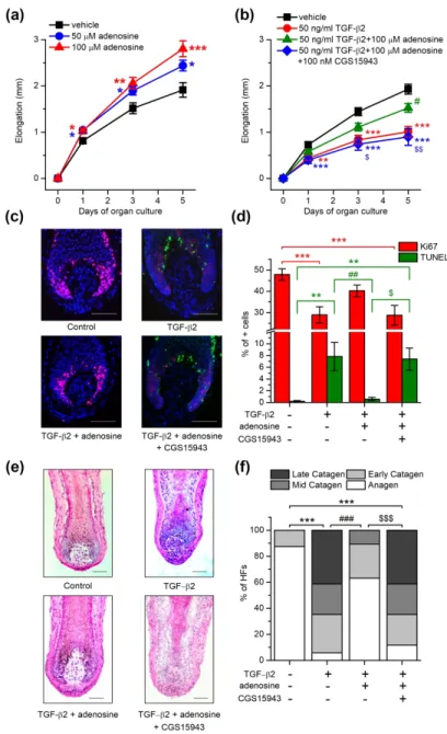
First, we treated microdissected human HFs with adenosine administered in the culturing medium for several days and measured the length of the HFs on the 1st, 3rd and 5th days. We found that the hair shaft elongation was stimulated in the presence of 50 or 100 µM adenosine (Figure 1a). These concentrations of adenosine increased cellular proliferation in HFs, especially in the matrix keratinocytes of the hair bulb as indicated by the increased number of the cells positive for the proliferation associated antigen Ki67 (Supplementary figure 1a-b). In good accordance with the above, adenosine slightly shifted the hair cycle by prolonging the duration of the anagen phase and inhibiting catagen entry: the ratio of HFs in the anagen stage was higher when 100 µM adenosine was added to the culture medium for 6 days, as assessed
by hair cycle staging based on the histomorphometric evaluation of the cultured HFs (Supplementary figure 1c-d).
Next, we wanted to further challenge the anagen-promoting effect of adenosine by the co-administration of the catagen inducer transforming growth factor β2 (TGF-β2) (Langan et al. 2015). As expected, TGF-β2 significantly inhibited hair growth, decreased the proliferation and induced the apoptosis of hair matrix keratinocytes in cultured HFs (Figure 21b-d). TGF-β2 also resulted in a striking catagen transition of the hair cycle and it practically abolished the anagen stage from the cultures in six days (Figure 21e-f, Supplementary Figure 2a-b). All these effects of TGF-β2 were mainly prevented by supporting the culture medium with 100 µM adenosine. Importantly, this blockade of TGF-β2 effect by adenosine was abrogated by the co- application of CGS15943, a pan-antagonist of ARs.
Therefore, we investigated the presence of ARs in human HFs. We isolated total mRNA from anagen human HFs from 3 donors, and determined the expression of ARs A1, A2A, A2B
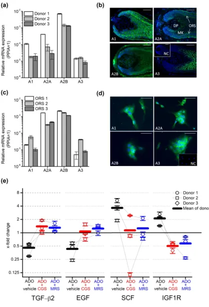
and A3 by Q-PCR following reverse transcription. We found that the transcripts of all four investigated receptors are expressed in HFs (Figure 2a) among which A2B was found to be the dominant isoform: its expression was at least 1 magnitude higher than any other isoform in the tested donors.
We studied the localization of AR proteins within the HF applying immunofluorescent labelling on frozen sections of isolated human anagen HFs (Figure 2b) using specific antibodies against ARs (Supplementary Figure 3). A1 showed a diffuse expression overall in the matrix and ORS keratinocytes, whereas A2A and A2B isoforms were highly expressed in the ORS and some cells in the dermal papilla in line with the previous results of Iino et al (2017) who investigated the expression of A2B in human scalp specimen and dermal papilla cells. The surrounding connective tissue sheath (CTS) was also positive for A2B and some A2A was also
detected in the inner root sheath (IRS). A3 was detected primarily in matrix keratinocytes above the Auber’s line, and a weak signal was observed in the ORS.
We isolated keratinocytes from the ORS, since immunofluorescence detected all ARs in that compartment and further studied the expression of ARs in the isolated cells. Q-PCR and immunofluorescent staining confirmed our results obtained in HF cultures: even in monolayer cultures, ORS keratinocytes expressed all four ARs, (Figure 2c-d.). Similar to HFs, the expression of the A2B transcripts was found to be higher than the expression of the other isoforms with the marked dominance of the A2B isoform (Figure 2c-d).
In our final experiments, we further dissected the potential mechanisms of adenosine on hair growth and the hair cycle. Since the ORS is well-known to synthesize both positive and negative paracrine regulators of hair growth and the hair cycle [e.g. TGF-β2, insulin like growth factor 1 (IGF-1) or stem cell factor/c-kit ligand (SCF), etc. (Bodó et al. 2005; Langan et al.
2015; Paus et al. 2014; Soma et al. 2002; Stenn and Paus 2001; Szabó et al. 2018)], we studied the effect of adenosine on the expression of selected, well established hair cycle regulators in ORS keratinocytes isolated from 3 donors. Detecting the specific mRNA transcripts, we found that adenosine downregulated the expression of the catagen inducing mediators TGF-β2 and epidermal growth factor. In contrast, expression of the anagen and pigmentation promoting mediator SCF was upregulated by adenosine treatment, as well as the expression of insulin like growth factor-1 receptor (IGF-1R) (Figure 2e). These alterations in gene expression induced by adenosine were observable in the samples of each donor. Importantly, the effect of adenosine was blocked by both the general AR receptor antagonist CGS15943 and MRS1754, an A2B
selective inhibitor, in case of each donor although the magnitude of the responses was variable among donors tested. These data demonstrated convincingly that adenosine can control the hair cycle via ARs expressed in human HFs. Moreover, our results also suggest that adenosine can
regulate complex intercellular signaling pathways in HFs acting on ARs (especially A2B) expressed by ORS keratinocytes beyond previously described dermal papilla cells.
Our findings describe the growth and anagen promoting effect of adenosine via ARs in human HFs and identify especially A2B expressed in ORS keratinocytes as a promising pharmacological target to influence various hair growth disorders, such as various forms alopecia.
CONFLICT OF INTEREST
MB is an employee of Monasterium Laboratory GmbH, TB and AO provide consultancy services to Phytecs Inc. (TB) and Botanix Pharmaceuticals Ltd. (AO). Botanix Pharmaceuticals Ltd., Phytecs Inc., Monasterium Laboratory GmbH, and the founding sponsors listed in the Acknowledgements section had no role in conceiving the study, designing the experiments, writing of the manuscript, or in the decision to publish it. Other authors declare no conflict of interest.
ACKNOWLEDGEMENTS
The presented work was supported by research grants of the National Research, Development and Innovation Office (K_120187, PD_121360, FK_125055, GINOP-2.3.2-15-2016-00050).
The project has received funding from the EU’s Horizon 2020 research and innovation program under grant agreement No. 739593. AO’s work was supported by the New National Excellence Program of the Ministry of Human Capacities (ÚNKP-18-4-DE-247). BIT, AO and AGSz are recipients of the János Bolyai research scholarship of the Hungarian Academy of Sciences.
CRediT STATEMENT
EL, BIT, AO, RP and TB conceptualized the project, EL, BIT, MB, ILSz, NZ, AO and AGSz, conducted the research presented in the manuscript. EL, BIT, and TB performed formal analysis of the results. The original draft was written by EL, BIT and BT whereas RP contributed significantly to the editing and review of the final version of the manuscript. BT was responsible for funding acquisition.
REFERENCES
Andrés RM, Terencio MC, Arasa J, Payá M, Valcuende-Cavero F, Navalón P, et al.
Adenosine A2A and A2B Receptors Differentially Modulate Keratinocyte Proliferation:
Possible Deregulation in Psoriatic Epidermis. J. Invest. Dermatol. 2017;137(1):123–31 Bodó E, Bíró T, Telek A, Czifra G, Griger Z, Tóth BI, et al. A hot new twist to hair biology:
involvement of vanilloid receptor-1 (VR1/TRPV1) signaling in human hair growth control.
Am. J. Pathol. 2005;166(4):985–98
Burnstock G, Knight GE, Greig AVH. Purinergic signaling in healthy and diseased skin. J.
Invest. Dermatol. 2012;132(3 Pt 1):526–46
Hwang K-A, Hwang Y-L, Lee M-H, Kim N-R, Roh S-S, Lee Y, et al. Adenosine stimulates growth of dermal papilla and lengthens the anagen phase by increasing the cysteine level via fibroblast growth factors 2 and 7 in an organ culture of mouse vibrissae hair follicles. Int. J.
Mol. Med. 2012;29(2):195–201
Iino M, Ehama R, Nakazawa Y, Iwabuchi T, Ogo M, Tajima M, et al. Adenosine stimulates fibroblast growth factor-7 gene expression via adenosine A2b receptor signaling in dermal papilla cells. J. Invest. Dermatol. 2007;127(6):1318–25
Iwabuchi T, Ideta R, Ehama R, Yamanishi H, Iino M, Nakazawa Y, et al. Topical adenosine increases the proportion of thick hair in Caucasian men with androgenetic alopecia. J.
Dermatol. 2016;43(5):567–70
Langan EA, Philpott MP, Kloepper JE, Paus R. Human hair follicle organ culture: theory, application and perspectives. Exp. Dermatol. 2015;24(12):903–11
Linden J. Adenosine in tissue protection and tissue regeneration. Mol. Pharmacol.
2005;67(5):1385–7
Oura H, Iino M, Nakazawa Y, Tajima M, Ideta R, Nakaya Y, et al. Adenosine increases anagen hair growth and thick hairs in Japanese women with female pattern hair loss: a pilot, double-blind, randomized, placebo-controlled trial. J. Dermatol. 2008;35(12):763–7
Paus R, Langan EA, Vidali S, Ramot Y, Andersen B. Neuroendocrinology of the hair follicle:
principles and clinical perspectives. Trends Mol. Med. 2014;20(10):559–70
Silva-Vilches C, Ring S, Schrader J, Clausen BE, Probst H-C, Melchior F, et al. Production of Extracellular Adenosine by CD73+ Dendritic Cells Is Crucial for Induction of Tolerance in Contact Hypersensitivity Reactions. J. Invest. Dermatol. 2019;139(3):541–51
Soma T, Tsuji Y, Hibino T. Involvement of Transforming Growth Factor-β2 in Catagen Induction During the Human Hair Cycle. J. Invest. Dermatol. 2002;118(6):993–7 Stenn KS, Paus R. Controls of hair follicle cycling. Physiol. Rev. 2001;81(1):449–94
Szabó IL, Herczeg-Lisztes E, Szegedi A, Nemes B, Paus R, Bíró T, et al. Transient Receptor Potential Vanilloid 4 is Expressed in Human Hair Follicles and Inhibits Hair Growth in Vitro.
J. Invest. Dermatol. 2018;
Watanabe Y, Nagashima T, Hanzawa N, Ishino A, Nakazawa Y, Ogo M, et al. Topical adenosine increases thick hair ratio in Japanese men with androgenetic alopecia. Int. J.
Cosmet. Sci. 2015;37(6):579–87
FIGURE LEGENDS
Figure 1. Adenosine enhanced HF elongation and prevented the hair growth-inhibiting and catagen-inducing effect of TGF-β2. (a) and (b) HF elongation curves. HFs were treated with vehicle (control) or with adenosine, TGF-β2, and adenosine receptor antagonist CGS15943 in combinations as indicated in the figure. Mean±SEM of N=36 (a) or N=28-29 (b) HFs/group from three individual donors. (c) Co-immunolabeling of proliferating (Ki-67+, red) and apoptotic (TUNEL+, green) cells along with nuclear staining (DAPI, blue). Representative images demonstrating the effect of the compounds indicated. (d) Statistical analysis of co- immunolabeled proliferating (Ki-67+) and apoptotic (TUNEL+) cells, following treatments with the indicated compounds applied in the same concentration as in panel (b) for 6 days.
Percentage of total cell number (DAPI+ nuclei). N=16-24 HFs of 3 donors/group. (e) Representative hematoxylin eosin-stainings showing the effect of the indicated compounds on the morphology of HFs. (f) Percentage of organ cultured HFs in anagen and catagen stages as determined by quantitative hair cycle histomorphometry based on hematoxylin eosin-stained sections. N=17-24 HFs/group from three different donors. *p<0.05, **p<0.01 and ***p<0.001 compared to control, #p<0.05, ##p<0.01 and ###p<0.001 compared to TGF-β2 treated group and
$p<0.05, $$p<0.01 and $$$p<0.001 compared to TGF-β2 and adenosine treated group as determined by One-way ANOVA and Dunnett (a) or Bonferroni post hoc tests (b) and (d), or compering the distribution of HFs in the different hair cycle stages pairwise by Fisher’s exact test (c). Scale bars:50 µm.
Figure 2. Adenosine Receptors (ARs) expressed in isolated human hair follicles (HFs) and cultured outer root sheath (ORS) keratinocytes controlled the expression of negative and positive hair cycle regulators in isolated ORS keratinocytes. (a) Expression of ARs’
transcripts in human HFs of three donors as assessed by Q-PCR. Data are expressed as
mean±SEM of three independent determinations. Data are normalized to the expression of cyclophilin A (PPIA) determined as endogenous control. (b) Immunofluorescent staining (FITC, green fluorescence) of ARs in organ-cultured human HFs. Nuclei were counterstained with 4’,6-diamidino-2’-phenylindole (DAPI, blue fluorescence). Scale bars represent 50 µm.
DP, dermal papilla; MK, matrix keratinocytes; ORS, outer root sheath. Inset: negative control (NC). (c) Expression of ARs’ transcripts in cultured ORS keratinocytes determined by Q- PCR as on panel (a). (d) Immunofluorescent staining (FITC, green fluorescence) of ARs in cultured human ORS keratinocytes. Scale bars represent 10 µm. Inset: negative control (NC). (e) ORS keratinocytes were isolated from 3 individual donors and cultured in the presence of 100 µM adenosine and adenosine receptor antagonist CGS15943 (CGS, 100 nM) or A2B selective blocker MRS1754 (MRS, 10 nM) as indicated for 3 hrs. Relative gene expression was determined and normalized to the expression values of the vehicle-treated (i.e. adenosine and inhibitor free) samples counted as 1 in each individual donor. Values of individual donors are presented as mean±SEM of three independent determinations. As endogenous control, the expression of PPIA was determined.
Figure 1: Adenosine enhanced HF elongation and prevented the hair growth-inhibiting and catagen-inducing effect of TGF-β2
199x320mm (300 x 300 DPI)
Figure 2: Adenosine Receptors (ARs) expressed in isolated human hair follicles (HFs) and cultured outer root sheath (ORS) keratinocytes controlled the expression of negative and positive hair cycle regulators in
isolated ORS keratinocytes.
199x299mm (300 x 300 DPI)
Supplementary material to the MS
ADENOSINE PROMOTES HUMAN HAIR GROWTH AND INHIBITS CATAGEN TRANSITION IN VITRO – ROLE OF THE OUTER ROOT SHEATH
KERATINOCYTES
by
Erika Lisztes1,2*(ORCID: 0000-0002-8517-6536), Balázs István Tóth3*(ORCID: 0000-0002- 4103-4333), Marta Bertolini4,5(ORCID: 0000-0002-5927-6998.), Imre Lőrinc Szabó3(ORCID:
0000-0002-9628-4372), Nóra Zákány3(ORCID: 0000-0003-4239-6106), Attila Oláh3 (ORCID: 0000-0003-4122-5639), Attila Gábor Szöllősi1,3 (ORCID: 0000-0001-6046-8236), Ralf Paus6,7(ORCID: 0000-0002-3492-9358), Tamás Bíró1,8(ORCID: 0000-0002-3770-6221)
1DE-MTA ‘Lendület’ Cellular Physiology Research Group, Department of Immunology, Faculty of Medicine, University of Debrecen, Debrecen, Hungary
2Doctoral School of Molecular Medine, Faculty of Medicine, University of Debrecen, Debrecen, Hungary
3Department of Physiology, Faculty of Medicine, University of Debrecen, Debrecen, Hungary
4Department of Dermatology, University of Münster, Münster, Germany
5Monasterium Laboratory - Skin and Hair Research Solutions GmbH, Münster, Germany
6Department of Dermatology & Cutaneous Surgery, University of Miami Miller School of Medicine, Miami, FL, USA
7Centre for Dermatology Research, University of Manchester, Manchester, UK
8Hungarian Center of Excellence for Molecular Medicine, Szeged, Hungary
SUPPLEMENTARY METHODS
Isolation and Maintenance of Hair Follicles (HFs):
Human skin samples were obtained from healthy individuals undergoing neurosurgery with written informed consent, adhering to Helsinki guidelines, and after obtaining the permission of the Institutional Research Ethics Committee’s and the Government Office for Hajdú-Bihar County (protocol No.: DE OEC RKEB/IKEB 3724-2012; document IDs: IX-R- 052/01396-2/2012, IF-12817/2015, IF-1647/2016, IF-778-5/2017). Human anagen VI HFs were isolated from the skin of male donors and maintained, as we have described before (Bodó et al. 2005; Telek et al. 2007; Bodó et al. 2009; Ramot et al. 2010; Borbíró et al. 2011; Szabó et al. 2018; Oláh et al. 2016). Briefly, isolated HFs were collected and maintained in Williams’
E medium (Life Technologies Corporation, Foster City, CA, USA) supplemented with 2 mM L-glutamine (Life Technologies), 10 ng/ml hydrocortisone, 10 mg/ml insulin, and antibiotics (all from Sigma- Aldrich, St. Louis, MO, USA). Culture medium was changed every other day, whereas treatment with various compounds was performed daily. For immunofluorescent staining and histomorphometry, follicles were frozen at -80 °C and further processed after 6 days in culture.
Culturing of Human HF-derived Outer Root Sheath Keratinocytes
Plucked human scalp HFs of several male volunteers were digested using trypsin to obtain outer root sheath (ORS) keratinocytes (Ramot et al. 2018). Similarly, human dermal fibroblasts (HDFs) were obtained from de-epidermized dermis of human skin samples using enzymatic digestion. ORS keratinocyte cultures were kept on feeder layer of non-proliferating HDFs treated with mitomycin C (Sigma-Aldrich) in a 1:3 mixture of supplemented Ham’s F12 and Dulbecco’s modified Eagle’s medium (both from Life Technologies) supplemented with 10%
Fetal Clone II (HyClone, South Logan, UT, USA), 0.1 nM cholera toxin, 5 μg/ml insulin, 0.4
μg/ml hydrocortisone, 2.43 μg/ml adenine, 2 nM triiodothyronine, 10 ng/ml epidermal growth factor, 1 mM ascorbyl-2-phosphate and antibiotics (all from Sigma-Aldrich) as described previously (Bodó et al. 2005; Borbíró et al. 2011; Ramot et al. 2018; Szabó et al. 2018; Telek et al. 2007).
Before experiments, ORS keratinocytes were harvested, and re-plated without a HDF feeder layer in 6-well plates (200,000 cells/well) which were previously coated with 1%
collagen (Sigma Aldrich) and kept in serum-free medium for 24 hours before treating them with different compounds. 3 hours after treatment cells were harvested using TRIzol (Life Technologies) and then RT-qPCR were performed as described below.
Measurement of Hair Shaft Elongation
Length measurements on individual cultured HFs were performed using a light microscope with an eyepiece measuring graticule. Elongation was calculated for each hair follicle separately, by subtracting the length measured on day 0 from the value of the relevant day.
Quantitative “Real-Time” PCR
Total RNA was isolated using TRIzol reagent (Life Technologies) and digested with recombinant RNase-free DNase-1 (Life Technologies) according to the manufacturer’s protocol. After isolation, 1 μg of total RNA was reverse-transcribed into cDNA using the High Capacity cDNA kit (Life Technologies) following the manufacturer’s instructions.
Quantitative real-time PCR was performed on a Stratagene Mx3005p sequence detection system (Agilent Technologies Inc., Santa Clara, CA, USA) by using 5’ nuclease assay. PCR amplification was performed using specific TaqMan primers and probes as follows; for
adenosine A1 receptor (ADORA1, Assay ID: Hs00379752_m1); for adenosine A2A receptor (ADORA2A, Assay ID: Hs00169123_m1); for adenosine A2B receptor (ADORA2B, Assay ID:
Hs00386497_m1); for adenosine A3 receptor (ADORA3, Assay ID: Hs00252933_m1); for transforming growth factor beta 2 (TGFB2, Assay ID: Hs00234244_m1); for epidermal growth factor (EGF, Assay ID: Hs01099999_m1); for stem cell factor (SCF/KITLG, assay ID:
Hs00241497_m1) and for insulin-like growth factor 1 receptor (IGF1R, Assay ID:
Hs00609566_m1) using the TaqMan Gene Expression Master Mix Protocol (Life Technologies). As internal control, transcripts of cyclophilin A (PPIA, Assay ID:
Hs99999904_m1) were determined. The amount of the above mentioned transcripts was normalized to the expression of the internal control gene, using the ΔCt method. Briefly: the threshold cycle (Ct) value of the target gene was subtracted from the average Ct value of the control gene resulting in the ΔCt value. ΔCt was then used as a power of two, which results in the relative expression of a given target gene compared to the control (i.e.: 2ΔCt). All experiments were performed in triplicates.
Immunolabeling of ARs
To detect the four type of ARs on isolated HFs and ORS keratinocytes, we performed indirect fluorescent immunolabeling. Cryosections of isolated HFs fixed with ice-cold ethanol:acetic acid (2:1) or acetone-fixed ORS keratinocytes grown on coverslips were first incubated with different primary rabbit antibodies (1:100 in DCS antibody diluent [DCS Innovative Diagnostik-Systeme, Hamburg, Germany] overnight, 4 °C) against A1, A2A (Abcam, Cambridge, UK, cat. numbers: ab124780 and ab3461, respectively), A2B and A3 (Alomone Labs, Jerusalem, Israel, cat. numbers: AAR-003 and AAR-004, respectively) receptors.
Sections and coverslips were then washed with phosphate-buffered saline (PBS), followed by incubation with Alexa Fluor 488 dye-conjugated goat anti-rabbit IgG (Life Technologies)
(1:500 in DCS antibody diluent, 45 min) at room temperature according to standard procedures.
Nuclei were counterstained with 4’,-6-diamidino-2-phenylindole (DAPI) (Life Technologies) (1 µg/ml in distilled water, 5 min), and sections were mounted with Fluoromount-G aqueous medium (Southern Biotech, Birmingham, USA). Images were acquired using an Eclipse E600 fluorescent microscope (Nikon, Tokyo, Japan). To verify the specificity of the antibodies used, paraffin embedded routine histology sections from tissues known to express different ARs were stained as positive controls. Human cerebral cortex served as positive control for A1 and A2A
(Latini et al. 1996; Luan et al. 2017; Svenningsson et al. 1997), human kidney for A2B (Zhang et al. 2013) and human cerebellum for A3 (Haeusler et al. 2015). Following deparaffination and antigen retrieval (in citrate-buffer, pH 6.0, at 750 W in microwave oven for 15 min), sections were incubated with the above primary rabbit antibodies against human ARs, then stained with HRP conjugated anti-rabbit IgG (1:500) (Bio-Rad, Hercules, CA, USA). Immunoreactions were visualized using DAB substrate kit (Vector Labs, Burlingame, California USA) and the sections were counterstained by hematoxylin (Sigma-Aldrich). For all immunostainings, the respective primary antibodies were omitted as negative controls.
Ki-67/TUNEL Double Labeling
To simultaneously detect proliferating and apoptotic cells in the HFs, Ki-67 immunolabeling and terminal dUTP nick end labeling (TUNEL) were performed in a double- staining protocol, respectively (Bodó et al. 2005; Borbíró et al. 2011; Langan et al. 2015; Purba et al. 2016; Szabó et al. 2018; Telek et al. 2007). Cryosections were fixed in formalin/ethanol/acetic acid and labeled with a digoxigenin-deoxyUTP (ApopTag Fluorescein In Situ Apoptosis detection kit; Millipore, Billerica, MA) in presence of terminal deoxynucleotidyl transferase (60 min, 4 °C) according to the manufacturers protocol, followed by overnight incubation with a mouse anti-Ki-67 antiserum (1:20, DAKO, Carpinteria, CA) at
4 °C. TUNEL+ cells were visualized by an anti-digoxigenin FITC-conjugated antibody (ApopTag kit), whereas Ki-67 was detected by an Alexa Fluor 568 dye-conjugated secondary antibody (Life Technologies, 1:500 at 4 °C for 45 min). Negative control stainings were performed by omitting terminal deoxynucleotidyl transferase and the Ki-67 antibody (data not shown). Cells positive for Ki-67 or TUNEL were counted per hair bulb (under the cross- sectional line perpendicular for the longitudinal axis of the hair shaft and tangential to the peak of the dermal papilla) and were normalized to the number of nuclei (DAPI+).
Histology and Quantitative Histomorphometry
Cryosections (6 µm) of cultured HFs were fixed in acetone, air-dried, and processed for routine histology. Hematoxylin and eosin (HE, Sigma-Aldrich) staining was used for studying HF morphology and hair cycle stage (anagen and different stages of catagen) of each HF was assessed according to defined morphological criteria (Kloepper et al. 2010; Langan et al. 2015).
Additionally, number of DAPI+ cells in a standardized area of the dermal papilla (DP) stalk was counted on Ki-67/TUNEL double labeled sections to further characterize hair cycle quantitatively (Kloepper et al. 2010).
Statistical Analysis
If not mentioned otherwise, values are presented as mean±SEM in every group. To compare the mean values of multiple groups, statistical analysis was subsequently performed by One-way ANOVA and Dunnett or Bonferroni post hoc tests, as appropriate. Significance was determined as *p<0.05, **p<0.01, ***p<0.001 compared to the control and/or $ or #p<0.05,
$$ or ##p<0.01, $$$ or ###p<0.001 compared to different treated samples as indicated. Differences in distribution of HFs among different hair cycle stages was compared pairwise by Fisher’s
exact test. Origin 9.0 (OriginLab Corporation, Northampton MA, USA) and IBM SPSS Statistics 23.0 (IBM Corporation. Armonk, NY, USA) were used to plot the data and perform statistical analysis, respectively.
Materials
Adenosine, CGS15943 (non-selective AR antagonist), MRS1754 (selective A2B
antagonist) and TGF-β2 were obtained from Sigma-Aldrich.
DATA AVAILABILITY STATEMENT
Datasets related to this article are freely available upon request. Requests should be addressed to the corresponding author.
SUPPLEMENTARY TEXT: DISCUSSION OF THE POTENTIAL MOLECULAR MECHANISMS UNDERLYING THE REGULATION OF HAIR GROWTH BY ADENOSINE
The favorable action of adenosine is supported by a growing body of evidence in HF biology but the potential mechanism of action has not been resolved yet. To get deeper insight into the cellular and molecular mechanisms of how adenosine can enhance human hair growth, we studied the effect of adenosine in an in vitro model of human hair growth using microdissected and organ cultured human HFs (Langan et al. 2015; Philpott et al. 1994). In good accordance with the previous clinical findings, we quantitatively measured that adenosine enhanced the hair shaft elongation in human HF cultures isolated from Caucasian male subjects in vitro. As a potential underlying mechanism, we found increased intrafollicular proliferation and also observed that the ratio of HFs in catagen stage was decreased and more HFs showed morphological signs characteristic for the growing anagen phase in the adenosine treated cultures. These results suggested that adenosine may have a regulatory effect on hair cycle. To explore how adenosine can influence the hair cycle, we induced the onset of the regressive catagen phase by adding TGF-β2 to the culture medium. Adenosine abolished the effects of TGF-β2: it prevented diminished hair growth, it reversed reduced intrafollicular proliferation and augmented apoptosis induced by TGF-β2, as well as inhibited catagen transition and kept the anagen morphology of the HFs. These findings strongly support that adenosine can have a significant impact on hair cycle regulation. Importantly, all the above effects of adenosine were inhibited by the general AR antagonist CGS15943 arguing for the specific role of adenosine related signaling in the hair growth control.
A complex molecular interaction between mesenchymal and epithelial cells of the HFs, e.g. dermal papilla cells and keratinocytes of the different layers, plays a crucial role in the regulation of hair cycle (Schneider et al. 2009; Stenn and Paus 2001). Although Wnt/β-catenin
signaling and several additional factors regulating the transition between the individual phases are relatively well-documented in the literature, the exact origin of the cyclic program and all the molecular details of the intercellular interactions are not resolved as of yet (Bernard 2012).
Earlier studies identified several positive regulator molecules of the hair cycle which promote hair growth and prolong anagen phase of hair follicle cultures or upregulate melanogenesis linked to anagen phase, e.g. β-catenin, IGF-I, fibroblast growth factor 7/keratinocyte growth factor (FGF7/KGF) or stem cell factor/KIT-ligand (SCF). On the other hand, negative hair cycle regulators like epidermal growth factor (EGF), TGF-β1, FGF5, and interferon gamma can initiate catagen transition and inhibit hair shaft elongation in vitro. Importantly, the expression of these regulatory molecules and their receptors are widely documented in various mesenchymal and epithelial components of the HFs providing an extended paracrine regulatory network for hair cycle and growth control (Langan et al. 2015; Paus et al. 2014; Stenn and Paus 2001). The modulation of these paracrine mechanisms regulating the hair cycle may be an effective tool to influence hair growth. For example, activation of β-catenin signaling activated a telogen to anagen transition and induced intense growth and intrafollicular proliferation of the epithelial components in HFs (Choi et al. 2013; Van Mater et al. 2003). Importantly, an intimate relationship is suggested between adenosine and Wnt/β-catenin signaling in the skin. It was shown that A2A promotes collagen type III synthesis via β-catenin activation in human dermal fibroblasts (Shaikh et al. 2016) and pharmacological blockade of A2A diminished the activity of Wnt/β-catenin pathway in a bleomycin-induced dermal fibrosis mouse model (Zhang et al.
2017). Moreover, Wnt/β-catenin signaling can promote extracellular adenosine generation via upregulating ecto-5'-nucleotidase and downregulating adenosine deaminase in Rat-1 cell line (Spychala and Kitajewski 2004). These data suggest that Wnt/β-catenin pathway can be a downstream target of adenosine receptor signalling as well as can act as a regulator of adenosine production i.e upstream of adenosine receptors. Moreover, in recent studies, adenosine was
reported to increase the expression of FGF2, FGF7, IGF1 and VEGF in cultured dermal papilla fibroblasts (Hwang et al. 2012). In human dermal papilla cell cultures, the upregulation of FGF7 by adenosine was abolished in the presence of the A2B antagonist alloxazine. In good accordance, A2B receptors were also detected by immunohistochemistry in the dermal papilla and outer root sheath of human hair follicles (Iino et al. 2007). In our study, we detected all AR subtypes in human cultured HFs. Importantly, our quantitative gene expression data also verified the A2B as the dominantly expressed AR in the HF. The expression pattern was similar to that found by Iino et al. (Iino et al. 2007): beyond the dermal papilla, the outer root sheath showed strong positivity for A2B receptors. This high expression was maintained in isolated outer root sheath keratinocytes in vitro, as well.
Treating the ORS keratinocyte cultures with adenosine, we revealed a marked alteration in the expression of hair cycle regulating factors: the receptor of the positive hair cycle regulator IGF1 and the anagen related pigmentation promoting SCF were upregulated, but the strong catagen inducer TGF-β2 and EGF were downregulated. These results suggest that the anagen and hair growth promoting effect of adenosine, next to the previously suggested dermal papilla cells, may be mediated by the outer root sheath, as well. These results suggest that adenosine can generally affect both sides of the local mesenchymal-epithelial paracrine communication in the HF.
Importantly, HFs seems not to be only “passive” targets of adenosine, but might produce adenosine locally, although this likely assumption still requires further experimental support.
However, earlier results suggested that a local, intrafollicular adenosine system can mediate the effect of hair growth promoting drugs. Minoxidil, a well-known hair growth promoting compound generally used to treat alopecia (Goren and Naccarato 2018), was shown to increase VEGF production in dermal papilla cells which effect was mimicked by adenosine and attenuated by inhibitors of A1 and A2 receptors expressed by dermal papilla cells suggesting
that local adenosine production mediates the beneficial effect of minoxidil on hair growth (Li et al. 2001). Although the mechanism of the local adenosine production is not known yet, a recent study reported expression of connexin and pannexin channels in the keratinocytes of the hair follicles (Cowan et al. 2012) which channels are known to release ATP (Lazarowski 2012), the precursor of extracellular adenosine synthesis by ectonucelotidases ecto-apyrase (CD39) and ecto-5’-nucleotidase (CD73) (Zimmermann 2000).
Although the detailed description of the intrafollicular adenosine system needs further studies, our results provide a deeper insight into the mechanisms on how adenosine can promote hair growth via increased intrafollicular proliferation and inhibition of catagen transition.
Moreover, our findings highlighted the potential role of outer root sheath keratinocytes and their adenosine receptors as a target of exogenously applied or endogenously produced adenosine. These results underline the role of the intrafollicular adenosine signaling as a potential therapeutic target to treat hair loss-associated diseases.
SUPPLEMENTARY REFERENCES
Bernard BA. The human hair follicle, a bistable organ? Experimental Dermatology.
2012;21(6):401–3
Bodó E, Bíró T, Telek A, Czifra G, Griger Z, Tóth BI, et al. A hot new twist to hair biology:
involvement of vanilloid receptor-1 (VR1/TRPV1) signaling in human hair growth control.
Am. J. Pathol. 2005;166(4):985–98
Bodó E, Kromminga A, Bíró T, Borbíró I, Gáspár E, Zmijewski MA, et al. Human female hair follicles are a direct, nonclassical target for thyroid-stimulating hormone. J. Invest.
Dermatol. 2009;129(5):1126–39
Borbíró I, Lisztes E, Tóth BI, Czifra G, Oláh A, Szöllosi AG, et al. Activation of transient receptor potential vanilloid-3 inhibits human hair growth. J. Invest. Dermatol.
2011;131(8):1605–14
Choi YS, Zhang Y, Xu M, Yang Y, Ito M, Peng T, et al. Distinct Functions for Wnt/β-Catenin in Hair Follicle Stem Cell Proliferation and Survival and Interfollicular Epidermal
Homeostasis. Cell Stem Cell. 2013;13(6):720–33
Cowan KN, Langlois S, Penuela S, Cowan BJ, Laird DW. Pannexin1 and Pannexin3 exhibit distinct localization patterns in human skin appendages and are regulated during keratinocyte differentiation and carcinogenesis. Cell Commun. Adhes. 2012;19(3–4):45–53
Goren A, Naccarato T. Minoxidil in the treatment of androgenetic alopecia. Dermatol Ther.
2018;31(5):e12686
Haeusler D, Grassinger L, Fuchshuber F, Hörleinsberger WJ, Höftberger R, Leisser I, et al.
Hide and seek: a comparative autoradiographic in vitro investigation of the adenosine A3 receptor. Eur. J. Nucl. Med. Mol. Imaging. 2015;42(6):928–39
Hwang K-A, Hwang Y-L, Lee M-H, Kim N-R, Roh S-S, Lee Y, et al. Adenosine stimulates growth of dermal papilla and lengthens the anagen phase by increasing the cysteine level via fibroblast growth factors 2 and 7 in an organ culture of mouse vibrissae hair follicles.
International Journal of Molecular Medicine. 2012;29(2):195–201
Iino M, Ehama R, Nakazawa Y, Iwabuchi T, Ogo M, Tajima M, et al. Adenosine stimulates fibroblast growth factor-7 gene expression via adenosine A2b receptor signaling in dermal papilla cells. J. Invest. Dermatol. 2007;127(6):1318–25
Kloepper JE, Sugawara K, Al-Nuaimi Y, Gáspár E, van Beek N, Paus R. Methods in hair research: how to objectively distinguish between anagen and catagen in human hair follicle organ culture. Exp. Dermatol. 2010;19(3):305–12
Langan EA, Philpott MP, Kloepper JE, Paus R. Human hair follicle organ culture: theory, application and perspectives. Exp. Dermatol. 2015;24(12):903–11
Latini S, Pazzagli M, Pepeu G, Pedata F. A2 adenosine receptors: their presence and neuromodulatory role in the central nervous system. Gen. Pharmacol. 1996;27(6):925–33
Lazarowski ER. Vesicular and conductive mechanisms of nucleotide release. Purinergic Signal. 2012;8(3):359–73
Li M, Marubayashi A, Nakaya Y, Fukui K, Arase S. Minoxidil-induced hair growth is mediated by adenosine in cultured dermal papilla cells: possible involvement of sulfonylurea receptor 2B as a target of minoxidil. J. Invest. Dermatol. 2001;117(6):1594–600
Luan G, Wang X, Gao Q, Guan Y, Wang J, Deng J, et al. Upregulation of Neuronal Adenosine A1 Receptor in Human Rasmussen Encephalitis. J. Neuropathol. Exp. Neurol.
2017;76(8):720–31
Oláh A, Gherardini J, Bertolini M, Chéret J, Ponce L, Kloepper J, et al. The Thyroid Hormone Analogue KB2115 (Eprotirome) Prolongs Human Hair Growth (Anagen) Ex Vivo. J. Invest.
Dermatol. 2016;136(8):1711–4
Paus R, Langan EA, Vidali S, Ramot Y, Andersen B. Neuroendocrinology of the hair follicle:
principles and clinical perspectives. Trends Mol Med. 2014;20(10):559–70
Philpott MP, Sanders D, Westgate GE, Kealey T. Human hair growth in vitro: a model for the study of hair follicle biology. J. Dermatol. Sci. 1994;7 Suppl:S55-72
Purba TS, Brunken L, Hawkshaw NJ, Peake M, Hardman J, Paus R. A primer for studying cell cycle dynamics of the human hair follicle. Exp. Dermatol. 2016;25(9):663–8
Ramot Y, Alam M, Oláh A, Bíró T, Ponce L, Chéret J, et al. Peroxisome Proliferator- Activated Receptor-γ-Mediated Signaling Regulates Mitochondrial Energy Metabolism in Human Hair Follicle Epithelium. J. Invest. Dermatol. 2018;138(7):1656–9
Ramot Y, Bíró T, Tiede S, Tóth BI, Langan EA, Sugawara K, et al. Prolactin--a novel
neuroendocrine regulator of human keratin expression in situ. FASEB J. 2010;24(6):1768–79 Schneider MR, Schmidt-Ullrich R, Paus R. The hair follicle as a dynamic miniorgan. Curr.
Biol. 2009;19(3):R132-142
Shaikh G, Zhang J, Perez-Aso M, Mediero A, Cronstein B. Adenosine A2A receptor
promotes collagen type III synthesis via β-catenin activation in human dermal fibroblasts. Br.
J. Pharmacol. 2016;173(23):3279–91
Spychala J, Kitajewski J. Wnt and beta-catenin signaling target the expression of ecto-5’- nucleotidase and increase extracellular adenosine generation. Exp. Cell Res. 2004;296(2):99–
108
Stenn KS, Paus R. Controls of hair follicle cycling. Physiol. Rev. 2001;81(1):449–94
Svenningsson P, Hall H, Sedvall G, Fredholm BB. Distribution of adenosine receptors in the postmortem human brain: an extended autoradiographic study. Synapse. 1997;27(4):322–35 Szabó IL, Herczeg-Lisztes E, Szegedi A, Nemes B, Paus R, Bíró T, et al. Transient Receptor Potential Vanilloid 4 is Expressed in Human Hair Follicles and Inhibits Hair Growth in Vitro.
J. Invest. Dermatol. 2018;
Telek A, Bíró T, Bodó E, Tóth BI, Borbíró I, Kunos G, et al. Inhibition of human hair follicle growth by endo- and exocannabinoids. FASEB J. 2007;21(13):3534–41
Van Mater D, Kolligs FT, Dlugosz AA, Fearon ER. Transient activation of beta -catenin signaling in cutaneous keratinocytes is sufficient to trigger the active growth phase of the hair cycle in mice. Genes Dev. 2003;17(10):1219–24
Zhang J, Corciulo C, Liu H, Wilder T, Ito M, Cronstein B. Adenosine A2a Receptor Blockade Diminishes Wnt/β-Catenin Signaling in a Murine Model of Bleomycin-Induced Dermal Fibrosis. Am. J. Pathol. 2017;187(9):1935–44
Zhang W, Zhang Y, Wang W, Dai Y, Ning C, Luo R, et al. Elevated ecto-5’-nucleotidase- mediated increased renal adenosine signaling via A2B adenosine receptor contributes to chronic hypertension. Circ. Res. 2013;112(11):1466–78
Zimmermann H. Extracellular metabolism of ATP and other nucleotides. Naunyn Schmiedebergs Arch. Pharmacol. 2000;362(4–5):299–309
SUPPLEMENTARY FIGURE LEGENDS
Supplementary Figure 1. Adenosine enhanced intrafollicular proliferation and prolonged anagen phase in human HF organ cultures. (a) Co-immunolabeling of proliferating (Ki67+, red) and apoptotic (TUNEL+, green) cells along with nuclear staining (DAPI, blue). Auber’s line is indicated through the bulbs. Scale bars represent 50 µm. (b) Mean ratio of Ki67+ cells as percentage of the total cell count (DAPI+ cells) in the bulb, N=25-31 HFs/group from three donors, *p<0.05, between the indicated groups by One-way ANOVA and Bonfferroni post hoc test. (c) Representative histological (hematoxylin-eosin) images illustrating the morphological changes (d) Percentage of organ cultured HFs in anagen and catagen stages as determined by quantitative hair cycle histomorphometry based on hematoxylin eosin-stained sections. N=25- 31 HFs/group from three donors.
Supplementary Figure 2. Adenosine prevents the effect of TGFβ2 as demonstrated by the decrease of the number of cells in the DP stalk, a quantitative marker of hair cycle- staging. (a) Representative images illustrating the region in which the number of DAPI+ nuclei were analyzed. In catagen stage, more DAPI+ cells migrate to the DP stalk. (b) Quantitative analysis on the number of cells in the DP stalk. N=17-24 HFs/group, **p<0.01 and ***p<0.001 comparing the indicated groups as determined by One-way ANOVA and Bonferroni post hoc tests (b).
Supplementary Figure 3. Specificity of the antibodies used to detect human ARs via immunolabaling. Various tissues described in the cited literature as expressing ARs were labeled with the primary antibodies used to stain ARs in HFs and ORS keratinocytes. (a) A1
stained in human cerebral cortex. Pyr.c.,: Pyramidal cell (b) A2a stained in human cerebral cortex. (c) A2b stained in human kidney. gl.: glomerulus, PCT: proximal tubule, DCT: distal tubule. (d) A3 stained in human cerebellar cortex. mol.lay.: molecular layer, gr.lay.: granular layer, Pur.c.: Purkinje cell. NC: negative control.
SUPPLEMENTARY FIGURE 1
40 50 60
(c)
control
adenosine adenosine50 M
Ki67+ cells (%)
100 M
*
control 50 M adenosine
100 M adenosine
(a)
(b)
0 25 50 75 100
catagen anagen
control
(d) Early Catagen
Anagen
% of HFs
Late Catagen Mid Catagen
control 50 M
adenosine adenosine100 M
control 50 M
adenosine
100 M adenosine
SUPPLEMENTARY FIGURE 2
0 5 10 15 20 25 30 35
+ CGS15943 TGF-2+adenosine
(b)
**
***
***
- -
Number of cells in DP stalk
50 ng/ml TGF-2 100 mM adenosine 100 nM CGS15943
-
- - + -
- -
- + +
+ + + (a) ***
vehicle TGF-2
TGF-2+adenosine
SUPPLEMENTARY FIGURE 3
TITLE PAGE
ADENOSINE PROMOTES HUMAN HAIR GROWTH AND INHIBITS CATAGEN TRANSITION IN VITRO – ROLE OF THE OUTER ROOT SHEATH
KERATINOCYTES
Erika Lisztes1,2*(ORCID: 0000-0002-8517-6536), Balázs István Tóth3*(ORCID: 0000-0002- 4103-4333), Marta Bertolini,4,5(ORCID: 0000-0002-5927-6998.), Imre Lőrinc Szabó3(ORCID: 0000-0002-9628-4372), Nóra Zákány3(ORCID: 0000-0003-4239-6106), Attila Oláh3(ORCID: 0000-0003-4122-5639), Attila Gábor Szöllősi1,3(ORCID: 0000-0001-
6046-8236), Ralf Paus6,7(ORCID: 0000-0002-3492-9358), Tamás Bíró1,8(ORCID: 0000- 0002-3770-6221)
1DE-MTA ‘Lendület’ Cellular Physiology Research Group, Department of Immunology, Faculty of Medicine, University of Debrecen, Debrecen, Hungary
2Doctoral School of Molecular Medicine, Faculty of Medicine, University of Debrecen, Debrecen, Hungary
3Department of Physiology, Faculty of Medicine, University of Debrecen, Debrecen, Hungary
4Department of Dermatology, University of Münster, Münster, Germany
5Monasterium Laboratory - Skin and Hair Research Solutions GmbH, Münster, Germany
6Department of Dermatology & Cutaneous Surgery, University of Miami Miller School of Medicine, Miami, FL, USA
7Centre for Dermatology Research, University of Manchester, Manchester, UK
8Hungarian Center of Excellence for Molecular Medicine, Szeged, Hungary
*these authors contributed equally to the work Correspondence to:
Tamás Bíró, MD, PhD, DSc
Department of Immunology, Faculty of Medicine University of Debrecen
H-4032 Debrecen, Egyetem tér 1.
Office Phone: +36 52 411-717/65242 FAX: +36 52 417-159
Email: biro.tamas@med.unideb.hu
Word count in main text: 1062 (without citations: 995) No. of references: 15
Displayed items: 2 Supplementary file: 1
Adenosine is a locally produced mediator exerting several cytoprotective effects via G-protein coupled cell membrane adenosine receptors (ARs) (Linden 2005). In the skin, adenosine can influence several (patho)physiological processes, such as wound healing, development of scleroderma, cutaneous inflammation, allergic reactions or barrier formation (Andrés et al.
2017; Burnstock et al. 2012; Silva-Vilches et al. 2019). A beneficial effect of adenosine on hair growth has already been reported in clinical studies: topical adenosine treatment was shown to alleviate the symptoms of alopecia by increasing hair thickness and promoting anagen hair growth (Iwabuchi et al. 2016; Oura et al. 2008; Watanabe et al. 2015); whereas adenosine also prolonged the anagen phase of mouse vibrissae cultures (Hwang et al. 2012). In parallel experiments, it upregulated the expression of fibroblast growth factor 7 (FGF-7) via activating A2B AR and stimulated the transcription of other growth factors in human dermal papilla (DP) cell cultures (Hwang et al. 2012; Iino et al. 2007). However, the influence of adenosine on the mechanisms of hair growth is not completely understood, and the role of ARs expressed in the different compartments of hair follicles (HFs) is not known in details. Therefore, we addressed the role of adenosine and ARs in hair cycle control studying isolated human HFs and primary outer root sheath (ORS) keratinocytes in vitro as described in the Supplementary methods.
Human HFs were isolated from human skin samples obtained from healthy individuals undergoing neurosurgery with written informed consent, adhering to Helsinki guidelines, and after obtaining the permission of the Institutional Research Ethics Committee’s and the Government Office for Hajdú-Bihar County (protocol No.: DE OEC RKEB/IKEB 3724-2012;
document IDs: IX-R-052/01396-2/2012, IF-12817/2015, IF-1647/2016, IF-778-5/2017).
First, we treated microdissected human HFs with adenosine administered in the culturing medium for several days and measured the length of the HFs on the 1st, 3rd and 5th days. We found that the hair shaft elongation was stimulated in the presence of 50 or 100 µM adenosine (Figure 1a). These concentrations of adenosine increased cellular proliferation in HFs,
especially in the matrix keratinocytes of the hair bulb as indicated by the increased number of the cells positive for the proliferation associated antigen Ki67 (Supplementary figure 1a-b). In good accordance with the above, adenosine slightly shifted the hair cycle by prolonging the duration of the anagen phase and inhibiting catagen entry: the ratio of HFs in the anagen stage was higher when 100 µM adenosine was added to the culture medium for 6 days, as assessed by hair cycle staging based on the histomorphometric evaluation of the cultured HFs (Supplementary figure 1c-d).
Next, we wanted to further challenge the anagen-promoting effect of adenosine by the co-administration of the catagen inducer transforming growth factor β2 (TGF-β2) (Langan et al. 2015). As expected, TGF-β2 significantly inhibited hair growth, decreased the proliferation and induced the apoptosis of hair matrix keratinocytes in cultured HFs (Figure 1b-d). TGF-β2 also resulted in a striking catagen transition of the hair cycle and it practically abolished the anagen stage from the cultures in six days (Figure 1e-f, Supplementary Figure 2a-b). All these effects of TGF-β2 were mainly prevented by supporting the culture medium with 100 µM adenosine. Importantly, this blockade of TGF-β2 effect by adenosine was abrogated by the co- application of CGS15943, a pan-antagonist of ARs.
Therefore, we investigated the presence of ARs in human HFs. We isolated total mRNA from anagen human HFs from 3 donors, and determined the expression of ARs A1, A2A, A2B
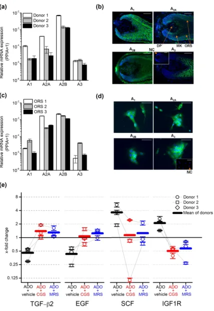
and A3 by Q-PCR following reverse transcription. We found that the transcripts of all four investigated receptors are expressed in HFs (Figure 2a) among which A2B was found to be the dominant isoform: its expression was at least 1 magnitude higher than any other isoform in the tested donors.
We studied the localization of AR proteins within the HF applying immunofluorescent labelling on frozen sections of isolated human anagen HFs (Figure 2b) using specific antibodies against ARs (Supplementary Figure 3). A1 showed a diffuse expression overall in the matrix
and ORS keratinocytes, whereas A2A and A2B isoforms were expressed in the ORS and some cells in the dermal papilla in line with the previous results of Iino et al (2017) who investigated the expression of A2B in human scalp specimen and dermal papilla cells. The surrounding connective tissue sheath (CTS) was also positive for A2B and some A2A was also detected in the inner root sheath (IRS). A3 was detected primarily in matrix keratinocytes above the Auber’s line, and a weak signal was observed in the ORS.
We isolated keratinocytes from the ORS, since immunofluorescence detected all ARs in that compartment and further studied the expression of ARs in the isolated cells. Q-PCR and immunofluorescent staining confirmed our results obtained in HF cultures: even in monolayer cultures, ORS keratinocytes expressed all four ARs (Figure 2c-d.). Similar to HFs, the expression of the A2B transcripts was found to be higher than the expression of the other isoforms (Figure 2c).
In our final experiments, we further dissected the potential mechanisms of adenosine on hair growth and the hair cycle. Since the ORS is well-known to synthesize both positive and negative paracrine regulators of hair growth and the hair cycle [e.g. TGF-β2, insulin like growth factor 1 (IGF-1) or stem cell factor/c-kit ligand (SCF), etc. (Bodó et al. 2005; Langan et al.
2015; Paus et al. 2014; Soma et al. 2002; Stenn and Paus 2001; Szabó et al. 2018)], we studied the effect of adenosine on the expression of selected, well established hair cycle regulators in ORS keratinocytes isolated from 3 donors. Detecting the specific mRNA transcripts, we found that adenosine downregulated the expression of the catagen inducing mediators TGF-β2 and epidermal growth factor. In contrast, expression of the anagen and pigmentation promoting mediator SCF was upregulated by adenosine treatment, as well as the expression of insulin like growth factor-1 receptor (IGF-1R) (Figure 2e). These alterations in gene expression induced by adenosine were observable in the samples of each donor. Importantly, the effect of adenosine was blocked by both the general AR receptor antagonist CGS15943 and MRS1754, an A2B
selective inhibitor, in case of each donor although the magnitude of the responses was variable among donors tested. These data demonstrated convincingly that adenosine can control the hair cycle via ARs expressed in human HFs. Moreover, our results also suggest that adenosine can regulate complex intercellular signaling pathways in HFs acting on ARs (especially A2B) expressed by ORS keratinocytes beyond previously described dermal papilla cells.
Our findings describe the growth and anagen promoting effect of adenosine via ARs in human HFs and identify especially A2B expressed in ORS keratinocytes as a promising pharmacological target to influence various hair growth disorders, such as various forms alopecia.
CONFLICT OF INTEREST
MB is an employee of Monasterium Laboratory GmbH, TB and AO provide consultancy services to Phytecs Inc. (TB) and Botanix Pharmaceuticals Ltd. (AO). Botanix Pharmaceuticals Ltd., Phytecs Inc., Monasterium Laboratory GmbH, and the founding sponsors listed in the Acknowledgements section had no role in conceiving the study, designing the experiments, writing of the manuscript, or in the decision to publish it. Other authors declare no conflict of interest.
ACKNOWLEDGEMENTS
The presented work was supported by research grants of the National Research, Development and Innovation Office (K_120187, PD_121360, FK_125055, GINOP-2.3.2-15-2016-00050).
The project has received funding from the EU’s Horizon 2020 research and innovation program under grant agreement No. 739593. AO’s work was supported by the New National Excellence Program of the Ministry of Human Capacities (ÚNKP-18-4-DE-247). BIT, AO and AGSz are recipients of the János Bolyai research scholarship of the Hungarian Academy of Sciences.
CRediT STATEMENT
EL, BIT, AO, RP and TB conceptualized the project, EL, BIT, MB, ILSz, NZ, AO and AGSz, conducted the research presented in the manuscript. EL, BIT, and TB performed formal analysis of the results. The original draft was written by EL, BIT and BT whereas RP contributed significantly to the editing and review of the final version of the manuscript. BT was responsible for funding acquisition.
REFERENCES
Andrés RM, Terencio MC, Arasa J, Payá M, Valcuende-Cavero F, Navalón P, et al.
Adenosine A2A and A2B Receptors Differentially Modulate Keratinocyte Proliferation:
Possible Deregulation in Psoriatic Epidermis. J. Invest. Dermatol. 2017;137(1):123–31 Bodó E, Bíró T, Telek A, Czifra G, Griger Z, Tóth BI, et al. A hot new twist to hair biology:
involvement of vanilloid receptor-1 (VR1/TRPV1) signaling in human hair growth control.
Am. J. Pathol. 2005;166(4):985–98
Burnstock G, Knight GE, Greig AVH. Purinergic signaling in healthy and diseased skin. J.
Invest. Dermatol. 2012;132(3 Pt 1):526–46
Hwang K-A, Hwang Y-L, Lee M-H, Kim N-R, Roh S-S, Lee Y, et al. Adenosine stimulates growth of dermal papilla and lengthens the anagen phase by increasing the cysteine level via fibroblast growth factors 2 and 7 in an organ culture of mouse vibrissae hair follicles. Int. J.
Mol. Med. 2012;29(2):195–201
Iino M, Ehama R, Nakazawa Y, Iwabuchi T, Ogo M, Tajima M, et al. Adenosine stimulates fibroblast growth factor-7 gene expression via adenosine A2b receptor signaling in dermal papilla cells. J. Invest. Dermatol. 2007;127(6):1318–25
Iwabuchi T, Ideta R, Ehama R, Yamanishi H, Iino M, Nakazawa Y, et al. Topical adenosine increases the proportion of thick hair in Caucasian men with androgenetic alopecia. J.
Dermatol. 2016;43(5):567–70
Langan EA, Philpott MP, Kloepper JE, Paus R. Human hair follicle organ culture: theory, application and perspectives. Exp. Dermatol. 2015;24(12):903–11
Linden J. Adenosine in tissue protection and tissue regeneration. Mol. Pharmacol.
2005;67(5):1385–7
Oura H, Iino M, Nakazawa Y, Tajima M, Ideta R, Nakaya Y, et al. Adenosine increases anagen hair growth and thick hairs in Japanese women with female pattern hair loss: a pilot, double-blind, randomized, placebo-controlled trial. J. Dermatol. 2008;35(12):763–7
Paus R, Langan EA, Vidali S, Ramot Y, Andersen B. Neuroendocrinology of the hair follicle:
principles and clinical perspectives. Trends Mol. Med. 2014;20(10):559–70
Silva-Vilches C, Ring S, Schrader J, Clausen BE, Probst H-C, Melchior F, et al. Production of Extracellular Adenosine by CD73+ Dendritic Cells Is Crucial for Induction of Tolerance in Contact Hypersensitivity Reactions. J. Invest. Dermatol. 2019;139(3):541–51
Soma T, Tsuji Y, Hibino T. Involvement of Transforming Growth Factor-β2 in Catagen Induction During the Human Hair Cycle. J. Invest. Dermatol. 2002;118(6):993–7 Stenn KS, Paus R. Controls of hair follicle cycling. Physiol. Rev. 2001;81(1):449–94
Szabó IL, Herczeg-Lisztes E, Szegedi A, Nemes B, Paus R, Bíró T, et al. Transient Receptor Potential Vanilloid 4 is Expressed in Human Hair Follicles and Inhibits Hair Growth in Vitro.
J. Invest. Dermatol. 2018;
Watanabe Y, Nagashima T, Hanzawa N, Ishino A, Nakazawa Y, Ogo M, et al. Topical adenosine increases thick hair ratio in Japanese men with androgenetic alopecia. Int. J.
Cosmet. Sci. 2015;37(6):579–87
FIGURE LEGENDS
Figure 1. Adenosine enhanced HF elongation and prevented the hair growth-inhibiting and catagen-inducing effect of TGF-β2. (a) and (b) HF elongation curves. HFs were treated with vehicle (control) or with adenosine, TGF-β2, and adenosine receptor antagonist CGS15943 in combinations as indicated in the figure. Mean±SEM of N=36 (a) or N=28-29 (b) HFs/group from three individual donors. (c) Co-immunolabeling of proliferating (Ki-67+, red) and apoptotic (TUNEL+, green) cells along with nuclear staining (DAPI, blue). Representative images demonstrating the effect of the compounds indicated. (d) Statistical analysis of co- immunolabeled proliferating (Ki-67+) and apoptotic (TUNEL+) cells, following treatments with the indicated compounds applied in the same concentration as in panel (b) for 6 days.
Percentage of total cell number (DAPI+ nuclei). N=16-24 HFs of 3 donors/group. (e) Representative hematoxylin eosin-stainings showing the effect of the indicated compounds on the morphology of HFs. (f) Percentage of organ cultured HFs in anagen and catagen stages as determined by quantitative hair cycle histomorphometry based on hematoxylin eosin-stained sections. N=17-24 HFs/group from three different donors. *p<0.05, **p<0.01 and ***p<0.001 compared to control, #p<0.05, ##p<0.01 and ###p<0.001 compared to TGF-β2 treated group and
$p<0.05, $$p<0.01 and $$$p<0.001 compared to TGF-β2 and adenosine treated group as determined by One-way ANOVA and Dunnett (a) or Bonferroni post hoc tests (b) and (d), or compering the distribution of HFs in the different hair cycle stages pairwise by Fisher’s exact test (c). Scale bars:50 µm.
Figure 2. Adenosine Receptors (ARs) expressed in isolated human hair follicles (HFs) and cultured outer root sheath (ORS) keratinocytes controlled the expression of negative and positive hair cycle regulators in isolated ORS keratinocytes. (a) Expression of ARs’
transcripts in human HFs of three donors as assessed by Q-PCR. Data are expressed as
mean±SEM of three independent determinations. Data are normalized to the expression of cyclophilin A (PPIA) determined as endogenous control. (b) Immunofluorescent staining (FITC, green fluorescence) of ARs in organ-cultured human HFs. Nuclei were counterstained with 4’,6-diamidino-2’-phenylindole (DAPI, blue fluorescence). Scale bars represent 50 µm.
DP, dermal papilla; MK, matrix keratinocytes; ORS, outer root sheath. Inset: negative control (NC). (c) Expression of ARs’ transcripts in cultured ORS keratinocytes determined by Q- PCR as on panel (a). (d) Immunofluorescent staining (FITC, green fluorescence) of ARs in cultured human ORS keratinocytes. Scale bars represent 10 µm. Inset: negative control (NC). (e) ORS keratinocytes were isolated from 3 individual donors and cultured in the presence of 100 µM adenosine and adenosine receptor antagonist CGS15943 (CGS, 100 nM) or A2B selective blocker MRS1754 (MRS, 10 nM) as indicated for 3 hrs. Relative gene expression was determined and normalized to the expression values of the vehicle-treated (i.e. adenosine and inhibitor free) samples counted as 1 in each individual donor. Values of individual donors are presented as mean±SEM of three independent determinations. As endogenous control, the expression of PPIA was determined.
Figure 1: Adenosine enhanced HF elongation and prevented the hair growth-inhibiting and catagen-inducing effect of TGF-β2.
199x320mm (300 x 300 DPI)
Figure 2: Adenosine Receptors (ARs) expressed in isolated human hair follicles (HFs) and cultured outer root sheath (ORS) keratinocytes controlled the expression of negative and positive hair cycle regulators in
isolated ORS keratinocytes.
199x299mm (300 x 300 DPI)
Supplementary material to the MS
ADENOSINE PROMOTES HUMAN HAIR GROWTH AND INHIBITS CATAGEN TRANSITION IN VITRO – ROLE OF THE OUTER ROOT SHEATH
KERATINOCYTES
by
Erika Lisztes1,2*(ORCID: 0000-0002-8517-6536), Balázs István Tóth3*(ORCID: 0000-0002- 4103-4333), Marta Bertolini4,5(ORCID: 0000-0002-5927-6998.), Imre Lőrinc Szabó3(ORCID:
0000-0002-9628-4372), Nóra Zákány3(ORCID: 0000-0003-4239-6106), Attila Oláh3(ORCID:
0000-0003-4122-5639), Attila Gábor Szöllősi1,3(ORCID: 0000-0001-6046-8236), Ralf Paus6,7(ORCID: 0000-0002-3492-9358), Tamás Bíró1,8(ORCID: 0000-0002-3770-6221)
1DE-MTA ‘Lendület’ Cellular Physiology Research Group, Department of Immunology, Faculty of Medicine, University of Debrecen, Debrecen, Hungary
2Doctoral School of Molecular Medine, Faculty of Medicine, University of Debrecen, Debrecen, Hungary
3Department of Physiology, Faculty of Medicine, University of Debrecen, Debrecen, Hungary
4Department of Dermatology, University of Münster, Münster, Germany
5Monasterium Laboratory - Skin and Hair Research Solutions GmbH, Münster, Germany
6Department of Dermatology & Cutaneous Surgery, University of Miami Miller School of Medicine, Miami, FL, USA
7Centre for Dermatology Research, University of Manchester, Manchester, UK
8Hungarian Center of Excellence for Molecular Medicine, Szeged, Hungary