A protein complex required for polar growth of rhizobial infection threads
Cheng-Wu Liu 1,8, Andrew Breakspear1, Nicola Stacey1, Kim Findlay1, Jin Nakashima2,
Karunakaran Ramakrishnan1, Miaoxia Liu3, Fang Xie3, Gabriella Endre4, Fernanda de Carvalho-Niebel 5, Giles E.D. Oldroyd6, Michael K. Udvardi2, Joëlle Fournier5& Jeremy D. Murray 1,7
During root nodule symbiosis, intracellular accommodation of rhizobia by legumes is a prerequisite for nitrogen fixation. For many legumes, rhizobial colonization initiates in root hairs through transcellular infection threads. InMedicago truncatula, VAPYRIN (VPY) and a putative E3 ligase LUMPY INFECTIONS (LIN) are required for infection thread development but their cellular and molecular roles are obscure. Here we show that LIN and its homolog LIN-LIKE interact with VPY and VPY-LIKE in a subcellular complex localized to puncta both at the tip of the growing infection thread and at the nuclear periphery in root hairs and that the punctate accumulation of VPY is positively regulated by LIN. We also show that an otherwise nuclear and cytoplasmic exocyst subunit, EXO70H4, systematically co-localizes with VPY and LIN during rhizobial infection. Genetic analysis shows that defective rhizobial infection in exo70h4is similar to that invpyandlin. Our results indicate that VPY, LIN and EXO70H4 are part of the symbiosis-specific machinery required for polar growth of infection threads.
https://doi.org/10.1038/s41467-019-10029-y OPEN
1Cell and Developmental Biology, John Innes Centre, Norwich Research Park, Norwich NR4 7UH, UK.2Noble Research Institute, 2510 Sam Noble Parkway, Ardmore, OK 73401, USA.3National Key Laboratory of Plant Molecular Genetics, CAS Center for Excellence in Molecular and Plant Sciences, Institute of Plant Physiology and Ecology, Chinese Academy of Sciences, 200032 Shanghai, China.4Institute of Plant Biology, Biological Research Centre, Szeged 6726, Hungary.5LIPM, Université de Toulouse, INRA, CNRS, 31326 Castanet-Tolosan, France.6Sainsbury Laboratory, University of Cambridge, 47 Bateman Street, Cambridge CB2 1LR, UK.7National Key Laboratory of Plant Molecular Genetics, CAS-JIC Centre of Excellence for Plant and Microbial Science (CEPAMS), CAS Center for Excellence in Molecular and Plant Sciences, Institute of Plant Physiology and Ecology, Chinese Academy of Sciences, Shanghai 200032, China.8Present address: Sainsbury Laboratory, University of Cambridge, 47 Bateman Street, Cambridge CB2 1LR, UK. Correspondence and requests for materials should be addressed to J.F. (email:joelle.fournier@inra.fr) or to J.D.M. (email:jeremy.murray@jic.ac.uk)
1234567890():,;
L
egumes form nitrogen-fixing symbiosis with soil bacteria called rhizobia, which reduce atmospheric nitrogen to ammonia in the nodule, a specialised plant organ developed from pericycle and cortical tissues of the root1,2. For nitrogen fixation to occur it is essential for rhizobia from the rhizosphere to colonize epidermal and outer cortical cells before being taken up into cells of the developing nodule3. This colonization process is called infection and is finely coordinated with nodule development4,5. In most legumes, including the model species Medicago truncatula and Lotus japonicus, rhizobial infection initiates in root hairs after perception of rhizobia-produced sig- nalling molecules known as Nodulation (Nod) factors by specific receptors of the host legume6,7. Downstream of these Nod factor receptors are various cellular components required for nuclear- associated calcium signalling, involving the generation and decoding of sustained intranuclear calcium spiking8–12. This so- called common symbiosis signalling pathway is shared by the arbuscular mycorrhizal symbiosis and is likely to also operate in theFrankia-actinorhizal symbiosis13,14. During nodulation, acti- vation of the common symbiosis signalling pathway triggers a transcriptional cascade which results in the cellular reprogram- ming required for intracellular rhizobial infection15–26.Rhizobial entry takes place via the infection thread, a trans- cellular tubular structure formed by targeted secretion of plant cell wall material and plasma membrane invagination in advance of rhizobial colonization27–29. Infection thread formation involves several sequential steps. Rhizobial attachment to the tip of a growing root hair leads to tip-growth reorientation, entrap- ment of the bacteria between root hair cell walls and the for- mation of a closed infection chamber29. A rhizobial microcolony is formed within the radially expanding infection chamber, from which the tubular infection thread initiates. The infection thread progressively elongates via polar tip growth, following the path of the migrating nucleus along and within the root hair cell.
Throughout this process, the nucleus remains in close proximity, connected initially to the infection chamber and then to the growing tip of the infection thread by an ER-rich cytoplasmic bridge27,30. This process is repeated in underlying root cortical cell layers until the infection threads ramify within the cells of the developing nodule. The rhizobia are then released intracellularly from the infection threads to form symbiosomes where they differentiate into nitrogen-fixing bacteroids31.
The development of the infection chamber and the infection thread involves extensive membrane expansion and cell wall remodelling which require targeted vesicle fusion and delivery of cargo to the extracellular space. One example is the ENOD11 protein fromM. truncatula, a proline-rich cell wall protein that is secreted into the infection chamber29. Another example is the cell wall modifying enzyme, Nodule Pectate Lyase (NPL), which is required for cell wall remodelling at early stages of rhizobial infection in bothL. japonicusandM. truncatula, and is delivered to the infection chamber and infection thread wall32,33. The delivery of materials necessary for infection chamber and thread development is thought to involve the host exocytosis machinery including Vesicle-Associated Membrane Proteins such as VAMP721d/e30,32. However, the molecular and cellular mechanisms leading to targeted exocytosis and the switch from infection chamber expansion to polar growth of the infection thread in root hairs remain to be established.
Genetic analyses in bothM. truncatulaand L. japonicushave identified several of the host components that regulate progres- sion of the infection thread following initiation. These include a coiled-coil protein Required for Polar Growth (RPG)34, several proteins involved in actin rearrangement35–38, an infection thread localized Cystathionine-β-Synthase-like 1 (CBS1)39, a putative E3 ligase LUMPY INFECTIONS (LIN)/CERBERUS and a protein of
unknown function called VAPYRIN (VPY)40–42. Amongst sev- eral transcription factors required for infection15–26, Nodule Inception (NIN) is necessary for the expression of several infection-related genes including NPL, RPG and CBS132,33,39. Furthermore, infection chamber development is defective in the ninmutant, due to the lack of targeted exocytosis revealed by the absence of VAMP721e accumulation in the surrounding mem- brane domain29.
vpymutants, defective in rhizobial infection, are characterized by underdeveloped small-sized nodules. While the infection chamber and rhizobial microcolony can form in vpy, the initia- tion of infection threads is either delayed or fails to occur42. VPY contains an N-terminal Major Sperm protein (MSP) domain and a series of ankyrin repeats at its C-terminus. Since both domains are predicted to mediate protein-protein interactions it is thought that VPY exerts its function by recruiting other protein part- ners42–45. VPY is also required for development of arbuscules during mycorrhization and during this process VPY interacts with and partially colocalizes with EXO70I, a subunit of the exocyst complex. The exo70i mutant has defective arbuscule development but lacks a nodulation phenotype46. No interacting proteins for VPY in nodulation have been reported to date. Here we describe a multi-protein complex comprising VPY, LIN and a component of the exocyst that localizes at the tips of growing infection threads and to a limited number of sites in the cyto- plasm. We propose that this infection-associated complex has a central role in polarized growth of the infection thread.
Results
VPY is required for infection thread initiation and growth.
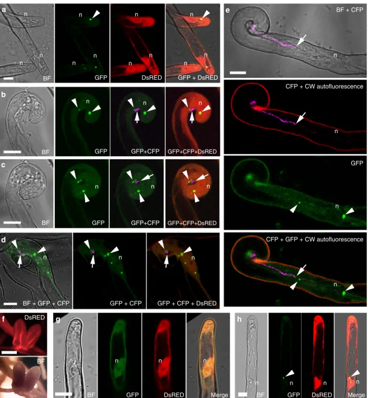
Previous work showed that VPY is required for rhizobial infec- tion of root hairs42. To better define how VPY contributes to infection thread development in root hairs, we used live cell imaging to compare the earliest stages of root hair infection in the M. truncatula vpy-1mutant and insunn-2plants after inocula- tion with the GFP-taggedSinorhizobium meliloti strain Sm2011 (Sm2011-CFP) (Fig.1a–h). Supernodulantsunn-2plants have a much higher number of infections compared to A17 but are nonetheless wild type-like for infection thread formation which is why they are preferred for in vivo microscopy30. Root hair curling and entrapment of rhizobia were normal in vpy-1 (Fig.1a, b).
However, after the formation of a microcolony within the infection chamber, infection threads did not form in the mutant up to 4 days post-inoculation (dpi) withS. meliloti(Fig.1c-e) in contrast to the wild type situation where infection threads already extended down the root hairs by 3 dpi (Fig. 1f-h). Instead, only swollen infection chambers hosting large microcolonies were found in the mutant 3–4 dpi (Fig.1c-e), suggesting that, in the absence of VPY, the ability to switch from radial to polar growth of the infection compartment has been lost. In addition, although the nucleus invpy-1curled root hairs was still connected to the developing infection chamber by a cytoplasmic strand, it was no longer in close vicinity of the enclosed rhizobia (Fig.1c, d insets), in contrast to its usual position adjacent to the developing infection chamber in sunn-2root hairs29(Fig.1f). These results indicate that VPY is also essential for positioning the nucleus close to the developing infection chamber.
Although most rhizobial infections are blocked in root hairs in vpy-2, small aberrant nodules do form in mutant plants47. We therefore investigated to what extent rhizobial infection was affected in these small nodules. Compared with wild type nodules at the same time point (47 dpi), the nodules ofvpy-2were much smaller and never elongated (Fig. 1i–k). Within vpy nodules, infection threads rarely entered cells and rhizobia frequently accumulated in large intercellular deposits (Fig. 1j, k).
Transmission electron microscopy of vpy-1 and vpy-2 nodule sections confirmed that these deposits contained large numbers of bacteria but also revealed large amounts of electron dense material (Fig. 1m, n) that was never observed in wild type nodules (Fig. 1l). This suggests that rhizobial infection thread construction is abnormal in vpy nodules, and that secretion processes are altered in the vpy mutant. Finally, very few symbiosomes were observed in nodules of vpy-1 and vpy-2 (Fig.1k, Supplementary Fig. 1), most likely as a consequence of defective cortical infection.
Altogether, our data indicate a clear role for VPY in infection thread formation in both root hairs and nodules, and that this role may involve the focused secretion of material that is required for the polar growth of infection threads. We next investigated the spatiotemporal pattern of VPY expression and VPY subcellular localization.
VPY expression is associated with rhizobial infection. The expression ofVPYinM. truncatularoot hairs is induced by both 2 dpi
2 dpi
vpy-1 vpy-1 vpy-1 vpy-1
sunn-2 sunn-2
sunn-2 vpy-1
n
i
f
c d e
a
b
l
*
*
*
* * *
WT
WT
4 dpi 3 dpi
4 dpi h
3 dpi g
vpy-2 vpy-1
j vpy-2 k vpy-2
m n
Nod factors and S. meliloti48. We further determined the spa- tiotemporal pattern ofVPYexpression using a fusion of theVPY promoter to theGUSgene (pVPY:GUS) in composite plants. GUS staining revealed that reporter activity was highest in the root hairs harbouring either microcolonies or developing infection threads (Fig. 2a, e, Supplementary Fig. 2a–b). During nodule development, GUS activity was found in young developing nodules and at the early symbiotic region of elongated nodules, but not in the interzone or nitrogen fixation zone (Fig. 2b–d).
Longitudinal sectioning showed that the GUS staining was con- fined to the group of cells harbouring or surrounding infection threads (Fig. 2f–h). Later, after zonation of nodules, the expres- sion of VPY was mainly found in the infection zone (Fig. 2i).
Thus, VPYexpression is correlated with active infection during both epidermal and nodule primordia infection as well as in mature nodules and is therefore coherent with the defective rhi- zobial infections observed in both root hairs and nodule cells in vpymutants.
Localization of VPY during rhizobial infection. The subcellular localization of VPY in root hairs was investigated in detail by live cell confocal imaging in roots of A17 or sunn-2 expressing a pVPY:VPY-GFP construct. Expression of this construct in vpy roots restored nodulation (about 30 nodules per plant 30 dpi with S. meliloti), indicating that fusion with GFP does not affect VPY function (Fig.3f, Supplementary Fig. 3a). Using live cell confocal imaging, we were able to follow the dynamics of VPY-GFP throughout the root hair infection process (Fig.3a–e). Following rhizobial inoculation, VPY-GFP localized to small puncta in the cytoplasm of most root hairs, notably growing root hairs (Fig.3a).
These puncta, although generally static, were occasionally mobile and were most often found in the vicinity of the nucleus (Fig.3a;
Supplementary Video 1). In curling root hairs, the VPY-GFP labelled puncta were similarly present in the vicinity of the nucleus, and in addition, several puncta were concentrated in a position facing the attached rhizobia, before and after curl closure (Fig. 3b, c) as well as during infection chamber development
Fig. 1VPYis required for rhizobial infection in the epidermal and nodule cells.a–bConfocal images ofvapyrin-1(vpy-1) mutant showing normal root hair curling and entrapment of rhizobia. Upper, merged image of brightfield and GFP; lower, merged pictures of cell wall auto-fluorescence and GFP.c–hRoot hair infection phenotypes ofvpy-1(c–e) and control plants (sunn-2) (f–h) at 2, 3 and 4 days post-inoculation (dpi) with Sm2011-GFP, showing exaggerated radial development of the infection chamber and abnormal nucleus positioning in the mutant. White arrows indicate nucleus inc,dandf. Blue arrowheads indicate the cytoplasmic bridge invpy-1(inset inc). White arrowheads indicate rhizobia in the infection chamber (green). Red arrowheads indicate elongated infection threads in control plants (g,h). Insets inc,dcorrespond to the same root hair at a lower magnification, showing the positions of nucleus. n, nucleus ina. White dashed line indicates the contour of the nucleus inf.i,jLongitudinal nodule sections of X-gal stained WT (i) andvpy-2(j) nodules.kToluidine blue-stained longitudinal nodule section ofvpy-2. Nodules were collected at 47 dpi. Blue colouri,jindicates X-gal staining of Rm1021- lacZ. Red arrows indicate abnormal intercellular accumulation of bacteria (j,k).l–nTransmission electron microscopy of nodule cells from WT (l),vpy-1 (m) andvpy-2(n) at 47 dpi showing intercellular rhizobial infection. Red asterisks indicate rhizobia. Arrowheads indicate plant cell wall. Arrows indicate unidentified electron dense material accumulating in the intercellular space occupied by rhizobia (l–n). Scale bars, 10μm (a–h), 100µm (i–k) and 1μm (l–n)
Interzone n
Fixation zone v
v
np
np
v
v v
v
v e
a Root hair Nodule primordia Young nodule Elongated nodule
Fixation zone
b c d
i h
g f
Fig. 2Expression ofVPYis tightly associated with rhizobial infection in root and nodule cells.a–iImages showingpVPY:GUSactivity during rhizobial infection in root hair (a,e) and in nodules from primordia to mature zonation stages (b–d,f–i). Composite transgenic plants were used at different stages after inoculation with Rm1021-lacZ.a–eare whole mount of GUS and/or X-gal stained roots,f–iare longitudinal sections of GUS and X-gal stained nodule primordia/nodule. Blue colour indicates staining of GUS activity and magenta colour indicates staining of rhizobia. Arrowheads indicate GUS stained root hairs (a,e) and arrows indicate infection threads in root hairs (a,e) or nodule primordia (f–h)/nodule (i). v, central vasculature of the root; np, nodule primordium; n, nodule. Scale bars, 100µm
n
n
n
n BF
BF GFP
BF
BF + GFP + CFP
n n
n n
n n
n n
n n n
n
n n n n
n n n n
n n n n
n
n n n
h g
f
e
d c
BF + CFP
CFP + CW autofluorescence
CFP + GFP + CW autofluorescence GFP
DsRED
BF
BF BF
b a
GFP + DsRED DsRED
GFP
GFP + CFP + DsRED GFP + CFP
n n n
Merge DsRED
GFP Merge
DsRED GFP
GFP+CFP+DsRED GFP+CFP
GFP+CFP+DsRED GFP+CFP
GFP
Fig. 3Subcellular localization of VPY during rhizobial infection.a–eLive cell confocal images showing localization of VPY-GFP driven byVPYpromoter in root hairs at different stages of rhizobial infection. The VPY-GFP fusion is observed in small dot-like bodies, in the vicinity of the nucleus, in elongating root hairs prior to deformation (a). During root hair curling around attached rhizobia and bacteria entrapment by curl closure, VPY-GFP labelled puncta are preferentially found close by the nucleus and in front of rhizobial attachment point (b,c), a localization pattern that persists during infection chamber remodelling (d); Finally, when polar elongation of the infection thread starts, one VPY-GFP punctus is found at the growing tip of the infection thread, situated a few micrometers ahead of the colonizing rhizobia (e). Images are representative of more than 100 root hairs from at least 20 A17 composite plants (a), more than 50 root hairs imaged in eight differentsunn-2composite plants (b–d) and more than 40 ongoing infection sites, monitored in 15sunn- 2composite plants (e) in three independent experiments.fNodules from transgenicvpy-2plants complemented bypVPY:VPY-GFPconstruct.
g,hSubcellular localization of MSP domain or ankyrin repeats containing domain of VPY in plants transformed withpLjUBQ1: GFP-MSP(g)or pLjUBQ1: GFP- ankyrin(h). All constructs contain a constitutively expressed DsRED marker (red colour). Root hairs were imaged from 2 dpi up to 7 dpi with Rm1021-CFP (a,gandh) or Sm2011-CFP (b–e) and nodules were examined at 60 dpi. Bright Field (BF), DsRED, GFP and CFP images were pseudo coloured in grey, red, green and magenta respectively, except inewhere wall autofluorescence is in red. Plants ina,gandhare in WT A17 background. Plants inb–eare insunn- 2background. n, nucleus. Arrowheads indicate GFP puncta. Arrows indicate CFP labelled rhizobia. Scale bars, 10µm (a–e,g–h) and 1000µm (f)
(Fig.3d). In the root hairs harbouring actively growing infection threads, VPY-GFP was consistently found to accumulate at the growing tip of the infection thread and in nuclear-associated puncta (Fig.3e; Supplementary Fig. 4). Closer examination of the infection thread tip-localizedfluorescence showed that VPY-GFP accumulates in a highly dynamic assembly capping the growing infection thread tip, and sometimes extending into the cytoplasm (Supplementary Video 2). In the absence of rhizobia, VPY-GFP localized uniformly to the cytoplasm and occasionally a weak GFP signal was found in the nucleus (Supplementary Fig. 5). The weak GFP signal is consistent with the weak expression ofVPYin root hairs before rhizobial inoculation42,48. This background localization was often observed in inoculated roots (Fig. 3) in addition to the specific punctate localization (Fig.3). However, VPY-GFP was not observed in puncta in non-inoculated roots (Supplementary Fig. 5), suggesting that accumulation of VPY- GFP in small subcellular domains is specifically induced following rhizobial inoculation. Altogether, our results indicate that, in response to inoculation, VPY accumulates in small cytoplasmic domains of unknown nature associated mainly with the nucleus and the apical tips of growing infection threads. This localization correlates well with thevpymutant phenotypes, further suggest- ing a role of VPY in polarized infection thread construction and related secretion.
To determine which domain of the VPY protein is responsible for the punctate localization, constructs were made to express GFP fused either to the N-terminal MSP domain (GFP-MSP) or to the C-terminal ankyrin repeat-containing domain (GFP- ankyrin) of VPY, both driven by the strong LjUBQ1 promoter in roots of composite plants. After rhizobial inoculation, GFP- ankyrin displayed the punctate pattern characteristic of full- length VPY, while GFP-MSP was found uniformly distributed in the cytoplasm and nucleus (Fig. 3g, h), arguing that the ankyrin repeats are responsible for the localization of VPY to the punctate, rhizobia inoculation-induced subdomains.
LIN and VPY co-localize during rhizobial infection. The MSP domain and ankyrin repeats of VPY are both predicted to med- iate protein-protein interactions, suggesting that VPY function in rhizobial infection may involve interactions with one or more other proteins. We have noticed that VPY exhibits striking similarities in terms of symbiotic phenotypes and expression patterns withLIN. Bothvpyandlinshow delayed initiation and development of infection threads and both of them form aberrant nodule-like structures with a characteristic central vasculature40,42,47,49. LINand VPYalso have similar expression patterns in nodules40(Fig.2). And when a construct containing theLINpromoter driving the GUS reporter gene (pLIN:GUS) was expressed inM. truncatula, strong GUS activity was detected in root hairs harbouring infection threads (Fig.4a), reminiscent of VPYexpression in root hairs (Fig.2a, e). Based on these simila- rities we investigated the possibility that VPY and LIN might act together during rhizobial infection.
Firstly, the sub-cellular localization of LIN was investigated by making use of an N-terminal fusion to GFP driven by either the LjUBQ1promoter or the nativeLIN promoter. BothpLIN:GFP- LINandpLjUBQ1:GFP-LINcomplementedlinmutants (Fig.4b, c; Supplementary Fig. 3; Supplementary Fig. 6). Live cell imaging of the roots of composite plants transformed with either construct and then inoculated withS. meliloti, showed similar localization patterns (Fig. 4; Supplementary Fig. 7). Since the fluorescent signal was much stronger for pLjUBQ1:GFP-LIN, this construct was used for subsequent experiments. No GFP signal for pLjUBQ1:GFP-LINwas observed under non-symbiotic conditions in either root hairs or roots, suggesting tight control of LIN
accumulation at the protein level. When root hairs were imaged in S. meliloti-inoculated roots, GFP-LIN localized primarily to a few puncta in close proximity to the nucleus, while weaker GFP fluorescence was visible throughout the cytoplasm (Fig. 4d–h).
After bacterial entrapment within curled root hairs or between touching root hairs, puncta were enriched around the developing infection chamber and the adjacent nucleus (Fig.4i–s). Finally, in root hairs with growing infection threads, GFP-LIN accumulated in the cytoplasm at the very tip of infection threads (Fig. 4t–y) with some puncta also found close to the nucleus in the same root hair (Supplementary Fig. 8). This localization of LIN during rhizobial root hair infection, highly reminiscent of that observed for VPY, prompted us to test whether these two proteins co- localize. For this, M. truncatula composite plant roots were generated using a construct containing bothpVPY:VPY-GFPand pLjUBQ1:mCherry-LIN. Live-cell imaging of root hairs in S.
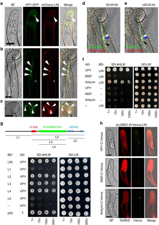
meliloti-inoculated roots showed that VPY-GFP co-localized perfectly with mCherry-LIN within the puncta and the cytoplasm (Fig.5a–e).
VPY interacts with LIN in yeast andin planta. By using a yeast two-hybrid assay we were able to show that a VPY prey could interact with LIN when used as a bait (Fig.5f). Additional yeast interaction assays indicated that LIN interacted with the VPY C- terminal fragment containing the ankyrin-repeat domain but not with one containing the N-terminal MSP domain (Fig.5f).
LIN is a putative E3 ubiquitin ligase with an unknown N- terminal domain, a U-box, an Armadillo-like domain and a C- terminal domain containing WD40 repeats40. Yeast two-hybrid tests using truncated LIN with different domain combinations showed that only the Armadillo-like domain was indispensable for the interaction with VPY (Fig.5g). The interaction between LIN and VPY was further evaluated using Bimolecular Fluor- escence Complementation (BiFC)50inM. truncatula, using C- terminal and N-terminal split-Venus fragments fused to VPY and LIN respectively. N-Venus-LIN was expressed from a strong constitutive promoter (pLjUBQ1:N-Venus-LIN) to improve detection and VPY-C-Venus was driven by the native VPY promoter (pVPY:VPY-C-Venus) to better reflect natural condi- tions. Strong Venus fluorescence was found in puncta in non- deformed root hairs (Fig. 5h) as well as curled root hairs (Supplementary Fig. 9) imaged in S. meliloti-inoculated com- posite plants expressing the BiFC fusions at 7 dpi, thus showing that VPY and LIN interact in planta following rhizobial inoculation. In root hairs undergoing rhizobial infection Venus fluorescence was also associated with the growing infection thread tip (Fig. 6). The small puncta labelled by the interacting proteins were alike in every aspect to those labelled by either GFP-LIN or VPY-GFP at identical stages (Figs.2,3). Additional BiFC experiments using N- or C-terminal truncated versions of VPY showed that the C-terminal ankyrin repeat-containing domain interacted with LIN, whereas the N-terminal MSP domain on its own did not (Fig. 5h), consistent with the yeast two-hybrid results and the localization of GFP-ankyrin (Figs. 5f, 3h). We therefore conclude that VPY interacts with LIN in rhizobia-inoculated plants, and that this interaction and its localization to puncta requires the C-terminal ankyrin repeat-containing domain.
VPY is regulated by LIN during rhizobial infection. Protein ubiquitination has diverse functions and can affect the degrada- tion, sub-cellular localization, activity or interaction of proteins51. The observed interaction of VPY and LIN led us to monitor whether VPY localization is dependent on LIN. To answer this, confocal imaging was used to testpVPY:VPY-GFPlocalization in
pLIN:GUS n n n n n
n n n n n
n n
n n
n n
n n
n n
n n n n n
lin-4/EV
BF
BF
GFP+DsRED merge
GFP+DsRED merge
Merge
Merge Merge Merge
(GFP+CFP)
Merge lin-4/GFP-LIN
y x
w v
u t
s r
q p
o n
j k l m
i b
c
h
d e f g
a
DsRED GFP
BF GFP DsRED
BF GFP DsRED CFP
DsRED
BF DsRED
BF GFP DsRED CFP Merge (GFP+CFP)
Fig. 4Expression ofLINand subcellular localization of LIN in root hairs during rhizobial infection.aExpression ofpLIN:GUSin a root hair harbouring an infection thread.b–cNodules fromlumpy infections-4(lin-4) mutants transformed with an empty vector (EV) (b) orpLIN:GFP-LINconstruct (c). Brightfield (BF), left and DsRED images, right inbandc.d–yLive cell confocal images of subcellular localization of GFP-LIN driven bypLjUBQ1promoter in root hairs throughout different stages of rhizobial infection: GFP-LIN accumulated in puncta associated to the nuclear periphery in non-deformed root hairs (d–h);
GFP-LIN labelled dot-like bodies were also found around rhizobial micro-colony and neighboring nucleus during infection chamber development (i–m,n–s);
andfinally, GFP-LIN also accumulated at the tip of infection threads, just ahead of the elongating structure (t–y). DsRED was used as a transgenic marker (red). Transgenic plants were either inoculated with Rm1021-lacZ (a–c) or Rm1021-CFP (d–y, magenta inq–s,w–y). Plants were imaged from 2 dpi up to 7 dpi (d–y). GUS staining is shown as blue. DsRED, GFP, CFP were pseudo coloured in red, green and magenta, respectively. n, nucleus. Arrowheads, GFP- LIN puncta. Arrows, CFP tagged rhizobia. Double arrowheads, auto-fluorescence. Scale bars, 10µm (a,d–y) and 500µm (b,c)
thelin-1mutant inoculated withS. meliloti. Compared with wild type, the VPY-GFP signal was much reduced inlin-1, both in the puncta and the cytoplasm (Fig.7a). In addition, quantification of the average number of VPY-GFP puncta per root hair revealed a
>2 fold decrease inlin-1compared to WT (Fig.7b). This could be
due to a genuine decrease in the number of labelled puncta, or from the difficulty of detecting puncta with lower fluorescence levels. To distinguish between these two possibilities, the fluor- escence intensity of the largest punctus per root hair was deter- mined for bothlinand WT plants. We found that the intensity of
LIN LIN – –
–
– LIN LIN
SD-AHLW
SD-AHLW
10×
BD-
LIN BD-
L1 L2 L3 L4 L5 –
p53 Ankyrin-C-VenusVPY-C-Venus MSP-C-Venus
h
Armadillo-like
L4 U box
L3 L5
WD40
L1 L2
g
Ankyrin
Ankyrin VPY
VPY AD-
pLjUBQ1:N-Venus-LIN MSP
MSP
n n n n
+00:03:54 00:00:00
VPY-GFP mCherry-LIN
VPY-GFP mCherry-LIN
e
BF d
c a
b
f
BF VPY-GFP mCherry-LIN Merge
SD-LW
1× 100× 1000× 10×1× 100× 1000×
10×1× 100× 1000× 10×1× 100× 1000×
DsRED Venus Merge SD-LW
AD- VPY VPY VPY VPY VPY VPY VPY T
Fig. 5Interaction of VPY and LIN.a–cLive cell confocal images of root hairs from transgenic plants containingpVPY:VPY-GFPandpLjUBQ1:mCherry-LIN showing co-localization of VPY and LIN after inoculation with rhizobia.d,eLive cell images from the root hair tip shown inb,cat two time points (0″and 3′54″) showing the movement and fusion of VPY-GFP/mCherry-LIN labeled puncta.f–gYeast two-hybrid assays: between LIN and VPY, MSP domain of VPY and ankyrin repeats of VPY, respectively (f) and between VPY and different domains of LIN (g). The combination of proteins expressed in either the AD vector (pGADT7) or the BD vector (pGBKT7) are indicated alongside the yeast colonies. Diploid yeast cells with a series of 10- fold dilutions were grown on either SD-Leu-Trp (SD-LW) or SD-Ade-His-Leu-Trp (SD-AHLW) media.hBimolecular Fluorescence Complementation (BiFC) experiments after inoculation with rhizobia. Split Venusfluorescent protein was used, with N-Venus fused to LIN driven bypLjUBQ1promoter and C-Venus fused to VPY, MSP domain or ankyrin repeats containing domain of VPY driven byVPYpromoter. DsRED was used as a transgenic marker. DsRED and GFP images were pseudo coloured in red and green, respectively. n, nucleus; Arrowheads, VPY-GFP/mCherry-LIN puncta. Scale bars, 10µm
the brightest punctus was greatly reduced in lin (Fig. 7c). To determine whether the observed reduction in VPY levels was a consequence of reduced VPY expression in the lin mutant we introducedpVPY:GUSinto wild type andlin-1backgrounds. We found similar GUS staining in infected root hairs for both gen- otypes suggesting that regulation of VPY by LIN is at post- transcriptional level (Supplementary Fig. 10). This, along with the finding that cytoplasmic VPY levels also appeared to be decreased inlin, suggests a role for LIN in the regulation of VPY abundance or stability, rather than its characteristic punctate localization.
This positive regulation of VPY by LIN is in line with the duo’s positive roles during rhizobial infection. To test whether VPY could affect the subcellular localization of LIN, we introduced pLjUBQ1:GFP-LIN into vpy-1 mutant and we found persistent punctate localization of LIN-GFP, as observed in wild type (Supplementary Fig. 11).
VPY-LIKE and LIN-LIKE may contribute to rhizobial infec- tion. The defective but not abolished rhizobial infection ofvpy and lin could result from genetic redundancy. Indeed, close homologs exist in M. truncatula for both LIN, which we have named LIN-LIKE (LINL; Medtr8g103227), and for VPY, called VPY-LIKE (VPYL)43 (Supplementary Fig. 12; Supplementary Fig. 13). We therefore investigated their expression during nodulation using promoter-GUS constructs (pLINL:GUS and pVPYL:GUS) in composite plants. Similarly to LIN and VPY fusions, thepLINL:GUSreporter also conferred high GUS activity levels in infected root hairs, in young nodules, and at the apical region of elongated nodules (Fig.8a–c). This suggests thatLINL may also contribute to rhizobial infection. In contrast, thepVPYL:
GUS fusion was constitutively expressed throughout the root (Fig.8f–h), including both infected and non-infected root hairs (Fig. 8f, g), consistent with our earlier published root hair pVPY:VPY-C-Venus + pLjUBQ1:N-Venus-LIN
BF a
b c d e f g
n n n n n
n
Merge
DsRED Venus CFP
Fig. 6VPY interacts with LIN during rhizobial infection thread formation.a–gLive cell confocal images showing a root hair undergoing infection in asunn-2 composite plant containingpVPY: VPY-C-Venus,pLjUBQ1:N-Venus-LINandpAtUBQ:DsREDas a transgenic marker. Bimolecular Fluorescence
Complementation (BiFC) experiments were performed using N-Venus fused to LIN driven bypLjUBQ1promoter and C-Venus fused to VPY driven byVPY promoter as in Fig.5h. One representative image is shown out of seven imaged ongoing infection sites in twosunn-2composite plants. Split Venus fluorescence was imaged 4 days after inoculation with Sm2011-CFP, in a root hair hosting an elongating infection thread. Venusfluorescence was associated notably to the tip of the growing infection thread. As in other root hairs, Venusfluorescence was also found associated to a number of other puncta in the nucleus vicinity or along the cytoplasmic bridge. DsRED, Venus and CFP were pseudo coloured in red, yellow and magenta respectively.
Dashed white line box inaindicates the regions shown in (b–g). Arrows, infection thread growing tip; arrowheads, infection thread tip-associated Venus- labelled puncta. n, nucleus. Scale bars, 10µm
0 50 100 200
150
0 1 2 3 4 5
Intensity of the biggest punctus
WT
lin-1
WT
Number of puncta per root hair
****
VPY-GFP DsRED
****
a b c
lin-1 WT lin-1
Fig. 7Regulation of VPY by LIN.aConfocal images of VPY-GFP driven byVPYpromoter in root hairs of wild type (WT) A17 orlin-1mutant under the same condition at 5 dpi with Rm1021-CFP. DsRED was used as a transgenic marker. DsRED and GFP were pseudo coloured in red and green respectively. Scale bars, 10µm.b,cQuantification of the numbers of VPY-GFP puncta per root hair (b), and integratedfluorescence intensity of the brightest punctus (c) in wild type (WT) A17 andlin-1mutant. Bars indicate standard error. ****p< 0.0001, Student’st-test.n=75 (WT) and 84 (lin-1) root hairs for (b) andn=47 (WT) and 46 (lin-1) for (c) Source data are provided as a Source Datafile
infectome data48.VPYLwas also expressed in young nodules and the apex of mature nodules (Fig.8h, i).
We then tested the sub-cellular localization of LINL (pLjUBQ1:
mCherry-LINL) together with VPY (pVPY:VPY-GFP). mCherry- LINL localized to puncta and cytoplasm in root hairs of transgenic composite plants inoculated with S. meliloti, and entirely co-localized with VPY-GFP (Fig. 8d). GFP-VPYL (pLjUBQ1:GFP-VPYL) showed a similar punctate pattern, and, as in the case of VPY-GFP, certain puncta were mobile (Fig.8j).
We further tested the interaction of VPY and LINL using a yeast two-hybrid assay and found that they were indeed able to interact (Fig. 8e). Further yeast two-hybrid tests showed that VPYL interacted with both full-length LIN and the C-terminus of LIN (Fig. 8k). Finally, VPYL also interacted with LINL in yeast (Fig.8e).
Taken together, the gene expression, subcellular localization and protein-protein interaction data obtained here suggest that LINL and VPYL may act together with LIN and VPY to promote rhizobial infection. We therefore propose that these proteins form complexes within punctate bodies in the cytoplasm and at the tip of growing infection threads.
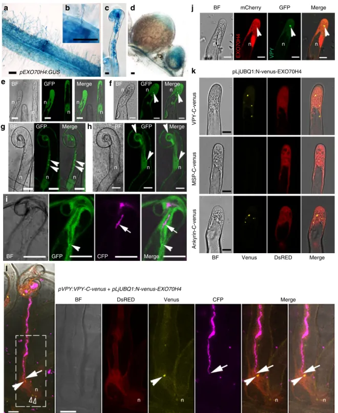
An exocyst subunit co-localizes with the VPY-LIN complex.
VPY is required for epidermal entry of AM fungi and arbuscule development during mycorrhization42–44. In addition, it has been shown that an exocyst component, EXO70I, is involved in mycorrhization and partially co-localizes with VPY in mycor- rhizal roots ofM. truncatulabut is dispensable for nodulation46. Our previous transcriptome profiling inM. truncatulashows that another EXO70 family member, EXO70H4 (Medtr4g062330) (Supplementary Fig. 14), is induced 3 and 5 dpi in root hairs after inoculation with rhizobia48 (Supplementary Fig 15). A pEXO70H4:GUSfusion revealed thatEXO70H4was constitutively expressed in Medicago roots, especially in root tips and lateral root primordia (Supplementary Fig. 16). After inoculation with rhizobia, EXO70H4 expression was enhanced in infected root hairs and the underlying cortical cells (Fig. 9a–c). EXO70H4 expression was also detected in young nodules, and in the apical region of elongated nodules, including the infection zone (Fig. 9d). Thus, the overall expression pattern of EXO70H4 is consistent with a potential contribution of the encoded exocyst component to rhizobial infection.
n n n n n n
n n n n
VPY VPYL
LINL –
e
VPY LINL
d
c b
a
BD-
SD-AHLW SD-AHLW
1×
k
0 min + 5 min
i
j
h g
f
BF BF
pLINL:GUS pVPYL:GUS
Merge mCherry GFP
GFP Merge BF GFP Merge
SD-LW 10× 100× 1000× 1× 10× 100× 1000×
1× 10× 100× 1000× 1× 10× 100× 1000×
AD- LINL LINL
LIN
LIN L2
VPYL VPYL L1
– – BD- AD-
VPYL VPYL
SD-LW
Fig. 8A protein complex containing VPY, VPYL, LIN and LINL.a–cExpression ofLINLin an infected root hair (a), in a young nodule (b) and in a mature nodule (c) as shown bypLINL:GUSactivity.dCo-localization of VPY (green) and LINL (red) in root hairs of rhizobia-inoculated hairy roots transformed with pVPY:VPY-GFP/pLjUBQ1:mCherry-LINL.eYeast two-hybrid assays between VPY, VPYL and LINL.f–iExpression ofpVPYL:GUSat infection site (f), in a root hair harbouring an infection thread (g), young and mature nodules (h,i).jLive cell images of localization of GFP-VPYL driven bypLjUBQ1promoter at two time points (0 min and 5 mins later) in a root hair after inoculation with rhizobia.kYeast two-hybrid assays between VPYL and LIN, C terminus of LIN (L2) and N terminus of LIN (L1). BD, pGBKT7; AD, pGADT7. SD-LW, SD-Leu-Trp; SD-AHLW, SD-Ade-His-Leu-Trp. GUS staining were shown as blue. DsRED and GFP were pseudo coloured in red and green respectively. Arrowheads, GFP/mCherry puncta. n, nucleus. Scale bars, 100µm (a–c,f, h,i) and 10µm (d,gandj)
pLjUBQ1:N-venus-EXO70H4
VPY-C-venusMSP-C-venusAnkyrin-C-venus
pVPY:VPY-C-venus + pLjUBQ1:N-venus-EXO70H4
BF mCherry
pEXO70H4:GUS
GFP Merge
BF GFP Merge
BF
BF BF
BF GFP
GFP Merge
Merge Merge
Merge CFP
Venus
Venus
DsRED
DsRED CFP
BF GFP Merge
BF GFP Merge
n n
n n
n n
n
n n
n n n n
n n
n n
n n n
n
n n n
VPYEXO70H4
a b c d
e f
g h
i
j
k
l
Fig. 9EXO70H4 co-localizes with VPY.a–dExpression ofEXO70H4in roots inoculated by rhizobia (a), at a rhizobial infection site (b), in a root hair containing an infection thread (c) and in nodules (d) as shown bypEXO70H4:GUSactivity.eSubcellular localization of GFP-EXO70H4 in root hairs without inoculation with rhizobia.f–iSubcellular localization of GFP-EXO70H4 in root hairs following inoculation with Rm1021-CFP (magenta).jCo-localization of VPY-GFP (green) and mCherry-EXO70H4 (red) at the same punctus in root hairs of rhizobia-inoculated composite plants transformed withpVPY:VPY-GFP /pLjUBQ1:mCherry-EXO70H4.kBimolecular Fluorescence Complementation (BiFC) experiments in root hairs following inoculation with rhizobia. Split Venusfluorescent protein was used, with N-Venus fused to EXO70H4 driven bypLjUBQ1promoter and C-Venus fused to VPY, MSP domain or ankyrin repeats containing domain of VPY driven byVPYpromoter.lLive cell confocal images showing a root hair undergoing infection in asunn-2composite plant containingpVPY:VPY-C-Venus, andpLjUBQ1:N-Venus-EXO70H4. Split Venusfluorescence was imaged 3d after inoculation with Sm2011-CFP, in a root hair hosting an elongating infection thread. Venusfluorescence (arrowheads inl) was associated notably to the tip of a growing infection thread (arrows);
Venusfluorescence (open arrowheads) was also found associated to a number of other puncta in the nucleus vicinity. Five ongoing infection sites where imaged in four plants inl. GUS staining is shown as blue. DsRED was used as a transgenic marker. DsRED, Venus, GFP and CFP were given pseudo coloured in red, yellow, green and magenta respectively. n, nucleus. Arrowheads, GFP/mCherry /Venus puncta. Arrows, CFP tagged rhizobia. Scale bars, 100µm (a,d) and 10µm (b,c,e–l)
The localization of EXO70H4 was then investigated by live-cell imaging. Since no GFP signal was detected using the native EXO70H4promoter driving expression of aGFP-EXO70H4gene fusion, we instead used the LjUBQ1 promoter. A strong GFP- EXO70H4 signal was detected in the cytoplasm and the nucleus under non-symbiotic conditions in root hairs and other root cells (Fig. 9e, Supplementary Fig. 17). After rhizobial inoculation, in addition tofluorescence in the cytoplasm and the nucleus, GFP- EXO70H4 was highly enriched in a small number of root hair puncta (Fig.9f). In curled root hairs, puncta were also found close to the nucleus, along the cytoplasmic bridge between the infection chamber and the nucleus, and close to the infection chamber (Fig. 9g, h). In root hairs hosting an infection thread, GFP- EXO70H4 accumulated at the infection thread tip (Fig. 9i), reminiscent of the complex labelled by VPY-GFP (Fig. 3e) and GFP-LIN (Fig. 4t–y).
The relative localization of EXO70H4 and VPY was investi- gated using a construct expressingpLjUBQ1:mCherry-EXO70H4 and pVPY:VPY-GFP. Upon rhizobial inoculation mCherry- EXO70H4 was found in the cytoplasm and nucleus and was highly enriched in puncta, where it co-localized with VPY-GFP (Fig.9j). Next we did BiFC with the combination ofpVPY:VPY- C-Venus and pLjUBQ1:N-Venus-EXO70H4 constructs. Strong Venus fluorescence was observed in cytoplasmic puncta in M.
truncatularoot hairs inoculated withS. meliloti(Fig.9k). Using truncated VPY proteins containing either the N- or C-terminal domains, we again found that only the ankyrin repeat-containing domain was able to show Venus fluorescence puncta with EXO70H4 (Fig.9k). Finally, strong Venusfluorescence was once more associated with the tip of the growing infection thread (Fig. 9l). Altogether, these results indicate that EXO70H4 is systematically co-localized with VPY and suggest a possible role for EXO70H4 as an additional component of the protein complex comprising VPY and LIN.
EXO70H4is required for rhizobial infection at early stages. To investigate whetherEXO70H4is required for rhizobial infection, two M. truncatula lines (NF10274 and NF14802) with the Nicotiana tabacum transposable element Tnt1insertions in the single exon ofEXO70H4were recovered from the Noble Research Institute mutant library52, designated asexo70h4-1andexo70h4- 2, respectively (Supplementary Fig. 18). Homozygous mutants were isolated and infection phenotypes were characterized alongside vpy-2 and lin-4 grown in sand: terra green by histo- chemical staining of roots inoculated with aHem:LacZexpressing strain ofS. meliloti(Rm1021-LacZ). Theexo70h4-1andexo70h4- 2mutants displayed about 50% fewer infection events than wild type at 3 and 5 dpi (Fig.10a). At 10 dpi, there was no significant difference between wild type and theexo70h4mutants, and at 16 dpi more infection events were present in both alleles ofexo70h4 compared to wild type (Fig. 10a). The dynamics of rhizobial infection of the exo70h4 mutants were very similar to those of vpy-2andlin-4, both of which had fewer infections at early time points and more infections at later times by comparison with wild type (Fig.10a). This pattern is likely the consequence of a feed- back mechanism resulting from initial failed infections subse- quently leading to increased infection attempts at later time points (Fig.10a–c, Supplementary Fig. 18). Consistent with this, exo70h4-1andexo70h4-2showed many defective infections, such as oversized microcolonies and blocked infection threads that sometimes burst leading to the release of rhizobia within root hairs (Fig. 10b). Sometimes branched infection threads were observed in exo70h4-1andexo70h4-2 (Fig.10b). Similar pheno- types were also frequently found in vpy-2 and lin-4 mutants (Fig.10b). Since occasionally“abnormal”infections could also be
observed for the WT, we quantified all types of defective infection at 5, 10 and 16 dpi. We counted infection events at all stages from microcolony (MC) to ramified infection threads (rIT) into the cortex. For microcolonies, only big swelled ones were counted as
b
c
WT exo70h4–1 exo70h4–2
lin-4
200 400 600 800
vpy-2
a
*
*
** ****
********
****
****
****
***
**
100
75
50
25
0 0
Percentage of defective infections per plant
3 dpi
Number of infection events per plant
5 dpi 10 dpi 16 dpi
exo70h4–2 exo70h4–1
exo70h4–2
exo70h4–2 exo70h4–1
exo70h4–1 vpy-2
vpy-2
vpy-2 lin-4
lin-4
lin-4 exo70h4–1
WT
WT
Fig. 10Rhizobial infection phenotypes ofexo70h4, vpyandlin.aRhizobial infection time course of WT,exo70h4-1,exo70h4-2,vpy-2andlin-4. All infection events from microcolony to ramified cortical infection threads were counted from 9 to 12 plants for each genotype at 3, 5, 10 and 16 dpi with Rm1021-lacZ respectively. Bars represent standard error.bTypical defective infection events ofexo70h4-1,exo70h4-2,vpy-2andlin-4(left) and a normal elongating infection event of WT (right). Top left, abnormal enlarged microcolonies (exo70h4-1,exo70h4-2,vpy-2andlin-4) and blocked infection in the epidermal cells (exo70h4-1). Bottom left, blocked infection threads, branched infection threads (arrowheads) and disintegrated infection threads resulting in rhizobia release (arrows) in the mutant root hairs ofexo70h4-1,exo70h4-2,vpy-2andlin-4. Scale bars, 10µm.
cQuantification of defective infection events in WT,exo70h4-1,exo70h4-2, vpy-2andlin-4at 5, 10, 16 dpi, respectively. All genotypes are in R108 background. Bars show the percentage of defective versus all infection events per plant. Error bars represent standard error. Two-tailed Student’s t-test, *p< 0.05; **p< 0.01; ***p< 0.001; ****p< 0.0001. Source data are provided as a Source Datafile
“defective”while the small ones were counted as normal infection events. We found that the percentage of abnormal infections was higher inexo70h4,vpyandlincompared to wild type (Fig.10c).
However, there was no significant difference in nodule number between wild type andexo70h4mutants (Supplementary Fig. 18), consistent with the relatively mild infection phenotype. The phenotype of exo70h4 mutants could be a result of genetic redundancy with members of the extensive EXO70 family (Sup- plementary Fig. 14), notably those which are also expressed in root hairs (Supplementary Fig. 15). Finally, in line with the lack of nodulation phenotype reported in Zhang et al. for the exo70i mutant46, we found no clear rhizobial infection phenotype for this mutant (Supplementary Fig. 19). Although the frequency of failed infections was much lower in exo70h4than in linorvpy, the phenotypes of all three of these mutants were very similar in terms of the nature of the root hair infection defects and infection dynamics. We conclude that puncta-resident VPY and LIN most likely function together with EXO70H4 to promote rhizobial infection.
Discussion
Nodulation involves two closely coordinated processes, nodule organogenesis and rhizobial infection5. While the main signalling pathway is well characterized in nodulation, the molecular and cellular mechanisms controlling rhizobial infection are far from clear53. Although a number of the components downstream of Nod factor perception and the common symbiosis signalling pathway have been identified, our knowledge about how these components function and cooperate to regulate rhizobial infec- tion is limited. Here we report on the co-localisation and capacity to interact of two symbiosis-specific proteins, VPY and LIN, in root hairs of M. truncatulaundergoing rhizobial infection. Fur- thermore, we have identified EXO70H4, a predicted subunit of the exocyst complex that regulates polar secretion and vesicle trafficking54,55, as an additional actor in rhizobial infection that, like VPY and LIN, is recruited to puncta associated to growing infection threads.EXO70H4is co-expressed withVPYandLINin infected root hairs and the infection zone of the nodule and exo70h4 mutants are partially defective in infection. The pre- dicted function of EXO70H4 suggests a link of the VPY-LIN interaction module with the cell machinery for polarised secre- tion. The fact that the infection phenotypes of vpy, lin and exo70h4 mutants are similar argues that a rhizobial infection- induced protein complex assembling VPY, LIN and possibly EXO70H4 is required for initiating and maintaining the polar growth of infection threads.
The development of a functional infection thread is essential for rhizobial colonization of the epidermal and outer cortical cell layers and the differentiating nodule tissues3,27. InM. truncatula, as in the majority of legumes, infection is preceded by entrapment of rhizobia within a closed root hair curl, followed firstly by infection chamber formation and then infection thread initia- tion29. These last two stages involve targeted exocytosis of cell wall and plasma membrane materials to the respective developing infection compartments, with a switch from isotropic growth of the infection chamber to polarised growth of the infection thread.
Enlarged microcolonies are often found invpy, lin andexo70h4 mutants, suggesting that whilst exocytosis leading to infection chamber formation is unaffected, there is a defect in the initiation of the polar growth of the infection thread, as shown in detail for vpy-142(Fig. 1). However, the existence of the close homologs VPYL andLINLmake it difficult to rule out the possible invol- vement of the protein complex during infection chamber for- mation. Double mutants for each gene type will now be required to resolve this issue. Nonetheless, narrow elongating infection
threads were rarely observed in mutants such as vpy and lin.
Furthermore, we have shown that VPY, LIN and EXO70H4 localize to punctate subcellular foci in growing root hairs after rhizobial inoculation and accumulate precisely at sites along the cell wall of the infection chamber where infections threads will initiate (Figs.3,4and9). When an infection thread develops in the root hair the protein complex localizes at the very tip of the infection thread while one or more additional foci are always found loosely associated with the nucleus. The tip localized pat- tern is consistent with a role of VPY, LIN and EXO70H4 in orchestrating the focused exocytosis leading to tubular infection thread tip growth. The dual localisation of the protein complex at both ends of the cytoplasmic bridge linking the nucleus to the elongating infection thread tip further suggests a link of the complex with the polarisation of the cytoplasm between these two poles, possibly through interaction with the actin cytoskeleton.
The defect in nuclear positioning invpyroot hairs after bacterial entrapment also supports a dual role of the infection protein complex both in maintaining an optimal distance between the developing infection compartment and the nucleus and in orientating the vesicle traffic. Both processes might involve the actin cytoskeleton. The relationships between the infection pro- tein complex and actin organisation during rhizobial infection will have to be investigated in the future.
The bursting of certain infection threads invpyandlinmutants and the large accumulation of matrix in the extracellular spaces of vpy nodules may also result from failure to correctly deliver extracellular materials or the delivery of incorrect or mistargeted materials to the infection compartment. Finally, the hyper- infection phenotypes in vpy,linandexo70h4at late time points following rhizobial infection may reflect a general mechanism of impaired feedback regulation, leading to higher numbers of epi- dermal infections. Hyper-infection and enlarged microcolonies have also been found in other mutants such ascbs1andnpl33,39, which are also required for the cell wall integrity of the infection thread.
VPY also localizes to punctate entities in AM fungal-colonized epidermal cells and arbuscule-containing cells during mycorrhization43,44. VPY homolog in petunia was reported in spherical structures associated with the vacuole, and thus termed
“tonospheres”44. In rhizobial-infected root hairs, we found no clear association of puncta with the vacuole since these were present solely in cytoplasmic regions. It is conceivable that tonosphere bodies are present in infected root hairs but that the fluorescent signal is below the detection threshold. In the case of M. truncatula, LIN is not required for arbuscule development (Supplementary Fig. 20)40. However, CERBERUS, the Lotus ortholog of LIN, is induced during mycorrhization and AM fungal colonization is lower in cerberus56,57. Further studies on LIN and LINL during mycorrhization should help to elucidate whether a similar cellular mechanism operates during mycor- rhizal infection and arbuscule development, as mycorrhization and nodulation not only share common signalling pathways but also exploit similar intracellular infection structures14,58–61.
The expansion of the EXO70 family in plants suggests func- tional diversification62–66. In Arabidopsis many EXO70s have been shown to be involved in different biological pathways, including roles for EXO70H proteins in cell wall maturation in trichomes and in plant-pathogen interactions67,68. Here we report that oneM. truncatulaEXO70 from the same H clade is required for symbiotic infection. It was recently reported that EXO70I, which belongs to a mycorrhizal-host specific clade, is also loca- lized at the tip of the infection thread in nodule cells69. This suggests that multiple EXO70s have been recruited to symbiotic infection. Some otherEXO70sare highly expressed in root hairs and some are slightly upregulated after rhizobial inoculation