THE ANATOMICAL STUDIES
ON TWO BIARUM (ARACEAE) SPECIES IN TURKEY
Y. Akyol1, C. Durmuskahya2, K. Yetişen3, O. Kocabaş3 and C. Özdemir3
1Manisa Hasan Türek Anatolian High School, Manisa, Turkey; E-mail: yurdanur45@gmail.com
2Forest Engineering Department, Forestry Faculty, İzmir Katip Çelebi University, İzmir, Turkey
3Biology Department, Art and Science Faculty, Celal Bayar University, Manisa, Turkey
(Received 13 May, 2017; Accepted 18 September, 2017)
In this study, anatomical properties of Biarum marmarisense (P. C. Boyce) P. C. Boyce and Biarum pyrami (Schott) Engl. var. pyrami were examined. The specimens were collected from Antalya province, South West of Anatolia. Degree of spathe tube margin connection, distribution of pistillodes and staminate-pistillate flower zone interstice length characters are important in Biarum Schott. taxonomy at specific level. The cross-sections of root, stem and leaf parts of the plant were examined and demonstrated by photographs. Although thicknesses of the cell wall of the endodermis and xylem strands are distinguishing fea- tures, raphide crystals are same feature in both species. B. marmarisense has observed para- cytic stoma types while B. pyrami var. pyrami has anomocytic stoma types. Most of the anatomical properties were similar to the other members of the Araceae family.
Key words: anatomy, Biarum, geophytes, Turkey
INTRODUCTION
Araceae family of 113 genera and over 3,170 species is most diverse in the New World tropics, although also distributed in the Old World tropics and north temperate regions (Anonymous 2013, Mayo et al. 1997). Some members of Araceae were used as ornamentals, house plants, food, medicinal, as arrow poisons and as biopesticide (Ajaib et al. 2011, Anonymous 2014, Mohammed et al. 2006, Rajput et al. 2010). Biarum is a genus of flowering plants in the Araceae family and comprises 21 species of dwarf tuberous-stemmed herbs that occurs in semi-arid and seasonally dry areas of southern Europe, North Africa, and the Near and Middle East. The centre of diversity is the Middle East, where 75% of the species occur as endemics (Boyce 2006). Biarum are often found growing in rock crevices and gravelled soil composed largely of limestone (Bown 2000). Biarum genus has been represented by 10 taxa in Tur- key (Akan and Balos 2008).
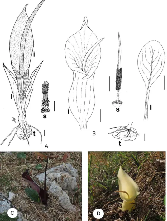
Biarum marmarisense and Biarum pyrami var. pyrami are distributed in the south of Turkey (Fig. 1). Biarum species is perhaps the most readily distin- guishable of the autumn flowering Biarum species due to the globose, strongly inflated, spathe tube, large spathe and greatly attenuated spadix appendix.
The bullate leaves of the typical variety are also unusual in the genus and pro- vide a ready means of identifying B. pyrami var. pyrami in Turkey, where no other species shares this character (Boyce 2008, Fig. 1). B. marmarisense, on the island of Simi in the eastern Aegean has shown that bees and queen wasps are attracted by the sweet lilac-like perfume emitted by the inflorescences (Boyce 2008).
Since the classical work by Solereder and Meyer (1928), there have been many publications on leaf and petiole anatomy of Araceae, and an extensive list of references can be found on French et al. (1995) and Keating (2000, 2003, 2004a). The recent extensive revision on the morphology of Biarum has been done by Boyce (2006, 2008). But, there is no study on B. marmarisense and B.
pyrami var. pyrami. The aim of the present paper to provide anatomical infor- mation about these species.
MATERIAL AND METHODS
Plants samples were collected from Antalya-Finike, which is situated in the SW part of Turkey, in October 2013. Taxonomic description of taxa was done according to Davis (1984). Some plants were dried according to her- barium techniques. Fresh samples and herbarium material were used in each case for experimental analysis and morphological measurements.
Plant samples were fixed in 70% alcohol for anatomical studies. Ana- tomical studies were carried out on fresh samples or samples kept in alcohol.
Hand-made cross-sections were prepared with razor blade from root, scape and leaves. The sections were stained by safranin/fast green dyeing method (Bozdağ et al. 2016). Anatomical measurements were made with the help of micrometric ocular. Results were presented by original drawings, photo- graphs and tables. Photographs were taken with Leica DM LB microscopy.
RESULTS Biarum pyrami var. pyrami
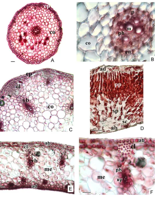
Root: The epidermis consists of elongated cells is two layered on the outer surface of the root. There is no cuticle layer on the epidermal cells. The cortex consisted of 6–8 layered parenchymatous cells has not got intercellu- lar spaces. The thickness of the cell wall of endodermis cells is not clear, but towards metaxylem, the cell walls are observed thicker. On the inside of the endodermis and the adjacent to it is a cylinder shape of parenchyma cells called the pericycle. This is one cell wide and consists of thin-walled cells.
Root xylem forms 4–5 strands which point toward the pericycle. There are one or more metaxylem on the midpoint of the vascular cylinder (Fig. 2A, B).
Fig. 1. Details and general appearance in natural habitat of Biarum pyrami var. pyrami (A, C) and B. marmarisense (B, D) (s = spadix, I = inflorescence, l = leaves, t = tuber. Scale bars: 1 cm)
Fig. 2. The cross-section of root (A, B), scape (C), leaf (D), spathe (E, F) of Biarum pyrami var.
pyrami (ab = abaxial epidermis, ad = adaxial epidermis, cl = collenchyma, co = cortex, en = endodermis, ep = epidermis, m = metaxylem, me = mesophyll, p = pericycle, ph = phloem, pp = palisade parenchyma, sc = substomatal cativity, sp = spongy parenchyma, vb = vascu-
lar bundle, xy = xylem. Scale bars: 50 µm)
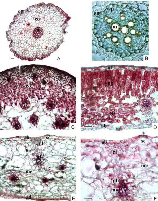
Fig. 3. The root of (A, B), scape (C), leaf (D) and spathe (E, F) cross-section of Biarum marma- risense (ab = abaxial epidermis, ad = adaxial epidermis, cl = chollenchyma, co = cortex, en = endodermis, ep = epidermis, m = metaxylem, me = mesophyll, p = pericycle, ph = phloem, pp = palisade parenchyma, s = stomata, sc = stomatal cavity, sp = spongy parenchyma, vb =
vascular bundle, xy = xylem. Scale bars: 50 µm)
Scape: The epidermis consists of elongated cells is a single layer of cell covering the entire of the scape. The presence of thick cuticle layer on the outer surface of the epidermal cells can be distinguished. The cortex com- posed of thin-walled and circular parenchyma cells with intercellular spaces
Table 1
Anatomical measurements of Biarum species (SD = standard deviation)
Width (µm) Length (µm)
min.–max. mean±SD min.–max. mean±SD
B. pyrami var. pyrami
Root, epidermis cell 13.60–27.20 18.20±4.37 10.88–19.04 15.54±2.89 Root, cortex cell (diam.) 32.64–59.84 44.20±5.70
Root, trachea (diam.) 9.52–16.32 12.58±2.36
Scape, epidermis cell 21.76–38.08 28.75±5.90 10.88–24.48 16.32±4.01 Scape, pith cell (diam.) 32.13–107.10 61.96±9.78
Scape, trachea (diam.) 9.52–21.76 14.96±4.41 Leaf, adaxial cuticle 2.72–5.44 4.08±1.07
Leaf, adaxial epidermis 13.60–21.76 18.49±3.01 16.32–27.20 23.39±4.00 Leaf, abaxial epidermis 13.60–21.76 18.49±3.02 16.32–24.48 20.12±2.48 Leaf, palisade parenchyma 10.88–19.04 14.96±2.51 21.76–35.36 28.10±4.32 Leaf, spongy parenchyma 10.88–27.20 19.04±5.26
Leaf, trachea 5.44–16.32 10.88±3.56
Leaf, abaxial cuticle 2.72–6.80 4.76±1.42 B. marmarisense
Root, epidermis cell 10.88–21.76 16.59±3.25 13.60–27.20 20.94±4.56 Root, cortex cell (diam.) 19.04–68.00 38.89±8.17
Root, trachea (diam.) 10.88–29.92 17.95±3.44
Scape, epidermis cell 10.71–32.13 17.24±4.15 10.71–53.55 26.77±5.78 Scape, pith cell (diam.) 42.84–117.8 70.60±9.64
Scape, trachea (diam.) 10.71–64.26 29.77±6.73 Leaf, adaxial cuticle 2.72–5.44 3.70±1.01
Leaf, adaxial epidermis 19.04–43.52 27.74±4.48 16.32–29.92 22.30±4.30 Leaf, abaxial epidermis 10.88–24.48 17.95±4.30 13.60–21.76 17.40±2.70 Leaf, palisade parenchyma 16.32–27.20 21.76±3.43 40.80–73.44 55.76±5.57 Leaf, spongy parenchyma 19.04–40.80 28.10±4.40
Leaf, trachea 5.44–13.60 9.61±3.01
Leaf, abaxial cuticle 2.72–8.16 5.44±1.82
is 8–10 layered under epidermis. A 10–12 collateral vascular bundles, which have thick-walled cells are arranged in a ring. The 8–9 layered collenchyma is present under the epidermis at the corners of scape. The xylem is observed in a larger area than the phloem (Fig. 2C). There are pith cells in the centre of scape. These cells are 32.13–107.10 µm in diameter (Table 1).
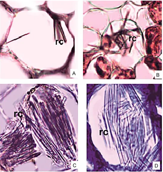
Leaf: The epidermis is a single layer of cells covering the abaxial and adaxial surfaces, where there is a thick cuticle layer. This layer is 2.72–6.80 µm. Two regions of the mesophyll are distinguishable, the uppermost con- sists of barrel-shaped palisade parenchyma cells that are in 2–3 rows and the lower region is consisting of loosely arranged spongy parenchyma cells are multi-layered with abundant air spaces between them (Fig. 2D). The raphides are seen in the mesophyll (Fig. 4A, B). The vascular bundles are arranged regularly in the centre of the mesophyll.
Spathe: Thick cuticle is present on adaxial and abaxial surfaces of the leaf. The abaxial epidermis is perforated by the stomata under which there is large substomatal cavity. Single layered hypodermis is situated on the in- side of the epidermis. The collenchymatous cells are present in the adaxial zones (Fig. 2E, F). Mesophyll layer is composed of homogeneous cells and also raphide crystals are observed there (Fig. 4A, B).
Biarum marmarisense
Root: The epidermis is two layered on the outer surface of the root. There is very thin or no cuticle layer on the epidermal cells. Cortex is composed of 10–12 layered parenchymatous cells and these cells become smaller towards to endodermis and epidermis. The cell wall is on the cortex-side of the endo- dermis is suberised. There is pericycle layer, which is formed thin-walled cells in a single row under the endodermis. Six–eight xylem strands are present on the periphery of the vascular cylinder of B. marmarisense (Fig. 3A, B).
Scape: There is a cuticular layer on the epidermal cells of the cross-sec- tion of the scape. The cortex, which is composed of multi-layered parenchy- ma cells adjacent to a single-layered epidermis, has some aerenchyma cells in which there are raphide crystals and tannin. Vascular bundles become com- pactly arranged towards to the pith (Fig. 3C).
Leaf: A thick cuticle layer covers the outer surface of the leaves. Abaxial cuticle is thicker than adaxial cuticle with 2.72–8.16 µm in size (Table 1). Leaf is bifacial, consisting of palisade and spongy parenchyma. Palisade paren- chyma is 3–4 layered and arranged very compactly. Spongy parenchymatic region is very narrow. Palisade parenchyma is 5–6 layered and covers almost equal area to spongy parenchyma (Fig. 3C, D, E). Raphide crystals are ob- served intensively in aerenchyma (Fig. 4C, D).
Spathe: There is a thick cuticle on both surfaces of the spathe. Towards to abaxial side, the collenchyma is situated on vascular bundles (Fig. 3E, F). Meso- phyll is homogeneous and raphide crystals are observed intensively (Fig. 4D).
DISCUSSION AND CONCLUSIONS
The anatomical analysis given in this work provides the first detailed description of Biarum pyrami var. pyrami and B. marmarisense, which is compa-
Fig. 4. Crystals in cross-section of Biarum pyrami (A, B) and B. marmarisense (C, D) (rc = raphide crystals)
rable with findings of some other examined Araceae members (Higaki et al.
1984, Mantovani and Pereira 2005, Mastroberti and Mariath 2003, Bercu and Făgăraş 2010).
Characteristically, the primary endodermis consists of slightly suberised cells possessing, at places, passing cells opposite xylem (Kroemer 1903). The present study showed that the thicknesses of the cell wall of the endodermis cells are not the same with each taxon; at Biarum marmarisense the cell walls are observed thicker on outwards, while at B. pyrami var. pyrami towards to the centre. Similarly Bercu and Făgăraş (2010) reported one-layered with slightly suberised cell walls in cross-section of root of Spathiphyllum wallisii Regel. B.
pyrami var. pyrami has 4–5 xylem strands, while B. marmarisense has 6–8 xylem strands. Bercu and Făgăraş (2002) and Batanouny (1992) have observed that the vascular system is quite well developed numbering in more than six other Araceae species, i.e. Monstera deliciosa Liebm. and Spathiphyllum Schott spp.
There is a hypodermis layer on the inside of the epidermis of scape of B. pyrami var. pyrami. The cross-section of the scape of B. marmarisense has large intercellular spaces in which there are some raphide crystals. Research- ers have observed druse and raphide crystals in mesophyll of Araceae before.
Also, they have noted that other crystals can be found in this layer (Genua and Hillson 1985, Keating 2003, 2004b, Mayo et al. 1997). Mantovani and Pereira (2005) have concluded that raphides of calcium oxalate crystals are found with low frequency, but druses are found very frequently. The raphides occur rarely and only on the spongy parenchyma while the druses are frequent in all mesophyll and in both surfaces in Anthurium Schott species. The presence, morphology and distribution of crystals within species are very important because it is under genetic control (Franceschi and Nakata 2005).
Epidermal cells in leaves of Biarum species are short, randomised, but the vascular bundles are elongated and distributed parallel to the longitudinal direction of the body as in Biarum species. This provision is generally found in leaves of monocotyledons (Metcalfe 1963). However, Mayo (1986) showed randomly distributed small cells in the epidermis of the spathe species of Philodendron Schott (Araceae). The number and distribution of cells subsidiar- ies of stomata are vary significantly in Araceae (French et al. 1995). In case of some plant species leaf anatomy and leaf epidermal anatomy can help identi- fication therefore it has got great importance sometimes (Hameed et al. 2002, Munsif et al. 2007). In this study paracytic stoma types have been observed in B. marmarisense, while B. pyrami var. pyrami has anomocytic stoma types.
The cross section of spathe of mesophyll is uniform, showing no differ- entiation between palisade and spongy parenchyma of spathe of both species.
The mesophyll cells have compact arrangement of Biarum species. The same feature has been observed in Anthurium species (Mantovani et al. 2010). Ac-
cording to Keating (2000), the expression of collenchyma appears to be com- plex in this family. That is, its position in an organ is reliable as an indicator of some suprageneric categories. On the adaxial surface of the palisade paren- chyma is usually interrupted by a collenchymatous tissue in both species. The same feature is observed among other members of the Araceae family (Man- tovani and Pereira 2005, Bercu and Făgăraş 2010). Batanouny (1992), Fahn (1990) and Bercu (2009) have concluded that it is a characteristic feature that for Araceae species both epidermis continuity is broken by the presence of stomata with large substomatal cavities. Druse crystals are seen in some of the mesophyll cells. This substomatal cavity has been seen in both Biarum taxa.
As a result, this work points out that B. pyrami var. pyrami is compared with B. marmarisense. However, further research with the other Araceae spe- cies as in a recently done study may illuminate whether any taxonomic im- plication is present or not within the genus Biarum or in the family. We expect these findings to contribute to the further phylogenetic and taxonomic studies into the Biarum species.
REFERENCES
Ajaib, M., Khan, Z., Khan, N., Abbas, A. M., Shahwar, D., Wahab, M. and Saddiqu, F. M.
(2011): Antibacterial and antioxidant activities of an ethnobotanically important plant Sauromatum venosum (Ait.) Schott. of district Kotli, Azad Jammu et Kashmir.
– Pak. J. Bot. 43(1): 579–585.
Akan, H. and Balos, M. M. (2008): Check-list of the genus Biarum Schott in the Flora of Turkey, with a new record for Turkey: Biarum syriacum (Spreng.) H. Riedl. – Turk. J.
Bot. 32: 305–310.
Algan, G. (1981): Microtechnics for the plant tissues. Nr 1. – Publication of Fırat Univ. Science et Art Faculty, Istanbul.
Anonymous (2013): The Plant List. Version 1.1. – Published on the Internet; http://www.
theplantlist.org/ (accessed 1st January).
Anonymous (2014): “Araceae”. – Iowa State University. (retrieved 17 January 2014.– http://
www.eeob.iastate.edu/classes/bio366/families/Araceae.pdf) Batanouny, K. H. (1992): Plant anatomy. – University Press, Cairo.
Bercu, R. (2009): Two aroids anatomical comparative features in accordance with their habit. – Analele Univ. Craiova. Ser. Biol., Hortic., Tehn. preluc. produ. agr., Ing. med. 14:
413–424.
Bercu, R. and Făgăraş, M. (2002): Lucrări practice de Fiziologie vegetală. – “Ovidius” Univer- sity Press, Constanţa, 118 pp.
Bercu, R. and Făgăraş, M. (2010): Anatomical aspects of the ornamental plant Spathiphyl- lum wallisii Regel. – Stud. şi Cerc. Ştiinţ. Biol., Ser. Biol. veg., Univ. din Bacău, 18: 13–17.
Bown, D. (2000): Aroids: plants of the Arum family. – Timber Press, Portland, 398 pp.
Boyce, P. C. (2006): A taxonomic revision of Biarum (Araceae). – Aroideana 29: 2–36.
Boyce, P. C. (2008): A taxonomic revision of Biarum (Araceae). – Curtis’s Botanical Magazine 25(1): 2–17. https://doi.org/10.1111/j.1467-8748.2007.00607.x
Bozdağ, B., Kocabaş, O., Akyol, Y. and Özdemir, C. (2016): Bitki Anatomisi Çalışmalarında El Kesitleri İçin Yeni Boyama Yöntemi. (A new staining method for hand-cut in plant anatomy studies). – Marmara Pharm. J. 20(2): 184–190. https://doi.org/10.12991/
mpj.20162044231
Davis, P. H. (1984): Flora of Turkey and the East Aegean Islands. Vol. 8. – Edinburgh Univ.
Press, Edinburgh.
Fahn, A. (1990): Plant anatomy. 4th ed. – Pergamon Press, Oxford.
Franceschi, V. R. and Nakata, P. A. (2005): Calcium oxalate in plants: formation and function. – Annu. Rev. Plant Biol. 56: 41–71. https://doi.org/10.1146/annurev.ar- plant.56.032604.144106
French, J. C., Chung, M. and Hur, Y. (1995): Chloroplast DNA phylogeny of the Ariflorae. – In:
Rudall, P. J., Cribb, P. J., Cutler, D. F. and Gregory, M. (eds): Monocotyledons: sys- tematics and evolution. Academic Press, London, pp. 255–275.
Genua, J. M. and Hillson, C. J. (1985): The occurrence, type and location of calcium oxalate crystals in the leaves of fourteen species of Araceae. – Ann. Bot. 56: 351–361. https://
doi.org/10.1093/oxfordjournals.aob.a087021
Hameed, M., Mansoor, U., Ashraf, M. and Rao, A. R. (2002): Variation in leaf anatomy in wheat germplasm from varying drought-hit habitats. – Int. J. Agr. Biol. 4(1): 12–16.
Higaki, T., Rasmussen, H. P. and Carpenter, W. J. (1984): A study of some morphological and anatomical aspects of Anthurium andreanum Lind. – Res. Ext. Ser. Hawaii Inst. Trop. Ag- ric. Hum. Resour. No. 030. Available from http://www.ctahr.hawaii.edu/oc/freepubs/
pdf/RS-030.pdf [accessed 7 July 2014]
Keating, R. C. (2000): Collenchyma in Araceae: trends and relation to classification. – Bot. J.
Linn. Soc. 134: 203–214. https://doi.org/10.1111/j.1095-8339.2000.tb02351.x
Keating, R. C. (2003): Anatomy of the Monocotyledons. IX. Acoraceae and Araceae. – Oxford University Press, Oxford, UK.
Keating, R. C. (2004a): Vegetative anatomical data and its relationship to a revised classifi- cation of the genera of Araceae. – Ann. Mo. Bot. Gard. 91: 485–494.
Keating, R. C. (2004b): Systematic occurrence of raphide crystals in Araceae. – Ann. Mo. Bot.
Gard. 91: 495–504.
Kroemer, K. (1903): Wurzelhaut, Hypodermis und Endodermis der Angiospermenwurzel. – Bib- liotheca botanica Vol. 59, Verlag Erwin Nägele, Stuttgart, 151 pp.
Mantovani, A. and Pereira, T. E. (2005): Comparative anatomy of leaf and spathe of nine species of Anthurium (section Urospadix; subsection Flavescentiviridia) (Araceae) and their diagnostic potential for taxonomy. – Rodriguésia 56(88): 145–160.
Mantovani, A., Filartiga, A. L. P. and Nadruz, M. A. C. (2010): Anatomia comparada da fol- ha e espata de espécies de Anthurium (Araceae) ocorrentes na Mata Atlântica. – Braz.
J. Bot. Rev. Bras. Bot. 33(1): 185–200. https://doi.org/10.1590/s0100-84042010000100016 Mastroberti, A. A. and Mariath, J. E. A. (2003): Leaf anatomy of Araucaria angustifolia (Ber- tol.) Kuntze (Araucariaceae). – Rev. Bras. Bot. 26(3): 343–353. https://doi.org/10.1590/
s0100-84042003000300007
Mayo, S. J (1986): Systematics of Philodendron Schott (Araceae) with special reference to inflores- cence characters. – PhD Thesis, University of Reading, Reading.
Mayo, S. J., Bogner, J. and Boyce, P. C. (1997): The genera of Araceae. – Kew Publishing, Kew, London.
Metcalfe, C. R. (1963): Comparative anatomy as a modern botanical discipline, with special reference to recent advances in the systematic anatomy of monocotyledons. – Adv.
Bot. Res. 1: 101–147. https://doi.org/10.1016/s0065-2296(08)60180-9
Mohammed, T. R., Khalifa, S. F. and Salah El-Dine, R. M. (2006): Leaf protein electrophoret- ic profiles and chromosome numbers of some Araceae. – Int. J. Agr. Biol. 8(2): 231–234.
Munsif, S., Khan, M. A., Ahmad, M., Zafar, M., Mujtaba, S. G. and Shaheen, N. (2007): Leaf epidermal anatomy as an aid to the identification of genera Lantana, Verbena and Vitex of family Verbenaceae from Pakistan. – J. Agr. Soc. Sci. 3(2): 43–46.
Rajput, M. T., Naqvi, S. N. H., Choudhary, I. M. and Abbas, A. (2010): Importance and im- plementation of essential oil of Pakistanian Acorus calamus Linn., as a biopesticide, – Pak. J. Bot. 42(3): 2043–2050.
Solereder, H. and Meyer, F. J. (1928): Systematische Anatomie der Monocotyledonen. Heft III.
Principles, Synathae, Spathiflorae. – Borntrager, Berlin, 100 pp.