VII. 3. FUNCTION OF PROTEIN DISULFIDE REDUCTASE IN CELLULAR DIVISION OF YEASTS
Walter J. Nickerson and G. Falcone
The Institute of Microbiology, Rutgers · State University, New Brunswick, New Jersey
I. Introduction 409 II. Polysaccharide-Protein Complexes in Cell Wall of Yeasts 410
III. Enzymatic Reduction of Disulfide Bonds in Protein Components of Yeast
Cell Walls 413 IV. Identification of Protein Disulfide Reductase as an Enzymatic Process Op-
erating Specifically in Cellular Division in Yeasts 414 1. Growth of C. albicans (Divisionless Strain) in a Sulfur-Deficient Medium 416
2. Form of Growth Under Conditions of Sulfur Deficiency 417 V. Physical Consequences of Cross-Linkage of Cell Wall Polymers . . . . 418
1. Establishment of a Localized, Highly Plastic Area of the Cell Wall . 418
2. Orientation of a Fibrillar Component of the Cell Wall 420
VI. Summary 421
I. Introduction
An essential, but largely unidentified, role in cellular division processes has long been attributed to the sulfhydryl (—SH) group (1-3; see reviews 4, 5). Experimental evidence for a role of —SH in cellular division (as distinct from its function in many enzymatic and group transfer reactions important in cellular growth) has been accumulating slowly. Rapkine (3) demonstrated a cyclic alteration in soluble —SH during division of sea urchin eggs in which a maximum in —SH concentration was attained im- mediately prior to division. The observation has been made with a variety of microorganisms, in which division could be selectively inhibited so that growth in the absence of cellular division occurred at a rate not appre- ciably less than in dividing material, that the addition of an exogenous source of —SH substance restored cellular division (6). As a corollary observation, failure of cellular division in Candida albicans is accompa- nied by a loss of cysteine from the cells and, between closely spaced streaks of growth, the concentration of secreted cysteine is usually suffi- cient to prevent filament formation ( 7 ) . On a polysaccharide medium, free of readily metabolizable carbon source, C. albicans readily develops in a filamentous form. Addition to the medium of either glucose or an —SH
409
substance resulted in restoration of cellular division and maintenance of the yeast phase (8). The existence of enzymatic reactions linking glucose metabolism and — S — S — reduction was postulated (9). Three disulfide reductases have been uncovered to date—i.e., glutathione reductase which operates via reduced triphosphopyridine nucleotide (TPNH) (10, 11), cystine reductase which employs D P N H (12, IS) and, most recently, pro- tein disulfide reductase (14) -
In purified cell wall preparations isolated from yeasts that had been mechanically disintegrated, the presence of protein, bound to polysaccha- ride, has been demonstrated (15). A glucomannan-protein was isolated from such cell wall preparations and its properties have been studied. The protein has a relatively high content of sulfur (2.1%) which is present largely as cystine-sulfur. An enzyme system was demonstrated to be pres- ent in mitochondria of yeast and to catalyze reduction of disulfide bonds in this glucomannan-protein. The enzyme was termed protein disulfide reductase ( P D S reductase), and shown to play a role in cellular division of yeasts (16).
II. Polysaccharide-Protein Complexes in Cell Wall of Yeasts
From cells of baker's yeast that had been mechanically disintegrated by agitation with glass beads in a Waring blendor (17), a fraction con- sisting of cell wall fragments was isolated (15). B y repeated washing and differential centrifugation, preparations of clean cell walls were obtained that were free of other particulate material, when examined in the light microscope or electron microscope (Fig. 1).
Analyses on the clean cell wall material reveal (Table I) it to be com- TABLE I
COMPOSITION OF ISOLATED, CLEAN CELL WALLS OF BAKER'S YEAST
Component Per cent by weight of dry cell wall Total reducing sugar 84.4
2.7 1.28 0.21 1.27 6.7 8.1 1.2 6.9 Hexosamine
Total nitrogen Hexosamine Ν Protein Ν Protein (Ν X 6.25) Total lipid
Free lipid Bound lipid
P D S R E D U C T A S E I N C E L L U L A R D I V I S I O N O F Y E A S T S 411
FIG. 1 (Top). Isolated cell wall fragment from Candida albicans strain 806 as seen in electron microscope ; 50,000 X. Fibrillar structures are apparent.
FIG. 2 (Bottom). Appearance of aggregates of glucomannan-protein I (GMP-I), from cell wall of baker's yeast, in electron microscope; 100,000 X. Preparation had been dissolved in water and evaporated on film.
posed principally of polysaccharide; in addition, there is present about 7% protein, and about 8% total lipid. Based on analyses for hexosamine, a small amount of chitin (about 3%) is also present. Assuming that the polysaccharide content of the wall cannot exceed 90% of the value for total reducing sugar, a maximum content of polysaccharide (other than chitin) of about 76% is present in the wall. The total for these four frac- tions amounts to about 95% of the dry weight of the cell wall. The sulfur present amounts to more than 2% of the protein content. In view of the presence of appreciable amounts of cysteic acid in hydrolyzates, the ab- sence of methionine, and failure to detect any other S-containing sub- stances, it is assumed that sulfur is present in the wall in cystine-cysteine.
The presence of appreciable absorption in the 2,500-2,600 c m .- 1 region (attributable to —SH stretching) in infrared absorption spectra of this cell wall material, supports this assumption.
Upon suspending cell wall preparations, from which lipid had not been removed, in Ν KOH, and warming the suspension briefly in a steam bath to 50°, approximately 75% of the wall material was solubilized. The solu- ble fraction was dialyzed against running tap water for 24 hours, and then lyophilized to give a white powder. This fraction was further purified by precipitating it from aqueous solution with ammonium sulfate. The precipitated materials was dialyzed, and lyophilized to yield a white powder that was readily soluble in water. On acid hydrolysis under ni- trogen, and subsequent chromatographic analysis, this material was found to be composed of polysaccharide and protein; 15 amino acids were iden- tified in the hydrolyzate; and mannose and glucose were present in approximately equal amounts. This fraction, therefore, will be termed glucomannan-protein I (GMP-I).
Examination of GMP-I in the ultracentrifuge revealed it to be mono- disperse with S2o = 4.3 X 1 0 ~1 3 (uncorrected). Electronmicrographs of solutions of GMP-I evaporated on film revealed it to be composed of aggregated globular particles, of rather uniform size (Fig. 2 ) .
Recently, in our laboratory, Dr. Gian Kessler has fractionated lipid- free cell walls of baker's yeast. When this starting material is suspended in Ν KOH for 1 hour at 25°, an almost clear solution of the entire wall is obtained that passes an ultrafine sintered glass filter. After dialysis for 24 hours against running tap water, followed by lyophilization, a white powder is obtained. The powder was suspended in water at 25° for 1 hour, then centrifuged. Approximately 70% of the powder had dissolved. The residue was washed and lyophilized. The centrifugate was saturated with ammonium sulfate and a precipitate was obtained that is similar to GMP-I. From the supernatant, after dialysis and lyophilization, a white powder was obtained that proved to be a polysaccharide-protein with
P D S R E D U C T A S E I N C E L L U L A R D I V I S I O N O F Y E A S T S 413 mannose and glucose present in a ratio of approximately 2:1. This fraction is termed glucomannan-protein II (GMP-II). The water insoluble residue proved to be still a third polysaccharide-protein fraction ; the polysaccha- ride is composed very largely of glucose (mannose was present only in small amount). This fraction, termed glucan-protein, comprised approxi- mately 30% of the weight of the wall material.
III. Enzymatic Reduction of Disulfide Bonds in Protein Components of Yeast Cell Walls
As previously mentioned, the major portion of the sulfur present in isolated, clean cell walls of baker's yeast appears to be present in — S — S — or —SH form. The same holds true for cell walls of normal and divisionless strains of C. albicans. In light of our earlier work on disulfide reductases, it was natural for us to examine the possibility that yeast possessed an en- zymatic mechanism for reducing disulfide bonds in its pseudokeratinous cell wall protein.
To this end, aqueous solutions of GMP-I were prepared; the —SH groups of the protein were oxidized with 0.001 M ferricyanide by the method of Anson (18), and the oxidized GMP-I was incubated with a mitochondrial particulate fraction from yeast (14)- To these components,
T A B L E I I
ENZYMATIC REDUCTION OF — S — S — IN CELL WALL PROTEIN (FRACTION G M P - I ) OF YEASTS
Δ Optical density SH-mercaptide& equivalent Reaction system0 255 πΐμ (as cysteine) (Χ 10~δ M) 1. Enzyme from baker's yeast
G M P - I (active enzyme) 0.124 1.4
G M P - I (boiled enzyme) - 0 . 0 0 2 zero 2. Enzyme from C. albicans (582)
G M P - I (582) 0.212 2.4
G M P - I (806) 0.063 0.8
3. Enzyme from C. albicans (806)
G M P - I (806) 0.017 0.18
G M P - I (582) - 0 . 0 0 7 zero
° The components indicated were added to a basal mixture containing succinate, 10 mg.; ethanol, 4.5 mg.; liver coenzyme concentrate (Armour), 0.5 mg.; Af/50 phos- phate buffer, pH 7.0, reaction volume 3.8 ml., and incubated at 37° for 2 hours.
b For determination of sulfhydryl content of protein, the above mixtures were centrifuged at 22,000 g for 20 minutes to remove particulate matter; 2.0 ml. samples of the clear supernatant were taken and added to 1.0 ml. of 0.3 M acetate buffer, pH 4.6, and 0.5 ml. 1.2 X 10~4 M p-chloromercuribenzoate. Mercaptide formation was allowed to proceed for 2 hours at 37°, and then determined at 255 τημ.
414
succinate and ethanol were added as hydrogen donors, and liver coenzyme concentrate as a source of cofactors. The system was incubated for 2 hours at 37°, after which time particulate matter was removed by cen- trifugation at 22,000 X g. Increase in the —SH content of GMP-I, con- tained in the supernatant, was estimated spectrophotometrically by mer- captide formation with p-chloromercuribenzoate, according to the method of Boyer (19).
As shown in Table II, the complete system from baker's yeast, con- taining oxidized cell wall protein (equivalent to about 10.8 X 1 0 ~5M sulfur), mitochondrial particulates, and 2 Χ 10~5M p-chloromercuriben- zoate, showed mercaptide formation equivalent to 1.4 X 1 0_ 5M (cys- teine) after a reaction time of 2 hours. The same system with heated par- ticulates had no greater absorbancy than the sum of its components. This analysis constitutes evidence for the formation of sulfhydryl groups in the cell wall protein on incubation with an active enzyme preparation. Disul- fide reductase systems have been described that operate on oxidized gluta- thione and on cystine, but this is the first system to be described that
accomplishes enzymatic reduction of disulfide linkages of a protein.
IV. Identification of Protein Disulfide Reductase as an Enzymatic Process Operating Specifically in Cellular Division in Yeasts
The existence of a pseudokeratinous type of protein bound to a gluco- mannan component of the cell wall of baker's yeast has been mentioned.
Enzymatic reduction of disulfide linkages in this protein has been accom- plished by the use of cell-free particulate preparations from baker's yeast
(14)' Pseudokeratinous glucomannans have recently been isolated from the clean cell walls of both normal and filamentous mutant strains of the yeast C. albicans (16). Mitochondrial particulates obtained from the nor- mal strain of C. albicans show powerful protein disulfide reductase activity toward its cell wall glucomannan-protein, whereas similar preparations from a divisionless (filamentous) mutant possess only slight protein di- sulfide reductase activity.
Previous work in this laboratory (20) had shown that the divisionless mutant of C. albicans suffers a biochemical lesion at a reductive reaction such that metabolically generated hydrogen is "spilled over" in quantity during growth for nonspecific reductions (added dyes, and tetrazolium compounds, with redox potentials as low as E0' = —0.150 volts, are readily reduced by growing cells of the mutant, but not by the parent strain).
This "waste" of reductive capacity by the mutant is not at the expense of demands of Η-acceptors participating in syntheses essential for growth,
PDS REDUCTASE IN CELLULAR DIVISION OF YEASTS 415
GMP-I of normal GMP-I of mutant
Enzyme of normal strain 24.0 8.0
Enzyme of mutant strain zero 1.0
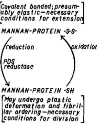
Enzymatic reduction of disulfide bonds in glucomannan-pseudokera- tin, a major structural component of the cell wall of yeasts, is thus shown to be a reaction essential for cellular division of yeasts. The cycle of oxi- dation-reduction of —SH ^± — S — S — in this complex substrate is dia- grammed in Fig. 3, and the consequences of breaking covalent bonds in this polymeric fabric are indicated. Further consideration of these effects is taken up in Section V of this paper. In the two subsections to follow, a completely different experimental approach is described that supports these conclusions.
since mutant and normal strains synthesize cell mass at approximately the same rate, nor is it at the expense of respiration since the mutant re- duces oxygen even more rapidly than does the normal strain (21). Our recent findings permit identification of the "H-acceptor participating in division" as the disulfide bond of the cell wall glucomannan-protein com- ponent, and the protein disulfide reductase, which catalyzes this reduction, may be termed a "cytokinetic enzyme."
The normal strain of C. albicans (582) and the filamentous, division- less mutant (strain 806) were grown in large batches in a synthetic me- dium with continuous agitation at 28° for 48 hours in order to obtain a sufficient weight of cells for the preparation of isolated clean cell walls, and to obtain cells from which mitochondrial particulates were isolated.
Glucomannan-protein was solubilized from isolated clean cell walls ; —SH of the protein was oxidized with ferricyanide, and the resulting oxidized cell wall protein was incubated with active or boiled mitochondria isolated from both strains of yeast. After incubation, the reaction mixture was centrifuged at high speed to remove particulates, and the —SH content of the protein was assayed spectrophotometrically by mercaptide forma- tion with p-chloromercuribenzoate.
As shown in Table II, mitochondrial particulates isolated from the normal strain of C. albicans exhibit vigorous protein disulfide reductase activity against the cell wall protein obtained from this strain and from the mutant. In contrast, the mutant exhibits only slight enzymatic reduc- tion of its own cell wall — S — S — linkages.
The relative activities, per unit weight of mitochondrial nitrogen, are as shown in the following tabulation.
Covalent bonded; presum- ably elastic-necessary conditions for extension MA NN A N-PRO TE IN -5-5-
MA NN A Ν-PRO TE IN SH May undergo plastic deformation and fibril- lar ordering-necessary conditions for division
FIG. 3. Outline of role of protein disulfide (PDS) reductase in chain of events re- sulting in cellular division in yeasts.
1. GROWTH OF C. albicans (DIVISIONLESS STRAIN) IN A SULFUR-DEFICIENT MEDIUM
Although sulfur is required for growth of all cells, it is not commonly a growth-limiting factor. Experimental conditions have been created whereby the growth of a yeast (strain 8 0 6 of C albicans) is proportional to the sulfur contamination of the medium. After more than 5 0 transfers of this yeast in a minus-S medium, growth to an optical density of 0 . 1 0 0 - 0 . 1 2 5 (approximately 2 3 - 2 5 mg. dry weight per 1 0 0 ml. of medium) has routinely been observed after 2 4 hours incubation—growth undoubtedly permitted by sulfur present as contamination (estimated to be somewhat less than 0 . 4 parts S per million) in the reagent chemicals employed in medium No. 2 - S . On the basis of culture turbidity, growth in a minus-S medium is about one-third of that produced in the complete medium. As will be described, cells grown in the minus-S medium are of the normal yeast form.
Analytical data comparing the content of sulfur, phosphorus, and ni- trogen of cells grown in the complete medium with those grown in the sulfur-deficient medium are shown in Table III. It will be seen that the sulfur content of cells grown in the sulfur-deficient medium is less than half that of cells grown in the complete medium. A corresponding reduc- tion in nitrogen content, and therefore presumably in protein, has been produced in the sulfur-deficient cells. In contrast, the phosphorus content of the two types is essentially the same, but the respective P / N ratios have been greatly altered.
Although most yeasts can utilize the sulfur of inorganic sulfate, far fewer can utilize organic compounds of sulfur for growth (22). In addi- tion to inorganic sulfate, C. albicans can utilize the sulfur of methionine,
PDS REDUCTASE IN CELLULAR DIVISION OF YEASTS 417 TABLE III
GROWTH AND COMPOSITION OF CELLS OF C. albicans 8 0 6 GROWN IN COMPLETE MEDIUM OR IN SULFUR-DEFICIENT MEDIUM FOR 2 4 HOURS AT 2 8 °
Measure
Complete medium (No. 2 )
Sulfur-deficient medium (No. 2-S) Sulfur content of medium 7 8 m g . / 1 0 0 ml. < 0 . 0 4 m g . / 1 0 0 ml.
Growth (mg. dry weight/100 ml.
medium) 1 2 0 mg. 2 3 mg.
Sulfur content of cells from 1 0 0 ml.
medium 0 . 2 5 mg. 0 . 0 2 1 mg.
Composition of cells"
Sulfur 0 . 2 0 7 0 . 0 9 3
Phosphorus 1.76 1.70
Nitrogen 4 . 7 6 2 . 7 6
° Per cent of dry weight.
cysteine, and glutathione. The addition of 200 /xg. of sulfur to medium No.
2-S results in more than twice the growth obtained in the absence of added sulfur. The sulfur of methionine and of sulfate are equally available to the organism.
2. FORM OF GROWTH UNDER CONDITIONS OF SULFUR DEFICIENCY In the absence of added sulfur the divisionless filamentous strain of C. albicans grows almost entirely in a budding yeast form. On the addition of a source of sulfur, there ensues a reversion to the filamentous form of growth. Thus, the condition of sulfur deficiency has brought about an almost complete reversion of the filamentous form to the yeast type of growth. After many passages in the sulfur-deficient medium, the division- less strain grows with only 10 to 20% of the cells in a filamentous form.
The form of growth is principally as a budding yeast, or a short chain of somewhat elongated cells that we term a "transition yeast form."
The cause of the promotion of a yeast form of growth in the sulfur- deficient medium is evidently through environmental control of a physio- logical process. Selection of a yeast type in the inoculum has been excluded by simultaneous transfer at each passage of the culture into complete, as well as into sulfur-deficient, medium. Strain 806 grows abundantly, in its typical filamentous fashion, in the complete medium even after more than 50 transfers in No. 2-S medium. Thus, there can be no question of genetic selection of a yeast form in the sulfur-deficient medium. The proportion of cell types appearing on transfer to complete and sulfur-deficient media gives no indication of selection, but, rather, demonstrates clearly the role of environmental control on the division process.
V. Physical Consequences of Cross-Linkage of Cell Wall Polymers In an attempt to analyze the physical consequences that follow upon the formation and breakage of covalent bonds among the molecular fibrils of which the cell wall fabrics of yeasts are composed, it will be interesting first to consider the "explosive" initiation of buds, as revealed by time- lapse cinematography.
1. ESTABLISHMENT OF A LOCALIZED, HIGHLY PLASTIC AREA OF THE CELL WALL
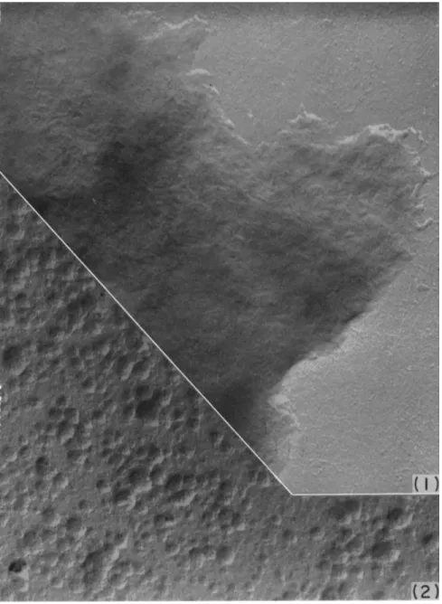
Examination of individual frames of time-lapse cinematographs (dark- field microscopy) of budding yeast reveals, in a most striking manner, that a bud-initial arises as a "blow-out" from the mother cell. As seen in con- secutive exposures at intervals of 30 seconds (Fig. 4 ) , from an apparently intact, thick cell wall, there is extruded in an explosive manner through a gaping hole a naked spherical bud-initial. From the wall of the mother cell at the base of the bud there rapidly ensues formation of wall substance which soon covers the surface of the expanding bud. These observations imply that the wall of a bud arises from pre-existing wall material by continuous growth of the latter.
From the sequence of photographs, shown in Fig. 4 , it may be deduced that a localized weakening of the cell wall resulted in a condition that could not withstand the internal fluid pressure; consequently, the cell wall ruptured at the weakened site, and a protoplasmic mass was extruded as a sphere (physical limitations dictate that the extruded liquid assume a spherical shape).
The manner in which a circumscribed portion of the cell wall appar- ently becomes the "target" upon which the plasticizing action of P D S reductase is concerted merits attention. In the first place, the location of the site of budding is not random, and the position of the bud with respect to the mother cell is not haphazard. A bud arises from a cell at the point of maximum curvature. Now, a static fluid exerts pressure uniformly against the wall of its container, irrespective of the shape of the container.
A fluid in motion, through inertial properties, exerts greatest force against the wall of its container in the region of maximal curvature of the con- tainer. But, selection of a point in a "permitted" annulus of maximal curvature, as the site against which plasticizing action is directed, dictates that the plasticizing action be concentrated in discrete particles of "point"
size, and excludes the possibility that the plasticizing action could be uniformly distributed in the liquid. The fact that P D S reductase activity is confined to the mitochondrial particulate fraction in cell-free prepara- tions of yeast is in agreement with the theoretical limitations.
PDS REDUCTASE IN CELLULAR DIVISION OF YEASTS 419
FIG. 4. Process of budding in baker's yeast (Saccharomyces cerevisiae), dark-field illumination; time-lapse sequence; rows A to C are in sequence with intervals of 30 seconds between each of the 12 exposures, photos in row D follow after a lapse of 15 minutes. First three exposures show parent cell before bud-initial emerges; fourth exposure (marked by arrow) shows gap in cell wall where bud has been "blown out."
The time interval between exposures 3 and 4 is 30 seconds. (The texture of the bud- initial is so thin that it can only be made visible by developing the print especially for it). The wall of the bud-initial thickens gradually. Particulate bodies can be seen to move into and out of the bud; in the cells marked by arrows in rows Β and C , particulates (mitochondria) are visible in the canal between the two parts of the cell.
In row D , the bud has enlarged and its wall has thickened ; the wall of the parent cell at the site of budding has been rebuilt. The canal between the two parts of the cell has narrowed considerably and the bud will shortly be completely cut off as an inde- pendent cell.
2. ORIENTATION OF A FIBRILLAR COMPONENT OF THE CELL WALL Electron micrographs of yeast cells reveal that polymer formation in the direction of growth develops as a densely intermeshed fibrillar net- work appropriate to an expanding prolate spheroid (Fig. 5 shows the pat- tern of fibrils in the glucan layer of the wall). It is important to note that covalent bonding between fibrils of the wall components (mannan and its
FIG. 5. Appearance of fibrils in glucan layer of yeast cell wall as seen in electron microscope; magnification indicated by micron scale. Note orientation of fibrils in cable-like fashion to create a "bud scar"; contrast with random disordered condition in other region of wall. From Houwink and Kreger (23).
sulfur-containing protein; glucan and the protein associated therewith) that are not linearly ordered, very likely serves to increase the modulus of elasticity in proportion to the number of covalent bonds. This feature of the wall fabric is undoubtedly essential for the expanding spheroid.
The "ring type" of ordered fibrillar structure observed in "bud scars"
serves to "constrict" the base of the bud in a plane normal to the direction of growth of the bud. This arrangement of fibrils is beautifully illustrated in electron microscope photographs of the yeast glucan layer by Houwink and Kreger (23). It is in the ordering of these fibrils that covalent bonding is believed to interfere. This may be a second stage in the division process
PDS REDUCTASE IN CELLULAR DIVISION OF YEASTS 421
during which maintenance of a sulfhydryl condition in a structural poly- mer is temporarily essential. Covalent — S — S — bonds might be formed, however, after the ordered arrangement has been achieved. The molecular events in cellular division that have been uncovered thus far bring into fold many scattered, apparently unrelated observations on the environmental control of cellular division. It is now intelligible, for example, that sulfhy- dryl substances applied externally to growing, but nondividing, cells might induce division in such cells (6"). It is becoming increasingly clear that a cell possesses a variety of systems, each with its degree of specificity, for maintaining functional —SH groups. Those employed in growth may be operative while a disulfide reductase essential for division may have failed.
In fact, this exact situation is met in the filamentous strain of C. albicans, which possesses both an active glutathione reductase and cystine reduc- tase, but is deficient in a protein disulfide reductase.
VI. Summary
The hypothesis has been advanced that growth of a cell in length re- quires the formation of stabilizing linkages between molecular fibrils of the form-determining matrix of the cell wall. On the other hand, division of a cell necessitates rupture of some of the linkages of the matrix so as to achieve a localized area of increased plasticity W). The nature of this matrix, and of a stabilizing intermolècular linkage, has been determined in one instance. In purified cell wall preparations isolated from yeasts that had been mechanically disintegrated, the presence of polysaccharide- bound protein has been demonstrated. A glucomannan-protein was iso- lated from such cell wall preparations and its properties investigated. The protein of this complex was found to have a relatively high content (2.1%) of sulfur—present largely as cystine-sulfur—and may be termed a pseudo- keratin.
Several lines of attack have been employed to investigate the role of cell wall polysaccharide-protein complexes in the growth and division of yeasts. Three of these approaches have been based on the pseudokeratinous nature of the glucomannan-protein. An enzyme capable of bringing about reduction of disulfide bonds of glucomannan-protein was found to be pres- ent in mitochondrial particulates of yeasts. This enzyme, termed protein disulfide (PDS) reductase, was found to be almost completely absent in a rapidly growing, divisionless mutant of a yeast. The participation of P D S reductase in the establishment of localized areas of increased plastic- ity in cell walls (by rupture of covalent bonds among fibrillar elements, as a result of enzymatic reduction of — S — S — to —SH) has been indicated.
A second approach grew out of the observation that addition of selenite
or tellurite to culture media served to overcome the enzymatic block in a divisionless strain of yeast. Selenium and tellurium, the more metallic elements of the sulfur family, were found to be incorporated into cell wall proteins. In contrast to the sulfhydryl (—SH) grouping, the selenhydryl (—SeH) and tellurhydryl (—TeH) groupings are more stable in the re- duced form, and thus appear to offset the deficiency of P D S reductase in the divisionless mutant (24).
Another experimental approach in our effort to gain an understanding of the role of pseudokeratinous wall proteins has been an attempt to con- trol the sulfur content of these structures through limitation of the sulfur content of the growth medium. Restriction in the amount of sulfur avail- able in a medium results in a lowered sulfur content of the cells grown therein. With a lower total sulfur content, the divisionless mutant yeast (despite its low P D S reductase activity) achieves relatively more exten- sive reduction of disulfide sulfur in its wall protein and, thereby, achieves in this wall component the requisite degree of plasticity that appears to be a necessary condition for establishment of a budding yeast form. Con- siderations, based on the behavior of other polymer systems [as described by Flory (25) ], of the fibrillar structure of the yeast cell wall as seen in electron micrographs, indicate that the circular ordering of fibrils compos- ing a bud scar can only result from the action of a force on a polymeric fabric in a plastic condition, i.e., one in which covalent or other type bond- ing between fibrils is minimal. Further, it might be predicted that limita- tion in the sulfur content of a growth medium would favor development of the yeast form in divisionless strain 806 of C. albicans, provided that the very low level of P D S reductase activity in the mutant was not abol- ished. Cells grown in a sulfur-deficient medium were found to have a diminished sulfur content and to develop predominantly in the yeast form.
REFERENCES 1. F. S. Hammett, Protoplasma 7, 297 (1929).
2. C. Voegtlin and H. W. Chalkley, Public Health Repts. (U. S.) 45, 3041 (1930).
3. L. Rapkine, Ann. physiol. physicochim. biol. 7, 383 (1931).
4. W.J. Nickerson, Nature 162, 241 (1948).
5. D. Mazia, in "Glutathione" (S. Colowick, A. Lazarow, E. Racker, D. R. Schwarz, Ε. Stadtman, and H. Waelsch, eds.), p. 209. Academic Press, New York, 1954.
6. W. J. Nickerson and N. J. W. Van Rij, Biochim. et Biophys. Acta 3, 461 (1949).
7. W. J. Nickerson, J. Infectious Diseases 93, 43 (1953).
8. W. J. Nickerson and C. W. Chung, Am. J. Botan. 41, 114 (1954).
9. W. J. Nickerson, Trans. Ν. Y. Acad. Sei. 13, 140 (1951).
10. L. W. Mapson and D. R. Goddard, Biochem. J. 49, 592 (1951).
11. Ε. E. Conn and B. Vennesland, Λ Biol. Chem. 192, 17 (1951).
12. W. J. Nickerson and A. H. Romano, Science 115, 676 (1952).
13. A. J. Romano and W. J. Nickerson, / . Biol. Chem. 208, 409 (1954).
P D S R E D U C T A S E I N C E L L U L A R D I V I S I O N O F Y E A S T S 423 14- W. J. Nickerson and G. Falcone, Science 124, 318 (1956).
16. G. Falcone and W. J. Nickerson, Science 124, 272 (1956).
16. W. J. Nickerson and G. Falcone, Science 124, 722 (1956).
17. C . Lamanna and M. F. Mallette, J. Bacteriol. 67, 503 (1954).
18. M. L. Anson, J. Gen. Physiol. 24, 399 (1940).
19. P. D. Boyer, J. Am. Chem. Soc. 76, 4331 (1954).
20. W. J. Nickerson, Λ Gen. Physiol. 37, 483 (1954).
21. J. M. Ward and W. J. Nickerson, J. Gen. Physiol. 41, 703 (1958).
22. A. S. Schultz and D. K. McManus, Arch. Biochem. 22, 1187 (1950).
23. A. L. Houwink and D. R. Kreger, Antonie van Leeuwenhock, J. Microbiol. Serol.
19, 1 (1953).
24. W. J. Nickerson, W. A. Taber and G. Falcone, Can. J. Microbiol. 2, 575 (1956).
25. P. J. Flory, Science 124, 53 (1956).
Discussion
WALLENFELS: Do these mannan containing proteins have any enzymatic activity?
NICKERSON: I have tried and we have not found any activity yet. I used the whole wall first. We have tried to see if it has invertase activity because this enzyme has been postulated (from kinetic considerations) to be on the wall or in the surface.
Neither invertase nor alkaline phosphatase activity has been detected in isolated walls of yeasts. Cell walls have been isolated in many laboratories from a variety of micro- organisms. Nobody has found any enzymatic activity in any of the walls that have been isolated. It is quite amazing. We are continuing the tests for invertase activity with these preparations under a variety of conditions.
MIDDLEBROOK : When you are testing activity how do you obtain it? You said you took the whole cell wall.
NICKERSON: As a possible source of invertase?
MIDDLEBROOK: Yes.
NICKERSON : We just incubate the isolated cell walls with sucrose, and determine the change, if any, in rotation of the solution after removing the cell walls. The mannan protein component of the wall would be soluble.
FRAENKEL-CONRAT: What is the relationship of the protein to the mannan? Is it a firm linkage and is protein supposedly the cross-link between the glucoside chains or did you say that the protein had at times been extracted, and all that remained was the carbohydrate.
NICKERSON : That is what I said about the one picture.
FRAENKEL-CONRAT: YOU can extract the protein away from the carbohydrate moiety?
NICKERSON : That one, the glucan. The others are soluble. You can extract every- thing from the cell except the glucan and get the picture that was shown. The poly- saccharide-protein fractions which we have obtained are firmly bound complexes that remain intact through the fractionation procedures—extraction of glucomannan pro- teins with dilute alkali, dialysis, ammonium sulfate precipitation, and so forth. In the ultracentrifuge the glucomannan-protein I appears to be a homogeneous entity. We have not yet been able to separate the protein from the polysaccharide by other than enzymatic methods. We are trying to establish the way in which the protein and the polysaccharide are linked.
BOYER: With relation to the enzymatic activity, if I recall correctly you used an alkali treatment along the way.
NICKERSON : Yes.
BOYER: Isn't it possible that you lose any of the enzymatic activity?
NICKERSON: That is one reason why we have not tested the wall fractions for enzymatic activity. The isolated cell wall has had no treatment other than mechanical rupture and differential centrifugation.
BOYER: Could you briefly summarize what you know about the specificity of the disulfide reductase? How widely have you tested this with other proteins besides those from your yeast cell wall?
NICKERSON : It stops right there. We have not tested for activity on other pro- teins.
RACKER: Where does the hydrogen come from?
NICKERSON: I don't know. I have no idea. We add succinate and ethanol when we incubate the mitochondrial preparation with G M P - I . We can now proceed to fractionate the mitochondrial preparations and get some information about the en- zyme. Up to now we have been far busier fractionating the wall.
EDSALL : Have you any histochemical evidence as to whether there is a larger con- centration of thiol groups in the region where this hole is being blown in the wall?
NICKERSON: The hole comes in 3 0 seconds or less. You could not get histochemi- cal information on such a fleeting target. We have not been alone in having this problem. Apparently the botanists find a similar "explosive budding"; Steward has found that plant tissue cultures may also grow in this way. We do have histochemical evidence that the wall of C. albicans strain 5 0 6 has practically all disulfide, while the wall of C. albicans strain 5 8 2 has some SH in it. The infrared spectra also support that point. The SH stretching band is appreciably more intense in C. albicans strain 582.
MAZIA : If the situation is comparable to that in plants, you should be able to re- lieve the pressure and prevent the blowout by increasing the osmotic pressure of the medium.
NICKERSON : I don't think so, because these yeasts will bud in media containing 8 0 % glucose. They grow perfectly well in such concentrations. So what the internal osmotic pressure must be is anybody's guess. Nobody has ever been able to measure it.
RACKER: T O come back to Dr. Edsall's question, couldn't you demonstrate it on the isolated wall? Can you demonstrate the —SH histochemically?
NICKERSON : We can do it histochemically and we have also done it by infrared.
There is a good band and the SH content of the isolated walls of the normal yeast is far greater than in the divisionless form. I don't see how we can get any information except by finding the bud scar. What we could do, and this might be a good idea, we could react it with some heavy metal and then take the electron microscope picture and see if the SH is higher at that site. But the bud scar is always an old site.