Ph.D. dissertation
D
EVELOPMENT OFLC-MS
METHODS FOR THE ANALYSES OF SELENIUM SPECIES OF NATURAL AND OF SYNTHETIC ORIGINOrsolya Egressy-Molnár
Supervisor:
Mihály Dernovics
Written at:
Corvinus University of Budapest Department of Applied Chemistry
Budapest, 2014
The Council of the Doctoral School of Life Sciences of Corvinus University of Budapest appointed the committee bellow for the PhD defence during its session on December 2, 2014:
Dissertation committee:
Chair:
Péter Biacs, DSc, BCE
Members:
1. Livia Simonné Sarkadi, DSc, BCE 2. László Lelik, CSc, r. assistant professor
3. Éva Kovács-Széles, PhD, MTA, Energiatudományi Kutatóközpont 4. Viktor Mihucz, PhD, ELTE
Opponents:
1. Mária Amtmann, PhD, BCE 2. Miklós Mézes, CMHAS, SZIE
Secretary:
Ágnes Woller, PhD, BCE
PhD School/Program
Name: Orsolya Egressy-Molnár, PhD School of Life Science
Field: Food Science
Head: Prof. József Felföldi
CORVINUS UNIVERSITY OF BUDAPEST
Supervisor: Mihály Dernovics
The applicant met the requirement of the PhD regulations of the Corvinus University of Budapest and the thesis is accepted for the defence process.
... ...
Head of PhD School Supervisor
Table of contents
Table of contents... 4
List of abbreviations... 7
1. Introduction... 9
2. Theoretical review... 11
2.1. About selenium in general ... 11
2.2. Bioavailability and biological role of selenium ... 13
2.2.1. Selenium in mushrooms... 13
2.2.2 Selenium in plants ... 16
2.2.3 Selenium in humans... 16
2.2.4 Selenoproteins and selenoenzymes ... 18
2.3. Selenium speciation analysis ... 19
2.3.1. The importance of selenium speciation ... 20
2.3.2. The importance of chirality... 21
2.3.3. The history of selenium speciation... 24
2.4. Sample preparation for selenium-speciation... 30
2.4.1 Complete digestion ... 30
2.4.2. Extraction with different solvents ... 31
2.4.3. Enzymatic digestion... 33
2.4.4. Chromatographic cleanup ... 36
2.5. Qualitative and quantitative aspects of selenium speciation ... 38
2.5.1 Quantification... 38
2.5.1.1 Standard addition – external calibration ... 38
2.5.1.2 Isotope dilution ... 40
2.5.2 Qualitative aspects... 43
2.5.2.1. Enantiomer-selective speciation ... 43
2.5.2.2. Identification of unknown compounds ... 43
2.5.2.3. Standard synthesis ... 45
2.5.2.4. The lack of selenised-yeast specific standards ... 46
3. Goal of research ... 47
4. Experimental: Materials and methods... 49
4.1 Instrumentation ... 49
4.2 Materials ... 51
4.3 Effect of sample preparation methods on the D,L-enantiomer ratio of extracted
selenomethionine ... 52
4.3.1. Clean-up of the protein fraction from the high selenium nut sample ... 52
4.3.2 Determination of total selenium content... 52
4.3.3. Sample preparation ... 53
4.3.3.1. Enzymatic preparation ... 53
4.3.3.2. Acidic hydrolysis ... 53
4.3.3.3. Cleanup with SAX – HPLC ... 53
4.3.3.4 Derivatisation and D,L-enantiomer separation ... 54
4.4. Validation of the 2,3-dihydroxy-propionyl group in selenium speciation by chemical synthesis and LC - MS analyses ... 55
4.4.1. Methods... 55
4.4.1.1. Desalting of glyceric acid... 55
4.4.1.2. Synthesis and clean-up of pentachlorophenol–glycerate ... 56
4.4.1.3. Coupling of pentachlorophenol-glycerate and selenocystine... 58
4.4.1.4. Conjugation of selenocystine with glutathione ... 58
4.4.1.5. Conjugation of (2,3-dihydroxi-propionyl)-selenocysteine-selenocysteine and di-N-2,3- DHP-Sec with glutathione ... 59
4.5. Metabolism of selenium in Hericium erinaceus (lion’s mane mushroom) ... 59
4.5.1. Production of Se-enriched H. erinaceus... 59
4.5.2. Methods... 59
4.5.2.1. Enzymatic sample preparation ... 59
4.5.2.2. Determination of total selenium content ... 60
4.5.2.3. Quantification of selenomethionine and Se-methylselenocysteine ... 60
4.5.2.4. Ultrasonic extraction... 61
4.5.2.5. Fraction collection from SEC separation and IP-RP-HPLC clean-up ... 61
4.5.2.6. HPLC-ESI-QTOF-MS analysis... 61
5. Results and discussion... 62
5.1. Effect of sample preparation methods on the D,L-enantiomer ratio of extracted selenomethionine ... 62
5.1.2. Sample preparation and cleaning-up of the selenomethionine fraction... 63
5.1.3. Derivatisation and quantification ... 64
5.1.4. Statistical analysis... 69
5. 2. Validation of the 2,3-dihydroxy-propionyl group in selenium speciation by chemical synthesis and LC - MS analyses ... 70
5.2.1. Conjugation of selenocystine with glutathione and the characterization of
selenocysteine-glutathione... 71
5.2.2. Synthesis and clean-up of pentachlorophenol-glycerate ... 76
5.2.3. Coupling of pentachlorophenol -glycerate to selenocystine and the characterization of the (2,3-DHP)- selenocysteine-selenocysteine and di-N-2,3-DHP- selenocysteine species ... 78
5.2.4 Conjugation and characterization of 2,3-DHP-selenocysteine-glutathione... 83
5.3. Metabolism of selenium in Hericium erinaceus (lion’s mane mushroom) ... 86
5.3.1. Quantification of total selenium and selenoamino acid content... 86
5.3.2. SEC separation and IP-RP-HPLC based clean-up of selenium species ... 87
5.3.3. HPLC-ESI-QTOF- MS analysis of the late eluting SEC fraction... 88
6. Conclusions... 96
7. Summary... 97
8. Scientific statements... 99
9. Appendix I: References ... 100
10. Appendix II: List of relevant publications... 115
11. Acknowledgements ... 116
List of abbreviations
(2,3-DHP)-Sec-Sec Di-N-2,3-DHP-Selenocystine 2,3-DHP 2,3-Dihydroxi-Propionyl
2,3-DOP Dihydroxy-1-oxopropyl
AAS Atomic Absorption Spectrometry
ACN Acetonitrile
AES Atomic Emission Spectroscopy
AFS Atomic Fluorescence Spectrometry
CRM Certified Reference Material
DCC N,N’-Dicyclohexylcarbodiimide
D-Met D-Methionine
DMF N,N’-Dimethylformamide
DMSeP Dimethylselenonium Propionate
DTT Dithiothreitol
EFSA European Food Safety Authority
EIC Extracted Ion Chromatogram
EPI Enhanced Product Ion
ESI Electro-Spray Ionisation
GC Gas Chromatography
GI Gastrointestinal
GSHPx Glutathione Peroxidase
HFBA Heptafluorobutyric-Acid
HG Hydride Generation
HILIC Hydrophilic Interaction Liquid Chromatography HPLC High-Performance Liquid Chromatography
ICP Inductively Coupled Plasma
ID Isotope Dilution
IP Ion-Pairing
IPD Isotope Pattern Deconvolution
IUPAC
International Union for Pure and Applied Chemistry
LC Liquid Chromatography
LOD Limit of Detection
MALDI Matrix-Assisted Laser Desorption-Ionization
MS Mass Spectrometry
MSA Methanesulphonic Acid
MSeAcG Se-Methyl-N-Acetylselenohexosamine
NIBC N-Isobutyril-Cysteine
NMM 4-Methylmorpholine
NMR Nuclear Magnetic Resonance
OPA O-Phthalaldehyde
PCP Pentachlorophenol
PTFE Polytetrafluoroethylene
Q Quadrupole
QQQ Triple Quadrupole
QTOF Quadrupole Time of Flight
RBV Relative Oral Bioavailability
RP Reversed Phase
SAX Strong Anion Exchange Chromatography
SBP2 SeCys Binding Protein 2
SCX Strong Cation Exchange Chromatography
SDS Sodium Dodecyl Sulphate
SEC Size Exclusion Chromatography
Sec2 Selenocystine
SeCys Selenocysteine
SeMet Selenomethionine
SePP Selenoprotein P
SeW Selenoprotein W
T4 Tetraiodothyronine
TCA Trichloroacetic Acid
TIC Total Ion Chromatogram
TRIS Tris(hydroxymethyl)Aminomethane
UHT Ultra High Temperature
USAED Ultrasonic Assisted Enzymatic Digestion
UV Ultraviolet
1. Introduction
In our age eating healthy is “trendy”. Consumers follow advertisements, jump from one fad to the other, trying to make sense of the tirade of half-information, half-truths, half-lies and occasionally outright lies that confront them. And why should they not? Food will never go out of fashion. In no age will it become unimportant, non-essential, or, not the source of enjoyment.
We often hear the saying: we are what we eat. It could not be truer, as our bodies build the new tissues, hair and muscles using the “building material” that we acquire through digestion. As no good-quality house can be built from rejected, third-rate material, no healthy body can be maintained on bad quality food. Health is an important issue for every single one of us. And the responsibility of choosing a suitable diet does not only lie with the consumer. It is the responsibility of manufacturers to produce good quality products, the responsibility of the controlling authorities to make the right regulations and monitor whether they upheld. And last, but not least, it is the responsibility of us, researchers, to produce data and results that would assist the law-makers in choosing the right regulations and controlling methods; data that would help us understand the workings of the human body and sources of sicknesses better.
The different topic I worked on can all relate to human health and food. My thesis focuses on one of the essential micro-elements, selenium. Molecules containing this element have important roles in a wide range of areas. It takes part in regulating a healthy bone growth, maintaining healthy nails, hair and skin, mental development, it has an important role in reproduction, pulse regulation, the prevention of depression and ensuring an energetic, healthy attitude. However, the minimal necessary and already poisonous levels of this element are close, and selenium poisoning is just as real of a danger as non-sufficient supplementation. In light of this it is easy to see why it is important to perfect Se-supplementation controlling methods and find out more about selenium pathways.
The most commonly used selenium supplements are selenised yeast and seleno- methionine. As selenomethionine is an amino acid, it naturally has two enantiomers, the D-, and L-enantiomers. While the L form can be easily utilised by the human body, the D-enantiomer can only be used in insignificant quantities. The importance of discriminating between the two was long recognised and several methods have been established for it through the years.
However researchers are still in disagreement about their accuracy and applicability. In the first part of my thesis I describe my experiments that were executed to compare the results obtained through different methods. I examined whether the different sample preparation techniques
influence the enantiomer ratio of selenomethionine in the sample and if the different methods yield different results.
The second part of my thesis is connected to the other commonly used selenium supplementing material: selenised yeast. To date this is the most effective supplement approved by the European Food Safety Authority. The reason behind this is the large number of Se-containing yeast metabolites that enhance and strengthen each others’ effects. To date more than seventy different such metabolites were found. On the other hand fermenting yeast is more expensive than simply synthesising racemic selenomethionine, and microbiological problems could possibly occur. It is of utmost importance that selenised yeast batches can be identified, both for unveiling forgery with selenomethionine and both for retracing problematic products.
The identification can be best done with the aforementioned metabolites, as their ratios change from yeast-batch to yeast-batch. However, for the measuring standards of the metabolites are necessary, and out of the seventy less than ten percent is commercially available. Therefore in the second part of my thesis I describe how I developed a method for synthesising four of these metabolites. After an optimisation these methods could not only be used for such regulations purposes, but for tests of the health effects of each metabolite as well.
The last part of my thesis work was part of a larger project started at the university, namely, to develop a functional food for selenium supplementation. For this purpose a mushroom autochthonous in Eurasia, used in Chinese medicine since the ancient times was chosen: Hericium erinaceus. After a selenium-enrichment and examining the selenium-metabolites, I managed to separate and extract three novel compounds, which led me to an interesting revelation: that this mushroom has a selenium-metabolism similar to that of yeast instead of other higher mushrooms, which opens up great possibilities of further research and use.
2. Theoretical review
2.1. About selenium in general
Selenium is a non-metal element, found in the sixth column of the periodic table of elements, in the group of chalcogens. It was discovered in 1817 by Jöns Jakob Berzelius as he was trying to produce sulphuric acid using the lead chamber process from pyrite mined in Falun, Sweden. He discovered a red precipitate in the chamber that smelled like horseradish when burned, and exhibited attributes similar to both sulphur and tellurium. Realising he came across a new compound of the chalcogen group, he named the element after the moon, as tellurium was named after earth.
Selenium can rarely be found in nature in elemental state or as pure ores. It occurs in several different forms, like selenides, selenates, and selenites, but they are rare. In the largest quantities it can be cleaned from different sulphide ores, where it substitutes the sulphur.
This element has three allotropes: red, black and silver-coloured ones. Given the correct heating and cooling is performed, these allotropes can morph into each other. When selenium precipitates during chemical reactions, it usually does so in the red, amorphous form. It has three different types, α, β, and γ forms, all of which are made of Se8 rings similar to that of S. The only difference is between these three forms is in the arrangement of the rings. The α type is the most densely packaged. When red selenium is heated, none of the forms exhibits the viscosity- change typical of sulphur [1].
If melting is executed rapidly, black, vitreous allotrope is formed. In this form selenium creates complex, irregular, polymeric rings of more than a thousand atoms, whose structure results in black, glossy but brittle crystals. The largest amount of commercially available selenium is sold in this allotrope. Black selenium has a melting point of 50 °C.
With gentle heating around 180 °C any of these two forms can be morphed into the silver-coloured allotrope, but it can also be created by the slow cooling of any molten selenium or by condensing vapours not far below the melting point. Silver is the most compact and stable form of selenium with a structure of helical polymeric chains creating a hexagonal crystal. This difference in structure is the reason that while other allotropes are insulators, this type is a semiconductor and has photo conducting abilities as well [2].
Selenium in its elemental form is rare; most often it can be found in four different oxidation states, those being -2, 2, +4, and +6.
Selenium has five stable isotopes, which are: 74Se, 76Se, 77Se, 78Se, and 80Se. It has twenty-five unstable isotopes too; the two most important ones are 79Se and 82Se. The former has a half-life of 327,000 years, the latter halves in about 1020 years, which in practical terms can be considered stable.
It can form two different oxides: SeO2 and SeO3. Selenium dioxide is a solid polymer, which if dissolved in water, forms selenious acid (H2SeO3). Selenium-trioxide however, is thermodynamically unstable unlike its sulphur analogue. Dissolved in water it forms selenic acid (H2SeO4), but because of the instability of the trioxide form, it is impractical. Selenic acid is usually prepared by oxidizing selenium compounds in lower oxidation states. Selenic acid shows a lot of similarities with sulphuric acid: it is highly toxic, corrosive and hygroscopic. It is a strong acid, though not as strong as sulphuric acid. However, it is a much stronger oxidizing agent, it can even dissolve pure gold [3].
It forms selenium-disulphide with sulphur. It has an interesting structure; it forms rings of eight atoms, with an approximate composition of SeS2; but the individual rings can vary. It is not a pure compound but a mixture of different rings with composition of SenS8-n.
Similarly to other chalcogens, selenium can also form hydrides [1]. Hydrogen-selenide is a toxic, colourless gas with a strong unpleasant smell. It is more acidic than H2S, and it hydrolyzes in water to HSe- or even Se2-. This dianion can form several different compounds, including those minerals which commercially available selenium can be acquired from, e.g., mercury selenide (HgSe), lead selenide (PbSe), zinc selenide. These minerals are all semi- conductors. Selenium reacts with alkali metal selenides resulting in polyselenides, Se2−x, which form chains.
Inorganic selenium is widely used in different areas. Selenium sulphide is an anti- dandruff agent in shampoos; the electro-wiring of electrolysis cells is made of selenium dioxide;
copper indium gallium selenide is used in the production of solar cells. It is used in photocopying, photocells and light meters as it is photovoltaic and photoconductive. It is also used in toning of photographic prints. On industrial scale, most selenium (50%) is used for colouring glass red. This suppresses the green or yellow tints caused by iron impurities that are typical for most glass.
2.2. Bioavailability and biological role of selenium
Selenium is a microelement that is essential for the human body to function properly. The main source of it is naturally the food we consume. The availability of selenium varies based on several factors such as geographical conditions, agricultural practice, type of diet (vegetarian or not), availability of fish, economy of the given country and the wealth of the consumer.
However, the selenium content of the soil is one of the most important factors [4].
The selenium content of soils varies by region, and certain factors can influence or change it. Plant and animal residues usually enrich soils in selenium. While mine spoils usually contain the same concentration of selenium as found in local soils, mine tailing and floodwater washouts can dissolve this selenium content and deposit it in the topsoil. Thus, selenium concentration might rise to toxic levels as it has happened in the northern Great Plain in America and in New Mexico, and can cause selenosis signs both in humans and animals. Volcanic activity along with sulphur introduces selenium into the atmosphere too, in form of SeO2 [5]. This molecule easily dissolves in water and is washed out from the atmosphere close to the volcanoes, resulting in a selenium-rich soil. Burning fossils rich in selenium introduces Se in the air. It has been estimated that in the USA around 3.6x10⁶ kg of selenium is released into the air. Soil additives such as ash, sewage sludge and fertilizers can also cause selenium enrichment [6].
The availability of selenium also depends on the pH, other elements and organic matter present in soils [7]. The Hawaiian topsoils for example contain an average of 2.7 mg/kg selenium, yet neither humans, animals nor plants show any sign of poisoning, while soils from Kansas, with lower than 1 mg/kg concentration can produce toxic vegetations. The reason behind is the high iron content of the Hawaiian soil that fixes the selenium and renders it unavailable [8].
2.2.1. Selenium in mushrooms
Healthy diet and nutrition is becoming a trend today, and it is important to be able to control what products, what compounds and in what form reach the consumers. While there are no specific regulations about higher mushrooms’ selenium content and speciation, there are European Food Safety Authority regulations about the forms of selenium that can be used for supplementation. From 2009 the European Union released a series of decrees with a list of selenium compounds allowed to be used, which includes inorganic selenium forms (Na-selenite, Na-hydrogen-selenite, selenic acid, Na-selenate, Na-hydrogen-selenate), selenised yeast, L-seleno-methionine and its hydroxi analogue. Clearly, the importance of speciation did not
elude the lawmaker organizations either. Therefore, any research aiming for the quantification of known selenium compounds or identifying new molecules is justified.
Enrichment of mushrooms with selenium has been documented since the 90s [9,10].
However, the observation of natural selenium accumulation in mushrooms dates back more than 30 years [11,12]. As reviewed recently [13,14], several mushroom species, including Agaricus, Albatrellus, Boletus and Lentinula ssp. can be considered as natural or artificial source of selenium. However, bioavailability studies [15,16] contradict in this statement. Selenium speciation should provide information necessary either to support or to decline the intention to use selenium containing mushroom in selenium supplementation.
Concerning the single cell fungi species, Saccharomyces cerevisiae, more than 100 selenium species have been identified with high species coverage (>90%) [17,18]. This is exactly the opposite of what is known about selenium speciation from higher fungi: up to now, only a few selenium species could be unambiguously identified in mushroom samples. Basically, selenomethionine, Se-methylselenocysteine and inorganic selenium are the forms detected with high performance liquid chromatography (HPLC)- inductively coupled plasma (ICP) – mass spectroscopy (MS) or HPLC-(UV decomposition)- hydride generation (HG) - AFS based techniques [19–23]. Also, most of the selenium usually remained either unidentified or in inorganic forms, even in cases when relatively high organic selenium concentrations were observed [19,24].
Recently, a mushroom species of Pleurotus genus (class Agaricomycetes) cultivated on selenium-rich wheat straw based compost has been found to contain 49% of accumulated selenium in the form of selenomethionine [25]. The reason for this high organic selenium ratio is uncertain as no speciation data is available for the straw, therefore, both the metabolism of inorganic selenium into selenomethionine and the uptake of selenomethionine from the compost could have occurred. Identification of other metabolites of the selenium biochemical pathways should be considered to reveal the origin of the abundant organic selenium compounds.
There is another selenium accumulating mushroom though, which attracted the interest of researchers. Hericium erinaceus (known as lion’s mane mushroom), also belonging to the mushroom class Agaricomycetes, is the most widespread edible species of the genus Hericium. It is autochthonous in Eurasia, and regarded as a parasitic mushroom of deciduous forests. In Figure 1 pictures of different types of this mushroom can be seen. Hericium erinaceus has been used as medicine in China since the ancient times, and it is becoming popular worldwide due to the nutritional values attributed to its special polysaccharide composition [26–28]. Another feature of this mushroom species is its relatively high protein content (~23.8 g/100 g d.w.)
[29,30]. In 2014 Wang et al. published a review [31] comparison about edible mushroom composition. Their results can be seen in Table 1.
Figure 1: Hericium erinaceus
Table 1: Proximate composition of some edible wild-grown mushrooms of China [31]
Proximate composition of some edible wild-grown mushrooms of China (mean values; % of dry matter)
Species Number of
samples (n) Carbohydrates Crude fibre
Crude protein
Crude
fat Ash
Boletus aereus 1 34 17 26.9 2.1 8.5
B. edulis 1 30.6 15.3 28.7 4.1 9.2
B. speciosus 1 28.6 21 28.1 2.9 7.6
Lactarius deliciosus 1 25 36.3 20.2 2.5 7.5
Lactarius hatsudake 1 38.2 31.8 15.3 1 7.3
Lactarius volemus 1 15 40 17.6 6.7 13.3
L. crocipodium 1 12.8 37.9 29.3 1 5.8
Lentinula edodes 1 30.2 39.4 17.1 1.9 4.3
Russula virescens 1 13.4 32.8 28.3 1.5 11.9
Sarcodon aspratus 1 64.6 5.1 12 2.8 10.4
Tricholoma
matsutake 3 36.7 29.1 14.3 5 8.9
2.2.2 Selenium in plants
While except for certain algae selenium is not essential for plants, certain bacteria do need it for the synthesis of selenoproteins, but that strongly depends on the linage. Up to now, only Gram-positive bacteria have been found specifically needing Se for their life circle. Such proteins are, for example, formate dehydrogenase in Moorella thermoacetica, glycine reductase PA in Clostridium sticklandii, glycine reductase PB in Eubacterium acidaminophilum. Unlike bacteria, fungi have lost selenoproteins during evolution [32].
Certain algae require Se for the production of selenoproteins, while higher plants have no such requirements [33]. Plants take up and metabolise selenium through the sulphur pathways because of the chemical similarity between the two elements; inorganic selenium compounds are reduced and converted into organic forms this way. The first species produced this way is selenocysteine. As this amino acid might be non-specifically incorporated into proteins rendering them dysfunctional, high levels of this compound can be toxic. To prevent selenosys, selenocysteine can be methylised into Se-methylselenocysteine, a non-toxic molecule as it cannot be accidentally incorporated into proteins. Selenocysteine may also enter the methionine synthesis pathway, resulting in selenomethionine, which like selenocysteine can be incorporated to various proteins, but the side effects are less harmful. Another pathway is to convert selenomethionine into dimethylselenide then dimethyldiselenide, which are volatile compounds used for Se-excretion. Plants with a tendency to accumulate S, like the Brassica species (cabbages and mustard) will likely accumulate Se too. These plants have no specific Se- pathways, they merely accumulate Se as a side effect. On the other hand, there is a group of plants that are Se-hyperaccumulators. They are found only in seleniferous areas, and they preferably take up Se over S, accumulating it up to 1% of dry weight, but to date no evidence has been found that these plants need Se for their metabolism [34].
2.2.3 Selenium in humans
In short, the amount of selenium available for humans depends on what is available from food: meat and plants (mostly crops). The source of selenium for animals is the feed they consume, so everything boils down to the plants grown in the region. According to literature, the average daily selenium intake all over the world varies between 10 and 200 µg, but if the areas with extremely high or low soil selenium content are included, these values range from 3-6500 µg/day. The richest sources are Brazil nuts, fish (salmon, halibut, tuna), sunflower seeds, shellfish, meat, eggs and certain mushrooms. Fifty percent of the selenium intake comes from five groups of foods: crops (bread), meat, poultry, fish and eggs.
The daily intake falling outside of the suggested limits (50-200 µg) for an extended amount of time has grave consequences [35]. The Keshan disease was first described more than a hundred years ago, but it took several decades before the reason behind it was found. This disease had a limited geographic distribution, covering the area from northwest to southwest China, and it was caused by the soils being acidic with a high organic matter and iron oxide content that resulted in the fixation of selenium in forms which are poorly absorbed by crops.
This caused a selenium deficiency in the inhabitants. Typical manifestations are loss of appetite, fatigue after even mild exercise, cardiac arrhythmia and palpitations, cardiac insufficiency and heart failure. The illness may appear after only three months exposure to selenium deficient food; but once established, selenium is of little or no therapeutic value. On the other hand, oral administration of selenium three months before the periods of exposure is highly effective. The symptoms and the number of patients showed fluctuation depending on seasons and weather (among other things), something typical of virus infection and not malnourishments [36].
Therefore, further experiments were executed and Chinese scientists found that the heart failure was the result of Coxsackie virus infections. In 2004, Melinda A. Beck et. al. completed a research about the connections between selenium deficiency and the symptoms caused by the infections [36]. They found that in selenium-deficient mice the non-virulent strain of the virus went through a mutation that resulted in the virulent strain. After the mutation, it caused sickness in non-selenium-depleted mice too. The Kaschin-beck disease is similar to the above-mentioned disease, and it is also described in regions of insufficient selenium-availability. This disease has been detected in children aged 5-13 years in China. Typical signs are the necrosis of joints. The limb joints are degraded, resulting in structural shortening of fingers, bone growth retardation and stanting. The selenium content of hair and blood became abnormally low. Selenium supplementation can lessen the symptoms. By 1986, the ratio of children suffering from this syndrome was reduced from 44% to 1%, which is attributed to improved food supplementation of the area [37].
Since then several other important roles of selenium have been discovered and several more sicknesses have been directly or indirectly ascribed to its deficiency, such as adult diabetes, grey cataract, column ulcer or cystic fibrosis.
Since replacement of S by Se in proteins and other S compounds disrupts the function of these molecules, selenium is toxic at elevated levels to most organisms. The difference between the amount of Se required as a nutrient and the amount that is toxic is small; as a consequence both Se deficiency and toxicity are common problems worldwide.
Studies focusing on Keshan disease and selenosis have shifted focus onto the importance of selenium in human metabolism. It was found that this microelement has a role in protecting
body tissues from oxidative stress, in regulation of growth and development and in the defence against infections.
2.2.4 Selenoproteins and selenoenzymes
To understand the effects of selenium in human health, we have to take a quick look into the human selenium metabolism. These pathways can be separated into two main groups:
regulated selenium metabolism, and non-specific incorporation. The reason selenium is a microelement essential for the human body is the presence of the selenocystine-containing enzymes [8]. These enzymes lose three orders of magnitude of their activity if Se replaced by S.
Selenium deficiency symptoms are consequences of the lack of properly working Se-enzymes.
The human body has only a limited capacity to store selenium safely [38]. Once the storage options are exhausted, which happens rather fast, Se is nonspecifically incorporated into body tissues in place of S. These substituted molecules lose their ability to function, causing selenosis.
Selenocysteine, inorganic selenate and selenite enters directly the regulated selenium metabolism. Given that selenocysteine poses a high risk if it is incorporated nonspecifically, all of these forms are first transformed into H2Se where the pathways diverge: one leads to selenium excretion through Se-methyl-N-acetylselenohexosamine (MSeAcG) and (CH3)3Se+ in urine or through (CH3)2Se in breath; the other leads to selenoprotein synthesis through Se-phosphate and selenocysteine tRNS. Selenomethionine, on the other hand, enters the amino acid pool and goes to the non-specific incorporation pathway unless no other, more easily available selenium source is present.
Up to now four main groups of selenoenzymes have been established. The first one contains peroxidases and thioredoxin reductase, responsible for controlling reactive metabolites carrying oxygen, which are necessary for the cell’s defence against infections but dangerous if overproduced. The mechanism of cytosolic enzyme glutathione peroxidase (GSHPx) family was first described in 1973. In cases of infection, stress or tissue injury, they protect against oxygen- rich free radicals, destroying hydrogen-peroxide and lipid-hydrogen peroxides [39].
The second group of selenoenzymes has a crucial role in converting thyroxin or tetraiodothyronine (T4) into their active form, triiodothyronine. Thus, selenium deficiency can result in iodine shortage, causing complex physiologic problems, like in the case of Kaschin- beck disease [37].
About 60-80% of selenoprotein in human plasma belongs to the group of selenoprotein P.
Selenium plays an important role in bone physiology, also demonstrated by Kashin-Beck disease. The already described symptoms, such as disfigured growth and stunting are the result of delayed bone development caused by mutations in selenocysteine binding protein 2 (SBP2).
This binding protein is a central factor for selenoprotein biosynthesis. While the function of circulating selenoprotein P (SePP) for bone homeostasis is not yet known, it is positively associated with bone turnover in humans. Bone Se is found exclusively in the organic matrix. In 2014 Nicole Pietschmann et al. analyzed murine models of altered Se metabolism. They found that most of the known selenoprotein genes and factors are expressed in bones, and they are needed for the selenoprotein biosynthesis. Their data highlighted the importance of selenoprotein P in the Se transport to bones. The results also implied that there is a hitherto unknown feedback mechanism for preferential uptake of Se in Se-deprived bones [40]. Several other selenoproteins exist. One is the component of the mitochondrial capsule of sperm cells, the lack of which causes deformed and dysfunctional sperm cell production, decreased progenitivity. Michaelis et al.
found that selenoprotein P is also needed for sperm production [41]. They examined selenoprotein P knockout mice, and found them to be infertile. They also examined the seminal plasma from different donors and found that selenoprotein P concentrations correlated positively to sperm density and fraction of vital sperm.
Enyzmes similar to the ones described above can be found in all mammals as well.
Another one of the most abundant selenoproteins is selenoprotein-W (SeW) This protein can be found in various animals and humans, including rats, mice, monkeys, sheep pig, fish and chickens. It shows the highest expression in skeletal muscle and heart (with the exception of rodents). The sequences of selenoprotein-W are identical in rats and mice, as well as in monkeys and in humans [42]. Up to date the rodent selenoprotein-W is the only one reported of containing four cysteines, others contain only two cysteines. In all eight species of animals, cysteine is present at residue number 9 and selenocysteine at residue number 13. The biological function of selenoprotein-W has not yet been indisputably described. It has been reported that it can serve as an antioxidant, responds to stress, it is involved in cell immunity, it is the specific target for methylmercury, and has thioredoxin-like function [43].
2.3. Selenium speciation analysis
It did not take a long time for researchers to realise the importance of selenium in human health; and soon focus shifted to it. At the dawn of selenium analysis only the total amount of selenium was determined. However, researches in the fields of other elements have highlighted the huge differences in biological effect between different forms. For example, while inorganic arsenic compounds are highly toxic, arsenobetaine passes through the human body without being metabolised. While inorganic tin is considered non-dangerous for human health, the organic
forms are highly toxic; therefore, they have to be measured separately. With that realisation the science of analytical speciation was born [8].
Finally, the International Union for Pure and Applied Chemistry (IUPAC) has published guidelines [44] or recommendations for the definition of speciation analysis:
“Speciation analysis is the analytical activity of identifying and/or measuring the quantities of one or more individual chemical species in a sample. The chemical species are specific forms of an element defined as to isotopic composition, electronic or oxidation state, and/or complex or molecular structure. The speciation of an element is the distribution of an element amongst defined chemical species in a system.”
2.3.1. The importance of selenium speciation
In case of selenium, there are no such huge differences between the lethal doses of the different oxidation states as in that of arsenic and tin. All forms can be metabolised and used in a selenium-deficient diet [35]. Once the intake reaches the absolute minimal necessary level, what happens with the “surplus” depends on the speciation of the given component. Biological availability, accessibility and possible accumulation are all speciation-dependent; given the small margin between the necessary and lethal selenium doses this is a reason for concern and further research.
It was Clark et al. who first recognised a connection between an increased Se-intake and cancer prevention in 1996 [45]. They completed a multicenter, double-blind, randomized, placebo-controlled cancer prevention trial in seven dermatology clinics in the eastern United States. They examined all-cause mortality and total cancer mortality, total cancer incidence, and the incidences of lung, prostate and colorectal cancers. Their results supported the hypothesis that supplemental selenium may reduce the incidence and mortality of carcinomas. To confirm or contradict this statement the so-called SELECT project was started in 2005 [46]. However, before they could complete the program it had to be terminated early – many of the patients developed diabetes as a result from the administration of high enough levels of selenium and vitamin E [47]. These two studies might seem antinomic. However, the latter study neglected a number of important factors. Clark has chosen his test subject from a population base of with a history of basal cell or squamous cell carcinomas of the skin, while the latter study examined average, healthy individuals. In the first study, patients received the selenium supplementation in form of selenised yeast. This contained 80% of selenomethionine, and a huge number of still undiscovered organic selenium-compounds. Later in 2005, patients were simply fed selenomethionine. The first study worked in areas that had a varying degree of selenium-
deficiency, the latter one picked their subjects from well-supplemented areas. Therefore the results cannot be compared, it can only be concluded that selenomethionine by itself has no anti- carcinogen effects.
Once anti-cancer effects have been attributed to selenium, several selenium-enriched products appeared on the shelves. They do not only differ in prices but also the speciation of the Se-containing compounds.
From those products that contain selenates only about 25% of all selenium-content is biologically available; the rest leaves the body through urine without alteration [48]. While inorganic selenites are biologically available, during uptake reactive oxygen radicals are formed, resulting in possible carcinogen effects [48]. Some products are claimed to contain organic selenium. However, many of them contain selenomethionine in largest quantities, the amino acid that is mostly used for non-specific selenium incorporation. While it can be used for selenium supplementation to prevent selenium-deficiency, in extra quantities it merely enters the protein synthesis pathways displaying no anti-carcinogen effects, and causing tissue selenium enrichment, something undesirable, potentially leading to selenosis. Whanger [49] and Block [50] both came to the conclusion that Se-methylselenocysteine and γ-glutamyl-Se- methylselenocysteine are the most effective anti-cancerous agents. Whanger used Se-enriched ramps to feed rats, and found that there was a ~43% reduction in chemically induced mammary tumours. Block used Se-enriched garlic and yeast to feed rats. He found that daily supplementation with selenised yeast (Se-yeast) led to a decrease in the overall cancer morbidity and mortality by nearly 50%; past research has also demonstrated that selenised garlic (Se-garlic) is very effective in mammary cancer chemoprevention in the rat model.
This is only true for mammals, but it was found that for birds and fishes it is the inorganic selenium that shows lower toxicity.
2.3.2. The importance of chirality
The issue of chirality is also of great importance. Almost exclusively the L-selenoaminoacids can be found in nature, but when they are produced chemically, a 50%-50%
racemic mixture is the result. However, racemization can occur around 100 oC, and the L-form is transformed into the D-enantiomer. Given that most food preparations include heat treatment of some kind the presence of the D-enantiomer in human metabolism cannot be ignored.
If significant percentages of the protein-bound amino acids are in the D-configuration the digestibility of the proteins decreases because of the stereospecificity of proteinases and peptidases [51]. The absorption can discriminate against D-amino acids [52] and the bioavailability of all amino acids can be diminished due to the lower D-amino acid oxidase
activity [53,54]. The efficiency of D-amino acid oxidases largely varies with species, age, organ, tissue and the substrate. It was found [53] that mammals can utilize only small ratios of the D-amino acids, and in some cases the D-stereoisomers of the essential amino acids caused growth inhibition and were mainly excreted through urine.
Table 2: Reports on the enantiomer ratio of selenomethionine in different matrices. LOD denotes for limit of detection
Reference Sample
preparation Instrumentation Sample D/L ratio
Mendez et al. [55] Protease GC-ICP-MS Selenized
yeast 15:85 Mendez et al. [56] Protease HPLC-ICP-MS Selenized
yeast 18:82 Sutton et al. [57] Gastric digestion HPLC-ICP-MS Selenized
yeast tablets D < LOD Mendez et al. [58] Protease HPLC-ICP-MS Selenized
yeast 17:83
Proteinase K Selenized
yeast D < LOD Montes-Bayón et
al. [59] Proteinase K and aminopeptidase M
HPLC-ICP-MS
Selenized
yeast tablets D < LOD Day et al. [60] Proteinase K CE-UV-ICP-MS Selenized
yeast D < LOD
Devos et al. [61]
Acidic digestion then extraction with 0.1 M HCl and chloroform
GC-ICP-MS
Selenized yeast formulation
~1:99
Bergmann et al.
[62] Water extraction HPLC-ICP-MS Antarctic
krill D < LOD Selenized
yeast 7:93
Huang et al. [63] Water extraction HPLC-UV-VIS
Garlic D < LOD Breast milk D < LOD Gómez-Ariza et al.
[64]
Fat and protein elimination
HPLC-MAD-HG-
AFS Formula milk 26:74
For D-methionine the value of relative oral bioavailability (RBV) is only 30% in humans [65]. From a nutritional point of view, racemization could result in the loss of protein, which is one of the most important components of food [66]. In general, D-amino acids are non- bioavailable for humans and most living organisms. The reason behind this is that “life itself is chiral”. Our own proteins are all formed from the L-variety amino acids, and our metabolism is only equipped to deal with this formation. Proteins that contain the D-enantiomers do not fit into our enzymes and cannot be digested. The undigested part lowers the value of the protein rendering more essential amino acids unavailable. Such sources are heat-treated protein products, especially if the treatment is combined with a higher pH, like in case of ultra high temperature treated milk.
If D-amino acids reside in toxins, their toxicity is elevated by an order of magnitude.
Studies indicate the different uptake of the two selenomethionine enantiomers [67] but similar bioavailability [68,69] in mice and rats. In humans, the bioavailability of D-methionine is considered almost equal to that of L-methionine [70]. However, utilization of D-selenomethionine in humans seems to be poorer compared to that of L-selenomethionine [71,72].
Accordingly, the ratio of D- and L-selenomethionine content of food and food related products can be an important quality parameter. This is of special significance in case of selenised feed and food supplements where the replacement of organic selenium source:
selenised yeast. This is the most widespread, European Food Safety Authority -approved food and feed additive that can used for Se-supplementation. Therefore, it is used by thousands of tons each year. In addition, through the animals, it enters the human food chain. The European Food Safety Authority order also decrees that at least 60% of selenium must be in the form of selenomethionine and the concentration of inorganic forms cannot exceed 1% of total Se- content.
When one single amino acid enters the human food sources in such high quantities, even a small scale of racemization can be a reason for concern; not to mention cheaper, synthetic and racemic selenomethionine that is sometimes added to the feeds, and inherently influences product quality. This is something to be monitored closely. A reliable and robust method, that avoids racemisation, is needed not only for the detection but also for the sample preparation. The importance of amino acid chirality is highlighted by the fact that the European Food Safety Authority (EFSA) has also published its opinion on the use of the natural, L-enantiomer of selenomethionine [73]. However, so far no law, order or decree has been released with an official sample preparation method.
Former studies have determined the enantiomer ratio of selenomethionine in different matrices.Table 2 shows the obtained direct quantification result [55–64]. These attempts applied various sample preparation and detection methods and resulted in several controversial achievements. The D,L-enantiomer ratios reported by different research groups do not only show remarkable differences, but so far also no relation has been established between the sample preparation and the reported D,L-ratio.
Considering these examples, it is obvious that the determination of total selenium content is far not informative enough to base decisions on, especially when human health is concerned.
2.3.3. The history of selenium speciation
In early times the full speciation of selenium required months of work. Without mass spectrometry detection available the structure had to be determined through a number of different chemical reactions and measurements, drawing conclusions from them. That is why relatively large amounts of the target component had to be purified from the samples, to provide sufficient mass for the reactions. One good example is the work of Barak [74], who completed the determination of selenomethionine from rat liver using paper chromatography coupled with neutron activation analysis. The samples were extracted with a special mixture of organic solvents, filtered, centrifuged, lyophilized, dissolved and placed on filtrate papers. They were irrigated for twelve hours, activated with ninhidrin, and the selenomethionine-containing peak area had to be cut out from the paper, irradiated and analyzed with a γ-ray spectrometer. The method was not only tedious and long, it was hard to reproduce; the chromatograms were destroyed during the process and the irradiation had strong interference with the O2 present.
Even a decade later, there was not much progress with the speed of speciation. In certain cases, it was not even attempted to identify the organic forms. However, it has already been recognised that the different oxidation state of the elements can have different levels of toxicity, especially in samples where the number of organic compounds is limited. This allowed for quicker methods to be developed and be used for different control purposes. Numerous articles have been published about the oxidation state speciations from different samples: drugs, foods, river and drinking water. They were usually performed with energy-dispersive X-ray fluorescence after different preconcentration steps [75]. Depending on the method applied, the oxidation state could change during preconcentration and all data regarding molecular weight or structure were lost in the process. A few years later HG AAS also became popular and gave more accurate and faster results. At the time, however, with most trace elements both fractionation methods and analytical procedures had to be combined and speciation had to be carried out by determining the elemental content in the separated fractions [76].
The appearance of both HPLC and ICP-AES meant a huge step forward in selenium speciation. While the importance of speciation had been understood for many years, it was only around this time that technical requirements became available complete such studies. While the detection of selenium with ICPremained non-specific, and lost all data relevant to molecular structure, the possibility to couple it directly to a separation technique allowed a relatively easy and fast determination compared to previous techniques. The new chromatographic methods shortened the elution time from twelve hours to about twenty minutes, and sped up not only the determination itself but the method development too. The relatively high detection limits of AES required both sample pre-concentration and the use of former methods, i.e., electrothermal AAS and neutron activation analysis. The drawback of the application of these more sensitive methods was that they worked with discrete sample volumes and had a discontinuous monitoring of the chromatographic effluent [77].
Atomic absorption spectrometry (AAS) and atomic fluorescence spectrometry (AFS) were also very popular as they could be used in conjunction with hydride-generating (HG) sample introduction to achieve suitable signal-to-noise ratios. Dozens of articles applying these instruments have been published. For example, in 1994 Pitts published his work on an on-line system for the determination of inorganic selenium species [78]. In 2013 six further articles used AAS for selenium speciation including the work of Sun et al. on the speciation of organic and inorganic selenium in selenium-enriched rice by graphite furnace AAS after cloud point extraction [79]. The method is rather sensitive, and the detection limits can be lowered by the application of HG, though it has certain drawbacks. HG process requires the selenium to be in the selenium (IV) oxidation state, the reduction of any selenium(VI) atoms present in the sample is necessary. This step is conventionally accomplished by maintaining the sample at an elevated temperature in the presence of hydrochloric acid. Although this approach suffers from a number of disadvantages including the loss of information on the original selenium species present in the sample [78]. Furthermore all information on molecular structure is lost.
Nuclear magnetic resonance (NMR) was the only available method for molecular-level detection or identification at the time. However, it was mostly used for examining already known components, since for a successful experiment several milligrams of pure compound are needed.
Such large amounts require immense amounts of sample and sample preparation. It was used in 1988 by Isab [80] for the characterization of selenomethionine. It was much later, only in 1998 that Fan et al. could use the technique for structure characterization of selenium metabolites [81].
At the beginning of the 1990s the advent of ICP-MS suddenly lowered the detection limits by several orders of magnitude and enabled isotopic separations and the measurement of different isotopes.
Due to the destructive nature of ICP-MS, no information of molecule composition can be acquired, let alone about molecule structure. It may seem to be an unfit method for speciation.
However, with the appearance of this hyphenated method the number of speciation articles published sky-rocketed, using ICP-MS with indirect speciation methods.
Block et al. compared different Se-enriched organic products and used liquid chromatography (LC) and gas chromatography (GC) - ICP-MS for the identification of organic Se-species [82]. Also in 1997, Bird could separate twenty different organic selenium compounds from Se-enriched yeast with IP-RP-HPLC, identifying selenocystine, selenomethionine and methylselenocysteine with the use of standards [83]. Unfortunately, he had no proposition for the rest of the components. In case of measuring volatile compounds, GC-ICP-MS became popular, not only because of the low detection limits, but also because of the ability of the GC system to transfer the entire injected sample to the detector. No loss of sensitivity caused by low efficiency nebulizers applied with HPLC had to suffer from. Species detection by ICP-MS brought a previously unseen, large linearity range rise covering several orders of magnitudes. On the other hand, selenium detection with ICP-MS suffers from problems including low ionisation efficiency and isobaric interferences. The former can be resolved by using ionization-promoting solutions, the latter by the application of collision cells.
Detection limits can further be lowered by ultrasonic nebulisers or post column HG. The greatest disadvantage of the ICP-MS method is still its standard-dependence, as the identification is usually based on retention time signal and spiking [84]. Since this new method still did not allow complete speciation, other systems were often used in parallel, such as X-ray chromatography, HG AAS, chatodic stripping voltammetry, etc. It was only later observed by Dernovics et al., that certain conditions may cause artefact formation, for example, acidic pH with methanol present can cause methylation. If such conditions were present ICP-MS results were easy to misinterpret [85].
The real significant change in Se speciation came with the application of molecular MS that enabled entire molecules to be measured and examined, though ICP-MS retained its leading role in identification until this technology, the so-called soft ionisation, become more widely available. The most user-friendly and efficient ion-source was the ESI-MS. Selenium has a typical, easy-to-recognise isotopic pattern, which makes MS the ideal tool for discovery, verification and quantification in Se-speciation. Identification and exclusion of “false positive”
Se-containing compounds is further facilitated by the relatively large mass defect of selenium.
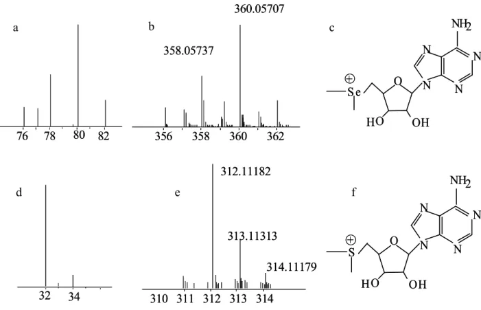
The Se-isotopic pattern can be seen in Figure 2a. The figure also contains a mass spectrum of a Se-containing compound, Se-dimethyl-5-selenonium-adenosine, (2b) where the phenomenon of the mass defect can be observed. The isotopologues 359 and 361 are the results of the molecule
76 78 80 82
76 78 80 82 356 358 360 362 360.05707
358.05737
356 358 360 362 360.05707
358.05737
312.11182
313.11313
310 311 312 313 314
314.11179 312.11182
313.11313
310 311 312 313 314
314.11179
N N N
N
NH2
S e O
HO OH
+
N N N
N
NH2
S e O
HO OH
+
N N N
N
NH2
S
O
H O OH
+
N N N
N
NH2
S
O
H O OH
+
32 34 32 34
b
d e f
containing 13C isotope. That is why there is no corresponding line in Figure 2a. In Figure 2c the structure of this molecule can be observed.
In comparison Figure 2d presents the mass spectrum of sulphur, and 2e shows the isotopic pattern of the S-analogue of the above mentioned compound, S-dimethyl-5-adenosine.
The differences between the mass defects and isotope-pattern complexity of the S and Se analogues can be observed.
Figure 2: 2a: The isotope distribution of Se. 2b: the isotopologue distribution of a Se- containing molecule, Se-dimethyl-5-selenonium-adenosine. 2c: molecule structure of Se- dimethyl-5-selenonium-adenosine. 2d: The isotope distribution of S. 2e: the isotope distribution
of a S-containing molecule, S-dimethyl-5-adenosine. 2c: molecule structure of S-dimethyl-5 – adenosine
In 1996 Crews was trying to identify unknown Se-compounds from a cod extract. In the process she injected selenomethionine, selenocystine, sodium selenite and sodium selenate standards into an ESI-MS system. While the standards were recognisable, she could not complete the identification of new components [86].
First it was used in 1999 by Casiot et al. who published an article on the identification of Se-adenosylselenohomocysteine, the major selenium species in an extract of a selenised yeast
a c
sample. Even though the mass accuracy still left a lot to be desired, another identifying attribute has been discovered: the fragmentation pattern [87].
The same year Kotrebai et al. used ICP-MS and ESI-MS in parallel experiments to identify major selenium compounds from Se-enriched yeast and garlic. It was found that selenomethionine and Se-adenosyl-selenohomocysteine made up 85% of the selenium content of garlic, and γ-glutamyl-Se-methyl-selenocysteine and γ-glutamyl-selenomethionine contributed to 90% of selenium in garlic [88]. Later that year, with the help of different colleagues, Kotrebai completed the optimization of an HPLC-ICP-MS system for the separation of Se-containing components extracted from organic sources. Once baseline separation was achieved, they applied the HPLC method for the characterization of amino acids with the use of ESI-MS [89]. A year later, also Kotrebai et al. has examined several ion-pairing agents for the separation of Se- containing compounds extracted from natural sources. With the optimized method and the γ-glutamyl-Se-methylselenocysteine and Se-methylselenocysteine standards oxidized with H2O2, several new oxidized products were identified from the extracts, using ESI-MS [90].
Even to date the leading indirect identification and speciation method is based on fragmentation-pattern, which by 2000 allowed McSheehy et al. to identify γ-glutamyl-Se- methylselenocysteine from garlic harvested in naturally seleniferous soil without the need for an authentic standard [91]. They also discovered a number of Se-containing compounds that they were unable to identify. In 2002 McSheehy et al. reported a large number of new Se-containing compounds found in selenised yeast but she could not yet identify their structure [92]. The same year Ogra et al. used ESI-MS detection for the identification of a new Se-containing compound, Se-methyl-N-acetylselenohexosamine from rat urine [93]. The method was also used for the characterization of non-protein selenium compounds from selenium accumulating plants by Montes-Bayón [94]. In 2002 Vonderheide et al. has identified several metabolites from Brazil nuts, including selenomethionine [95]. In 2007, coupling ESI with tandem MS enabled Ogra et al. to identify selenohomolanthionine from pungent radish, detecting this component from natural sources for the first time [96]. ESI-MS is a widely used method even today and a multitude of different samples have been examined with it. The drawback of the method is that molecule composition cannot be determined solely based on molecular mass, as the mass accuracy of the instrument might not be high enough. While ICP-MS is a robust method, and samples could be analyzed after minimal sample treatment, ESI-MS requires further purification, and the linear range of detection is much smaller.
The problem of the mass accuracy could be solved when the previous Q-MS and QQQ-MS systems were upgraded to time of flight (TOF) system. Molecular weight data acquired with a quadrupole time of flight (QTOF) detector is accurate enough to use for the
determination of molecule or fragment composition. This higher mass accuracy coupled with the fragmentation databases may provide enough information for the accurate determination of most compounds. In 2008, with the help of higher mass accuracy of QTOF Dernovics et al. [97] was able to tentatively assign structure to the Se-compounds McSheehy et al. had discovered in 2002 [92].
Another of the soft ionization techniques is matrix-assisted laser-desorption/ionization (MALDI). Owing to a lower detection limit and superior matrix tolerance to electrospray MS, MALDI allowed a successful detection of selenocompounds in samples where ESI-MS had failed. In 2003 Encinar et al. [98] found several previously unidentified Se-containing components in selenised yeast, even though they did not have a proposed structure for most of them yet. The higher mass accuracy and better separation techniques also allowed previous works to be re-observed, finding new compounds, unveiling artefacts and indentifying so far unknown components. During the following years, several articles were published identifying selenoproteins with this method. The advantage of the technique is the high mass accuracy;
while its drawback is that in numerous occasions the presence of Se cannot be confirmed. That is why in many cases ICP-MS is used in parallel, to gain quick and accurate information about the retention time, the concentration of the Se-containing compound, its location in gel electrophoretic media, artefact formations and enzymolysis. That was the case in 2007 when Ballihaut et al. reported his findings on the detection of bovine glutathione peroxidase selenoprotein using several hyphenated techniques [99].
One of the numerous selenoproteomic studies must be highlighted. In 2005 Giusti et al.
has examined the efficiency of enzymatic digestion with trypsine [100]. The digest was separated with HPLC, and the effluent was split into two branches, one going to ICP-MS for quantification with isotope dilution, the other entering ESI-MS for identification. Since the tryptic peptides, miscleaved and/or oxidized peptides, incompletely digested protein and undigested protein could be determined in one run, the method allowed the precise evaluation of the efficiency and quality of tryptic digestion using several nanolitres of sample only.
In 2006 a new detector, the hybrid linear ion trap/orbital ion trap Msn has appeared on the market, raising mass accuracy into unseen heights, which facilitated molecular structure identification. The coupling of a normal bore (4.6 mm) hydrophilic interaction liquid chromatography (HILIC) column with a hybrid linear ion trap/orbital ion trap mass spectrometer allowed the detection of the selenium-isotopic pattern in mass spectra down to the intrascan abundance of 0.001 with the low-and sub-part per million (ppm) mass accuracy regardless of the concentration [101]. In 2010 a method was developed for low-molecular weight component measurements, such as cysteine, homocysteine, selenocysteine, glutathione, selenomethionine
and cysteinyl-glycine. With the Orbitrap instrument their detection limits fall into the fmol range [102]. In 2011 a novel Se-metabolite was discovered from human urine despite of the concentration level barely reaching ppb magnitude [103]. A year later Arnaudguilhem et al. [17]
identified numerous Se-containing components discovered by McSheehy in 2000 [91]. The method developed enabled the detection of 64 metabolites including 30 SeSe or SeS conjugates (3 triple S/Se/S ones) and 14 selenoethers. Aureli et al. [104] discovered nine selenosugars from staple crops grown on soils naturally rich in selenium. Among the identified compounds, Se- containing monosaccharides (hexose moiety, m/z 317 and m/z 358) or Se-containing disaccharides (hexose-pentose moiety, m/z 407 and m/z 408) were the first selenosugars reported in edible plants. In 2013 Ouerdane et al. [105] could detect and characterize over 30 Se species, also with the help of the Orbitrap, from black mustard seeds (Brassica nigra) grown on Se-rich soil.
To date hybrid linear ion trap/orbital ion trap Msn has remained the most effective and most accurate method for detection. However, it must not be forgotten that simpler techniques are often needed for effective sample cleanup. In certain cases, with high enough target compound concentrations and well-known fragmentation patterns simpler and cheaper detections may also be sufficient.
2.4. Sample preparation for selenium-speciation
Speciation analysis, naturally, requires sample preparation. The more sensitive and accurate the analytical system the cleaner the samples have to be to enable measuring. There are a number of different methods widely used in Se-speciation; which one is chosen for a particular sample depends on different factors, like complexity, fat content and selenium concentration of the sample, whether the sample is liquid or solid, what kind of instrumentation is intended to be used, what type of components are expected to be found, etc.
2.4.1 Complete digestion
It might seem redundant to talk about complete sample digestion in speciation, as the method is supposed to bring all different species into the same form, losing all information even on the oxidation state of selenium let alone the molecule structure. Yet, it is a method rarely missing from speciation articles, because it gives a frame for the entire analytical process. With the determination of total selenium content the efficiency of extraction can be monitored, the
![Table 1: Proximate composition of some edible wild-grown mushrooms of China [31]](https://thumb-eu.123doks.com/thumbv2/9dokorg/840539.43692/15.892.130.811.731.1069/table-proximate-composition-edible-wild-grown-mushrooms-china.webp)
![Figure 3 : Derivatisation method with NBIC and OPA by Bergman et al. [62]](https://thumb-eu.123doks.com/thumbv2/9dokorg/840539.43692/54.892.125.832.884.1127/figure-derivatisation-method-nbic-opa-bergman-et-al.webp)