The Zn-finger of Saccharomyces cerevisiae Rad18 and its adjacent region mediate interaction with Rad5
Orsolya Frittmann,1,2,†Vamsi K. Gali,1,‡Miklos Halmai,1Robert Toth,1Zsuzsanna Gyorfy,1Eva Balint,1and Ildiko Unk1,*
1Biological Research Centre, Szeged, Eotvos Lora´nd Research Network, The Institute of Genetics, Szeged, H-6726, Hungary
2Doctoral School of Biology, University of Szeged, Szeged, H-6720, Hungary
†Present address: York Structural Biology Laboratory, Department of Chemistry, University of York, York, YO10 5DD, UK
‡Present address: Institute of Medical Sciences Foresterhill, University of Aberdeen, Aberdeen, AB25 2ZD, UK
*Corresponding author: Biological Research Centre, Szeged, Eotvos Lora´nd Research Network, Szeged, Temesvari krt. 62, H-6726, Hungary. unk.ildiko@brc.hu
Abstract
DNA damages that hinder the movement of the replication complex can ultimately lead to cell death. To avoid that, cells possess several DNA damage bypass mechanisms. The Rad18 ubiquitin ligase controls error-free and mutagenic pathways that help the replication complex to bypass DNA lesions by monoubiquitylating PCNA at stalled replication forks. InSaccharomyces cerevisiae, two of the Rad18 governed pathways are activated by monoubiquitylated PCNA and they involve translesion synthesis polymerases, whereas a third path- way needs subsequent polyubiquitylation of the same PCNA residue by another ubiquitin ligase the Rad5 protein, and it employs template switching. The goal of this study was to dissect the regulatory role of the multidomain Rad18 in DNA damage bypass using a structure- function based approach. Investigating deletion and point mutantRAD18variants in yeast genetic and yeast two-hybrid assays we show that the Zn-finger of Rad18 mediates its interaction with Rad5, and the N-terminal adjacent region is also necessary for Rad5 binding.
Moreover, results of the yeast two-hybrid andin vivoubiquitylation experiments raise the possibility that direct interaction between Rad18 and Rad5 might not be necessary for the function of the Rad5 dependent pathway. The presented data also reveal that yeast Rad18 uses different domains to mediate its association with itself and with Rad5. Our results contribute to better understanding of the complex machinery of DNA damage bypass pathways.
Keywords:Rad18-Rad5 interaction; yeast genetics; DNA damage tolerance; yeast two-hybrid
Introduction
Due to the continuous exposure to environmental agents that can harm or modify the inheriting material, cells are equipped with several DNA repair mechanisms that preserve the integrity of DNA by removing any alterations and restoring the original sequence. Some lesions, however, escape repair and pose an obstacle to the moving replication fork. Cells avoid the fatal consequences of stalled replication by activating the so-called DNA damage tolerance (DDT) mechanisms that facilitate replica- tion through DNA lesions without removing them. DDT can be error-free or error-prone depending on the type of damage and the mechanism of bypass, and accordingly, it can contribute to genomic stability or it can destabilize the genome by introducing mutations during the bypass process.
In the yeastSaccharomyces cerevisiae, the Rad6/Rad18 complex governs at least three subpathways of DDT on UV-damaged DNA (Torres-Ramoset al.2002). Specialized translesion synthesis (TLS) DNA polymerases constitute the REV1-dependent error- prone subpathway together with the Def1 protein. The TLS poly- merases of this branch, the Rev1 protein and the Rev3-Rev7 formed DNA polymerasef, either introduce incorrect nucleotides during lesion bypass with high frequency or extend from it,
respectively, resulting in mutagenesis (Lawrence and Maher 2001). The Def1 protein enables the exchange between the repli- cative and TLS polymerases by promoting proteasomal degrada- tion of the catalytic subunit of the stalled replicative polymerase (Darabaet al.2014). The second branch consists of the RAD30 encoded TLS DNA polymerase g that is uniquely error-free opposite pyrimidine dimers, the most frequent UV-induced DNA lesions (McDonaldet al.1997;Johnsonet al.1999). The third branch involves theRAD5, theMMS2, and theUBC13genes, and it oper- ates through template switching using the newly synthesized strand of the sister chromatid as a template resulting in error- free bypass (Johnsonet al.1992;Broomfieldet al.1998;Hofmann and Pickart 1999;Zhang and Lawrence 2005;Blastya´ket al.2007).
The two subbranches involving TLS polymerases are activated by PCNA monoubiquitylation at lysine 164 (K164) by Rad6/Rad18, whereas the MMS2 branch requires PCNA polyubiquitylation at the same residue performed by Rad5 together with Mms2 and Ubc13 (Hoegeet al.2002;Stelter and Ulrich 2003). In the poly- ubiquitylation reaction, the Mms2/Ubc13 complex acts as an ubiquitin conjugase, and Rad5 acts as an ubiquitin ligase (Hofmann and Pickart 1999).
Rad18 together with Rad6 controls an early step in the activa- tion of DNA damage bypass mechanisms by forming an ubiquitin
Received:August 28, 2020.
VCThe Author(s) 2021. Published by Oxford University Press on behalf of Genetics Society of America.
This is an Open Access article distributed under the terms of the Creative Commons Attribution License (http://creativecommons.org/licenses/by/4.0/), which permits unrestricted reuse, distribution, and reproduction in any medium, provided the original work is properly cited.
2
DOI: 10.1093/g3journal/jkab041Advance Access Publication Date: 11 February 2021 Investigation
Downloaded from https://academic.oup.com/g3journal/article/11/4/jkab041/6133228 by guest on 03 February 2022
conjugase/ligase complex that monoubiquitylates PCNA at DNA damage-stalled replication complexes (Bailly et al.1994; Hoege et al.2002). In the complex, a homodimer Rad18 binds a single Rad6 (Huanget al.2011;Masudaet al.2012). The ubiquitin conju- gase Rad6 is a small protein, but the ubiquitin ligase Rad18 has several domains. At the N-terminus, Rad18 contains a RING do- main characteristic of a group of E3 ubiquitin ligases, which is es- sential for its function (Tateishiet al. 2000). In humans it was found to contribute to Rad6 binding and to mediate homodimeri- zation (Tateishiet al.2000;Huanget al.2011;Masudaet al.2012).
In the middle of the protein, there is a Zn-finger the exact func- tion of which is still debated. The Zn-finger of human Rad18 was shown to bind ubiquitin, even the crystal structure of their com- plex was resolved (Bish and Myers 2007;Notenboomet al.2007;
Huanget al.2009;Rizzoet al.2014). However, another study found that the Zn-finger mediates the association of human Rad18 with itself, as the C207F amino acid change affecting one of the core cysteine residues of the Zn-finger prevented homodimerization (Miyaseet al.2005). Moreover, HLTF and SHPRH the two human homologs of Rad5 were shown to bind Rad18 through its Zn- finger (Unket al.2006,2008;Zeman et al. 2014). The SAP (SAF-A/
B, Acinus and Pias) domain was identified as a DNA binding do- main, and it was found to have the same role in human Rad18 (Aravind and Koonin 2000; Notenboom et al. 2007; Tsuji et al.
2008). Close to the C-terminus Rad18 has a sequence responsible for Rad6 binding (Bailly et al.1997b; Watanabe et al. 2004;
Notenboomet al.2007). Both DNA and Rad6 binding are essential for the role of Rad18 in DDT. Beside these, two small motifs have been identified in the Rad18 sequence. One is a nucleotide bind- ing motif situated N-terminal to the Rad6 binding site, the so- called Walker type A box characteristic to nucleotide binding pro- teins, and indeed, Rad18 has a single stranded DNA dependent ATP-ase activity (Baillyet al.1994, 1997a). The other is a SUMO interacting motif between the RING and Zn-finger domains, which confers higher ubiquitylating activity toward sumoylated PCNA (Parker and Ulrich 2012).
In spite of the accumulating data, the exact choreography of Rad18 action in DDT is elusive since the functions of some of its known domains are still debated and specific parts of the protein mediating its several interactions have not been identified. In this study, we set out to investigate how regions in Rad18 lacking well-defined domains contribute to its role in DNA damage toler- ance. Based on our results, we could assign a novel function to the Zn-finger motif of yeast Rad18 and refine the current model of DDT.
Materials and methods Yeast strains and plasmids
Yeast strains used in the genetic studies are derivatives of EMY74.7 (MATa, his3-D1, leu2-3, -112, trp1D, and ura3-52).
Deletions were constructed by gene replacement. For ectopic complementation of theRAD18deletion, the wild-type (wt) and the mutant variants ofRAD18with their promoter and termina- tor sequences, from600 to 300 nucleotides (nt) after the STOP codon, were cloned into the yeast centromeric vectors YCplac33 and YCplac111 having theURA3andLEU2marker genes, respec- tively. The C-terminal deletions of RAD18 (C1–C3) were con- structed by replacing the XbaI-StuI fragment of the wt RAD18 with XbaI-StuI flanked PCR products having the shortened sequences and a STOP codon at the desired position. For the N- terminal deletions (N1, N2), the MscI-NdeI fragment of the wt RAD18 was exchanged to MscI-NdeI flanked PCR products
containing the shortened sequences. The CC190,193GG point mutations in RAD18 were generated by a PCR based method according to the Stratagene “Quick Change Site Directed Mutagenesis” protocol. The wt and the mutantRAD18genes in the YCplac33 vector were C-terminally tagged with three copies of the hemagglutinin epitope tag (3-HA) by a PCR-based strategy (Knopet al.1999) in EMY74.7, previously maderad18D. Strains car- rying Rad18 expressing constructs were always grown in syn- thetic dropout media to maintain the plasmids. All mutations were confirmed by sequencing.
For the two-hybrid assays, the yeast strain PJ69-4A (MATa trp1-901 leu2-3,112 ura3-52 his3-200 gal4Dgal80DMET2: GAL7-lacZ LYS2: GAL1-HIS3 GAL2-ADE2) (James et al.1996), and its rad18Dpol30-K164Rvariant were used, where the wtPOL30gene at the genomic locus was replaced by a mutant coding for the K164R mutant PCNA. The whole coding regions of the wt and mu- tantRAD18,RAD5, andRAD6genes were cloned into the Gal4-BD vector pGBT9þ1 resulting in pID685 (wt Rad18), pID771 (N1), pID704 (N2), pID1047 (Zn*) pID623 (Rad5), and pID231(Rad6). The wt and mutant RAD18, the RAD5, and the POL30 genes were cloned into the Gal4 activation domain fusion vector pGAD424þ1 resulting in pID684 (wt Rad18), pID768 (N1), pID700 (N2), pID1046 (Zn*), pID145 (Rad5), and pID999 (Pol30) plasmids.
A region ofRAD18coding for amino acids 170–230 consisting the wt, or CC190,193GG mutant Zn-finger was cloned into pGBT9þ1 resulting in plasmids pID1149 (Znfþp), and pID1150 (Znf-p), re- spectively.
Detection ofin vivoPCNA ubiquitylation was carried out in a DF5a strain (MATa, trp1-1, his3–200, ura3-52, lys2-801, andleu2-3,2- 112) (Finley et al.1987) that was maderad18D and pol30D, and expressed an N-terminally tagged 7His-PCNA under its own pro- moter as the sole source of PCNA from the YCplac33 plasmid.
Sensitivity assays
For qualitative assays,rad18Dcells carrying theRAD18gene or its mutant derivatives on a plasmid were grown in synthetic com- plete media lacking uracil (SC-ura) overnight at 30C. Cells were counted using a Bu¨rker chamber, then the cultures were adjusted to the same density and a series of 10-fold dilutions of the cul- tures were spotted onto SC-ura plates. For UV sensitivity, the plates were exposed to different doses of UV (254 nm) radiation and incubated at 30C in the dark for 3 days. Methyl methanesul- fonate (MMS), bleomycin and hydroxyurea (HU) sensitivities were assayed on plates supplemented with the indicated amount of the chemicals (Sigma-Aldrich).
For quantification of UV sensitivity, cells were spread onto SC- ura plates at appropriate dilutions and irradiated with UV for given times to apply the required dosages. Colonies were counted after incubating the plates in the dark at 30C for 3–5 days.
UV-induced and spontaneous mutation rates
UV irradiation induced forward mutation frequencies at the CAN1 locus were measured by comparing the numbers of can1Rcolonies at different UV doses, selected on SC-leu/arg plates containing canavanine, with the number of colonies on SC-leu plates exposed to the same UV doses. Spontaneous forward mu- tation frequencies were measured also at theCAN1locus. In par- allel, ten single colonies for each strain were diluted in water, and approximately 10 cells from each colony were inoculated into 500ml SC-leu in a microtiter plate and grown for 3 days at 30C. The cultures were plated onto canavanine containing SC- leu/arg plates, and the number of mutants in case of each culture was counted for 107plated cells. Mutation frequencies for each 2 | G3,2021, Vol. 11, No. 4
Downloaded from https://academic.oup.com/g3journal/article/11/4/jkab041/6133228 by guest on 03 February 2022
strain were determined using the median value of mutations of the ten cultures and a chart based on the Lea-Coulson fluctua- tion model. The mean values of the median mutation frequencies and standard deviations were calculated from five experiments.
Yeast two-hybrid
The binding and activation domain fusion constructs were co- transformed into the wt or the modified PJ69-4A strains, and transformants were selected on SC-leu/trp medium. Single colo- nies of the transformants were resuspended in water at equal densities and spotted onto SC-leu/trp plates to demonstrate equal growth, and onto SC-leu/trp/his plates to assess theHIS3 gene expression that reflects protein-protein interaction. Where indicated, plates were supplemented with the given doses of 3- amino-1,2,4-triazole to test the strength of the interactions.
Plates were incubated at 30C for 3–6 days.
Protein techniques and antibodies
To show the level of Rad18 gene expression, 10 ml yeast cultures grown in SC-ura were harvested at A600:1. Whole cell extracts were prepared by glass-bead lysis method in 1XPBS (137 mM NaCl, 2.7 mM KCl, 10 mM Na2HPO4, and 1.8 mM KH2PO4)supplemented with 1 mM EDTA, 10% glycerol, and prote- ase inhibitors. Cell lysates were quantified by Bradford. Equal amounts of whole cell lysates were separated by SDS-PAGE and analyzed by western blotting. Rad18 forms were visualized using anti-HA (5000X, GeneTex GTX21208) primary and anti-rabbit (10 000X, Thermo Scientific 31460) secondary antibodies, and PGK with anti-PGK (10 000X, Molecular Probes A6457) primary and anti-mouse (10 000X, Thermo Scientific 31430) secondary anti- bodies.
Denaturing Ni-nitriloacetic acid (Ni-NTA) chromatography was applied to detect in vivo PCNA ubiquitylation (Ulrich and Davies 2009). Whole cell extracts were prepared according to a protocol (Jianget al.2019). Briefly, 200–200 ml of cell cultures co- expressing 7His-PCNA and wt or mutant Rad18 were grown in SC-leu medium, since selection for the plasmid expressing the es- sentialPOL30gene is not necessary. At A600:1 half of the cultures were treated with 0.02% MMS for 90 minutes. Then cells were col- lected and washed, and resuspended in 1.5 ml of 0.3 M NaOH.
After a 2 minutes incubation at RT, cells were collected and resuspended in 350ml of lysis buffer [60 mM Tris/HCl pH 6.8, 5%
glycerol, 2% SDS, and 4% 2-mercaptoethanol (ME)] and boiled at 100C for 8 minutes. Samples were clarified by centrifugation at 1,500 g for 20 minutes, and the supernatants were transferred into new tubes containing 1 ml of dilution buffer (10 mM Tris/HCl pH 8, 100 mM sodium-phosphate buffer pH 8, and 8 M urea), 30ml of Ni-NTA beads, 15 mM imidazole and 0.05% Tween-20. Samples were incubated at 4C for 4 hours on a rotator and then washed three times with 1–1 ml of dilution buffer containing 0.05%
Tween-20, and then three times with wash buffer (10 mM sodium-phosphate buffer pH 8, 500 mM NaCl, 0.5% NP-40, and 0.05% Tween-20). His-tagged PCNA was eluted and denatured by incubating the Ni-beads for 10 minutes at 60C in 30ml of HU buffer (8 M urea, 200 mM Tris/HCl, pH 6.8, 1 mM EDTA, 5% SDS, 0.1% bromophenol blue, and 1.5% dithiothreitol). Samples were resolved on a 12% SDS gel and transferred onto a PVDF mem- brane (Immobilon-P, Merck Millipore Ltd.). After blotting, mem- branes were pre-treated either in 6 M guanidyl-HCl, 20 mM Tris/
HCl pH 7.5, 5 mM 2-ME for 30 minutes at 4C for labeling with anti-Ub antibody (1500X, P4D1, Cell Signaling Technology), or 2 6 minutesat RT in stripping buffer (0.2 M glycine, 0.1% SDS, 1%
Tween-20) for labeling with anti-PCNA antibody (1000X, Abcam
5E6/2). Anti-mouse (10 000X, Thermo Scientific 31430) and anti- rabbit (10 000X, Thermo Scientific 31460) secondary antibodies were used, respectively.
Statistical analysis
Student’st-test using Excel (Microsoft, Redmond, WA, USA) was applied to compare separate groups.P-values of<0.05 were con- sidered statistically significant. *P<0.05, **P<0.01, ***P<0.001.
Results
Design of the Rad18 deletion mutants
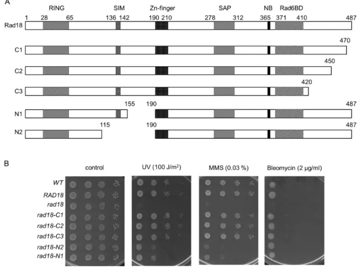
To better understand the role of Rad18 in DDT, we aimed to in- vestigate the contribution of extended, uncharacterized regions of the protein to its function. At the C-terminus, residues be- tween 371 and 410 were shown to be necessary for binding Rad6 (Bailly et al.1997b). However, C-terminal to this Rad6 binding domain there is an acidic tail of 77 amino acids without any assigned function. Therefore, we created three overlapping truncations in this region, the longest eliminating almost the complete C-terminal sequence following the Rad6-binding do- main. At the N-terminus, Rad18 has a RING domain characteris- tic of a group of E3 ubiquitin ligases and essential for the function of Rad18 in DDT, and a Zn-finger in the middle of the protein whose function is still not clear. We engineered two overlapping deletions between the RING and Zn-fingers with the bigger one removing more than half of the region between the two domains. The schematics of the wt Rad18 and the deletion mutants, referred to as C1, C2, C3, N1, N2, are shown in Figure 1A.
The C-terminal truncations do not influence the in vivo function of Rad18 in DDT
The mutant proteins were ectopically expressed under the native RAD18promoter in haploid yeast cells, from which the genomic copy ofRAD18was removed. Thein vivoeffects of the deletions were assessed by treating the cells with DNA damaging agents like ultraviolet (UV)light, the methylating agent MMS, and the ra- diation mimetic drug bleomycin. Surprisingly, as Figure 1B shows, all three C-terminal mutants displayed wt sensitivity to all three agents, even at high doses. These results suggested that the C-terminal acidic tail did not significantly contribute to the function of Rad18 in DNA damage response.
The N-terminal deletions in Rad18 affect the Mms2-dependent sub-pathway
Contrary to the C-terminal truncations, the two N-terminal deletion mutants, N1 and N2, conferred almost identical mild sensitivities to yeasts upon UV and MMS, however, they did not influence the sensitivity to bleomycin (Figure 1B). First, we veri- fied that the deletions did not significantly alter the expression level ofRAD18resulting in the observed sensitivities. Therefore, we tagged the wt and mutantRAD18genes with three copies of the haemagglutinin tag (3HA) at the C-terminus, and examined their cellular expression level in whole cell extracts. AsFigure 2A shows, both N1 and N2 displayed similar expression level as the wt Rad18 ruling out that the deletions affected the intracellular abundance of Rad18. Next, we investigated whether the in- creased sensitivities could be linked to selective inactivation of a sub-branch of the Rad18 governed DDT. For this reason, we sepa- rately inactivated the three sub-branches in the rad18-N1 or rad18-N2strains by deleting a prominent gene of the given sub- branch, and investigated their genetic relations to the mutant
Downloaded from https://academic.oup.com/g3journal/article/11/4/jkab041/6133228 by guest on 03 February 2022
RAD18genes by epistasis analysis. In these experiments, the N2 mutant conferred slightly higher sensitivities than the N1 mutant to different strains upon UV and MMS treatment.
We surmised that this might be because the deletion in this mutant removed the SIM motif, as well. The analysis showed, that additional deletion ofREV1orRAD30resulted in increased sensitivities over the single mutants suggesting that these sub- branches were functional in therad18-N1andrad18-N2strains (Figure 2, B and C). In accordance, induced mutagenesis was active in these strains confirming that the N-terminal deletions in Rad18 did not disturb the mutagenic sub-pathway involving the TLS polymerases (Figure 2E). Moreover, at lower doses mu- tagenesis was higher in the mutants compared to the wt sug- gesting that the mutagenic TLS pathway gained more ground over the error-free pathway. At higher dose the rates were very similar probably due to the activation of additional, RAD18 independent pathways like homologous recombination. Next, we examined the relationship of therad18mutants to the third, error-free branch. We found, that upon deleting MMS2 the resulting double mutant strains displayed similar sensitivities as themms2Dsingle mutant strain suggesting that the N-termi- nal deletions in RAD18 affected the MMS2-dependent sub- pathway (Figure 2D).
Amino acids 155-190 of Rad18 are necessary for Rad5 binding
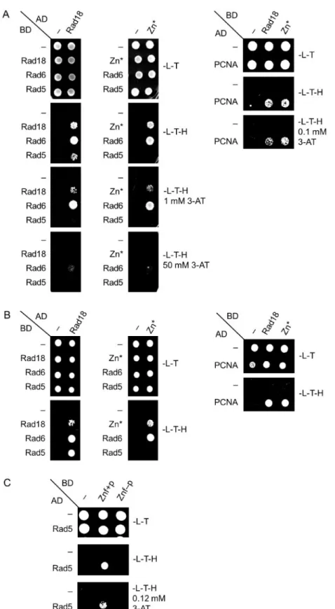
Rad5 together with Mms2 and Ubc13 acts in the error-free branch of DDT promoting template switch. Among these proteins, Rad5 has been shown to directly interact with Rad18 (Ulrich and Jentsch 2000). Therefore, we set out to investigate whether the N- terminal deletions in Rad18 inhibited the formation of the Rad18- Rad5 complex, by applying yeast two-hybrid assays. As controls, we included known binding partners of Rad18 in the experiments like Rad6, PCNA, and the Rad18 protein itself. The results showed that the wt Rad18, and also both N1, and N2, could form dimers with themselves and they could bind Rad6 and PCNA, too (Figure 3) The mutations did not detectably affect the strength of the binding either, except with PCNA where slight weakening of the interactions could be observed in the presence of 3-AT.
(Figure 3B). However, contrary to the wt, neither N1, nor N2 showed interaction with Rad5 (Figure 3A). Similar experiments using arad18Dstrain expressing K164R PCNA from the genomic locus corroborated that the detected interactions were direct and they were not mediated by the endogenous Rad18 or ubiquity- lated PCNA (Figure 3, C and D). Taken together, these results sug- gested that the region in Rad18 between amino acids 155–190 missing from both N1 and N2 was necessary for Rad5 binding.
Figure 1The C-terminal truncations do not affect Rad18 function. (A) Schematic representation of wt and deletion mutant Rad18 proteins engineered in this study. RING, RING finger domain; SIM, SUMO interaction motif; Zn-finger, Rad18-type Zinc finger domain; SAP, SAF-A/B, Acinus, and PIAS domain; NB, Walker-A nucleotide binding motif; Rad6BD, Rad6 binding domain. Amino acid numbers are marked above the schemes and the mutants are named on the left. (B) Sensitivity of therad18strains to DNA damaging agents. Tenfold serial dilutions of the indicated strains were spotted on plates either exposed to the indicated UV dose, or containing the indicated amount of MMS or bleomycin. Several UV, MMS, and bleomycin doses were applied, but only plates with the most appropriate doses are presented. Genotypes are in italic, with the expressed Rad18 variants indicated.
4 | G3,2021, Vol. 11, No. 4
Downloaded from https://academic.oup.com/g3journal/article/11/4/jkab041/6133228 by guest on 03 February 2022
The Zn-finger of Rad18 mediates the interaction with Rad5
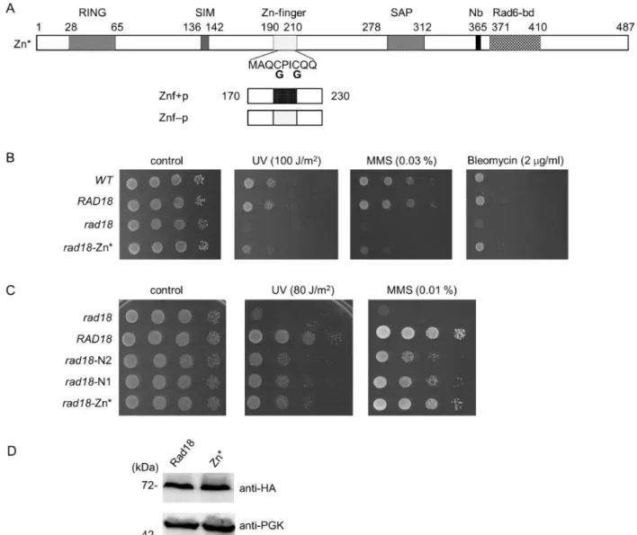
As the Zn-finger is situated next to the deletions in the N1 and N2 proteins, we aimed to investigate whether it was also involved in mediating the association with Rad5. For this purpose, we disrupted the Zn-finger by replacing the N-terminal two zinc- coordinating cysteine residues of the Zn-finger with glycines (Figure 4A). The functionality of the resulting CC190,193GG Rad18 mutant protein, which we named Zn*, was checked in sen- sitivity assays. As it is shown in Figure 4B, therad18-Zn* strain exhibited the same sensitivity pattern as the rad18-N1 and
rad18-N2strains: it showed mild sensitivities to UV and to MMS but none to bleomycin, compared to the wt strain. Next, we con- firmed that the CC190,193GG mutations did not perturb the intracellular expression level of Rad18 (Figure 4D). Interestingly, the rad18-N1, rad18-N2, and rad18-Zn* strains exhibited very similar sensitivities to UV irradiation, however, upon MMS treatment therad18-Zn* strain was more resistant (Figure 4C).
Even so, when we examined the genetic relations of the Zn* mu- tant to the three sub-branches of the Ra6/Rad18 pathway, we got the same results as with the N1 and N2 mutants (Figure 5).
Deletion of RAD30 or REV1 in the rad18-Zn* strain further Figure 2Genetic characterization of the N-terminal mutants. (A) Intracellular expression level of wt Rad18, N1, and N2 mutants. Whole cell extracts were prepared from strains expressing the C-terminally tagged proteins that were visualized with anti-HA antibody. PGK served as a loading control.
(B–D) Epistasis analysis with theREV1,RAD30, andMMS2dependent sub-pathways. Quantitative analysis of UV survival of single and double mutants with (B)rev1(C)rad30, and (D)mms2. Results represent the averages of three experiments. Standard deviations are indicated. (E) UV-induced mutagenesis. Forward mutation rates at theCAN1locus were determined after exposing the cells to the indicated doses of UV. Averages of three experiments and standard deviations are shown.P-values representing the significance of difference are shown as *,P<0.05; **,P<0.01; ***,P<0.001.
Downloaded from https://academic.oup.com/g3journal/article/11/4/jkab041/6133228 by guest on 03 February 2022
sensitized the single mutants indicating that the function of the TLS polymerases was not affected by the CC190,193GG rad18mutations (Figure 5, A and B). Contrary to that, themms2D rad18-Zn*strain was not more sensitive than themms2Dsingle deletion strain indicating that the CC190,193GG mutations in Rad18 disturbed the Mms2-dependent branch (Figure 5C).
Taken together, the above data suggested that the mutations in the N1, N2, and Zn* proteins affected the same biological func- tion of Rad18.
Therefore, we investigated the effect of the CC190,193GG mutations on the interaction between Rad18 and Rad5 by applying yeast two-hybrid assays. Indeed, the results of these experiments indicated that the absence of the two C residues pre- vented the formation of the Rad18-Rad5 complex (Figure 6, A and B). Nevertheless, self-dimerization or the interaction with Rad6 or
PCNA was not affected. Based on these, we concluded that with- out the Zn-finger Rad18 was not able to bind Rad5.
To obtain direct evidence on the role of the Rad18 Zn-finger, we tested the Rad5-binding capability of a short Rad18 fragment spanning from aa. 170 to 230 that consisted the Zn-finger (Figure 4A), in yeast two-hybrid experiments. Figure 6Cshows, that this Znfþpeptide interacted with Rad5, whereas the Znf peptide having the CC190,193GG amino acid changes did not show interaction. These experiments confirmed that the Zn- finger of Rad18 mediated the association with Rad5.
DNA damage induced PCNA polyubiquitylation occurs in rad18-N1 and rad18-Zn* cells
Formation of the Rad18-Rad5 complex is generally considered to be necessary for PCNA polyubiquitylation, hence to the Figure 3Formation of the Rad18-Rad5 complex is inhibited by the N-terminal deletions. The interactions of N1 and N2 were tested (A), (C) with Rad6, Rad5, and self, as well as (B), (D) with PCNA by yeast two-hybrid assay. Experiments were performed in (A) and (B) using wt PJ694A strain, while in (C) and (D)rad18 pol30-K164Rstrain. AD, fusion with Gal4 activation domain; BD, fusion with Gal4 DNA binding domain; -L-T, -Leu -Trp control medium; - L-T-H, -Leu-Trp-His test medium; 3-AT, 3-aminotriazol.
6 | G3,2021, Vol. 11, No. 4
Downloaded from https://academic.oup.com/g3journal/article/11/4/jkab041/6133228 by guest on 03 February 2022
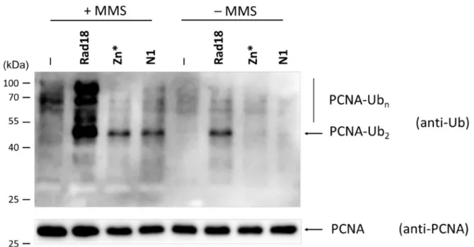
functionality of the Rad5 branch. However, yeast strains express- ing our Rad18 mutant proteins unable to interact with Rad5 displayed only mild sensitivities to UV and to MMS; they were less sensitive than themms2Dstrain. This prompted us to check the activity of the Mms2 subpathway in the different strains by probing the DNA damage induced polyubiquitylation of PCNA. As Figure 7shows, MMS treatment triggered PCNA polyubiquityla- tion in the wt and, though to a lesser extent, also in therad18-N1 andrad18-Zn*strains indicated by the appearance of bands corre- sponding to di- and polyubiquitylated forms of PCNA. The mono- ubiquitylated form could not be observed since the applied anti- ubiquitin antibody could not recognize it, as already reported by others (Hoegeet al.2002). Altogether, these findings raised the possibility that Rad5 could promote PCNA polyubiquitylation even without interacting with Rad18, though with a decreased ef- ficiency.
Spontaneous mutagenesis is affected in the N1, N2, and Zn* expressing strains
Besides DDT, another process that is influenced by Rad5, Mms2, and Ubc13 is spontaneous mutagenesis. In the lack of either Rad5, Mms2, or Ubc13, spontaneous mutation rate is highly ele- vated (Broomfield et al.1998; Liefshitz et al.1998; Brusky et al.
2000). We aimed to investigate, how the absence of interaction between Rad18 and Rad5 affected this process. AsFigure 8shows, a2,5-1.5-fold raise in the rate of spontaneous mutations was observed in the rad18-N1, rad18-N2, and rad18-Zn* expressing strains, whereas a4-fold increase was detected in themms2D strain over the wt rate. Deletion of MMS2in the rad18mutant strains resulted in a spontaneous mutation rate similar to that measured in themms2Dsingle mutant indicating that the muta- tions were epistatic tomms2Din respect of spontaneous muta- genesis, as well.
Figure 4Characterization of the Zn-finger mutant Rad18. (A) Schematic structure of the Zn* mutant and the Znfþand Znf- peptides. Domains designated as inFigure 1A. The positions of cysteines substituted to glycines are indicated. (B) Sensitivity of therad18-Zn*strain to different DNA damaging agents. The assay was performed as described inFigure 1B. (C) Comparing the sensitivities of therad18-N1,rad18-N2, andrad18-Zn*mutants upon UV and MMS. Tenfold serial dilutions of the indicated strains were spotted on plates either irradiated with UV or containing the indicated dose of MMS. (D) Equal expression of the wt Rad18 and the Zn* mutant. The experiment was performed as described forFigure 2A.
Downloaded from https://academic.oup.com/g3journal/article/11/4/jkab041/6133228 by guest on 03 February 2022
Discussion
This study identifies Zn-finger of yeast Rad18 as the Rad5 binding domain, and shows that its N-terminal neighboring sequence is also important for complex formation. By engineering internal deletions and point mutations in the full-length RAD18 gene, we investigated the contribution of different regions of the protein to DNA damage bypass. Our yeast two-hybrid experi- ments show that mutations that abolish the conserved cysteine residues of the Rad18 Zn-finger, or remove its N-terminal nearby sequence prevent the interaction with Rad5. Our genetic data with theRAD18mutant strains are consistent with a defect in the Rad5 dependent error-free subpathway. However, these strains are considerably less sensitive to UV and to MMS treatments than themms2Dstrain that is completely devoid of DNA damage induced PCNA polyubiquitylation. Also, spontaneous mutagen- esis in the mutants increased to a smaller extent compared to themms2Dstrain. These results suggested that DNA damage in- duced PCNA polyubiquitylation still occurred in the N1, N2, and Zn* expressing strains, which we could confirm inin vivoexperi- ments . One possible explanation is that the Rad18-Rad5 com- plex is not absolutely required for PCNA polyubiquitylation. In its absence, probably the direct interaction between Rad5 and PCNA, reported earlier (Hoegeet al.2002), could promote a de- creased rate of PCNA polyubiquitylation resulting in reduced ac- tivation of the error-free damage bypass pathway. Another possibility that we cannot exclude is that a weak interaction undetectable by yeast two-hybrid still takes place between the mutant Rad18 proteins and Rad5, which can support some level of PCNA polyubiquitylation.
There are conflicting results in the literature about the role of the Rad18 Zn-finger. Peptides containing the Zn-finger of human Rad18 were shown to bind ubiquitin, bothin vitroandin vivo(Bish
and Myers 2007;Notenboomet al.2007;Huanget al.2009;Rizzo et al.2014). It was reported, that replacing one of the conserved cysteine residues (C207F) in the Zn-finger abolished homodimeri- zation of human Rad18in vivo(Miyaseet al.2005), whereas an- other study found that the same mutant could form homodimers in vitro(Notenboomet al.2007). Moreover, structural studies with human Rad18 identified the RING finger as the dimerization domain that mediates the assembly of an asymmetric complex containing two Rad18 and one Rad6 molecules (Huanget al.2011;
Masudaet al.2012). Also, HLTF and SHPRH were both shown to bind human Rad18 via its Zn-finger, in a competitive manner with ubiquitin (Zemanet al.2014). In the case of yeast Rad18, the N-terminal half of the protein containing the Zn-finger was shown to mediate both self-dimerization and Rad5 binding (Ulrich and Jentsch 2000). Our data indicate that, like its human homologue, yeast Rad18 binds Rad5 through its Zn-finger.
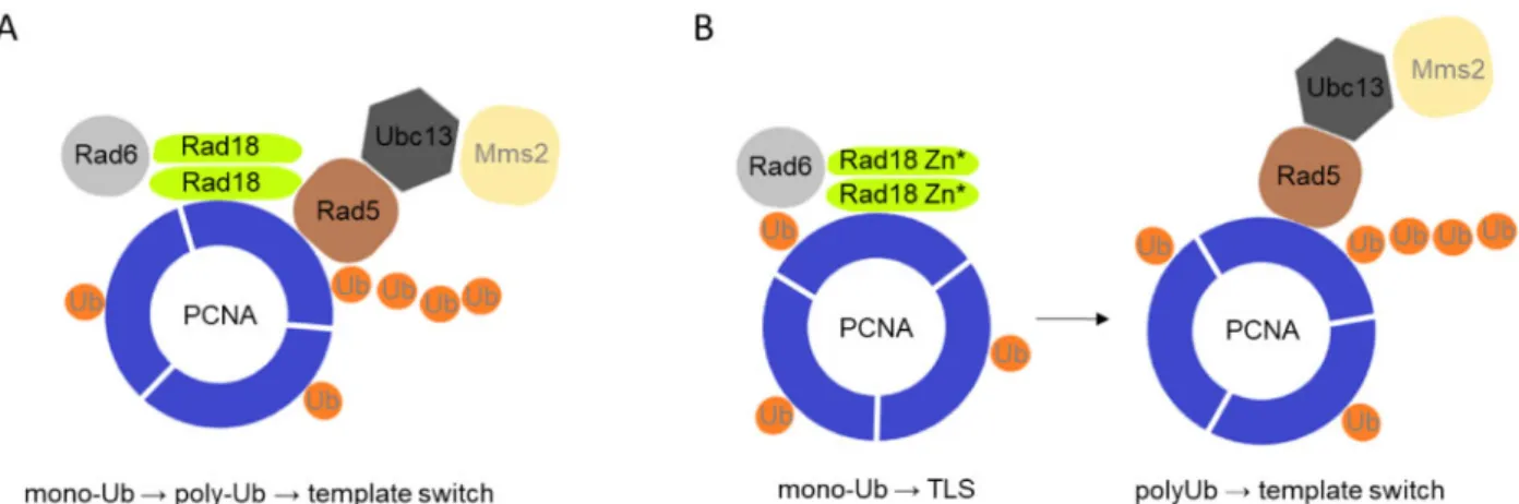
However, as the Rad18 mutant proteins defective in Rad5 binding could still form homodimers, self-assembly must be mediated by other regions of the protein implying that Rad18 can bind itself and Rad5 at the same time. These results support the model that the two ubiquitin ligase-conjugase complexes, one composed of two Rad18 and one Rad6, and the other of Rad5-Mms2-Ubc13, could assemble into a single multiprotein complex (Figure 9). In such complex, PCNA mono- and polyubiquitylation would be spatially brought and linked together ensuring that once PCNA is monoubiquitylated, polyubiquitylation would follow, giving pri- ority to the Rad5 mediated error-free branch over the mutagenic branch mediated by the TLS polymerases.
Our data also show, that neither the N-terminal deletions, nor the Zn* mutant confer additional sensitivity to bleomycin that causes DNA strand breaks. This is in agreement with previous results showing that even though both Rad18 and Rad5 play a Figure 5Epistasis analysis of therad18-Zn*mutant. MMS sensitivities of the double mutants containing deletions of (A)RAD30, (B)REV1, and (C)MMS2 were examined. Tenfold serial dilutions of the indicated strains were spotted on plates containing the indicated concentration of MMS.
8 | G3,2021, Vol. 11, No. 4
Downloaded from https://academic.oup.com/g3journal/article/11/4/jkab041/6133228 by guest on 03 February 2022
role in the repair of DNA strand breaks, their functions in that process is independent from each other (Friedl et al. 2001).
Unexpectedly, our experiments indicate that removing the last 67 residues from the C-terminal tail of Rad18 does not confer any sensitivity to UV, to MMS, or to bleomycin suggesting that the
C-terminal tail of Rad18 does not have a significant role in DNA damage processing. Further experiments are needed to discover the function of this part of the protein, and to identify other structural elements mediating interactions between Rad18 and its numerous partners.
Figure 6Yeast two-hybrid assay to test the interaction of the Zn* mutant, and the Znfþ, and the Znf- peptides. (A) and (B) Interaction of the Zn* mutant with itself, with Rad6, with Rad5, and with PCNA. Experiments were performed in (A) with wt PJ694A strain, (B) withrad18 pol30-K164Rstrain. (C) Interaction between Rad5 and the Znfþand Znf- peptides. Labels are as inFigure 3.
Downloaded from https://academic.oup.com/g3journal/article/11/4/jkab041/6133228 by guest on 03 February 2022
Figure 7DNA damage induced PCNA polyubiquitylation is decreased in therad18-N1andrad18-Zn*strains. The strains expressing the indicated Rad18 variants were untreated or treated with 0.02% MMS for 90 minutes. His-tagged PCNA from extracts prepared from 100 ml cultures were bound to Ni- beads, and ubiquitylated forms were visualized by Western blotting using anti-ubiquitin antibody (upper panel). PCNA was visualized on Ni-beads by anti-PCNA antibody (lower panel). The positions of di- and polyubiquitylated PCNA forms are labeled. Markers are shown on the left.
Figure 8Spontaneous mutation rates are increased in the N1, N2, and Zn* expressing strains. Forward mutation rates at theCAN1locus were determined using 10 parallel cultures for each strain. Genotypes are indicated in italic. Averages of minimum five experiments and standard errors are shown.P-values representing the significance of difference are shown as *,P<0.05; **,P<0.01; ***,P<0.001.
10 | G3,2021, Vol. 11, No. 4
Downloaded from https://academic.oup.com/g3journal/article/11/4/jkab041/6133228 by guest on 03 February 2022
Data availability
Strains and plasmids are available upon request. The authors af- firm that all data necessary for confirming the conclusions of the article are present within the article, figures, and tables.
Acknowledgment
We thank Szilvia Minorits and Aniko´ Bozo´-To´th for technical as- sistance.
Funding
This work was supported by grants from National Research, Development and Innovation Office GINOP-2.3.2-15-2016-00001 and GINOP-2.3.2-15-2016-00024.
Conflicts of interest:None declared.
Literature cited
Aravind L, Koonin EV. 2000. SAP – a putative DNA-binding motif involved in chromosomal organization. Trends Biochem Sci. 25:112–114.
Bailly V, Lamb J, Sung P, Prakash S, Prakash L. 1994. Specific complex formation between yeast RAD6 and RAD18 proteins: a potential mechanism for targeting RAD6 ubiquitin-conjugating activity to DNA damage sites. Genes Dev. 8:811–820.
Bailly V, Lauder S, Prakash S, Prakash L. 1997a. Yeast DNA repair pro- teins Rad6 and Rad18 form a heterodimer that has ubiquitin con- jugating, DNA binding, and ATP hydrolytic activities. J Biol Chem.
272:23360–23365.
Bailly V, Prakash S, Prakash L. 1997b. Domains required for dimeriza- tion of yeast Rad6 ubiquitin-conjugating enzyme and Rad18 DNA binding protein. Mol Cell Biol. 17:4536–4543.
Bish RA, Myers MP. 2007. Werner helicase-interacting protein 1 binds polyubiquitin via its zinc finger domain. J Biol Chem. 282:
23184–23193.
Blastya´k A, Pinte´r L, Unk I, Prakash L, Prakash S,et al.2007. Yeast Rad5 protein required for postreplication repair has a DNA heli- case activity specific for replication fork regression. Mol Cell28:
167–175. [10.1016/j.molcel.2007.07.030]
Broomfield S, Chow BL, Xiao W. 1998. MMS2, encoding a ubiquitin-conjugating-enzyme-like protein, is a member of the
yeast error-free postreplication repair pathway. Proc Natl Acad Sci USA. 95:5678–5683.
Brusky J, Zhu Y, Xiao W. 2000. UBC13, a DNA-damage-inducible gene, is a member of the error-free postreplication repair path- way inSaccharomyces cerevisiae. Curr Genet. 37:168–174.
Daraba A, Gali VK, Halmai M, Haracska L, Unk I. 2014. Def1 promotes the degradation of Pol3 for polymerase exchange to occur during DNA-damage-induced mutagenesis in Saccharomyces cerevisiae.
PLoS Biol. e1001771.12:
Finley D, O¨zkaynak E, Varshavsky A. 1987. The yeast polyubiquitin gene is essential for resistance to high temperatures, starvation, and other stresses. Cell48:1035–1046.
Friedl AA, Liefshitz B, Steinlauf R, Kupiec M. 2001. Deletion of the SRS2gene suppresses elevated recombination and DNA damage sensitivity inrad5andrad18mutants ofSaccharomyces cerevisiae.
Mutat Res. 486:137–146.
Hoege C, Pfander B, Moldovan G-L, Pyrowolakis G, Jentsch S.
2002.RAD6-dependent DNA repair is linked to modification of PCNA by ubiquitin and SUMO. Nature419:135–141.
Hofmann RM, Pickart CM. 1999. NoncanonicalMMS2-encoded ubiq- uitin-conjugating enzyme functions in assembly of novel polyu- biquitin chains for DNA repair. Cell96:645–653.
Huang A, Hibbert RG, de Jong RN, Das D, Sixma TK,et al. 2011.
Symmetry and asymmetry of the RING-RING dimer of Rad18. J Mol Biol. 410:424–435.
Huang J, Huen MSY, Kim H, Leung CCY, Glover JNM, et al.2009.
RAD18 transmits DNA damage signalling to elicit homologous re- combination repair. Nat Cell Biol. 11:592–603.
James P, Halladay J, Craig EA. 1996. Genomic Libraries and a Host Strain Designed for Highly Efficient Two-Hybrid Selection in Yeast. Genetics 144:1425–1436.
Jiang Q, Zhang W, Liu C, Lin Y, Wu Q,et al.2019. Dissecting PCNA function with a systematically designed mutant library in yeast. J Genet Genomics46:301–313.
Johnson RE, Henderson ST, Petes TD, Prakash S, Bankmann M,et al.
1992.Saccharomyces cerevisiae RAD5-encoded DNA repair protein contains DNA helicase and zinc-binding sequence motifs and affects the stability of simple repetitive sequences in the genome.
Mol Cell Biol. 12:3807–3818.
Johnson RE, Prakash S, Prakash L. 1999. Efficient bypass of a thymine-thymine dimer by yeast DNA polymerase. Poleta Sci.
283:1001–1004.
Figure 9Suggested model of the effect of the Rad18-Rad5 interaction on PCNA ubiquitylation. (A) Monoubiquitylation and polyubiquitylation of PCNA is coupled in a single protein complex when Rad18 can interact with Rad5, giving preference to the error-free template switch pathway. (B) When Rad18 is not able to bind Rad5, the mono- and poly-ubiquitylation steps are carried out by two separate complexes and become uncoupled, and due to that the error-prone TLS pathway becomes prevalent.
Downloaded from https://academic.oup.com/g3journal/article/11/4/jkab041/6133228 by guest on 03 February 2022
Knop M, Siegers K, Pereira G, Zachariae W, Winsor B,et al.1999.
Epitope tagging of yeast genes using a PCR-based strategy: more tags and improved practical routines. Yeast15:963–972.
Lawrence CW, Maher VM. 2001. Mutagenesis in eukaryotes depen- dent on DNA polymerase zeta and Rev1p.Philos Trans R Soc Lond B Biol Sci. 356:41–46.
Liefshitz B, Steinlauf R, Friedl A, Eckardt-Schupp F, Kupiec M. 1998.
Genetic interactions between mutants of the “error-prone” repair group ofSaccharomyces cerevisiaeand their effect on recombina- tion and mutagenesis. Mutat Res. 407:135–145.
Masuda Y, Suzuki M, Kawai H, Suzuki F, Kamiya K. 2012.
Asymmetric nature of two subunits of RAD18, a RING-type ubiq- uitin ligase E3, in the human RAD6A-RAD18 ternary complex.
Nucleic Acids Res. 40:1065–1076.
McDonald JP, Levine AS, Woodgate R. 1997. TheSaccharomyces cerevi- siae RAD30gene, a homologue ofEscherichia coli dinBandumuC, is DNA damage inducible and functions in a novel error-free postre- plication repair mechanism. Genetics147:1557–1568.
Miyase S, Tateishi S, Watanabe K, Tomita K, Suzuki K,et al.2005.
Differential regulation of Rad18 through Rad6-dependent mono- and polyubiquitination. J Biol Chem. 280:515–524.
Notenboom V, Hibbert RG, van Rossum-Fikkert SE, Olsen JV, Mann M,et al.2007. Functional characterization of Rad18 domains for Rad6, ubiquitin, DNA binding and PCNA modification. Nucleic Acids Res. 35:5819–5830.
Parker JL, Ulrich HD. 2012. A SUMO-interacting motif activates bud- ding yeast ubiquitin ligase Rad18 towards SUMO-modified PCNA.
Nucleic Acids Res. 40:11380–11388.
Rizzo AA, Salerno PE, Bezsonova I, Korzhnev DM. 2014. NMR struc- ture of the human Rad18 zinc finger in complex with ubiquitin defines a class of UBZ domains in proteins linked to the DNA damage response. Biochemistry53:5895–5906.
Stelter P, Ulrich HD. 2003. Control of spontaneous and damage-induced mutagenesis by SUMO and ubiquitin conjuga- tion. Nature425:188–191.
Tateishi S, Sakuraba Y, Masuyama S, Inoue H, Yamaizumi M. 2000.
Dysfunction of human Rad18 results in defective postreplication
repair and hypersensitivity to multiple mutagens. Proc Natl Acad Sci USA. 97:7927–7932.
Torres-Ramos CA, Prakash S, Prakash L. 2002. Requirement ofRAD5 and MMS2 for Postreplication repair of UV-damaged DNA in Saccharomyces cerevisiae. Mol Cell Biol.22:2419–2426. [10.1128/
MCB.22.7.2419-2426.2002]
Tsuji Y, Watanabe K, Araki K, Shinohara M, Yamagata Y,et al.2008.
Recognition of forked and single-stranded DNA structures by hu- man RAD18 complexed with RAD6B protein triggers its recruit- ment to stalled replication forks. Genes Cells13:343–354.
Ulrich HD, Davies AA. 2009.In VivoDetection and Characterization of Sumoylation Targets in Saccharomyces cerevisiae. Methods in Molecular Biology.497:81–103.
Ulrich HD, Jentsch S. 2000. Two RING finger proteins mediate cooper- ation between ubiquitin-conjugating enzymes in DNA repair.
EMBO J. 19:3388–3397.
Unk I, Hajdu´ I, Fa´tyol K, Hurwitz J, Yoon J-H,et al.2008. Human HLTF functions as a ubiquitin ligase for proliferating cell nuclear anti- gen polyubiquitination. Proc Natl Acad Sci USA. 105:3768–3773.
[10.1073/pnas.0800563105]
Unk I, Hajdu´ I, Fa´tyol K, Szaka´l B, Blastya´k A,et al.2006. Human SHPRH is a ubiquitin ligase for Mms2-Ubc13-dependent polyubi- quitylation of proliferating cell nuclear antigen. Proc Natl Acad Sci USA. 103:18107–18112. [10.1073/pnas.0608595103]
Watanabe K, Tateishi S, Kawasuji M, Tsurimoto T, Inoue H,et al.
2004. Rad18 guides poleta to replication stalling sites through physical interaction and PCNA monoubiquitination. EMBO J. 23:
3886–3896.
Zeman MK, Lin J-R, Freire R, Cimprich KA. 2014. DNA damage-specific deubiquitination regulates Rad18 functions to suppress mutagenesis. J Cell Biol. 206:183–197.
Zhang H, Lawrence CW. 2005. The error-free component of the RAD6/RAD18DNA damage tolerance pathway of budding yeast employs sister-strand recombination. Proc Natl Acad Sci USA.
102:15954–15959.
Communicating editor: N. Rhind 12 | G3,2021, Vol. 11, No. 4
Downloaded from https://academic.oup.com/g3journal/article/11/4/jkab041/6133228 by guest on 03 February 2022