SHORT REPORT
Usp14 is required for spermatogenesis and ubiquitin stress responses in Drosophila melanogaster
Levente Kovács1,2,*, Ágota Nagy1,2, Margit Pál1,2and Peter Deák1,2,‡
ABSTRACT
Deubiquitylating (DUB) enzymes free covalently linked ubiquitin moieties from ubiquitin–ubiquitin and ubiquitin–protein conjugates, and thereby maintain the equilibrium between free and conjugated ubiquitin moieties and regulate ubiquitin-mediated cellular processes.
Here, we performed genetic analyses of mutant phenotypes in Drosophila melanogaster and demonstrate that loss of Usp14 function results in male sterility, with defects in spermatid individualization and reduced testicular free monoubiquitin levels.
These phenotypes were rescued by germline-specific overexpression of wild-type Usp14. Synergistic genetic interactions with Ubi-p63Eand cycloheximide sensitivity suggest that ubiquitin shortage is a primary cause of male sterility. In addition,Usp14is predominantly expressed in testes inDrosophila, indicating a higher demand for this DUB in testes that is also reflected by testis-specific loss-of-function Usp14 phenotypes. Collectively, these results suggest a major role of Usp14 in maintaining normal steady state free monoubiquitin levels during the later stages of Drosophila spermatogenesis.
This article has an associated First Person interview with the first author of the paper.
KEY WORDS: Deubiquitination, Deubiquitylaton, Usp14,Drosophila spermatogenesis, Ubiquitin equilibrium
INTRODUCTION
Deubiquitylating enzymes (DUBs) are ubiquitin-specific proteases that hydrolyze isopeptide bonds between ubiquitin–ubiquitin and ubiquitin–protein conjugates, and thereby act as key regulators of ubiquitin-dependent processes (Amerik and Hochstrasser, 2004).
Opposing ubiquitylation and deubiquitylation activities maintain a dynamic intracellular equilibrium between free and conjugated ubiquitin forms, in which monomeric ubiquitins are indispensable for normal cell physiology and development. About 100 DUB genes have been identified in humans, and these are classified into five or six conserved families of DUBs according to their catalytic domains (Nijman et al., 2005; Suresh et al., 2016). Basic DUB activities include processing of ubiquitin precursors and ubiquitin
recycling following deubiquitylation and/or polyubiquitin chain editing (Kim et al., 2003; Reyes-Turcu et al., 2009).
The evolutionarily conserved proteasome-associated DUBs Rpn11, Uch-L5 and Usp14 have prominent roles in protein recycling, and release ubiquitin from proteasome-bound substrate proteins prior to translocation and proteasomal degradation (Guterman and Glickman, 2004). Binding to the proteasome was previously associated with disassembling activities of Ubp6, the yeast ortholog of Usp14, and deletion of this protein led to reduced monoubiquitin levels (Leggett et al., 2002). Several pleiotropic effects of Usp14/Ubp6 loss have been identified, including synaptic transmission defects, ataxia and premature death in mice (Wilson et al., 2002). Dual roles of Ubp6 in regulating ubiquitin levels and proteasome function have been described in yeast (Hanna et al., 2007), and loss of this protein reduced the yeast prion [PSI+] expression and heightened sensitivity to a broad range of toxic drugs (Chernova et al., 2003).Usp14inactivation delays cell proliferation in mouse embryonic fibroblasts and the developingDrosophilaeye (Lee et al., 2018). Moreover, gross perturbations of ubiquitin equilibria and reduced free monoubiquitin pools are common to all of these Usp14/Ubp6-related abnormalities.
In our previous studies of Usp5 inDrosophila, we found that deficiencies of free monoubiquitins triggered ubiquitin stress responses that were correlated with increased Usp14 deubiquitinase expression (Kovács et al., 2015). To investigate the biological function of this response, loss-of-function Usp14 mutants were isolated and subjected to phenotypic characterization.
We found that male Usp14-null flies were sterile and had sperm individualization defects and low free monoubiquitin levels in the testes. Reduced ubiquitin levels in these mutants was confirmed by high sensitivity to the translational inhibitor cycloheximide and an additive phenotype in Usp14 Ubi-p63E double-mutants. These phenotypes are consistent with the predominant Usp14 expression in testes.
RESULTS AND DISCUSSION Usp14mutant males are sterile
TheDrosophilaortholog of Usp14 is encoded by theCG5384gene and has 49% overall similarity with yeast Ubp6 and 71% similarity with both mouse and human USP14 ubiquitin proteases. Catalytic ubiquitin-specific protease and proteasome binding ubiquitin-like (UBL) domains of these proteins are highly conserved (Tsou et al., 2012; Kovács et al., 2015). Our previous study showed increased expression ofUsp14inUsp5mutants, suggesting roles of Usp14 in ubiquitin stress responses to ubiquitin shortages (Kovács et al., 2015), as described in yeast (Hanna et al., 2007).
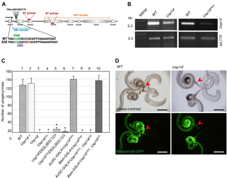
To investigate the physiological consequences of Usp14 depletion, we analyzed phenotypes following PiggyBac-mediated insertional mutation of Usp14 (to give the allele Usp14f). The PiggyBac element was inserted at the start of the second exon (Fig. 1A) and resulted in substantial reductions inUsp14mRNA
Received 2 August 2019; Accepted 13 December 2019
1Department of Genetics, University of Szeged, H-6726 Szeged, Hungary.
2Institute of Biochemistry, Biological Research Centre, H-6726 Szeged, Hungary.
*Present Address: Department of Genetics, University of Cambridge, CB2 3EH, UK
‡Author for correspondence (deakp@bio.u-szeged.hu)
L.K., 0000-0002-3226-3740; M.P., 0000-0001-5587-1637; P.D., 0000-0002- 8849-0352
Journal of Cell Science
expression levels, indicating that Usp14f is a relatively strong hypomorphic allele (Fig. 1B). AlthoughUsp14fhomozygotes were viable, all eclosed males were sterile (Fig. 1C, column 3) and despite normal mating behaviors, their wild-type (WT) female mating partners laid mostly unfertilized eggs. In phase-contrast and fluorescent microscopy analyses ofUsp14fhomozygote testes, all stages through spermatid elongation were indistinguishable from those in WT flies. Although sperm bundles were formed inUsp14f testes, seminal vesicles were almost empty and contained few individualized sperms (Fig. 1D). In addition, most sperm bundles were accumulated before the entrance of seminal vesicles, suggesting defects in sperm individualization. Male sterility was also observed whenUsp14f was situatedin transto a deficiency uncovering theUsp14locus (Fig. 1C, column 5). However, male fertility was completely restored in revertants ofUsp14f(Fig. 1C, column 2). Viability and fertility of females were not affected by Usp14mutations (data not shown).
For more accurate analyses of Usp14 gene functions, we generated null alleles using CRISPR/Cas9-mediated mutagenesis
(Port et al., 2014). Among several indel mutants, we selected the point mutant alleleUsp14Δ32for detailed analysis. In this mutant, insertion of a cytosine after the second codon generates a frameshift and a premature stop codon at 16 base pairs downstream of the start site (Fig. 1A). Despite the resulting absence of functional Usp14, Usp14Δ32null homozygotes were viable but males were sterile, as observed with the hypomorphic alleles (Fig. 1C, column 4).
Usp14Δ32mutant testes imitated individualization defects inUsp14f males, indicating no clear differences between the phenotypes of null and the strong hypomorphic alleles. These results suggest that Usp14 is involved in the coordination of late spermatogenesis.
Usp14mutant male sterility is germline dependent
The male sterile phenotype observed in Usp14 mutants may exclusively reflect requirements of Usp14 in germline cell lineages and/or testis-forming somatic cells. To identify affected cell types following Usp14 depletion, we used the GAL4/UAS system in Drosophila(Duffy, 2002). Several GAL4 cell lines have specific spatio-temporal expression patterns in fly testis (White-Cooper,
Fig. 1. Male-sterile phenotype ofUsp14mutants.(A) Diagram of theUsp14gene; arrowhead shows the position of the PiggyBac insertion (denoted asUsp14f in text). The insertion of a single cytosine nucleotide inUsp14Δ32mutants is highlighted in red. The protospacer adjacent motif (PAM) is in green. (B)Usp14 expression levels in mutant and knockdown flies were determined using semi-quantitative RT-PCR analyses of WT, mutant and RNAi-transfected flies.
Usp14RNAiwas controlled by the ubiquitousAct5C-GAL4driver andrpL17Awas used as a loading control. (C) Quantification of the fertility of the males with indicated genotypes (n=10 individual males/genotype). Results are mean±s.d.,n=10. *P<0.01 compared with WT (two-tailed Student’st-test). (D) Micrographs of internal male genitalia in WT andUsp14fmutant flies (upper panels) and protamineB–GFP-labeled nuclei of elongated spermatids (lower panels). Arrowheads indicate seminal vesicles. Scale bars: 250 µm.
Journal of Cell Science
2012). Herein, we used the bam-Gal4 driver, which showed a germline-restricted expression pattern in previous studies (Chen and McKearin, 2003). TheAct5c-Gal4driver was used exclusively to drive expression in somatic cells (White-Cooper, 2012).
Usp14 specific transgenic RNA interference was induced by mean of the germline-specificbam-Gal4driver and resulted in male sterility (Fig. 1C, column 8) and resulted in individualization defects that were similar to those seen in theUsp14hypomorphic and null mutants. However, induction of the RNAi with the soma- specific Act5C-Gal4 driver did not lead to male sterility or phenotypic abnormalities (Fig. 1C, column 7). Conversely, the male sterility ofUsp14Δ32homozygotes was rescued by transgenic Usp14 expression under the control of the bam-Gal4 driver (Fig. 1C, column 10),but not with the somaticAct5C-Gal4driver (Fig. 1C, column 9). These results suggest that germline-specific expression ofUsp14is required for proper sperm development.
Usp14mutation disrupts individualization complexes The germline-dependent male sterility and deficient processing of sperm bundles to mature sperms in Usp14 mutant testes suggest that Usp14 facilitates sperm individualization. Cytoskeletal-membrane individualization complexes (ICs) mediate sperm individualization in elongated spermatid bundles. ICs contain a cluster of 64 actin-rich structures, known as actin cones. During sperm individualization, these move down the bundles and push excess cytoplasm and organelles into so called‘cystic bulges’that serve as waste bags.
Concomitantly, spermatids are sheathed in plasma membranes and become individualized and move into seminal vesicles for storage until mating (Noguchi et al., 2006). Severe disturbances of this process lead to individualization defects and stagnation of sperm bundles in front of seminal vesicles.
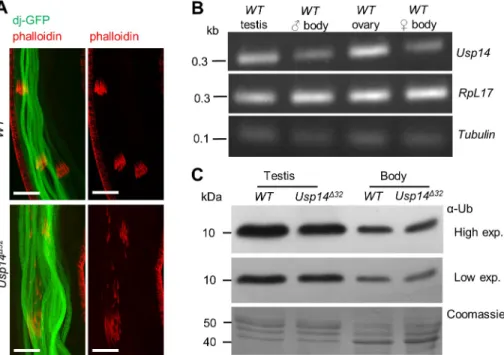
To determine whether structures of the IC are affected inUsp14 mutants, actin cones were visualized in WT and Usp14Δ32testes after staining with fluorescently labeled phalloidin. In WT testes, triangular shaped actin cones move synchronously (Fig. 2A, upper panels) and actin cones behind the moving IC can only be observed occasionally. However, in Usp14Δ32 testes the majority of ICs lacked synchronous movements of actin cones, and a high
proportion of individual actin cones were spread along the sperm bundles (Fig. 2A, lower panels). Hence, Usp14 is likely required for synchronous migration of actin cones, and disruption of ICs leads to male sterility. Strikingly similar individualization defects were observed following mutation of the testis-specific proteasome subunit Prosα6T (Zhong and Belote, 2007). Because the proteasome interactions of Usp14 are well established in Drosophila (Lundgren et al., 2005; Lee et al., 2011), the resemblance of Prosα6T and Usp14 mutant phenotypes may suggest that both subunits are required for normal testis-specific proteasome function.
Usp14 is predominantly expressed in testis
Previous studies have shown that many proteasome subunits and associated proteins have testis-specific orthologs in Drosophila (Belote and Zhong, 2009; Ma et al., 2002; Zhong and Belote, 2007), suggesting important roles of the proteasome inDrosophilatestes.
Because Usp14 is a transient, but conserved, subunit of the regulatoryDrosophilaproteasome particles (Lundgren et al., 2005;
Lee et al., 2011), and its mutants show testis-specific phenotypes, we investigated the possibility of differential expression ofUsp14in testis. To investigate the expression levels of Usp14, testis and gonadectomized bodies of WTDrosophilawere analyzed through semiquantitative RT-PCR experiments.Usp14transcripts were more abundant in testis than in other tissues (Fig. 2B). Because accessory glands and other organs were removed from testes before mRNA extraction, somatic cell representation was very low in this sample, including only epithelial cells of the testis surface. With observations ofUsp14rescue by germline drivers (Fig. 1C), these data indicate thatUsp14is expressed predominantly in the male germline.
Loss of Usp14 and reduced free ubiquitin levels in testes Loss of Usp14 function in yeast and mouse models previously resulted in considerable reductions in monoubiquitin levels (Leggett et al., 2002; Anderson et al., 2005). Usp14 recycles monoubiquitins from protein substrates that are targeted to the proteasome, and prevents their degradation together with the proteins targeted to the proteasome (Hanna et al., 2003;
Fig. 2. Individualization defects and reductions in monoubiquitin pools in the absence of Usp14.(A) confocal micrographs of sperm bundles; dj-GFP (green) labels sperm tails and phalloidin (red) individualization complexes. Scale bars: 5 µm. (B) Expression levels ofUsp14in testis. Semiquantitative RT-PCR analyses of WT gonadectomized body, testis and ovary samples;
rpL17Aand tubulin were used as loading controls.
(C) Monoubiquitin levels inUsp14mutant somatic (body) and testis tissues (also shown on Fig. 3B);
western blots show a monoubiquitin band (α-Ub) at 8.5 kDa. Lanes were loaded with 4 µg aliquots of total protein, as determined using Bradford assays.
Journal of Cell Science
Anderson et al., 2005). We determined monoubiquitin protein expression levels in WT and Usp14 mutant testes and gonadectomized body samples by western blotting. We primarily observed significant differences between body and testis monoubiquitin levels (Fig. 2C, compare lane 1 and 3). Pixel density analysis of western blot images demonstrated that the monoubiquitin levels in testis are more than 3-fold higher than in the rest of the body. Greater than 80% of the monoubiquitin pool is present in testis, suggesting that ubiquitylation is prevalent during spermatogenesis. Pixel density analyses of immunoblots (Fig. 2C) revealed up to a 30% decrease in monoubiquitin concentrations inUsp14mutant testes, but no differences in other tissues from WT and Usp14 mutant flies (see Fig. S1). These data show that monoubiquitin pools in testes are especially sensitive to the loss of Usp14 function, likely contributing to male sterility and individualization defects.
The effects of monoubiquitin deficiencies on spermatogenesis were exacerbated when theUsp14mutation was accompanied by a hypomorphic allele of the testis-specific ubiquitin geneUbi-p63E. InDrosophila,Ubi-p63Eis the polyubiquitin gene that provides most of the newly synthetized ubiquitins in testis (Lu et al., 2013).
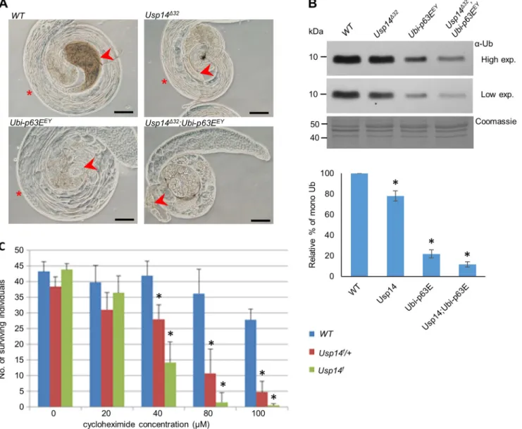
Ubi-p63Enull mutant spermatocytes show meiotic arrest with loss of monoubiquitins. The Ubi-p63EEY hypomorph P element insertion allele, however, has moderate effects on monoubiquitin pools and permits progress of spermatogenesis to the elongated spermatid stage (Lu et al., 2013). After combiningUsp14Δ32null and Ubi-63EEY hypomorph mutations, we observed an increased severity of the testis phenotype compared with that in single mutants (Fig. 3A). In particular, spermatocytes from the Usp14Δ32 Ubi- 63EEYdouble mutants had arrested meiosis and no postmeiotic and elongated spermatocytes were observed (Fig. 3A). This phenotype is consistent with theUbi-p63Enull phenotype (Lu et al., 2013),
Fig. 3. Lack of Usp14 leads to monoubiquitin deficiency.(A) Phase-contrast micrographs of testes from males with indicated genotypes; arrowheads show seminal vesicles. Asterisks indicate the presence of elongated spermatids that are absent inUsp14;Ubi-p63Edouble mutants. Scale bars: 100 µm.
(B) Monoubiquitin levels in testis ofUsp14andUbi-p63Emutants were measured using western blots with an anti-ubiquitin antibody (α-Ub). After determining protein concentrations using Bradford assays, 4 µg aliquots of total protein were loaded onto gels. Monoubiquitin contents in mutants were calculated using data from densitometric analyses and are presented as percentages of that in WT controls (mean±s.d.,n=4) is shown in the graph. *P<0.01 compared to WT (two-tailed Student’st-test). (C) Cycloheximide sensitivity ofUsp14mutants. Data are expressed as means±s.d. deviations of three independent replicates.
*P<0.01 compared to WT data at a given concentration (two-tailed Student’st-test).
Journal of Cell Science
with considerable reductions in monoubiquitin levels (Fig. 3B).
PerhapsUsp14Δ32andUbi-63EEYsynergistically maintain adequate free monoubiquitin pools in the testis. Most likely, loss ofUbi-p63E caused severe free ubiquitin shortage leading to early arrest of meiosis, whereas a less severe shortage of ubiquitin inUsp14null mutants manifests in a later stage of spermatogenesis.
Usp14 mutants are cycloheximide sensitive
The antibiotic cycloheximide is widely used as a eukaryotic protein synthesis inhibitor. It has toxic side effects that are related to rapid depletion of ubiquitin pools in treated cells. Accordingly, the expression levels of ubiquitin genes are exceptionally sensitive to this drug (Hanna et al., 2003). Previous studies have also identified mutations that reduce intracellular ubiquitin levels and lead to cycloheximide sensitivity in yeast andDrosophila(Leggett et al., 2002; Chernova et al., 2003; Kovács et al., 2015).
While previously studying the physiological roles of the Drosophila DUB Usp5, we observed a ubiquitin stress response as had been described in yeast (Hanna et al., 2007). Another study also indicates that Usp14 has an essential role (Kovács et al., 2015) that is unrelated to its testis-specific function.
Because loss of Usp5 leads to reduced monoubiquitin levels and triggers the expression of Usp14 to counteract ubiquitin degradation and boost ubiquitin recycling, it was possible that loss-of-function Usp14mutants are cycloheximide sensitive. To test this possibility, we compared the effects of cycloheximide in WT and hypomorph Usp14fmutants.
Cycloheximide treatments ofUsp14fhomozygote larvae (Fig. 3C) led to death in a dose-dependent manner, with an LD50of 32 µM.
Viability of WT animals was not affected at this concentration.
Usp14f/+ heterozygotes also showed high sensitivity to cycloheximide (LD50=42 µM), indicating that one copy ofUsp14 does not compensate for ubiquitin shortages sufficiently after cycloheximide treatments. These data suggest that Usp14 functions in somatic tissues in situations when the ubiquitin equilibrium is disturbed by reduced ubiquitin expression due to cycloheximide treatment, or by abolished Ub recycling following elimination of Usp5 (Kovács et al., 2015). Demand for Usp14 activities in somatic tissues under stressed conditions indicate that Usp14 is not a testis- specific subunit, because it also has roles in somatic tissues. It is more likely that normal spermatogenic processes have high ubiquitin demands and, hence, resemble quasi-ubiquitin stress conditions in which proper functioning of Usp14 and other proteasome subunits is heavily required. Usp14 dependence of spermatogenesis may also be evolutionarily conserved becauseUsp14mutantataxiaJmice are also sterile and had defects in spermatogenesis (Crimmins et al., 2009).
However, loss of Usp14 function also severely affects the neuromuscular system in mice (Wilson et al., 2002; Anderson et al., 2005), suggesting that, unlike in flies, neuronal tissues of more- complex organisms are hypersensitive to ubiquitin homeostasis.
Taken together, our data demonstrate pleiotropic roles of the Drosophila Usp14 deubiquitylase in spermatogenesis and in the ubiquitin stress responses. Detailed phenotypic analyses of loss-of- function alleles show thatUsp14is essential for normal spermatid differentiation. Accordingly, homozygousUsp14mutant males are sterile and have defective sperm individualization processes. In agreement, the phenotype associated with loss of Usp14 function is consistent with its expression pattern in testes.
Finally, we show that individualization defects were strikingly similar to the mutation of the testis-specific proteasome subunit Prosα6T (Zhong and Belote, 2007). Given that interaction of Usp14 with the proteasome is well established inDrosophila (Lundgren
et al., 2005; Lee et al., 2011), the resemblance of Prosα6Tand Usp14 mutant phenotypes may suggest that both subunits are required for normal testis-specific proteasome function.
MATERIALS AND METHODS Drosophilastocks and methods
Drosophilalines were maintained on standard yeast and cornmeal medium at 25°C. The PiggyBac element insertion lineUsp14f00779 was obtained from BloomingtonDrosophilaStock Center (BDSC stock number: 18368).
The transgenic RNA interference lineUsp14KK102888was obtained from ViennaDrosophilaResource Center (VDRC stock number: v110227). All genetic markers are described in FlyBase at http://flybase.org. Thedj-GFP line was obtained from BloomingtonDrosophilaStock Center (BDSC stock number: 5417). TheprotamineB-GFPstock was kindly provided by Helen White-Cooper (School of Biosciences, Cardiff University, Cardiff, UK).
The WT strainw1118was used for comparisons in all experiments.
To reverse theUsp14f00779PiggyBac insertion, we crossed stocken mass with a PBac transposase source (BDSC 8285) and established two candidate revertant stocks. Restoration of male fertility was observed in both revertants, but onlyUsp14frv were maintained and used in experiments.
CRISPR/Cas9-mediated mutagenesis was used to generateUsp14Δ32null mutants using previously published tools (Port et al., 2014) with the targeting guide RNA sequence 5′-GCTACCTTTAAATGCGGGCA-3′. In detail, the targeting guide RNA sequence was cloned into pCFD3 vector containing the U6:3 ubiquitin promoter and the guide RNA backbone. This construct was injected into the y[1] w[*] P{y[+t7.7]=nos-phiC31
\int.NLS}X; P{y[+t7.7]=CaryIP}su(Hw)attP1 (BDSC 35567) line and, through integrase-mediated site-specific transformation, we obtained a fly line constitutively expressingUsp14-specific guide RNAs. The guide RNA expressing line was crossed to anos-Cas9expressing line (BDSC 54591) and the resulted‘cut starter’G0 progeny was further crossed to aw;CyO/Sco balancer line. From the subsequent generation, 100 males were individually crossed to aw;CyO/Scobalancer line and balanced candidate stocks were established. Candidates showing male sterility in homozygotes or over a deficiency were sequenced. In lineΔ32(Δrefers to the absence of gene function; 32 is the serial number of the candidate) we identified a single base pair insertion 3 bp downstream of the start codon resulting in a frameshift and premature stop codon 16 bp after the transcription start.
Male fertility was tested by placing individual 1-day-old males in vials with two 4-day-old WT virgin females. After 6 days, adults were discarded and the numbers of the eclosed progeny were scored.
Semiquantitative RT-PCR
Testes of 0–1-day-oldDrosophilamales were dissected in ice-cold PBS and tubes containing either 50 testes or 10 gonadectomized males were frozen at
−80°C. Total RNA was isolated using Tri Reagent extraction kits (Sigma- Aldrich). RNA samples were treated with RQ1 RNase-Free DNase (Promega) and were reverse transcribed using Fermentas cDNA synthesis kits. Samples of cDNA were normalized to the rpL17A level using PCR with rpL17A forward primer, 5′-GTGATGAACTGTGCCGACAA-3 and rpL17A reverse primer, 5′-CCTTCATTTCGCCCTTGTTG-3′. Usp14 mRNA expression levels were then determined in 20–25-cycle PCRs with the exon-specificUsp14RT forward primer 5′- ACGGTGGTGCCCTTCTCC- 3′, andUsp14RT reverse primer 5′-GGCGCTGTGGTCCTGTTG-3′.
Fluorescence microscopy
Prior to native observations of GFP fluorescence, testes were dissected and mounted without extensive squashing in PBS and were then directly observed and imaged using an Olympus BX51 upright microscope with phase contrast and UV GFP filters.
Confocal microscopy was performed with testes of 0–1-day-old males expressing the sperm tail marker dj-GFP. In these experiments, WT or Usp14mutant flies were dissected at room temperature in PBS and were immediately fixed for 20 min in 4% formaldehyde. Testes were stained with Alexa Fluor 647-conjugated phalloidin (1:400; ThermoFisher Scientific,
# A22287) and DAPI and were then examined under an Olympus FV 1000
confocal microscope.
Journal of Cell Science
SDS-PAGE and western blot analyses
Testes were dissected in ice-cold PBS buffer and were frozen in liquid nitrogen and stored at−80°C until use. Protein samples were then prepared from homogenates of 25 testes in Buffer I F, which contained 100 mM Tris- HCl pH 7.6, 150 mM NaCl, 1 mM EDTA, 100 mM N-ethylmaleimide (NEM, Sigma-Aldrich), 20 µM MG132 (Calbiochem) and 1× EDTA-free Complete protease inhibitor cocktail (Roche). Homogenates were centrifuged (21,000g for 10 min at 4°C) and boiled in 1× Laemmli buffer. Samples containing 4 µg of total protein were loaded onto 14% SDS- acrylamide gels, were electrophoresed and subjected to western blotting.
Protein concentrations were determined using Bradford assays (Bradford, 1976). Monoubiquitin forms of 8.5 kDa were detected using a mouse monoclonal anti-ubiquitin primary antibody (Sigma-Aldrich, U0508, 1:3000 dilution) and a peroxidase-conjugated AffiniPure goat anti-mouse- IgG (Jackson Immuno Research, 115-035-003, 1:30,000 dilution) secondary antibody. Western blots were developed on X-ray films, and were scanned and processed using ImageJ software.
Cycloheximide treatment
Cycloheximide (Sigma-Aldrich) was dissolved in ethanol to obtain stock solution (17.6 mM). First-instar larvae were collected and maintained in vials containing 3.5 ml of standardDrosophilamedium supplemented with 60μl aliquots of cycloheximide solution in ethanol. Final treatment concentrations were 0–100μM in the final volume of 3.5 ml. Eclosing adults were scored and statistically analyzed using a Microsoft Office Excel software.
Acknowledgements
Drosophilastocks obtained from the BloomingtonDrosophilaStock Center (NIH P40OD018537) and the ViennaDrosophilaResource Center (VDRC, www.vdrc.at) were used in this study. We also thank Flybase for providing database information (McQuilton et al., 2012).
Competing interests
The authors declare no competing or financial interests.
Author contributions
Conceptualization: P.D.; Methodology: L.K., A.N., P.D.; Validation: A.N., P.D.;
Formal analysis: L.K., P.M.; Investigation: L.K., A.N., P.M.; Resources: P.D.; Data curation: A.N., P.D.; Writing - original draft: P.D.; Writing - review & editing: L.K., P.D.;
Visualization: L.K., A.N.; Supervision: P.M., P.D.; Project administration: P.M., P.D.;
Funding acquisition: P.D.
Funding
This work was funded by grants from the Hungarian Scientific Research Fund (OTKA-K116372), Ministry for National Economy of Hungary (GINOP-2.3.2-15- 2016-00032).
Supplementary information
Supplementary information available online at
http://jcs.biologists.org/lookup/doi/10.1242/jcs.237511.supplemental
Peer review history
The peer review history is available online at
https://jcs.biologists.org/lookup/doi/10.1242/jcs.237511.reviewer-comments.pdf
References
Amerik, A. Y. and Hochstrasser, M. (2004). Mechanism and function of deubiquitinating enzymes.Biochim. Biophys. Acta1695, 189-207. doi:10.1016/
j.bbamcr.2004.10.003
Anderson, C., Crimmins, S., Wilson, J. A., Korbel, G. A., Ploegh, H. L. and Wilson, S. M.(2005). Loss of Usp14 results in reduced levels of ubiquitin in ataxia mice.J. Neurochem.95, 724-731. doi:10.1111/j.1471-4159.2005.03409.x Belote, J. M. and Zhong, L.(2009). Duplicated proteasome subunit genes in
Drosophila and their roles in spermatogenesis. Heredity (Edinb)103, 23-31.
doi:10.1038/hdy.2009.23
Bradford, M. M. (1976). A rapid and sensitive method for the quantitation of microgram quantities of protein utilizing the principle of protein-dye binding.Anal.
Biochem.72, 248-254. doi:10.1016/0003-2697(76)90527-3
Chen, D. and McKearin, D. M.(2003). A discrete transcriptional silencer in the bam gene determines asymmetric division of the Drosophila germline stem cell.
Development130, 1159-1170. doi:10.1242/dev.00325
Chernova, T. A., Allen, K. D., Wesoloski, L. M., Shanks, J. R., Chernoff, Y. O. and Wilkinson, K. D.(2003). Pleiotropic effects of Ubp6 loss on drug sensitivities and yeast prion are due to depletion of the free ubiquitin pool.J. Biol. Chem.278, 52102-52115. doi:10.1074/jbc.M310283200
Crimmins, S., Sutovsky, M., Chen, P.-C., Huffman, A., Wheeler, C., Swing, D. A., Roth, K., Wilson, J., Sutovsky, P. and Wilson, S.(2009). Transgenic rescue of ataxia mice reveals a male-specific sterility defect.Dev. Biol.325, 33-42. doi:10.
1016/j.ydbio.2008.09.021
Duffy, J. B.(2002). GAL4 system in Drosophila: a fly geneticist’s swiss army knife.
Genesis34, 1-15. doi:10.1002/gene.10150
Guterman, A. and Glickman, M. H.(2004). Deubiquitinating enzymes are IN(trinsic to proteasome function). Curr. Protein Pept. Sci. 5, 201-211. doi:10.2174/
1389203043379756
Hanna, J., Leggett, D. S. and Finley, D.(2003). Ubiquitin depletion as a key mediator of toxicity by translational inhibitors.Mol. Cell. Biol.23, 9251-9261.
doi:10.1128/MCB.23.24.9251-9261.2003
Hanna, J., Meides, A., Zhang, D. P. and Finley, D.(2007). A ubiquitin stress response induces altered proteasome composition.Cell129, 747-759. doi:10.
1016/j.cell.2007.03.042
Kim, J. H., Park, K. C., Chung, S. S., Bang, O. and Chung, C. H.(2003).
Deubiquitinating enzymes as cellular regulators.J. Biochem.134, 9-18. doi:10.
1093/jb/mvg107
Kovács, L., Nagy, O., Pál, M., Udvardy, A., Popescu, O. and Deák, P.(2015). Role of the deubiquitylating enzyme DmUsp5 in coupling ubiquitin equilibrium to development and apoptosis in Drosophila melanogaster. PLoS ONE 10, e0120875. doi:10.1371/journal.pone.0120875
Lee, M. J., Lee, B. H., Hanna, J., King, R. W. and Finley, D.(2011). Trimming of ubiquitin chains by proteasome-associated deubiquitinating enzymes.Mol. Cell.
Proteomics10, R110.003871. doi:10.1074/mcp.R110.003871
Lee, J. H., Park, S., Yun, Y., Choi, W. H., Kang, M.-J. and Lee, M. J.(2018).
Inactivation of Usp14 perturbs ubiquitin homeostasis and delays the cell cycle in mouse embryonic fibroblasts and in fruit fly Drosophila.Cell. Physiol. Biochem.
47, 67-82. doi:10.1159/000489750
Leggett, D. S., Hanna, J., Borodovsky, A., Crosas, B., Schmidt, M., Baker, R. T., Walz, T., Ploegh, H. and Finley, D.(2002). Multiple associated proteins regulate proteasome structure and function.Mol. Cell10, 495-507. doi:10.1016/S1097- 2765(02)00638-X
Lu, C., Kim, J. and Fuller, M. T.(2013). The polyubiquitin gene Ubi-p63E is essential for male meiotic cell cycle progression and germ cell differentiation in Drosophila.Development140, 3522-3531. doi:10.1242/dev.098947
Lundgren, J., Masson, P., Mirzaei, Z. and Young, P.(2005). Identification and characterization of a Drosophila proteasome regulatory network.Mol. Cell. Biol.
25, 4662-4675. doi:10.1128/MCB.25.11.4662-4675.2005
Ma, J., Katz, E. and Belote, J. M.(2002). Expression of proteasome subunit isoforms during spermatogenesis in Drosophila melanogaster.Insect Mol. Biol.
11, 627-639. doi:10.1046/j.1365-2583.2002.00374.x
McQuilton, P., St Pierre, S. E. and Thurmond, J. and FlyBase Consortium (2012). FlyBase 101–the basics of navigating FlyBase.Nucleic Acids Res.40, D706-D714. doi:10.1093/nar/gkr1030
Nijman, S. M., Luna-Vargas, M. P. A., Velds, A., Brummelkamp, T. R., Dirac, A. M. G., Sixma, T. K. and Bernards, R.(2005). A genomic and functional inventory of deubiquitinating enzymes.Cell123, 773-786. doi:10.1016/j.cell.2005.11.007 Noguchi, T., Lenartowska, M. and Miller, K. G.(2006). Myosin VI stabilizes an
actin network during Drosophila spermatid individualization.Mol. Biol. Cell17, 2559-2571. doi:10.1091/mbc.e06-01-0031
Port, F., Chen, H.-M., Lee, T. and Bullock, S. L.(2014). Optimized CRISPR/Cas tools for efficient germline and somatic genome engineering in Drosophila.Proc.
Natl. Acad. Sci. USA111, E2967-E2976. doi:10.1073/pnas.1405500111 Reyes-Turcu, F. E., Ventii, K. H. and Wilkinson, K. D.(2009). Regulation and
cellular roles of ubiquitin-specific deubiquitinating enzymes.Annu. Rev. Biochem.
78, 363-397. doi:10.1146/annurev.biochem.78.082307.091526
Suresh, B., Lee, J., Kim, K.-S. and Ramakrishna, S.(2016). The importance of ubiquitination and deubiquitination in cellular reprogramming.Stem Cells Int.
2016, 6705927. doi:10.1155/2016/6705927
Tsou, W.-L., Sheedlo, M. J., Morrow, M. E., Blount, J. R., McGregor, K. M., Das, C. and Todi, S. V.(2012). Systematic analysis of the physiological importance of deubiquitinating enzymes. PLoS ONE 7, e43112. doi:10.1371/journal.pone.
0043112
White-Cooper, H. (2012). Tissue, cell type and stage-specific ectopic gene expression and RNAi induction in the Drosophila testis.Spermatogenesis2, 11-22. doi:10.4161/spmg.19088
Wilson, S. M., Bhattacharyya, B., Rachel, R. A., Coppola, V., Tessarollo, L., Householder, D. B., Fletcher, C. F., Miller, R. J., Copeland, N. G. and Jenkins, N. A.(2002). Synaptic defects in ataxia mice result from a mutation in Usp14, encoding a ubiquitin-specific protease.Nat. Genet.32, 420-425. doi:10.1038/
ng1006
Zhong, L. and Belote, J. M. (2007). The testis-specific proteasome subunit Prosalpha6T of D. melanogaster is required for individualization and nuclear maturation during spermatogenesis.Development134, 3517-3525. doi:10.1242/
dev.004770