Aging As a Consequence of Growth Cessation
ROBERT R. KOHN
Institute of Pathology, Western Reserve University, Cleveland, Ohio
Introduction
In comparison with many other areas of biology, useful concepts regard- ing aging processes have been evolving very slowly. While much informa- tion has accumulated, there is little agreement concerning the significance of various observations. Few of the most active workers in this field would agree on the specific questions to be asked and the types of experiments to be performed to obtain a better understanding of the mechanisms of aging. The slow progress in aging research has been partly due to over- simplifications, advancing of general theories which allegedly explained diverse and probably unrelated phenomena, and confusion in regard to the kinds of processes to be categorized under the unsatisfactory term of
"aging." Progress has also been slow because of difficulties inherent in the study of aging processes: the frequent necessity for maintaining organ- isms for long periods, difficulties in distinguishing between diseases and aging, and the lack of adequate control systems, i.e., systems existing in time but not undergoing change. In addition, perhaps many of the most competent biologists have ignored aging processes because on superficial examination these processes do not appear to contain the programming and certainty so attractive to most investigators.
In this discussion, we will develop the thesis that enough information is available to classify and make clear distinctions between different types of aging processes and that for some types we know enough to construct conceptual frameworks which can tell us what questions to ask and what kinds of investigations would be most informative. In association with this thesis, several arguments will be advanced: Aging processes, which are widespread in nature, should be considered "normal" biology and as such are worth studying and understanding; aging is closely related to growth cessation, but only when certain cell or tissue constituents are retained;
a primary distinction should be made between aging of an intact meta- zoan and aging of an organism's cells and tissues. For example, almost
291
all of the cells a hydra has today will have aged and died in 6 weeks but the hydra itself will still be vigorous at that time (Burnett, 1961). On the other hand, if we study a very old human being who is about to die, we will find that essentially all of his cells appear normal by metabolic and chemical criteria. Some questions provoked by these observations are:
What is the mechanism of cell death in hydra; why does the human being die when his cells appear viable; and, what cell or tissue changes could explain the age-related death in a complex higher animal? In this dis- cussion it will be argued that the common denominator in cell and ani- mal aging is growth cessation, that mechanisms in the two cases are quite unrelated, and that enough is known, particularly about aging of some higher animals, for us to propose plausible descriptions of these mechanisms.
Scope of Aging
An aging process can be usefully defined as one occurring in all mem- bers of a population and which has its onset, or progresses more rapidly, after maturity. Maturity, therefore, is the border between traditional developmental biology and gerontology. Aging processes progress and are not reversible under usual physiological conditions. Progression also distinguishes aging processes from other biological reactions which either stop or are maintained at equilibria by feedback mechanisms. Reactions which progress are generally harmful to the system involved, since homeo- stasis depends on the establishment of equilibria or steady states. Such progression in biological systems frequently results in death; and death is therefore a convenient endpoint in the study of aging processes. In a human context, it can be argued that aging processes are responsible for the only 100% fatal disease everyone has. Biological processes which con- stitute a barrier to human immortality should be of more than casual interest.
Aging processes are exceedingly widespread, both in nonliving systems and in living systems which have attained certain levels of complexity and organization. Among the nonliving systems, almost all man-made materials and machines age, some at rates and by mechanisms apparently analogous to the aging of higher animals. Conspicuous aging occurs in plastics, paper, and rubber. It is perhaps significant that these materials are high polymers, as are substances in higher animals which show promi- nent age-related changes. In living systems aging processes as defined above occur at all levels from the macromolecule to the intact mammal.
AGING AS A CONSEQUENCE OF GROWTH CESSATION 293
In many cases rates of aging and occurrence of death endpoints can be predicted and described with the same precision as developmental stages, growth rates, and metabolic reactions.
Bacteria in culture age, die, and lyse in a predictable fashion. In some species bacterial aging has been associated with characteristic changes in morphology and cell wall composition (Chatterjee and Williams, 1962;
Collins, 1964). Among the protozoa, Tocophrya infusorium degenerates and dies by aging processes (Rudzinska, 1951). Similarly, gametes which do not participate in fertilization pass through a sequence of degenera- tive processes ending in death. In tissue culture, under optimal condi- tions of serial passage, diploid cell strains die out in 4 to 10 months (Hayflick and Moorhead, 1961; Miles, 1964). Hayflick and Moorhead (1961) favor the hypothesis that an intracellular factor necessary for survival is not replicated as fast as the cells are replicated. It is not likely that such rapid and continuous cell division occurs in nature, and this example of aging processes in proliferating cells may not provide informa- tion about other aging systems.
In metazoans a distinction must be made between aging of the intact organism and aging of the various cell populations within the organism.
While both aging phenomena satisfy the criteria of aging processes, they proceed independently of each other and appear to involve different mechanisms. Aging of intact animals, characterized by clearly denned loss of function, degenerative structural changes, and a specific type of age-related mortality rate, is seen in most phyla. It is most conspicuous in animals showing both a species-specific size and maintenance of stable tissues in the adult. Such aging has been studied most thoroughly in insects, laboratory mammals, and man; it will be dealt with in some detail below.
Many cell populations in both embryos and adult animals undergo an apparently clearly programmed senescence. In development, cellular degeneration and death are noted in such instances as the involution of ascidian and anuran larval tails, loss of gills, involution of insect tissues during metamorphosis, disintegration of chrondrocytes in osteogenesis and involution of Müllerian and Wolffian ducts in male and female mammals respectively. An orderly sequence of cellular degenerations oc- curs in tissue invagination and separation, in the formation of lumina, and in tissue and organ modeling. In most of these cases cell degeneration and death are probably essential for subsequent normal development.
Such degenerations are programmed to the extent that transplantation experiments can be performed with presumptive degenerative tissue.
Some types of developmental degeneration appear to result from intrinsic cellular factors while others are influenced by hormones or other tissues (Biggers, 1964; Zwilling, 1964). After organ systems have developed and throughout the life of an animal, many cell populations demonstrate phases of cell division, cell specialization, degeneration, and death. T h e last two mentioned phases satisfy the criteria of aging processes and will be considered in some detail.
Growth and Aging
From a priori considerations, we would predict that aging processes would be more conspicuous after growth cessation. For a system to change with time, either different components must be added or subtracted at different times, or there must be changes in components which remain.
Substances which are constantly turning over and being replaced would not be expected to contribute to an aging process. Similarly, a growing system is constantly adding to itself new (and young) components which, even though added to a cell or tissue present for some time, would not contribute to aging until present long enough to undergo change them- selves. It follows that the aging rate of organelle, cell, tissue, or intact organism would be proportional to the amount of biologically important nonrenewable materials present and the time during which such materials are present. The aging rate would of course be a manifestation of the changes occurring in these materials which alter their biological properties.
Accumulated data have supported the notion that aging character- istically follows growth cessation. In nature, cell populations which degenerate on schedule and show an age-related increase in death rate are those which stop dividing and which presumably contain some stable elements. The metazoans which demonstrate the most exact life-spans and sequence of degenerations are those which attain a size characteristic of the species and grow no more, and which retain significant amounts of nonrenewable body constituents. It has been noted that senescence is the necessary price paid for evolutionary attainment and maintenance of an ideal size in higher animals (for discussions of evolution and aging, see Strehler, 1962, and Comfort, 1956). Experimentally, it has been possible to lengthen the lifespan of mammals considerably by prolonging the growth phase (McCay, 1952). Factors causing cessation of growth appear to represent the last interest of traditional developmental biology and the first interest of gerontology, although neither growth nor aging can be
AGING AS A CONSEQUENCE O F GROWTH CESSATION 295
appreciated without some understanding of their interrelationships. The remainder of this discussion will be devoted to examples of the two most conspicuous types of aging in nature: aging of intact higher animals, and aging of certain cell populations within metazoans.
Animal Aging
From a consideration of the relationship between growth and aging, it follows that aging of higher animals can best be studied in those with a species-specific size. Human beings satisfy this criterion and will serve as examples in much of the following discussion. Humans are also valuable in considerations of aging because so much is known about alterations in their physiological processes with time, age-related changes in morphology, chemical composition, disease incidence, and mechanisms of death and death rates of large populations. For the construction of hypotheses ex- plaining aging mechanisms, it is essential that something be known about the factors which precede the death endpoint. This is particularly true when lifespans are altered by some experimental procedure or when genetic variations in life-span are under study. Too frequently differences in lifespan alone resulting from genetic variation or special treatment, are used in generalizations about aging. Such differences may be due in some cases to changed patterns of disease or specific lesions which have little relationship to aging in a natural population.
Some Characteristics of Aging Populations
If death is considered the endpoint of human aging, some inferences regarding mechanisms can be drawn from the inspection of death rates of different populations plotted in different ways. If the percentage sur- viving is plotted as a function of age, different types of curves are obtained from different populations. In Fig. 1, the death rate of the British India population yields a curve somewhere between the logarithmic die-away curve expected from random nonage-related deaths and the more rec- tangular curve for the New Zealand population. In general, the more highly developed societies show the most rectangular types of curves. This results largely from the reduction of deaths due to infectious diseases which either affect a young population or show a poor correlation with age, allowing the population to die from age-related processes. T h e curve for the New Zealand population demonstrates very clearly the age-related increase in the probability of dying. Such rectangular curves, indicating an increasing death rate with increasing age, are characteristic of popu-
lations having members that wear out, such as a population of auto- mobiles. The various curves in Fig. 1, in spite of their shapes, all end at approximately the same place on the age axis, indicating that the maxi- mum life-span is about the same in the different populations. It may be inferred from these curves that continuing progress in sanitation and immunity programs and in treatment of disease will result in survival curves becoming more rectangular, but will not lengthen the human life-span.
Information concerning reasons for the dying out of human popula- tions can be obtained by referring to accepted causes of death. Some reser-
New Zealand, 1934-1938 United States, whites, 1939-1941
FIG. 1. The number of survivors out of 100,000 male live births; from life tables for selected countries (Comfort, 1956).
vations are required in interpreting these data since in many individuals different diseases progress concurrently and it may be fortuitous that one disease appears to kill rather than another. It is also likely that the effects of one disease may be altered by the presence of another. In Fig. 2 the age-specific deaths for all causes yield a straight line when plotted logarith- mically as a function of age after maturity. This type of age-specific death rate is an important feature of an aging population (Gompertz, 1825). It indicates that in a human population the rate and probability of dying double about every 8 years after maturity. Curves for the main causes of death in Fig. 2 include some which parallel or rise faster than the curve
AGING AS A CONSEQUENCE OF GROWTH CESSATION 297
for all causes indicating a very strong age-dependence for deaths due to the disease involved, and some which rise more slowly or drop off with increasing age indicating less of a relationship to age. Most deaths are due to specific lesions such as varieties of arteriosclerosis, malignancy, and hypertension. It can be shown, however, that if these diseases did not exist or were all cured, mean life expectancy would increase very little
ALL CAUSES ALL- NEOPL.
CARDIOVASC.-RENAL ALL-AS
ALL-AS-NEOPL.
ARTERIOSCLEROTIC HEART DIS.
VASC. LES. OF CNS MALIGN. NEOPL.
HYPERTEN.
ACCIDENTS FLU & PNEUMONIA CHRON. RENAL DIS.
DIABETES HERNIA. OBSTRUCT.
ULCER -TUBERCULOSIS LIVER CIRRHOSIS APPENDICITIS SYPHILIS, SEQUELLA
50 60 70 YEARS
FIG. 2. Mortality from selected causes by age (Kohn, 1963).
and maximum life-span probably not at all. A population without cancer would have its curve for all causes shifted to the right as indicated in Fig. 2 (curve ALL-NEOPL.), increasing the life expectancy of the popula- tion 1-3 years. Similarly, conquest of arteriosclerosis would increase life expectancy about 7 years (curve ALLAS). Absence of both neoplasms and arteriosclerosis would not add more than 10 years to life expectancy
(curve ALL-AS-NEOPL.). The population would then die off from a large number of pathological processes, headed by respiratory infections and accidents. In later decades of life the curves for influenza and pneu- monia and for accidents rise more rapidly than the all causes curve (Fig. 2) and these processes would be largely responsible for maintaining the present maximum life-span. This can be seen more clearly when fre-
White males - Deaths in cohort of 100,000 born 2 0 0 0 Γ
1600 1200
0
100 Motor vehicle
White females . . Deaths in cohort of 100,000 born 5 0 0 r Can :er
J,
i
#, 1
It II 1 1 11 I
! 1 11 t I
Π 1 *
M
Tuberculosis.
Suicide
20 4 0 60 80 100
Age 20 4 0 60 80 100
Age
FIG. 3. Life-table deaths from the principal causes, United States, 1939-1941 (Dublin et al, 1949).
quency distributions of deaths due to different processes are examined (Fig. 3). The mode value for populations which die only from respiratory infections and accidents (females) is approximately 81 years. Thus, 81 years may be taken as a value for the life-span of man.
Aging of a human population is programmed to the extent that the death rate doubles at regular intervals and a large number of pathologic
AGING AS A CONSEQUENCE O F GROWTH CESSATION 299
processes evolve and occur on schedule. Although the processes causing debility and death in older populations appear to be quite different from one another, they progress and cause death at approximately the same rates and during the same late periods in life. In addition, some of these processes represent the development of specific lesions while others ap- parently result from a decline in resistance factors. Deaths resulting from pneumonia are an example of the latter. T h e observation that different types of lesions and declining resistance characterize an aging population, suggests that one or a small number of underlying basic aging changes are occurring and that causes of death are complications of the basic changes.
Considerable attention has been directed toward age-related changes in physiological functions and processes responsible for homeostasis in human populations. Many careful studies have been performed on functional capacities in populations of varying ages. These studies have been reviewed previously (Shock, 1960a,b; Hobson, 1957; Kohn, 1963) and will not be described in detail here. The pattern which emerges is one of progressive decline in all functions from the optimal level attained in young adulthood. Respiratory, cardiovascular and excretory systems show a marked decline in efficiency with age. T h e aging human being becomes progressively less effective in regulating body temperature, blood pH, blood glucose levels, gland secretions, and reactivity of special senses.
Maintenance of homeostasis becomes increasingly precarious, particularly when equilibria are upset by metabolic or environmental stresses.
We have now characterized an aging population as one with a sharply defined life-span, a population which dies off at a rate that doubles at regular intervals, develops a number of specific pathological lesions, and demonstrates a progressive decline in resistance factors and in efficiency of essentially all physiological mechanisms. Any changes at the cell or tissue level proposed as causes of aging must explain these characteristics of an aging population.
Role of Cells in Animal Aging
The characteristics of aging populations just described are by no means very subtle. An aging animal changes so drastically that just by observa- tion we can recognize this change and can frequently guess its age quite closely. We would therefore expect an early agreement on the part of investigators as to which systems, cells, or tissues undergo the important underlying alterations. Such agreement has not been reached although enough data is now available to enable us to discard some untenable views at least until some unanticipated supporting evidence appears.
When an age-related alteration is discovered at the cellular or tissue level, the question arises whether or not it has an important causal role in the various debilities of aging. In most cases it is practically impossible to devise experiments which could prove or disprove such a causal rela- tionship, and use must be made of indirect methods. Is the basic alteration a true aging process? Does it demonstrate universality within a popula- tion, and does it progress? Reference can be made to postulates analogous to those of Koch relating microorganisms to disease: Is the basic change always present when the debility is present and does it always precede the debility? Is the debility always present when the basic change is present, and is there a positive correlation between extents of the two?
When two or more basic changes are considered as causes of a given debility, the one which best satisfies such postulates should be favored.
A theory which does not appear to age itself states that a higher animal ages and dies because its cells age and die; aging is the sum total of cellular aging and when we know why a cell ages we will know why a human being ages. T h e very remarkable achievements in studies of the DNA code and the sequence of events resulting in protein synthesis have helped to popularize this theory. Since these intracellular mechanisms can provide answers to so many important questions in biology, it has been assumed by many that their study will result in a solution to "the aging problem." Aging is frequently discussed in terms of somatic muta- tions, altered instructions, defects in information readout, etc. Although this type of approach is of value in considerations of growth cessation and of cellular aging, it will be argued that the aging of higher animals is not caused by aging of their cells, and that other more plausible explanations are available.
The role of cellular aging should be considered against a background of two concepts. First, aging of a higher animal is a generalized process occurring throughout the body. This is apparent from data on diseases and physiological mechanisms discussed above. No available information enables us to ascribe age-related debilities to a single organ, and indi- viduals seldom, if ever, die from anything that could be considered un- complicated aging of a given organ. Second, the reserve of cells in various organs is enormous. It is well known that a human being can survive without symptoms with less than 40% of his liver, part of one kidney, one lung, fractions of stomach and intestine, etc. Thus, if aging of the intact animal results from aging of its cells, we would expect to find cell death or loss of cellular function in organs throughout the aging
AGING AS A CONSEQUENCE O F G R O W T H CESSATION 301
body, and we would expect the loss of very large numbers of cells—
perhaps 50% or more of those present in the young adult.
Even though an animal is aging, many of its cells actually appear to be young. These are the cell populations which are constantly being re- placed. Epithelial surfaces such as those of the skin and gastrointestinal tract consist of relatively young cells which continually replace degener- ate cells. The same is true of both erythroid and myeloid elements of the blood. It has been estimated, for example, that the epithelium of the human gastrointestinal tract is replaced every 3 to 6 days (Lipkin, 1965).
There is no evidence that such cell turnover is significantly hindered with increasing age (Grant and Le Grande, 1964), or that the newly formed cells in an older individual are themselves aged. Other cell populations which have a slower or less clear-cut turnover rate, demonstrate a striking capacity to react to injury or cell loss by accelerated cellular proliferation resulting in regeneration and restored organ function. The liver has been particularly well studied in this regard, and its ability to reconstitute quickly the original mass after removal of 70% has been frequently docu- mented. This regenerative capacity is not significantly diminished in old age (Bûcher and Glinos, 1950). Loss of cells or cellular functions with age in any organ or tissue in which there are cells capable of dividing should not result in any long term changes in cell number or in organ or tissue function. Cells which might be expected to show some significant changes related to the age of the animal are those which normally do not divide in the adult. Some skeletal muscle proteins, however, have a high rate of turnover and it has been shown that some proteins of neurons have turn- over rates comparable to those of plasma proteins (Davison, 1961).
Organelles have also been shown to turn over faster than the cells which contain them (Fletcher and Sanadi, 1961) indicating that many com- ponents of old cells are not necessarily old themselves. Other com- ponents, however, such as proteolipids of the central nervous system (Davison, 1961) are known to have essentially no turnover in fixed post mitotic cells. If cell aging occurs to a degree sufficient to cause debilities in the intact animal it should occur in these cells and as a result of changes in their nondynamic constituents.
Many studies of age-related changes in cells of the body have been carried out, including fixed post mitotic cells, and consideration of the findings should help us determine the role of cell aging in the aging of a higher animal. These findings have been reviewed by several authors (Bourne, 1957; Strehler, 1962; Kohn, 1963) and will not be described here.
Cells at scattered sites in aging animals have been reported to show a variety of ultrastructural alterations, accumulation of pigmented sub- stances, and slight changes in composition and enzyme content. It would perhaps be more useful to emphasize what has not been described in aging tissues. No overall loss of cells has been noted which could begin to depreciate the known reserve in specific vital organs. Similarly, neither losses of enzymes nor of metabolic activity severe enough to depreciate the reserve or to result in functional debility of an organ, or to demonstrate the occurrence of numerous changes such as somatic mutations have been described. These conclusions on the triviality of cell changes in aging of the animal are based on a comparison between the generalized deterioration of an aging mammal in terms of physiological mechanisms, diseases and death rates, and the extent of cellular changes which has actually been found. When the postulates mentioned above are con- sidered, relating basic change to debility, the relationship between cell and animal aging becomes even more tenuous.
The notion that cell aging causes animal aging has been kept viable largely by findings in two closely related areas which cannot be ignored in a discussion of aging mechanisms. First, if mammals are given low doses of ionizing radiation early in life their lifespans are shortened in a manner which simulates accelerated aging (Upton et al., 1963; Jones and Kimeldorf, 1964). The second line of evidence comes from the work of Curtis (1963) who by elegant procedures demonstrated an increasing number of chromosomal aberrations in liver cells with increasing age of mice. Relating his findings to the radiation data, Curtis advanced the thesis that aging in general could be best explained on the basis of somatic mutations. Mice, however, do not die of or with liver failure, and of all sites in the body, the liver is probably the one with the least age- related change in terms of function or disease, presumably because of the cellular reserve and regenerative capacity mentioned above. Thus, chro- mosomal aberrations in this organ cannot be used to explain the aging of a mouse. Curtis (1963) also found that irradiated mice very quickly demonstrated large numbers of chromosomal aberrations in the liver, but were not rapidly aged by any criteria; in time, their tissues showed a reduction in the number of altered cells. These results suggested to Curtis that, while the liver was useful in demonstrating the cellular alterations, the important mutations causing aging were in organs with different rates of cell division. Curtis, however, also presented data showing that mice receiving near-lethal doses of the mutagen, nitrogen mustard, for two-
thirds of their life-span, did not have shortened life-spans and did not
AGING AS A CONSEQUENCE OF GROWTH CESSATION 303
have altered liver cells although mutations occurred in several other sites.
This led Curtis to the conclusion that the brain, which was not studied, was a possible site of the important aging mutations. There is no evidence available, however, that relates the debilities of aging to brain failure.
Rather than trying to keep alive the somatic mutation theory of aging by making numerous assumptions, we should perhaps account for the ob- servations by making the single assumption that, while aberrations occur in cells with time, they are probably of no great importance in the aging of the intact animal.
In regard to life shortening induced by ionizing radation, the pat- terns of disease in irradiated mice differ in several ways from those in a naturally aging population (Alexander and Connell, 1963). These authors point out that in acceleration of normal aging the latent period of all diseases should be advanced and their incidence should be unaffected. In addition to the possibility that radiation appears to accelerate only certain aspects of aging, there is a strong likelihood that radiation does not affect aging by causing cellular changes of the type usually considered to be mutations. Casarett (1963) has described radiation effects on small blood vessels which result in a widespread arteriolocapillary fibrosis. From the discussion to follow, there are reasons to believe that interference in vascular function by connective tissue is an important cause of animal aging. Since both ionizing radiation and natural aging are associated with generalized changes in connective tissue, it would not be surprising to find similarities in the consequences of the two.
Role of Connective Tissue in Animal Aging
In comparison with changes of equivocal significance found in cells as a function of the age of the animal, very striking alterations have been described in connective tissue fibrous elements. These changes appear to be generalized, and when considered in relation to the distribution, metabolism, and function of connective tissue proteins, they appear suffi- cient to explain most of the major manifestations of mammalian aging. A great deal has been learned about collagen largely as a result of early work by chemists in the leather, glue, and gelatin industries who determined the composition and properties of this substance, while biologists devised methods for straining out or otherwise eliminating connective tissue so that they could study cellular processes. Less is known about elastin, but in regard to age changes those so far described for elastin appear analo- gous to the changes in collagen which will be described in some detail below. T h e most conspicuous alterations in aging elastin are an increased
mineralization, increased cross linking, and an increased accumulation of a fluorescent material which may participate in the cross links (Partridge et a/., 1963; LaBella and Lindsay, 1963; Yu and Blumenthal, 1963;
Eisenstein et al, 1964; Miller et al, 1964). Studies of collagen have progressed beyond those of elastin, and emphasis will be placed on collagen in this discussion. Alterations in collagen are undoubtedly of importance in their own right, and age-altered collagen might also serve as a model for aging of the closely related reticulin and other stable fibrous proteins including elastin.
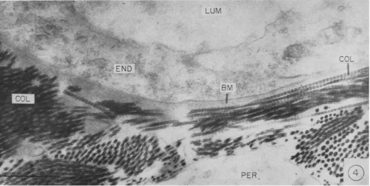
Collagen constitutes about one-third of the total body protein. Histo- logical sections stained by aniline blue show collagen distributed around cells, within and around blood vessel walls throughout the body, and as the major component of bone, cartilage, tendon, and skin. In organs it can be viewed as part of a matrix in which the cells are imbedded. The intimate association between collagen fibrils and small blood vessels can be appreciated in electron micrographs (Fig. 4). Collagen is an unusual protein in that glycine contributes about one-third of the amino acid residues and proline and hydroxyproline another third. Tropocollagen, the collagen molecule, has a molecular weight slightly over 300,000 and is released from cells into the extracellular space as a rod with a length of 2800 Â and a diameter of 14 Â. Extracellularly, tropocollagen molecules undergo an end-to-end polymerization and lateral aggregation to form fibrils and fibers. The characteristic 640 Â periodicity of fibrils seen in the electron microscope is caused by adjacent molecules being out of phase with each other by one-forth the length of a molecule (Schmitt, 1959). The collagen molecule is composed of three polypeptide chains of about 100,000 molecular weight each in a helical arrangement. In some collagens two of the chains appear identical (Piez et al, 1961), while at least one type of collagen has been reported to contain three chains all differing in amino acid composition (Piez, 1964).
When extracellular collagen matures to form fibrils and fibers there is an increase in covalent cross links between polypeptide chains within tropocollagen and the formation of intermolecular cross links between tropocollagen molecules (Piez et al, 1961). There is some evidence that both intra- and intermolecular cross links form by single continuous processes (Bornstein et al, 1964). As collagen matures, it becomes in- creasingly insoluble. A high proportion of newly synthesized collagen is extractable by neutral salt solutions and dilute acids while only traces of fully mature collagen are extracted by these agents. A similar maturation process in which fibrils form from soluble collagen subunits and become
>
o
o
M C W Ω W
o *1
o *3
o
H X
Ω W C/3 >
H 3
FIG. 4. Portion of capillary of hamster cheek pouch. The basement membrane (BM) is seen as a homogeneous band limited by collagen fibrils (COL); LUM, lumen; END, endothelium; PER, perivascular cell (Fernando et al., 1964).
increasingly insoluble when incubated at 37°C can be demonstrated in vitro (Gross, 1958b). When an animal is growing it has tissue pools of newly synthesized soluble collagen. When growth stops, either naturally, or because of dietary restriction, new collagen is not added to the pools and the soluble collagen disappears, presumably because it is incorporated into the mature insoluble collagen (Gross, 1958a). The relationship of growth to aging of collagen can thus be appreciated. So long as an animal is growing it will contain some young collagen, the amount of which will be dependent on the growth rate.
Mature collagen has an extremely low turnover rate at most sites under the usual physiological conditions. It is quite likely that most of the body collagen does not turn over at all (Neuberger and Slack, 1953; Thompson and Ballou, 1956). Collagen is not degraded by the common proteolytic enzymes of tissues at physiological p H levels, and collagen morphology and composition have been found well maintained in fossils (Wyckoff et al., 1964). Mature fibers are easily deformed by laterally acting forces but cannot be significantly extended; they have a very high Young's modulus (Kohn and Rollerson, 1959b; Harkness, 1961). Such a meta- bolically inert and insoluble material would be expected to undergo change with the passage of long periods of time.
The most conspicuous age changes in collagen which occur at quite precise times and rates are in physical properties. Although scattered references were previously made to age-related changes in collagen much recent work was stimulated by the observations of Verzâr (1957) and Banfield (1956). Varzâr made use of the phenomenon of thermal shrinkage of collagen fibers. He found that with increasing age of rat tendon fibers the amount of weight required to inhibit such shrinkage increased markedly. Banfield observed that with increasing age human tendon frag- ments lost the ability to swell in dilute acid. Both of these observations can be explained on the basis of an increased cross linking between collagen molecules.
We have found the swelling properties of collagen to be useful mani- festations of subunit organization and have extended Banfield's work through studies of age-related variation in such properties in human collagen. When collagen is placed in a hydrochloric acid solution at p H 2.5 it imbibes water and swells to a marked degree. This separation of subunits is caused by the establishmnt of a Donnan equilibrium charac- terized by an excess of diffusible ions inside the tissue which, by their tendency to diffuse out, exert pressure on components of the tissue. The extent of swelling provides a measure of the modulus of elasticity (Proctor
AGING AS A CONSEQUENCE OF GROWTH CESSATION 307
and Wilson, 1916) and indicates the degree to which factors are present which hold subunits together. When swelling capacity was studied as a function of the age of human collagen (Kohn and Rollerson, 1958), it was found to remain at a high level until approximately 30 years of age when it decreased quite rapidly until 50 years of age and thereafter declined at a slower rate (Fig. 5). By comparison, Young's modulus, determined by
; ,.r
T
Under 30 years old
Over 5 0 years old
1 1 1 1 1 1 1 1 1 1 0 5 0 100 150
Young's modulus, Kg/mm'
0 ' i ■ ' 1 i i i i 1
0 25 50 75 100
Age (years)
FIG. 5. T o p : Young's modulus of h u m a n tendon fibers as a function of age. Bottom:
Osmotic swelling ability of h u m a n tendon at p H 2.5 as a function of age, expressed as percentage increase in weight (Kohn and Rollerson, 1958, 1959b).
measuring the increase in length of collagen fibers when weighted at one end, was found to undergo a slight decrease with age which was hardly of statistical significance (Fig. 5). These changes were consistent with the view that cross linking occurred between adjacent collagen molecules with age. It was of particular interest that the changes began at 25-30 years
of age, e.g., after growth had ceased and after all of the collagen had matured. In subsequent studies it was found that swelling caused by thermal denaturation varied in both rate and extent as functions of age.
By correlating thermal and Donnan equilibrium swelling data, it could be shown that collagen passes through four stages of increasing rigidity during the human lifespan (Kohn and Rollerson, 1959a). This increasing cohesion of collagen subunits was ascribed to intermolecular cross links which form shortly after synthesis of the collagen, develop to the greatest extent between the ages of 30 and 50, and continue to form after the age of 50.
A large number of studies have now been carried out involving changes with age in different collagenous tissues from different animals. Properties studied include elasticity and tensile strength, thermal shrinkage, solu- bility, and susceptibility to degradation by proteolytic enzymes. Accu- mulated data support the view that collagen throughout the mammalian body becomes increasingly cross linked with age (Kohn, 1959; Kohn and Rollerson, 1960; Fry et al, 1964; Kulonen et al, 1963; Schaub, 1963).
In approaching the problem of collagen cross linking at the molecular level, several questions arise immediately. Cross linking, both intra- and intermolecular, takes place in the course of collagen maturation. This suggests the possibility that aging results in more of the same type of links which form in maturation. Other possibilities are that with age different types of cross links are added to those already present, or that new types of links are formed in aging, coincident with the rupture of bonds formed during maturation. Rigby (1964) has shown that cyclic stresses applied to collagen fibers in vitro cause an increased shrinkage temperature and a more oriented structure, changes which simulate natural aging. This sug- gests that cyclic stresses of the kind that occur in tissues may over long periods of time influence rates and types of cross linking. The interesting observation has been made by Sinex (1957) that stable substances such as collagen are incubated at body temperature for many years and would be expected to undergo some thermal denaturation which might contribute to age changes. This possibility has been tested by experiments based on the assumption that if thermal denaturation at body temperature was a significant cause of collagen aging it should be possible to incubate collagen at elevated temperatures, i.e. 56°C, and reproduce all of the age- related changes in vitro. It turned out that elevated temperature did in fact cause agelike changes in swelling capacity and solubility. Thermo- dynamic calculations based on certain long range extrapolations suggested that thermal denaturation might explain the in vivo changes with age
AGING AS A CONSEQUENCE O F GROWTH CESSATION 309
(Kohn and Rollerson, 1959a,b). One of the striking age-related changes in collagen, however, a decreased susceptibility to the action of collagenase, was not reproduced by thermal denaturation (Kohn and Rollerson, 1960).
Although it is difficult to conceive of thermal denaturation not occurring in a protein incubated at 37°C for 50 years, and some denaturation may take place in collagen, collagen aging cannot be explained on the basis of denaturation alone. T h e possibility exists that heat plays a partial role in causing the rupture of intramolecular bonds, for example, and freeing reactive groups which then participate in intermolecular cross links.
The bulk of our knowledge of the chemistry of collagen cross linking has come from studies dealing with the chemistry of collagen per se rather than from studies motivated by the desire to learn something about aging.
This has resulted in the majority of investigations being carried out on collagen molecules and molecular subunits in solution. A large variety of different kinds of bonds at different sites has been proposed as a result of observations of such systems. These include bonds formed by a non- collagenous pepsin-sensitive pep tide (Rubin et al., 1963), ester links (Joseph and Bose, 1962; Bello, 1960), and aldehyde-mediated links in tyrosine-rich regions (de la Bürde et al., 1963; Schlueter and Veis, 1964).
Although links of these types appear to form in maturation, the extent to which they form in the transition of mature collagen to old collagen is unknown. Old human collagen has been treated with pepsin in an attempt to rupture the proposed noncollagenous peptide links. In addi- tion, old collagen has been treated with hydroxylamine to rupture ester or other hydroxylamine-sensitive bonds. According to the results of swelling experiments, neither of these treatments has caused "rejuvenation" of the collagen (Kohn, 1962). When a soluble fraction is obtained from col- lagenous tissue by extraction with neutral salt solutions or acidic buffers it contains fractions of young collagen regardless of the age of the indi- vidual from which the sample was obtained. Conclusions regarding aging of collagen have been drawn from studies of soluble collagen which represented less than 1% of total collagen in the tissue (Bakerman, 1964).
In a recent study of total collagen, it was reported that a fluorescent sub- stance and a yellow pigment increase with age while the tyrosine content decreases. This suggested that tyrosine residues become oxidized to a quinoid which participates in cross linking (LaBella and Paul, 1965). Old collagen is one of the most insoluble of biological substances. Very harsh treatment is required to solubilize it for study and there is a possibility that such treatment will abolish or alter the age-related changes. Non- destructive analytical methods such as wide-line nuclear magnetic
resonance and electron spin resonance spectroscopy and deuterium or tritium-hydrogen exchange may prove of value in describing the impor- tant intermolecular cross links which form as insoluble mature collagen is transformed into insoluble old collagen.
If it is assumed that intermolecular cross links form in collagen with age and thereby cause collagenous tissue to become more rigid throughout the body, it becomes necessary to relate such alteration to the debilities of aging, e.g., the decreased efficiency of homeostatic mechanisms, decline in resistance, and increased incidence of certain diseases. The establishment of such a relationship depends on knowledge of the physiological role of collagen. Because of its physical properties and distribution, we would assume that collagen is of importance in providing strength and main- taining the structure of organs. It is also apparent from the distribution that blood vessels pulsate and muscle cells contract within a framework of collagen, and that any substance passing between cells and blood vessels must move through collagenous extracellular material. Several studies have indicated that collagen fibers are oriented so that they bear stresses and provide structural limits to deformation, particularly in such mobile organs as the lungs and blood vessels (Harkness, 1961; Mead, 1961; Wolin- sky and Glagov, 1964). Growing animals given a lathyrogenic agent such as ß-aminopropionitrile develop collagen which does not mature properly because of inadequate cross linking (Martin et al., 1963). Such animals with defective collagen are unable to maintain elevated blood pressure when injected with norepinephrine (Kohn and Rivera-Velez, 1965). This suggests that mature collagen plays a role in stabilizing or potentiating reactions of small blood vessels although the changes caused by lathyro- genic agents are not understood well enough to exclude other possible mechanisms of action.
The relating of collagen cross linking to debilities of age depends on the argument that such cross linking causes a greater rigidity of tissues and results in defective diffusion of substances between cells and vascular spaces. Defective diffusion could result from loss of tissue elasticity and movement within tissues so that substances are slowed in their passage, or could result from barriers provided by the more densely cross linked collagen per se. T h a t age-altered collagen causes the organ or tissue which contains it to become more rigid is suggested by the observation that human myocardium loses osmotic swelling ability with age (Kohn and Rollerson, 1959c) and that the age-related difference is abolished by treatment of the tissue with bacterial collagenase (Kohn and Rollerson, 1959d). The collagenase used in these studies may have contained other
AGING AS A CONSEQUENCE OF GROWTH CESSATION 311
proteinases and more work of this type should be performed with a highly purified enzyme. From this and from the studies of age-related changes in collagen described above, such as a decrease in Donnan equilibrium swell- ing, it would appear that both collagen itself and tissues which contain it do become more rigid with age. Because of the distribution and sug- gested physiological role of collagen, an age-related increase in rigidity would be expected to cause alterations in diffusion. Some evidence is accumulating which demonstrates altered tissue permeability with age although the role of collagen has not been defined. Thus, diffusion of a silver proteinate into cartilage, a tissue rich in collagen, decreases with age (Stockwell and Barnett, 1964). When a labeled protein is injected intravenously, the amount found within perfused myocardium decreases with age, suggesting the presence of a barrier to diffusion (Sobel et al., 1964). In the absence of methods by which proof of the causal role of collagen in altered diffusion can be obtained, it is necessary to acquire an overwhelming amount of circumstantial evidence. A useful experiment might involve in vivo determination of age changes in metabolic or phys- iological activity in an intact organ in which blood vessel, connective
tissue, and cell relations are intact, followed by a study of biochemical processes in homogenates in which the metabolic systems are freed from their connective tissue environments. If significant age changes in intact organs were not observed in homogenates, support would be obtained for the view that the extracellular environment or cell membranes were causes of aging. In a sense these types of experiments have already been performed. From the discussion earlier, it is clear that the functional activity of organs such as kidney declines markedly with age, but studies of kidney metabolism by biochemical techniques have not revealed any very striking age-related changes.
A conclusion consistent with available data is that in addition to in- creased rigidity of collagenous tissue with age, increased cross linking of collagen causes altered diffusion in tissues. T h e probable relationships of these changes to debilities of aging have been described previously (Kohn, 1963) and will only be summarized here. It has been argued that connec- tive tissue alterations are sufficient to explain almost all of the characteris- tics of aging human beings. The age incidences of hypertension and arteriosclerosis could be explained, respectively, on the basis of increasing rigidity of small vessels and on decreasing diffusion of nutrients and other materials within vessel walls with consequent inflammation and complica- tions caused by the trapping of minerals and lipids. Decreased resistance to infection and trauma might be due to the inability of hormones, nu-
trients, and antibodies to reach critical sites in the appropriate concentra- tions and inefficient removal and detoxification of harmful substances.
Similarly, the decline in efficiency of physiological and homeostatic mechanisms would be due to the lowered reactivity of small vessels because of their more rigid connective tissue matrix and to the sluggish passage of metabolites, hormones, and chemical messengers from cell to cell or cell to vessel through connective tissue.
A significant age-related change which, at the present time, is difficult to explain on the basis of connective tissue alterations, is the increased incidence of malignant neoplasms. It has been pointed out, however, that malignancies lack the universality of a true aging process within a popu- lation, do not follow a Gompertzian type curve in regard to cause of death, and do not play a very important role in determining the mean life-span of a human population (Kohn, 1963).
A more serious problem encountered by the view that aging connective tissue causes aging in animals is the difference in life-span between different species of mammals. T h e mean life-span of the rat may be 2\
years and that for man 70 years. When the rat dies, its oldest collagen is the same age as infantile human collagen. Aging of collagen has been described as an extracellular phenomenon analagous in some ways to crystallization. Although cross linking of rat collagen with age has fre- quently been described, apparently no one has attempted to determine whether collagen from a given site of a 2^-year-old rat is cross linked to the same extent as a similar sample from a 70-year-old man. It is not clear why the rat collagen should be any more cross linked than very young human collagen. A possible explanation is based on the knowledge that collagen molecules and fibers are laid down parallel to lines of stress in tissues, that cyclic stresses cause a more oriented structure of fibrils prob- ably resulting in more cross linkages (Rigby, 1964), and that the life-span of mammals has a clear-cut inverse relationship to metabolic rates of different species (Sacher, 1959). The rat, having a higher metabolic rate than man, would have a greater rate of vessel pulsation and, in general, greater rates of tissue movement and a higher frequency of tissue stresses.
This could result in a more rapid alignment of molecules and fibrils and more rapid formation of collagen cross links to the extent that rat col- lagen at 2^ years is really comparable to human collagen 70 years old.
Some support for this hypothesis has been gained by the recent observa- tion that aging of collagen is slowed in hypophysectomized rats (Olsen and Everitt, 1965). A decreased metabolic rate would be one of the prominent changes in such animals.
AGING AS A CONSEQUENCE O F GROWTH CESSATION 313
We have some methods for testing the collagen theory of aging, as well as possibly lengthening the life-span. It should be possible to keep animals on a diet deficient in ascorbic acid so that maturation of collagen is slowed, but not deficient enough to produce symptoms of scurvy. Such animals should have a lengthened life-span. Also, the lathyrogen ß-amino- propionitrile mentioned above, can be given to animals from the time of weaning in doses sufficient to slow down collagen cross linking but not enough to cause overt lathyrism. We have experiments utilizing the nitrile in progress. Groups of rats and mice are receiving ß-aminopropionitrile fumarate in their drinking water from the time of weaning and their life- spans and mechanisms of death will be noted. The groups of animals are small since they are kept pathogen-free in isolators. Preliminary studies have been required to determine the dosage which does not cause lathyrism over long periods. T h e first group of rats, decimated by an air conditioning failure, now consists of four control and four experimental animals which were started on the nitrile in August, 1963.
Aging of Cells within Animals
Although the role of cell aging in the aging of a higher animal has been deprecated, aging of cell populations is a widespread biological phenom- enon which satisfies the criteria of universality, progression, increasing age-specific death rates, and growth-relatedness. Very little work has been performed in an attempt to understand the mechanisms by which cells degenerate and die on schedule after they have stopped dividing and have attained a certain high level of specialization. In our present state of ignorance we can do little more than describe the phenomena of cell aging and consider some preliminary experimental data which might be useful in directing future investigations.
There are enormous differences between life-spans of different cell- populations within a higher animal. In man, neurons are capable of living as long as the individual while the granular leukocyte life-span is measured in days or hours. In terms of differentiation, differences such as these in cells which have both come from the zygote and presumably have the same genetic makeup, probably represent a maximum divergence in controls of gene expression. Cells which die in a higher animal appear to belong to at least two different types of populations. In one, cell life-span is quite distinct and degeneration appears programmed. In the second, the life-span cannot be defined and cell death appears to be a random-hit process with a probability of occurrence not directly related to cell age.
Cell death in the latter case appears to be accidental and is presumably due to breakdowns and failures of the machinery of cells which do not synthesize new DNA and cannot repair themselves. Death of neurons in the central nervous system appears to be such a random process. In the human being, neuron death is a steady process which is not accelerated with increasing age (Wright and Spink, 1959). In accord with this view of randomness is the observation that neurons of mice do not show a very significant dropping out over the life-span (Wright and Spink, 1959), probably because there is not sufficient time for a large number of intracellular accidents to occur.
T h e cell populations which age and die on schedule have been men- tioned earlier in this discussion. Such populations may constitute almost the entire animal as in the case of hydra (Burnett, 1961), or exist at many sites in higher animals as exemplified by epithelial tissues and leukocytes in mammals. Characteristically, in these populations there is a focus of stem cells which are periodically or continuously dividing, giving rise to cells which no longer divide but which undergo morphological and chemical specialization. After passing through one or more well-defined and predictable phases of specialization, the cells degenerate and die. This sequence is not necessarily irreversible. At every step in the sequence, questions arise which are pertinent to an understanding of growth, differentiation, and aging processes. We have answers to none of these questions. No more is known of factors which cause cells to stop dividing than of factors responsible for division in the first place, although extra- cellular factors such as functional demand and negative feedback mech- anisms appear to exercise control of cell division in some systems.
Even if a cell is not dividing there is no theoretical reason why it should not remain viable indefinitely. Intracellular accidents may be invoked but it then becomes necessary to explain how such accidents kill all mature granulocytes and cells of the gastrointestinal tract in a matter of days, but not all neurons in 80 years. Many other hypothetical mechanisms can be advanced: toxic metabolites accumulate, degradative enzymes are syn- thesized, or essential molecules are used up and not resynthesized. An old idea which has never been tested, is that in highly specialized cells so much of the cell's machinery is engaged in carrying out the special functions that there is not enough energy production left over for the maintenance of factors necessary for life, such as semipermeability of membranes or synthesis of enzymes. Finally, when a cell is no longer viable, the processes by which it degenerates are poorly understood. These are the processes
AGING AS A CONSEQUENCE OF GROWTH CESSATION 315
responsible for the degradation of cellular constituents and are pre- sumably the same processes which occur in various types of cell injury and atrophy, and in normal turnover of organelles and proteins. From studies of protein turnover in atrophying cells, it would appear that such degradative processes are at least as important as synthesizing reactions in determining the size of some cells and the amount of cellular constituents present at a given time (Slack, 1954; Simon et al., 1962).
Many measurements have been made of cell life-span, and various stages of differentiation and specialization occurring after the last division have been described for a variety of cell types. From the viewpoint of cell aging, however, it would appear that very few attempts have been made to discover why a cell degenerates and dies, or to discover the mechanisms by which its constituents are degraded. Our own work in this area was started with neutrophils obtained from the peritoneal cavity of the rat following injection of a saline solution. These cells do not divide, and constitute a population which degenerates on schedule and has a life-span of a few days. They were maintained under conditions believed to be very favorable and various metabolic and morphological characteristics were followed as a function of time in vitro (Kohn and Fitzgerald, 1964). T h e purpose was to define cell death in terms of specific cell function or prop- erty and to detect the earliest degenerative change. The latter might suggest an initial cause of degeneration and be a cause itself of subsequent alterations.
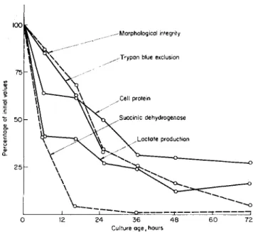
The cells degenerated both metabolically and morphologically at rapid rates as soon as they were obtained and placed in a tissue culture environ- ment. Deterioration occurred earliest in lactate production and in succinate dehydrogenase activity, while alterations in cell permeability, morphology by light microscopy, and cell protein occurred at slower rates (Fig. 6). It was subsequently found by electron microscopy that all com- ponents of the cells underwent marked degeneration during 8 hours in culture. However, the cells could incorporate labeled amino acids into trichloracetic acid-insoluble material at a steady rate for 20 hours in vitro (Kohn, 1964b).
It thus appeared that death in these cells is not an all-or-none phenom- enon, but has a different time course depending on the property under observation. Deterioration can be detected earliest in morphology by electron microscopy and in glycolysis and succinate dehydrogenase activ- ity. It was of interest that although glycolysis and succinate oxidation depend on different enzymes, the ability to carry out both of these proc-
esses declines at approximately the same rapid initial rate. These results suggest either earlier defects in ability to synthesize various enzymes con- cerned with intermediary metabolism, or earlier accelerated rates for their degradation.
The very rapid degeneration of these cells in vitro suggested the possi- bility that they were not dying natural deaths but were being killed by the experimental procedures. Lactate production was used as a measure of viability, and the cells were treated in different ways including culture on siliconized and ordinary glassware, incubation in a complete culture medium, in serum, and in Ringers solution, and incubation in air versus
0 12 24 36 4 8 6 0 72 Culture age, hours
FIG. 6. Percentages of initial values for various leukocyte properties as functions of time in vitro (Kohn and Fitzgerald, 1964).
a mixture of nitrogen, oxygen, and carbon dioxide. None of these vari- ables appeared to alter significantly the rate of decline in lactate produc- tion, suggesting that degeneration and death were processes intrinsic to the cells. Any additional role played by trauma or environmental change might be comparable to that played by similar factors in vivo.
Attempts were made to obtain information on mechanisms of cell degradation in dying neutrophils. Particular attention was given to the possible role of lysosomes because of the attractive hypothesis that such bags of hydrolytic enzymes are stimulated to release their contents which then degrade constituents of cells in cases of atrophy or cell injury (see
AGING AS A CONSEQUENCE O F GROWTH CESSATION 317
reviews edited by de Reuck and Cameron, 1963). Lysosomes appeared especially large and numerous under the electron microscope while neu- trophils degenerated rapidly. Attempts to demonstrate increased intracel- lular protease activity by determinations of protein degradation products released into the medium during degeneration, however, yielded equivocal results. Increased autolysis could usually be detected but it varied in amount and time of onset from one experiment to another.
Another system was then chosen for the study of degradative processes.
Skeletal muscle cells do not normally age and die, but when deprived of their innervation they provide a degenerating system which has several advantages over the leukocyte cultures: Large, uniform samples are ob- tainable, atrophy is very rapid and reproducible, and a great deal is known about muscle enzymes and structural proteins. T h e mechanism of muscle atrophy is also of interest because of the disease, progressive mus- cular dystrophy, which is characterized by atrophy and disappearance of cells in ways which simulate aging of a cell population. In addition, muscle contains the largest amount of mobilizable protein in the body.
Mechanisms by which such protein is degraded in starvation, disuse and denervation atrophy, and muscular dystrophy are presumably identical or very closely related.
Previous studies suggested that muscle wasting in both denervation atrophy and dystrophy resulted from the accelerated breakdown of protein rather than inhibited synthesis (Slack, 1954; Simon et al., 1962).
In addition, an increase in lysosomal enzymes has been demonstrated in muscular dystrophy (Tappel et ah, 1962). Our work on denervated muscle was initiated by the supposition that we could use as substrate a well- studied major structural protein such as myosin which is known to dis- appear from muscle in atrophy and dystrophy, and isolate from control and atrophying muscle the lysosomal proteases which degrade it. This system could then be used to study factors which cause synthesis or activa- tion of the degradative enzymes.
After section of the sciatic nerve in rats, lower leg muscles lose 50% of their protein in 12 days. Although 50% of the myosin is lost during this period, studies of extracted myosin revealed no intermediate stage in its degradation, and exhaustive attempts to identify a myosin-degrading enzyme in muscle were unsuccessful (Kohn, 1964a). We did obtain a partially purified protease from muscle granules which digested denatured hemoglobin but which appeared to have no activity toward pooled muscle proteins. This is consistent with a report by Bodwell and Pearson (1964) which states that muscle cathepsin does not digest the major structural
proteins of muscle. It was tentatively concluded at this point that loss of protein could not be explained on the basis of generalized proteolysis, that when muscle is removed from the animal important differences between atrophying and control muscle might be abolished, and that func- tion per se might be an important factor in the regulation of protein loss.
The latter view is supported by the observation of Cotlar et al. (1963) that stretching of a denervated muscle inhibits its wasting. A possible explana- tion of this is that in a flaccid muscle the highly polymerized proteins might be allowed to separate from one another and depolymerize spon- taneously, or expose sites for enzymatic cleavage which would result in depolymerization to fragments which were still proteins but in a form which could leave the muscle. In muscle under tension, the polymers would be held in linear array, close enough to each other for short range forces to keep them in position. Such a mechanism would require no new enzyme system, but could utilize systems present in all muscle. This hy- pothesis was tested by incubating fiber bundles, both flaccid and under tension, and determining protein efflux into the medium. Tension was found to inhibit protein efflux markedly (Kohn, 1964a).
Although no degradation of myosin could be demonstrated in muscle, it was found that low levels of protein degradation occurred in homo- genates of both control and atrophying muscle, and that autolysis was slightly but consistently greater in preparations from atrophying muscle.
Recombination experiments with subcellular fractions indicated that at least three components were required for autolysis: one in the residue or myofibrillar fraction, one in the mitochondrial fraction, and one in the soluble fraction. The increased autolysis in atrophying muscle was de- pendent on all three components being obtained from atrophying muscle (Kohn, 1965). Electron microscopy has revealed no significant number of lysosomes in the mitochondrial fraction, but has demonstrated a differ- ence between mitochondrial fractions from control and atrophying muscle in that the fraction from atrophying muscle contains fragments of myofi- brillar protein. Recent experiments in which protein shifts have been followed during in vitro incubations of recombined fractions followed by refractionation, have indicated that in atrophying muscle there is an accelerated shift of residue or myofibrillar protein into the mitochondrial fraction, and that this shift requires the presence of both the mito- chondrial and soluble fractions.
Our current working hypothesis, which seems consistent with all ob- servations, is that the degradation of muscle structural proteins is initiated by the action of enzymes in the soluble and mitochondrial fractions which cause a depolymerization to fragments which are still large pro-
AGING AS A CONSEQUENCE O F GROWTH CESSATION 319
teins, and also cause the release of a small number of free amino acids or peptides. The fragments of myofibrillar proteins are further degraded, but not into units smaller than actin or myosin, after which they leave the muscle. The first depolymerization step is influenced by the physiological state of the muscle since fibrillar proteins in a flaccid muscle are more susceptible to attack by depolymerizing enzymes. Such an alteration in physiological state could be the major difference between atrophying and normal muscle. Zak and Drahota (1960), who followed the release of labeled methionine from muscle, similarly concluded that degradation was enhanced when the utilization of energy was limited and not depend- ent on greater amounts of proteolytic enzymes.
Our studies have not implicated lysosomes in degradation of muscle protein and there is some evidence that they do not participate in kidney necrosis (Nagel and Willig, 1964). Lysosome proteases are generally assayed according to their ability to degrade certain peptides and de- natured hemoglobin, and apart from the demonstration that preparations from liver can digest some liver proteins (Sawant et al., 1964), there is little reason to accept the view that lysosomes play an important role in the degradation of cell constituents throughout the body. Cathepsins appear in increased amounts in virtually every tissue undergoing degener- ation, but whether this represents an increase in their synthesis or merely the fact that they are spared from degradation, and whether they play a trivial or important role in degradation is not known. There is no reason why mechanisms of degradation in different types of cells must be identi- cal or even closely related, but it would seem quite uneconomical for a large number of different mechanisms to evolve to carry out similar processes in different cell populations.
In regard to cell aging, cells may degenerate and die because alterations in physiological state allow degradative processes to become dominant.
Such altered states could arise because energy in specialized cells is used for specialized functions and there is not enough available for the mainte- nance of structural proteins in their proper state of alignment. Present knowledge enables us to consider these problems in only the most general terms, and probably the appropriate generality to be gained from the above discussion is that problems of cell aging constitute a neglected and challenging area in developmental biology.
Summary
An aging process can be defined as one which occurs in all members of a population under consideration, which has its onset or accelerates with