Aspects of Connective Tissue
P. F . MILLINGTON, T. GIBSON, J . H. EVANS AND J . C. B A R B E N E L
BioEngineering Unit, University of Strathclyde, Glasgow, Scotland
I. Introduction . . . . .
I I . Basic Organization of Connective Tissue . A. The fibrous elements
I I I . The Fibre Arrangements in Connective Tissue
A. The cornea . . . . .
B. Ligaments and tendon
C. Skin (integument) . . . .
D. Cartilage . . . . .
IV. Mechanical Characteristics of Connective Tissue A. Mechanical properties of fibrous and mucoproteins B. Interfibrillar material and ground substance C. Mechanical chacteristics of tissues
D. The test environment . . . . E. Skin
F . Cartilage . . . . . .
G. Relation between mechanical response and structure V. The Mechanical Response of Living Tissue
A. Deformation response in the plane of the skin B. Deformation normal to the skin surface
C. Skin-fold techniques . . . .
D. In situ techniques . . . . .
Acknowledgements . . . . . .
References . . . . . . .
I. INTRODUCTION
189 190 191 194 194 194 196 208 215 215 216 217 218 220 223 228 229 229 239 239 240 243 243
CONNECTIVE tissue elements form the structural framework in which all parts of the body have continuity. They build the skeleton and also serve as the binding material between various organs. They are not, however, simple passive substances. They form tough capsules for protection and maintenance of shape and size. They serve as pathways through which vessels and nerves pass; they also penetrate into as well as between the organs, carrying with them the vascular and nervous components. They are associated with the basal regions of epithelia and in some instances serve as storage depots. I t also appears t h a t they may retain within themselves a reservoir of unspecialized cells capable of a variety of lines of development. In performing these tasks they also assist in perhaps the most important body function, movement.
189
1 9 0 P . F . MILLINGTON, T. GIBSON, J . H. EVANS AND J . C. BARBENEL
I I . BASIC ORGANIZATION OF CONNECTIVE T I S S U E
These complex tissues have been classified on the basis of the type of intercellular matrix: fluid as in blood; highly viscous or semi-solid as in the fibrous connective tissues, e.g. dermis; solid and elastic as in cartilage; solid and rigid as in bone and dentine. Although complex they may be seen as a simple transition from one state to the other; for example, the intercellular substance in blood may remain as a colloid sol in which albumins, globulins and fibrinogen become involved in a solidifying process—clotting. On the other hand, the intercellular sub
stance may become much more viscous with the addition of muco- proteins and muco-polysaccharides which may vary in the degree of polymerization becoming a gel. In this state, we may find fibrous ele
ments differing in structure and function also present. In general, we tend to use the term "connective" or "fibrous" for these more solid forms of supporting tissues. Finally, the addition of calcium and mag
nesium salts or salt complexes to the intercellular fibre containing ground substance leads to the formation of the more rigid systems.
Changes from one type to another are not uncommon; indeed, bone may be formed at almost any supporting tissue site.
Histologically eleven cell types can be identified in connective tissue.
These cells play only a small role in the immediate mechanical response of the material although their presence and activity are vital to the continuing behaviour of the tissue under stress. In mechanical terms, cells may be considered as points of discontinuity in an otherwise regular though not necessarily uniform system, or series of systems.
Interpretatation of mechanical behaviour in continuum terms con
centrating only on boundary conditions will give little information relating to zonal changes within any system. Since many of the con
nective tissue systems are very extensive and zonal variations do occur interpretation must, therefore, be based on an understanding of the spatial relationships of the various tissue components. This is not to say t h a t a fully comprehensive statistical analysis using point-by-point techniques is required, although such an approach is an obvious choice under certain conditions.
The other components of the connective tissues, namely the matrix and fibrous elements, are important in terms of mechanical behaviour.
The behaviour of these elements, however, are closely related and difficult to separate. They are also difficult to separate and identify bio-chemically and structurally. Only in recent years have we begun to assess the origin and nature of the components. Broadly speaking, these are now known in the matrix material as mucopolysaccharides,
e.g. hyaluronic acid, proteo-glycons, the glyco-proteins and sclero proteins. Many of these substances have an enormous affinity for water in which their mechanical properties may change from solid to gel to solution. Since the water content of tissue may vary from one place to another the local mechanical response may also vary directly in relation to this one component.
A. The Fibrous Elements
The fibrous elements of connective tissue are of the three basic types;
reticular, collagen and elastic. Of these only the more readily accessible collagen fibres have been studied in any detail. Most of the data relating to reticular fibres have been derived from histological studies. The reticular fibres are basically protein similar to t h a t in collagen; they are fine, highly refractile, branching fibres of varying calibre. They form feltworks of intermeshing fibres as well as true networks, but such is their relationship to the matrix material t h a t the form in which they exist in the unfixed condition remains uncertain. They have a particular affinity for silver, reducing silver salts rapidly and, as a consequence, are often called argyrophil fibres. In the electron microscope the fine fibrillar nature is clearly evident. The fibrils are about 10 nm thick and normally show a periodicity of 64 nm in their transverse striations.
The elastic component also exists in the form of fibres which appear first as fine highly refractile branching elements forming a netlike structure. Later they may aggregate together to form very thick fibres (Fig. 1) and even fuse together to form elastic laminae. The fibres if torn show their elastic properties by recoiling. By electron microscopy the fibres show similar features to those described already for reticulin, but the periodicity may be as low as 45 nm.
The third and final fibrous component, collagen, develops first as thin, wavy unbranched fibres. Later they may come to lie in larger and larger bundles which may even take on the appearance of a lamina structure.
These bundles may extend unbranched over long distances or they may split into smaller bundles which, after a short or long course, may join the other bundles. In most long chain proteins it is the order of the amino acid residues in the polypeptide chains which largely determine the general mode of organization. This order while at present not under
stood in detail is specified by the cell genes during biogenesis. However, the relationship between collagen and mucopolysaccharide production is apparently linked. In the healing of flexor tendons, for example, the production of collagen and mucopolysaccharide is not only interrelated but interdependent (Munro, Lindsay and Jackson, 1970). Thus, while it
7*
1 9 2 P . F . MILLINGTON, T. GIBSON, J . H. EVANS AND J . C. BARBENEL
may be true to say t h a t it is the arrangement of the sequence of the side chain groups t h a t determine the spontaneous generation of higher structures, the production of large fibres is not merely a matter of producing more and more protein of a certain type. The resultant chain conformation at the molecular level is determined by the balance between internal and external interactions (Cooper and Russell, 1969).
F I G . 1. L i g h t m i c r o g r a p h of h u m a n d e r m a l collagen a n d elastin fibres s t a i n e d W e i g e r t ' s resorcin-fuchsin. E l a s t i n fibrils h a v e a g g r e g a t e d t o g e t h e r t o form large, dense b u n d l e s . M a g . x 150.
I t is now commonly accepted t h a t collagen contains three polypeptide chains in what is known as the triple helix form. I t has a molecular weight of 300,000 and dimensions of approximately 280 nm x 1-5 nm.
J u s t how the fibril or collagen structure is achieved is not really known.
One theory suggests t h a t the triple helices align themselves end to end with a repeat period of 64 to 71 nm. However, two other types are known to exist, the fibrous long spacing fibril (repeating unit 280 nm) and the segment long spacing particles (length 280 nm, but not occurring as fibrils). The original quarter-stagger concept has been criticized (Olsen, 1963; Tromans, 1963) since it imposes restrictions on the method of macromolecular alignment. A modified theory involving an overlap has also met with some criticism (Grant, H o m e and Cox, 1965) but, with the stagger theory, it is possible to achieve a model with a " h o l e "
structure, a feature important in bone formation (Glimscher and Krane,
1968). Densitometric analysis of both positively and negatively stained collagen has shown t h a t it is possible to interpret the electron micro
scope data in terms of non-porous and porous regions (Bairati, Petruc- ciolo and Tarelli, 1969). Structures involving more random aggrega
tions of the tropocollagen have been suggested (Grant et ah, 1965) which allow for greater flexibility, but in order to achieve the required stability, packing of groups of macromolecules become important. For example, Veis et ah (1967) based their ideas on the presence of 4 macro- molecules whereas Smith (1968) used a 5-unit structure. I t is possible, however, t h a t the detailed packing of these macromolecules may change in the course of growth or in response to new or unusual stimuli, and until we know more about the mechanical response of collagen and collagen biosynthesis this will remain an area of speculation.
Although collagen fibrils can be formed in vitro without participation of non-collagenous proteins (Ross and Benditt, 1961; Ross, 1968; Miller and Martin, 1968) extracted collagen does contain small amounts of the polysaccharides in association with protein and in addition some carbohydrates which are bound to the tropocollagen polymer (Lowther, 1963). However, the removal of the proteo-glycons from tissue contain
ing collagen fibres results in a breakdown of the organization of the collagen bundles (Lowther and Bialkower, 1970) The ground substance or matrix then is closely associated with the fibre formation and does exert an influence on maturation, fibrillar diameter stability and the distribution of water and electrolytes (Gustavson, 1956; Schultz-Haudt and Aas, 1962; Sirsat and Khanolkar, 1962; Matthews, 1965).
The turnover of collagen varies not only from species to species but also from site to site. Goldstein et ah (1964) showed t h a t in the rat, collagen of the aorta and tendon was relatively inert but in liver and gut the turnover was between 30 and 60 days, whereas, in the dermis 50% of the labelled collagen is still present after 300 days. The question of collagen production is of particular interest in wound healing. Ogilvie and Douglas (1964) showed t h a t resutured wounds contained more collagen than primary wounds of the same age and believed t h a t the added increment of collagen explained the rapid increase in tensile strength. Madden and Smith (1970) in a study of the rate of conversion of radioactive proline to hydroxyproline reached the conclusion t h a t in primary wounds elevated synthesis of collagen occurs but may be delayed for up to 48 hours. I t reaches its maximum between 7 and 21 days.
The breakdown of collagen is believed to be primarily the result of attack by collagenases, defined by Seifter and Gallop (1966) as specific enzymes t h a t catalyse the hydrolytic cleavage of undenatured as well
1 9 4 P . F . MILLINGTON, T. GIBSON, J . H. EVANS AND J . C. BARBENEL
as denatured collagen. The role of inflammation in the denaturation process in human skin has been summarized by Weissman (1969) in his review of the physical characteristics of collagen. I t has become in
creasingly obvious t h a t a delicate homeostatic balance exists between the arrangement of the various tissue fibres, the ground substance and the cells present in connective tissue. An awareness of these relationships in association with clinical problems has been largely responsible for the initiation of the studies undertaken by the Bioengineering Unit at Strathclyde University (Gibson and Kenedi, 1963; Kenedi, Gibson and Daly, 1965; Gibson, 1966; Kenedi, 1967a, b, c).
I I I . T H E F I B R E ARRANGEMENTS IN CONNECTIVE T I S S U E
A. The Cornea
I t is well known t h a t the arrangement of the fibrils in the cornea are very regular in diameter and spacing. The cells are infrequent and do not disturb the arrangement of the fibrils (see, for example, Schwarz and Keyserlingk, 1969). From transmission electron micrographs the fibrils would appear to alternate with a glycoprotein moiety, and various models have been developed to demonstrate their interrelation. Calcu
lations of the spatial distribution of fibres in such a system by both theoretical and real measurement show remarkable agreement (Farrell and Hart, 1969). The equations so deduced can also be used to demon
strate transparency properties of the tissue. Future theoretical studies based on known spatial parameters might reveal many interesting relationships between a number of properties, but such an approach requires more certain data on tissue structure.
B. Ligaments and Tendon
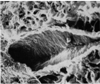
In ligaments and tendon the fundamental development is the com
mon parallel alignment of coarse collagen bundles. The tissue is rela
tively avascular and has few autonomic nerve elements. By scanning electron microscopy using formaldehyde (10% solution, buffered p H 7-2) fixed specimens, the fibres appear to be kinked in two planes as in Fig. 2 (Evans, Millington, Gibson and Kenedi, 1970). One possible explanation of this arrangement of fibres is based on the assumption t h a t the fibres are slowly coiled with a long periodicity. If this is so, then mechanical tests should show about a 3 % extension under low- load conditions before the tissue stiffens. In the section dealing with the mechanical response of tissue the close correlation between structure
50^.m
I I
F I G . 2. Scanning electron micrograph of relaxed tendon in sheet form showing the undulations in the fibres.
F I G . 3. Scanning electron micrograph of tendon after loading. At higher magnifi
cation the individual fibres can be seen. These are now straight.
1 9 6 P . F. MILLINGTON, T. GIBSON, J . H. EVANS AND J . C. BARBENEL
and mechanical behaviour will be seen. When the tissue has been loaded (Fig. 3) the fibres are seen to be straightened fully. There is normally sufficient elastic tissue present in ligaments to enable the tissue to re
turn to its normal rest condition with the collagen fibres in their
"coiled" configuration.
C. Skin (Integument)
The skin or a modification of it covers the whole external surface of the body and, as such, constitutes the largest organ. I t forms a protec
tive barrier interposed between the outside world and the organs it covers. This barrier is an efficient one against many dangers, osmotic,
FOOT £§§£> FAT CELLS
F I G . 4. D i a g r a m a t i c r e p r e s e n t a t i o n of some differences in skin a t v a r i o u s sites on t h e b o d y .
thermal, mechanical, etc. The skin is one of the means whereby in
formation concerning the external environment is obtained but is limited to a response to the immediately adjacent external situation.
In adults the skin forms about 16% (11 kg) of the total body weight (70 kg) and has a surface area of about 1-5 to 2-0 m2. Its structure varies widely in different areas of the body, for example, on the heel pad the
keratinized layer is very thick and at the ends of the fingers the epi
dermis is modified to form a very dense thick horny layer, the nail.
The skin is replaced at differing rates, on different areas of the body.
A person in good health may grow as much as 3 to 4 cm of finger
nail a year. Examples of differences in structure are shown in Fig. 4.
This generalized histological picture, however, has not taken into ac
count differences in the connective tissues present, a factor which will obviously play an important role in determining regional variations in tissue response to load.
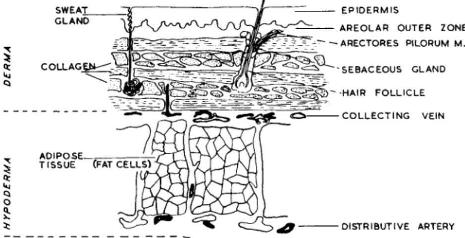
Figure 5 is a diagrammatic representation of skin from the thoracic
F I G . 5. Diagram of skin and subcutaneous tissue typical of the abdominal region in man.
or abdominal areas. The skin is characterized by a thin epidermis, short elastic fibres parallel to the surface and few eccrine glands. The dermal region (dermis or corium) may be divided into two zones, a thin outer layer, often called the papillary layer, and a thick inner layer, sometimes referred to in older obsolete texts as the reticular layer. This inner zone contains the dense connective tissue but whenever they are penetrated by structures from the surface (e.g. sweat glands, hair follicles), the loose tissue of the outer zone descends also. In some earlier histological studies of skin, it was customary to remove a thin piece of epidermis with forceps and to mount this as a " s p r e a d " directly on to the slide where it was thin enough to view after fixation and staining without further manipulation. I n such a preparation the reticular, elastic and collagen fibres are loosely arranged. The reticular fibres are seen
1 9 8 P. F. MILLINGTON, T. GIBSON, J. H. EVANS AND J. C. BARBENEL
immediately subjacent to the basal layer of epithelial cells (stratum basale) and in close association with capillaries and vessels. The collagen is present only as fine bundles.
The dense connective tissue of the inner zone is composed very largely of thick bundles of collagen fibres which are arranged in very irregular patterns, so t h a t few bundles lie parallel for any length of their course. In some sites of the body (e.g. the back), thick elastic fibres are present in the deep layers (see Fig. 1). Most of the tissue to be described below, however, was taken from regions of the body where the elastic fibres were few in number and thin relative to the collagen bundles.
1. Histological studies of stressed skin
Craik and McNeil (1965) showed t h a t when post-mortem abdominal human skin was stretched, the collagen fibres of the dermis became oriented in the plane of stress. They also demonstrated t h a t the red dye of the trichrome stain (Masson's Haematoxylin-Ponceau-Fuchsin- Light Green) had an increased affinity for the stressed collagen. At low-load levels after relaxation the fibres retained their red-staining properties despite the recovery physically of the normal resting arrangement of the collagen bundles. Greater stress did, of course, produce irreversible changes both in organization and staining proper
ties.
There are two technical problems associated with these discoveries which require elaboration before proceeding further. The first is t h a t when collagen is fixed in the presence of acetic acid, or when merely exposed to acetic acid, the collagen bundles swell. If this procedure is allowed to continue the bundles disappear from view in a homogeneous hyaline mass. When teased out in this condition the individual fibres show constricting annular rings. Craik and McNeil used a formal ace
tate fixative in their experiments.
Secondly, when tissue is stained by the trichrome procedure all the collagen fibres become stained with the red Ponceau Acid Fuchsin, which is then removed by treatment with 1% phosphomolybdic acid.
The accepted procedure is to continue this process until all the collagen fibres have been decolorized. The persistence of the red stain in collagen related therefore to a standardized staining procedure in which the collagen has been "swelled" by some indeterminate amount.
The experiments of Craik and McNeil have since been repeated on a number of occasions and under varying conditions. Ten per cent buffered formalin contracts tissue by about 10-20%, depending on the type of tissue being fixed. When this fixative was used together with a
standardized staining procedure, the altered staining reaction was again recorded. Other fixatives, dehydration procedures and embedding materials have been tried without any alteration in staining reaction changes. The possibility t h a t at least part of the effect could be due to stresses imparted during sectioning has been investigated by cutting the same block face of embedded unstressed tissue in different direc
tions. Only on very rare occasions have we noted any change in collagen fibre reactivity on cutting, whereas the reaction change to load is con
sistent.
No valid explanation has yet been advanced to account for the staining reaction change. There appears to be no alteration in the amount or orientation or staining properties of the elastic fibres under different load conditions prior to rupture. Precurser elements in skin have also been studied by staining reactions and these too give no in
dication of stress changes. While further studies are being pursued on two levels: (1) carbohydrate involvement in the staining reaction, and (2) water content of the fibres, the staining changes remain one of the mysteries of tissue stress reaction.
2. Structural changes accompanying load-deformation
Analysis of results obtained from in vitro and in vivo studies of the mechanical response of skin to load was originally based on bulk tissue information and light microscopic observations (Kenedi et ah, 1965,
1966). These analyses were valuable since they led to the first mathe
matical planar network model incorporating visco-elastic features. To obtain a more specific understanding of the load-deformation character
istics it was necessary to view the dermis at a level greater than t h a t possible with the light microscope. By about this time the Cambridge Stereoscan electron microscope had become generally available, and its great depth of focus was seen as an important development for use in this field. As with any new technique, methods of specimen preparation had to be re-evaluated and, since these are still not generally accepted, a brief review follows:
In the description of electron micrographs t h a t accompany this sec
tion, all the specimens used were human and obtained either directly from the operating theatre, from tissue storage banks or from the mor
t u a r y at autopsy. In most cases subcutaneous fat was removed with a Ross-Gibson (1965) dermatome or, where very small pieces of tissue were obtained, by section with a scalpel. The skin was then clamped in a relieved or predetermined stress state and placed in a solution of either 4 % , 5 % or 10% formaldehyde in normal saline. In more recent experiments the fixative was also buffered to p H 7-2. Fixation was
2 0 0 P. F. MILLINGTON, T. GIBSON, J . H. EVANS AND J . C. BARBENEL
allowed to proceed for not less than 12 hours and the tissue was then washed in running tap water to remove excess formalin and salt.
After fixation, one of four routine methods was adopted for further processing:
The split skin was mounted directly on to aluminium stubs with the deeper dermal collagen uppermost.
The tissue was frozen to about — 150°C by immersion in a vacuum flask containing Arcton 12 refrigerant. Thick sections were cut and mounted on aluminium stubs or, in order to avoid surface smearing by the knife, the tissue was freeze-fractured before mounting.
The tissue was embedded in wax using normal histological procedures involving dehydration in acetone, etc. The embedded skin was then sectioned (15 to 20 μπι) and, after dewaxing, was mounted on the stub.
Thick sections (8-10 μιη) cut from wax-embedded specimens here mounted on glass slides and photographed by conventional optical microscopy after staining with a Masson's trichrome or an elastin- trichrome stain. The section was then removed from the slide and mounted on the Stereoscan stub.
In order to see the collagen fibres more clearly in the Stereoscan a number of preparations were incubated in hyaluronidase at 37°C for 12 hours removing much of the mucopolysaccharide. Provided fixation of the specimen had not been prolonged unduly this post-fixation removal of polysaccharide takes away enough ground substance to give a "clean "
surface to the fibres. All specimens were attached to the stubs with either Evostick or similar low-vapour-pressure glue. Final drying out of the glue was achieved by leaving under high vacuum in a coating unit prior to the evaporation of a thin layer of gold-palladium (60/40) on to the surface. Specimens were rotated sequentially first to the right and then the left while coating in order to obtain a more uniform layer, and sufficient metal was evaporated to give a thickness of 20 nm.
Stereoscan microscopy of skin
The Stereoscan electron microscope has revealed a number of new features relating to the structure, ageing effects and load response of skin. In young children the collagen of the dermis exhibits a coiled appearance with random orientation (Fig. 6). At higher magnifications the large collagen bundles are seen to contain smaller fibres and be
tween the bundles fine intertwined fibres with a degree of coiling.
These small fibres could well be elastic. When uniaxially loaded there is a significant orientation of the fibres along the direction of loading, thus confirming the light microscope observations. Where two layers of fibres at different depths can be seen (Fig. 7), each layer is now oriented in a single direction. This type of picture has led to the suggestion t h a t there may be a "scissor a c t i o n " during realignment (Finlay, 1969).
F I G . 6. Scanning electron micrograph of dermal collagen. In tissue from a young individual (2 years) the fibres exhibit a random, coiled appearance.
Some of the tightly packed layers, particularly in older people, may split along their length as a result of load action (Fig. 8). Extensive study of the dermal region of skin indicates t h a t the collagen bundles run approximately parallel to the surface, and it is reasonable to ex
pect t h a t those bundles lying in the direction of the load align themselves to it. When the load is applied at an oblique angle to the direction of the fibre bundles the possibility of rotation is much less.
In unstressed tissue the dermal components from the epidermis (glands, hair follicles, etc.) are readily identified when comparison is made with the light microscope. Sectioned and fractured tissue allows the observer to investigate the nature of the internal surfaces of a
202 P. F. MILLINGTON, T. GIBSON, J. H. EVANS AND J. C. BARBENEL
F I G . 7. Scanning electron micrograph of dermal collagen prepared after uni
axial loading. Fibres in alternating layers tend to orient in the direction of load.
Their movement relative to one another appears as a "scissor" action.
F I G . 8. Scanning electron micrograph of dermal collagen after uniaxial loading.
Fibre bundles at right angles to the load direction are re-oriented but sometimes split along their length.
10
F I G . 9. Scanning electron micrograph of a hair sheath. I t has a smooth surface which in this preparation appears to have been thrown up into folds. Such folding may well be caused by fixation shrinkage.
F I G . 10. Scanning electron micrograph of part of an accrine gland in human skin. Although the cut surfaces of the cells are smeared, within the lumen cell surfaces can be seen, including the raised appearance of the cell boundaries.
204 P. F. MILLINGTON, T. GIBSON, J. H. EVANS AND J. C. BARBENEL
F I G . 11. Scanning electron micrograph of a large blood vessel cut obliquely.
Red cells remain in the vessel and lie against the endothelial wall.
FIG. 12. Scanning electron micrograph of two small arterioles originally identified by comparison with their appearance under the light microscope and the circumferential layering of the vessel walls. Collagen bundles cut end-on show some of their composite fibrils.
number of components such as the hair shaft (Fig. 9), gland cell sur
faces (Fig. 10) and even the indigenous blood vessels (Fig. 11). Small vessels, however, proved more difficult to identify (Millington and Brown, 1970), but it was possible to record examples of small arterioles (Fig. 12). Unfortunately, the effect of load on the skin tends to close down the vessels so t h a t under conditions of stress the vessels become almost impossible to identify in the Stereoscan electron microscope. The effect of stress on these components is, however, important in relation
F I G . 13. Scanning electron micrograph of dermal collagen from a young person. In this case the coiling of the fibres is very prominent, but most of them seem to lie in one plane.
to their function. For example, Sellotape stripping of the epidermis results in increased permeability followed by slowing of the blood flow and dilation of the vessels (Ryan and Kurban, 1970). Pressure applied at this time could result in a number of disturbing consequences, in addition to the well-known blanching effect.
As described above, the collagen fibres of young skin are randomly coiled and interlaced but as the skin changes with age we can also record changes in the collagen fibre pattern. By the time the person has reached the age of 30 or 40 years the bundles have become less coiled and tend to aggregate into sheets. In very much older people the fibres are diffi
cult to distinguish from each other and are almost fully extended
2 0 6 P . F . MILLINGTON, T. GIBSON, J . H. EVANS AND J . C. BARBENEL
before load application (Brown, 1971). The sequence of changes is illus
trated in Figs. 13, 14 and 15. Their relationship to the mechanical properties will be described later.
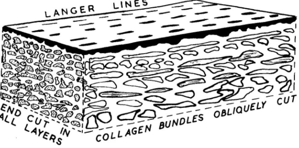
Langer (1861) and later Cox (1941) examined the orientation of fibres in skin by the simple procedure of puncturing the tissues with a sharp, round-bodied objects such as an awl. The holes produced were not round as might be expected but were distinctly ellipsoidal. By joining together the long axes of the ellipses a line could be drawn
F I G . 14. S c a n n i n g e l e c t r o n m i c r o g r a p h of d e r m a l collagen from a m a t u r e per
s o n (42 y e a r s ) . T h e collagen fibres a r e still coiled b u t n o longer so readily identified.
T h e y a p p e a r t o be forming a m o r p h o u s s h e e t s .
across the surface of the body. These so-called Längeres lines were taken as indicating the direction of fibres, and surgeons used these lines to indicate direction of cut when making an incision. Recent studies by scanning electron microscopy of tissue punctured in a similar manner have demonstrated t h a t this simple idea is true only in part.
Brown (1971) has shown t h a t in uniformly pre-stressed skin the punc
ture lines still follow the same directions as those demonstrated by Langer and Cox. In specimens from an oedematous arm removed at surgery, he showed t h a t the tissue when sectioned at right angles to Langer's lines always displayed cut ends of fibres, even though studies
FIG. 15. Scanning electron micrograph of dermal collagen from an older person (62 years). The collagen fibres are no longer coiled; they appear much less rounded and many have aggregated together to form sheet-like layers.
L A N * V.}^*—^^^^*^-
3&
$£
&^ΜΒΆ
CA**'«'—COLLAGE
^--^,IQU£ObLW LY
FIG. 16. Diagram showing the relationship of Langer's lines to the orientation of dermal collagen in human skin.
2 0 8 P . F . MILLINGTON, T. GIBSON, J . H. EVANS AND J . C. BARBENEL
on other planes of section clearly indicated t h a t the layers of fibres were quite randomly disposed relative to each other. Brown (1971) also showed that the puncture sometimes rotated in orientation as it passed from one layer to another through the tissue. I t would appear then t h a t while Langer's lines may indicate a tendency for fibres to align in the direction of the split, the direction recorded may only be indicative of the superficial collagen layer orientation. The application of this phenonema to in vivo studies will be described later, but Fig.
16 shows the relationship between fibre layer and Langer line at dif
ferent orientations, as seen in the stereoscan electron microscope.
D. Cartilage
Articular cartilage is another connective tissue containing collagen fibres, some elastic tissue and a considerable amount of mucopoly- saccharide in the protein of the ground substance or matrix.
In embryonic growth of vertebrates cartilage serves as the main material of the endo-skeleton before bone appears. Where it remains at the ends of bones it forms the bearing material of the joint. This type of cartilage is known as permanent hyaline cartilage since it remains in this form throughout life, though it may come to contain larger and larger amounts of calcium salts without the formation of bone. One feature t h a t distinguishes articular cartilage from other hyaline car
tilages is the definite regular arrangement of cells with reference to the sy no vial surface.
Although some earlier workers (Hunter, 1743; Ran vier, 1877) attempted to describe the fibrous nature of articular cartilage, Hult- kranz (1898) tried to demonstrate fibre orientation by means of splits induced in the surface in a manner similar to t h a t used by Langer for skin. He believed t h a t such prick-patterns followed joint movement.
Benninghoff (1925) extended this work and found t h a t the patterns did not follow joint movement and further suggested they connected the edges of the articular surface by the shortest routes and the least curvature. He also studied the arrangement of fibres throughout the depth of the cartilage and postulated the "arcade t h e o r y " of fibre orientation (Fig. 17). Controversy continued on whether cartilage was truly hyaline and homogeneous or fibrous; even in 1928 Shipley re
corded t h a t he believed cartilage to be "quite without structure", while others attempted to distinguish alternative fibre arrangements (MacConail, 1951). I t was the advent of the electron microscope in the late 1950's t h a t eventually led to a better understanding of collagen fibre orientation. Little et al. (1958) described fibres parallel to the sur-
face in foetuses and newborn but described the remaining organization as random. This they suggested changed by middle age to include a zone close to the subchondral bone where fibres were perpendicular to the articular surface.
TANGENTIAL 'Superficial Loynr)
TRANSITIONAL
PERPENDICULAR
^PRESSURE ZONE*)
FIG. 17. Diagram illustrating the "arcade" structure suggested by Benninghoff for articular cartilage.
These studies led to the development of interest in two distinct as
pects of cartilage structure: (i) the structure of the surface in relation to lubrication, and (ii) the structure of the mid- and deep zones of cartilage in relation to mechanical response to load and attachment to the bone.
1. Surface structure
Animal studies by Zelander (1959), Cameron (1958) and Little (1958) indicates intersecting bundles of fine fibres tangential to the surface. Davies et al. (1962) confirmed that the superficial zone contained many fibres which tended to run parallel to the surface. The most superficial region was almost non-fibrous to a depth of 0*2 μιη and was thought to correspond to the Lamina splendens of MacConail (1951).
The surface itself was said to be very smooth with no depressions visible.
More recently Bullough and Goodfellow (1968) again showed that the surface fibres lay parallel to the articular surface, despite the fact that
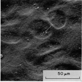
210 P. F. MILLINGTON, T. GIBSON, J. H. EVANS AND J. C. BARBENEL Weiss et al. (1969) claimed that in human femoral cartilage the 3 μιη- thick superficial zone was composed of a dense network of randomly oriented fine fibres. With regard to the surface itself, Walker described surface irregularities in human cartilage ranging from 0-75 to 5 μπι at intervals from 25 to 250 μπι presumed to be due to the presence of surface ridges.
The use of the scanning electron microscope has not provided answers with the same degree of certainty evident in the skin studies. Both McCall (1969) and later Graham (1969) found ridges on the surface of
FIG. 18. Scanning electron micrograph of the surface of human hip articular cartilage showing many oval-shaped depressions. Some of these depressions have a central ridge giving them a "figure-of-eight" appearance.
femoral cartilages, but these are now believed to be artifacts of pre
paration (Clarke, 1970). Gardner (1969) still maintains that there are surface ridges on guinea-pig cartilage but admits to species differences.
Woodward et al. (1969) also describe numbers of blunt processes, rubus ideaus in rat and pig synovial tissues which they suggest might be related to similar structures on the cartilage surface.
Clarke has described numerous oval-shaped depressions, 20 to 40 μπι (Fig. 18), in diameter, similar to that described by Gardner (1969).
These depressions occurred both singly and in pairs. When pairs existed each depression was separated from the other by a narrow isthmus
similar to t h a t seen in pairs of chondrocyte containing lacunae. I t appeared therefore t h a t the surface depression could overlie the chond- rocytes whose distribution was oriented relative to the surface. Detailed comparison of tissue by both light and scanning electron microscopy has shown a close correlation between the size and frequency of both the underlying chondrocytes and the surface depressions. From such studies it was concluded t h a t the depressions were probably formed as a result of the thin surface membrane sinking into the lacunae of the chondrocytes.
While this explanation of surface depressions seems plausible, it remains t h a t Gardner (1970) has shown t h a t freshly displayed cartilage can develop raised surface irregularities following arrest of the blood supply. J u s t exactly what structure exists in the intact normal joint is unknown. Cartilage is largely an avascular tissue but is very sensitive to blood-supply failure to the adjacent tissue. I t is probable therefore t h a t a number of changes occur in the tissue structure after death or in the period between exposure and fixation.
The fibrous structure of the region immediately subjacent to the surface has now been established by scanning electron microscopy.
Clarke found t h a t dry fracture of specimens of human cartilage often left a small tag of the superficial zone free. This tag could be removed for inspection in the microscope. Mital (1970) further developed this technique by removing wedges of the surface material for study of the structure in depth. I t would now appear (Millington et al., 1970; Mital and Millington, 1971) t h a t quite a specific sequence of orientation exists near the surface of the femoral head cartilage.
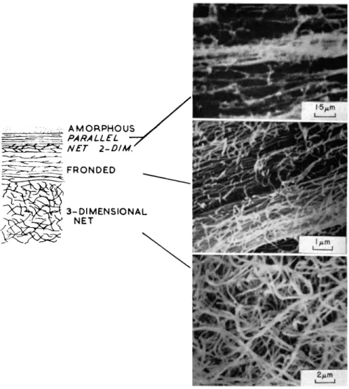
At the surface there appears to be a thin layer of material not re
moved by treatment with hyaluronidase. This amorphous layer overlies a layer of fibrils oriented parallel to the surface. These fibrils are cross- linked by short branches. As the depth of the peeling increased, fronded fibrils still oriented parallel to the surface became evident. The fronds were probably longer strands extending between fibres at different depths which were broken during fracturing. This eventually gave way to a more random interlaced pattern of fibrils still parallel to the surface.
A true three-dimensional random arrangement of fibrils was not found until much deeper in the specimen. The changes in fibril pattern is illustrated in the composite Fig. 19, which shows the amorphous layer, the oriented fibrils, fronded fibrils and the two-dimensional net struc
ture. Thus in summary it appears t h a t the random organization of the mid-zone, well known from previous studies (Silberberg, 1961; Little et al. 1958; Bullough and Goodfellow, 1968; McCall, 1969), gives way to a netlike structure in which the predominant organization of the net
212 P. F. MILLINGTON, T. GIBSON, J. H. EVANS AND J. C. BARBENEL
FIG. 19. Composite diagram showing the various fibre layers near the surface of human femoral cartilage. Some of the layers are also illustrated by scanning electron micrographs of appropriate areas.
lies parallel to the surface, which in turn gives way to a parallel fibre arrangement at the surface.
2. Mid-zone and deep layers of cartilage
As already indicated above, the mid-zone of articular cartilage is now considered to be a three-dimensional random array of fibrils.
Bullough and Goodfellow (1968) demonstrated by transmission electron
microscopy t h a t split lines occurred only when the superficial surface was present. Inspection of fractures through the depth of a hole or split produced by a round-ended awl by scanning electron microscopy has confirmed these observations (Millington et al., 1970). The super
ficial fibres lay parallel to the split direction, but in the mid-zone of cartilage no orientation with respect to the hole was observed, even though the fibres pushed aside by the awl could be detected by dif
ferences in packing.
The basal zone structure in femoral head cartilage is quite distinct.
McCall (1969) described it as a radial pattern of coarse fibres. In some
F I G . 20. Scanning electron micrograph of a channel in the subchondral bone extending into the basal regions of the articular cartilage. I t is channels like these that may form nutrition pathways for the deeper cartilage layers.
specimens, arrays of fibrils were seen extending from the mid-zone coming closer and closer together to form tufts of fibrils at the point of insertion into the subchondral bone plate. The aggregation of the fibrils into tufts left larger spaces between the remaining fibrils and speculation arose on the significance of this feature.
Cartilage, while being avascular, receives sufficient nutrients by diffusion to enable it to survive for the life of the individual under normal conditions. In some cartilages, particularly those in human and other large animals, diffusion pathways from the synovial surface would
214 P. F. MILLINGTON, T. GIBSON, J. H. EVANS AND J. C. BARBENEL be extensive. Alternate routes have therefore been suggested from time to time. Nutrition pathways arising from the subchondral plate and its vascular system have been postulated from dye transport and labelling experiments (Greeenwald and Hayes, 1970). Evidence for the existence of such channels in humal femoral cartilage has been presented by Mital and Millington (1970) from scanning electron microscope studies.
But the extent of these channels has not been determined nor the zone
FEMORAL HEAD FOVEA
A C E T A B U L U M
F I G . 21. Composite diagram of slits formed by pin-pricks into the surface of femoral head and acetabular cartilage. The central "load-bearing" region indi
cates relative fibre orientation at right angles to each other.
of cartilage which is dependent primarily on this route of nutrition. An example of the type of channel seen by scanning electron microscopy is shown in Fig. 20.
From studies made on human femoral cartilage it is possible to de
scribe fairly accurately its fibre arrangement, but there is evidence which suggests t h a t all cartilages are unique in fine structure. From clinical evidence it is certainly safer to regard each cartilage as unique (Gibson, 1967). Studies on the acetabular cartilage (Clarke, 1970;
Mital and Millington, 1971) have revealed a number of differences in structure when compared with its apposed femoral cartilage. The superficial structures are similar but, in the acetabulum, orientation of the surface parallel fibres change frequently over the cartilage surface.
The pin-prick patterns of the two surfaces indicates these differences (Fig. 21). The mid-zone of the acetabular cartilage is relatively thin but the basal zone is by comparison a thick layer. The fibres are not oriented at right angles to the surface as in the femoral head but lie obliquely to it. Thus it is suggested t h a t before any detailed interpre
tation of mechanical response of cartilage to load is attempted the structure must first be determined as accurately as possible.
IV. MECHANICAL CHARACTERISTICS OF CONNECTIVE T I S S U E
The mechanical behaviour of load-bearing connective tissue is determined by the physical properties of the extracellular materials and their morphology. Daly (1966) established t h a t the epidermis of skin, although several cells thick and tightly adherent to the fibrous dermis, contributes little to the tensile behaviour of the whole skin. I t is probably justifiable to assume t h a t the influence of cells, capillaries, glands and hair follicles on the mechanical properties is negligible.
However, those tissues involving muscle fibres exhibit complex charac
teristics which are significantly influenced by both the passive and ac
tive constituent proteins.
Most connective tissues are comparatively pliant and soft, except bone and ageing cartilage where rigidity is imparted by deposits of calcium salts in the protein matrix.
A. Mechanical Properties of Fibrous and Mucoproteins To appreciate the composite nature of the supporting tissues some knowledge of the mechanical properties of their constituents is essen
tial. The majority of the data on the mechanical behaviour of these tissues and constituents have been ellicited in vitro. Study has been made of tissue characteristics on the living person, but recourse has had to be made to animal experiment for some essential data. I n the following account it will be assumed t h a t all results relate to in vitro studies unless otherwise stated.
1. Collagen
In all fibrillar assemblies the macromolecules of collagen are embedded in ground substance and the mechanical interaction is significant
8 A.B.E.
2 1 6 Ρ. F. MILLINGTON, T. GIBSON, J . H. EVANS AND J . C. BARBENEL
but not well understood. The mechanical behaviour of collagen fibres themselves has been extensively investigated by the leather trades research teams. An excellent review of this work and many other aspects of the behaviour of collagenous tissue has been written by Harkness (1968). However, only a small proportion of this work can be said to be relevant to our results, and these aspects will be emphasised when necessary.
2. Elastin
It has been suggested that the regular coiling of the fibres plays an important role in the mechanical behaviour of elastin (Gross, 1949).
Ramachandran and Santhanam (1957) have suggested that the elastin molecule has a triple helix structure which is thermally contracted at body temperatures. Incidentally, thermal shrunk or acid-swollen collagen exhibits a diffraction pattern similar to that of elastin (Rama
chandran, 1963).
Because it is covalently cross-linked, elastin is a very stable protein which mechanically resembles lightly vulcanized rubber (Ayer, 1964).
It is weaker, however, than collagen. I t exhibits long-range elasticity but negligible time-dependence. Carton et al. (1962) extracted single elastin fibres from bovine ligament which were subsequently subjected to tension. Typically, the fibres extended readily to 130% before reaching rupture strain. An empirical expression for the behaviour is of the form:
Strain = 1 · 3 - A exp " B tension
where A and B are constants.
King and Lawton (1960) have investigated the behaviour of elastin rich tissues in terms of elastomer theory and derived the pressure- volume relation for an elastomeric sphere and closed cylinder. The comparison of this data with that obtained from inflated bladder and aorta showed good agreement over a large strain range. Further work indicated that the number of molecular cross-linkages increases with advancing age.
B. Interfibrillar Material and Ground Substance
Although the mechanical function of the interfibrillar and ground substance materials can be established in part, no direct measurements of any consequence have been made of their mechanical behaviour. As they consist of long-chain molecules and are apparently amorphous
under the electron microscope, it is assumed t h a t these molecules are in a contracted state. In the form in which they occur in the body, they probably behave as highly viscoelastic liquids similar to unvulcanized rubber.
Fluids expressed from tissue are unlikely to represent the ground substance completely. The streaming of fluids within a tissue on the application of a pressure gradient has been studied in articular car
tilage, but these studies are still in preliminary form. Isolation of the ground substance materials by selective enzymatic degradation of fibrillar components has not yet proved possible since the enzymes also damage the mucoproteins themselves. I t is probable t h a t the intact tissue studies will remain the only way in which the mechanical pro
perties of ground substance materials can be determined in any really meaningful form.
C. Mechanical Characteristics of Tissues
Most of the mechanical properties of connective tissues can be shown to be related to function in the body. The more varied the functional role, the more complex is the mechanical behaviour. What is sometimes not appreciated is t h a t as mechanical complexity increases so also does the structure. " S i m p l e " materials, such as tendon, have a limited functional role and are structurally simple. Skin is a much more com
plex material in its functional, structural and mechanical properties.
In some tissues, the mechanical properties may be determined largely by either the collagen, elastin or ground substance, whereas in others there exists a more complex interaction between the components.
There is now evidence to suggest t h a t the response of connective tissues to high tensile stresses relies on the properties of collagen, whereas the intermediate response may be dictated by the structure of the fine meshworks and mucoproteins.
1. Tendon and ligament
As described earlier, the undeformed or resting structure of most tendon is t h a t of nearly parallel bundles of collagen fibres or pleated sheets. I t is probable t h a t the natural course of the individual fibres is helical, but close packing restricts them more nearly to a plane wave.
On uniaxial extension these waves gradually disappear and the fibres themselves are extended. There exists, however, a range of initial wave
lengths and wrave amplitudes (Viidik, 1968), and so not all fibre bundles are straightened at the same overall extension. The relative ease with which the fibres are straightened is evident in the initial compliance of
2 1 8 Ρ. Γ. MILLINGTÖN, T. GIBSON, J. H. EVANS AND J. C. BARBENEL
the whole tendon (Elliot, 1965). During extension there is a progressive stiffening as a greater proportion of bundles become straight. In addi
tion, the effect of lateral resistance, whatever the cause, will be more significant the smaller the amplitude to wavelength ratio, i.e. the more nearly straight the fibre. Independent assessments of the extension occuring during this straightening phase (Viidik, 1968; Evans et al., 1970) both indicate a mean value of 3 % (Fig. 22).
The initial extension is normally reversible and the buckling or folding of the collagen fibres is not solely due to stresses within the collagen. Wood (1954) has demonstrated, however, t h a t the initial phase becomes irreversible after treating the tissue with elastase.
The behaviour of tendon and collagen rich ligaments in the second phase of response (the stifFer part of the curve) closely resembles t h a t of isolated collagen fibres (Fig. 22), but the time-dependence of the stress- strain relation is evident to a much greater degree. Extension of tendon in excess of 3 % normally causes irreversible deformation (Partington and Woods, 1963).
The failure stress of tendon appears to be markedly less than t h a t of a single collagen fibre. There appears to be two reasons for this obser
vation: firstly, not all fibre bundles are equally strained owing to the initial configuration, and, secondly, the methods of test generally do not strain the tissue uniformly. The peripheral fibres take a dispro
portionately large force because of shear deformation.
Measurements of rupture stress and strain have been frequently reported but the values cover a very wide range. Lesions artificially produced in muscle-tendon-bone or bone-ligament-bone systems commonly indicate a weakness at the tissue junctions. However, clinically observed lesions are normally found in the body of the tendon or ligament and are usually associated with suddenly applied tensile or shear forces. This anomaly has not been resolved. Indeed, the esti
mated maximum force transmitted by a tendon when rapidly extending a fully contracted muscle is only 50% of the rupture strength as measured in vitro (Elliott, 1965). I t would seem t h a t the most significant variable feature of in vitro testing is the test environment itself, and perhaps a word about this factor might now be appropriate.
D. The Test Environment
Each research team appears to have used different media and tem
perature when carrying out tests on biological material. More commonly, we find tests carried out in water-saturated air or isotonic buffered solutions. For most collagenous tissues, the use of water-saturated air
101
z
9 5
Ul I -
/co LLAGEN T l
ft / / /
X /
/ / / / / / /ENDON /
O-Ot 0 · 0 2 Ο Ό 3 S T R A I N
0 - 0 4 O-O^
S T R A I G H T E N E D
F I G . 22. Stress-strain curves for collagen a n d t e n d o n . Projection back t o t h e abscissa of t h e stiffer p a r t of t h e t e n d o n curve indicates a 3 % extension of t e n d o n a t low load. This degree of extensibility corresponds almost exactly w i t h t h e w a v e p a t t e r n of t e n d o n as seen in t h e scanning electron microscope (viz. Fig. 2.)
2 2 0 P . F . MILLINGTON, T. GIBSON, J . H. EVANS AND J . C. BARBENEL
would seem to be satisfactory, but is difficult to obtain at an ambient temperature of 37°C. When tissue is immersed in a solution it is almost impossible to determine accurately the amount of water and ions taken up by the tissue. Where buoyancy effects can be used, however, the advantages may outweigh the disadvantages.
In contained or partially contained systems the amount of fluid present could determine the type of mechanical response observed.
Veronda and Westmann (1970) showed, for example, t h a t when skin was uniaxially stressed there was an initial increase in volume, but as the tissue response became stiffer the volume ratio decreased so t h a t by the time the incremental load was almost proportional to the ex
tension increments the volume became less than at the start of the experiment. Correlating these observations with experimental condi
tions, they suggested t h a t an explanation could be found in fluid loss from the tissue as the load increased.
E. Skin
Mechanically, the skin must contain the organs of the body and protect them from physical damage while allowing mobility. Local functions, as we have already indicated, are numerous but generally associated with the cellular components. Daly (1966) showed t h a t where the epidermis is thin it contributes little to the tensile behaviour, but more recent studies have indicated t h a t the influence of the epider
mis is measureable and may be of interest to the dermatologist (Daly, 1970).
Kenedi (1964) distinguished between the mechanical response of the superficial layer (papillary layer), the deeper layers (hypodermis) and the dermis itself. The dermis, a denser region, is much less compliant t h a n the other regions. Its mechanical response to uniaxial load is now fairly well established, but a simple model for the region has not been developed satisfactorily. One reason for this is t h a t the dermis cannot be considered as a two-dimensional structure, one layer lying on top of the other. Some fibres do migrate from one layer to another through the depth of the tissue, and in addition we know t h a t the sum of the mechanical properties of the layers of the dermis is less than t h a t of the whole. Ward and Brooks (1965) have also shown similar properties for leather.
When skin is subjected to in vitro uniaxial loading the stress-strain curves follow a well-established pattern (Kenedi et ah, 1966). A typical graph of tension vs. extension is shown in Fig. 23. I t is seen t h a t the graph exhibits three ranges of behaviour; the primary in which con-
siderable extensions result at low tension; the secondary (transitional), during which the incremental increase in extension corresponding to incremental increases in tension continues to diminish; followed by the tertiary, where the tension-extension relationship approaches linearity and culminates in failure of the specimen. Accompanying the extension, application of tension also results in a decrease in specimen width, shown as a lateral contraction in Fig. 23. The physiological load limit lies in the transition phase.
- 0 - 4
PRIMARY [SECONDARY | TERTIARY (transitional)
j Physiological limit 0-2 0*4 0-6 0-8
CONTRACTION EXTENSION in
F I G . 23. Uniaxial extension and lateral contraction in tension of human skin.
Specimen was taken from a post-mortem sample of abdominal skin (68 years old).
(After Kenedi et ah, 1966.)
Specimens of skin subjected to suddenly applied extension which is then maintained display very pronounced stress relaxation phenomena.
Figure 24 shows a typical response with the tension applied, relaxing to as little as 10% of the initial value.
All the mechanical properties of skin appear to be time-dependent.
The histological studies have indicated t h a t tissue fluid is increasingly displaced as the load increases and the whole structure compacts. The effect of fluid movement in the in vitro experiments, however, has never been fully elucidated, and so the value of the data so far obtained is limited. Other published data, whilst copious, are also of limited value since earlier workers tried even less sophisticated techniques with an
2 2 2 P. F. MILLINGTON, T. GIBSON, J. H. EVANS AND J. C. BARBENEL
almost total disregard for environmental control or the time depend
ence of the tissue response. Nevertheless, the reader may wish to con
sult the excellent review by Harkness (1968) which also includes the site dependence of the tissue properties.
„ 0-3 E
O
0-2
«0
NORMAL . SKIN
*"
1
/ 4
•
r-~±
/ ' 1 / ' 1 1 ,
/ | / , / I / 1 / ,
f * / * / I
*
1~' 1
SKIN
0-2 0 · 4 0·6 0 · 8 STRAIN (Ai/g\
F I G . 24. Comparison of low-load behaviour of human skin before and after treat
ment with elastase. (Adapted from Daly, 1969.) 1. Enzyme degradation of skin
The response of skin to mechanical manipulation can be altered by enzymatic activity. When collagen is fully degraded the tissue has no continuity, breaking up under extremely low loads. Elastin, on the other hand, can be removed with consequent changes in the load- deformation curves. Normally, when skin is subjected to uniaxial load and stress cycled slightly beyond the transition stage, a hysteresis loop is produced which returns to the initial loading curve and then back to the original strain zero position. There is no permanent deformation.
Daly (1969) demonstrated t h a t tissue treated with elastase behaved quite differently. Enzyme-treated skin showed a large initial deforma
tion at almost zero stress before it became stiff. Then the hysteresis loop produced on load cycling failed to return to the zero strain position.
The tissue was no longer elastic (Fig. 24). From these and other ex
periments, it would seem t h a t the initial load response of skin is de
termined at least in part by the elastic component of the dermis.
2. Multiaxial loading
The behaviour of skin in biaxial tension has received little attention.
Dick (1951) clamped a circular skin sample at the periphery and subjected it to a differential pressure while recording the maximum displacement in the central region. By taking the skin in its natural state of strain and neglecting anisotropy, he attempted to establish the average tension in the tissue before excision. Ragnell (1954) tried a more sophisticated approach to the determination of resting tension.
Point loads were applied at the periphery of an excised skin sample.
Considerable anistropy was observed. More recently, Veronda and Westland (1969) have attempted to measure the triaxial strain result
ing from a uniaxial load applied to animal skin. Although recognizing t h a t skin is a non-linear, anistropic, rate-dependent material, they developed time-independent stress-strain relations using strain energy or work functions. I t seems t h a t the difficulties associated with multi- axial testing and subsequent analysis are considerable, and it is not surprising t h a t most in vivo tests have adopted the simplest approach possible. Only now, with growing interest in the dynamic behaviour of skin, are more elaborate techniques being developed. Incidentally, at high deformation rates, the failure of skin even when impacted by sharp instruments normal to the surface appears to be t h a t of tensile rupture (Gadd, Peterson and Lange, 1966). The load-absorption characteristics of skin which appear to be high (Gadd et al., 1970) has been largely neglected in head injury studies, where most work has been carried out.
F. Cartilage
Human articular cartilage is subjected to cyclic loading during most normal activity. The hip joint, for example, carries loads varying from zero to many times body weight during walking. From the work of Paul (1969) and Rydell (1966) we now know t h a t the forces are not applied symmetrically with time. The data (Paul, 1965, 1966, 1967) indicate t h a t two peaks varying between 1-7 and 9-2 times the body weight act on the femoral head and acetabulum at an angle of 15-20°
to the longitudinal reference axis. Rydell's results, while varying some
what with those obtained by Paul, showed an average peak force of between 2-3 and 2-8 times the body weight.
The forces applied to cartilage are transmitted via the bones, which themselves are deformable (Evans and Lissner, 1953, 1955; Pederson, Evans and Lissner, 1949; Kuntscher, 1935). Under dynamic loading the acetabule tend to displace outwards creating tensile strains within them.
9 + A.B.E.
224 Ρ. Γ. MILLINGTON, T. GIBSON, J. H. EVANS AND J. C. BARBENEL Hammond and Charnley (1960) in their investigations of the femoral head found that it was almost a true sphere, particularly the carti- lagenous surface. They suggested that it could be completely accom
modated under the load of body weight through compliance of the two layers of articular cartilage. Bullough et al. (1968) showed, however, that neither the femoral head nor acetabulum were completely spheri
cal, especially towards the tips of the "horse-shoe " articular area. They also showed that there was a significant decrease in sphericity with increasing age.
#c m
Ό O i-
| io
a O li. ai
a Γ 2 3 4 5
hr
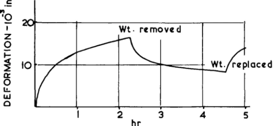
FIG. 25. Time-dependent deformation response of cartilage to static load. A spherical indenter J inch in diameter was loaded to 5 lb. Recovery and reloading response is also shown. Snbchondral bone was left attached to the cartilage.
(Graham, 1969.)
Stiffness of human femoral cartilage has been measured by Kempson, Swanson and Freeman (1967, 1969). They applied a static physiologi
cally representative load normal to the surface via a plane-ended indenter. Maps were obtained showing indentation stiffness and creep modulus at 2 sec. They also showed a 3:1 ratio between the stiffest and most compliant areas, the stiffest area being the superior aspect of the head containing the load-bearing region described by Rydell. With age, the overall plasticity increased even though the cartilage appeared normal. Fibrillated and ulcerated regions, of course, had diminished stiffness. These changes in stiffness were associated with a decrease in mucopolysaccharide content. Maroudas (1967, 1969) has further sug
gested that the cartilage owes its elasticity and resilience to the presence of negatively charged mucopolysaccharides. She found that the fixed- charge density reached a maximum between the ages of 30 and 40 years, and decreased thereafter. The particles in cartilage have a mobili-
replaced