Emergence of Form and Function in the Embryonic Heart
ROBERT L. DEHAAN
Department of Embryology, Carnegie Institution of Washington, Baltimore, Maryland
INTRODUCTION
It is not the ultimate goal of the embryologist to learn how differ
ential readout of genetic information leads to the synthesis of protein A in one embryonic cell and protein B in another ( as important as that question is ) ; nor is it to understand the mechanisms that regulate with exquisite precision the localized rate of mitosis in different regions of a developing tissue; nor again is it to become familiar with the forces that cause a flat sheet of cells to fold into a tube or ball. The goal of the embryologist is more complex. It is to be able to describe as ac
curately as possible, at all levels of consideration, how a functional organ arises from a single progenitor cell.
To do this, of course, we must have all the information mentioned above—and much more. Since the development of any organ repre
sents the sum of the changing properties and interactions of the cells that comprise it, what we must seek, in analyzing the process, are the primary properties of cells that underly their behavior and lead, step by step, to their organization into the functional configuration of the organ in question. Fortunately, when we examine the behavioral reper
toire of cells, we find a fairly limited range of activities. An embryonic cell can divide; it can change its shape through pseudopodial activity or contraction of its surface; it can alter its adhesive properties, and thereby its contact relations with neighbors; it can obtain directional information from its surroundings; and, of course, it can synthesize a variety of intracellular structural and enzymatic components, and extracellular products, which result in its histodifferentiation.
Each of these capacities presents the cell, at each moment in its history, with a set of alternatives: it can maintain its adhesion to a neighbor, it can strengthen that adhesion and thereby draw closer, or
208
F O R M AND FUNCTION IN T H E EMBRYONIC HEART 2 0 9
it can weaken it; it can continue its progress through the mitotic cycle, it can hasten that cycle, or it can block in d or G2. And we assume that the course a cell takes in each of these alternatives is forced upon it by information it reads out of its genome, by the structure of the cell at that moment, and by influences impinging upon it from its environment.
Although this view of organogenesis recognizes the complexity of the developmental events involved, it emphasizes that through the specification of a finite number of alternatives open to each cell at any moment—probably a relatively small number—complex and specific organizations can be formed.
Faced with this complexity, Grobstein (1962) has pleaded elo
quently for multilevel analyses of developmental events. We must explore the process of differentiation, for example, at the level of molecular species, supramolecular aggregates, intracellular organelles, cells, and cell groups, to gain real understanding. But the essence of comprehension of biological processes—and especially of develop
mental processes—is simultaneity and interdependence. We must not only investigate the mechanisms underlying differentiation. We must also learn how the processes of differentiation, at each of these levels, control and are controlled by the mitotic activity of cells, their motility, their adhesion and deadhesion, their spatial relationships and modes of communication with one another.
The differentiative state of a cell at any time is dependent upon the particular DNA sequences or opérons being transcribed as RNA mes
sages (for reviews see Ebert and Kaighn, 1966; McClintock, 1967).
But gene activity is influenced by the properties of the cytoplasm in which the cell nucleus happens to reside (Gurdon and Woodland, 1968). And the characteristics of that cytoplasm are in turn modified by a host of environmental factors: diffusible agents such as hormones and inducers, as well as the chemical and physical attributes of the substratum and other surfaces with which the cell is in contact.
Likewise, division of a cell is dependent upon gene action (e.g., Tobey et al., 1966), but it is also controlled by components dissolved in the liquid environment, as well as the contact relations the cell has with its neighbors (Stoker, 1967). Finally, the spatial relationships a cell exhibits in a group are a function of the size of the cell population, i.e., how much mitotic activity it has undergone, and its adhesivity.
The latter is, in turn, dependent upon the structure of the cell surface
■—a result of synthetic activity on the part of the cell itself—and reciprocal adhesiveness of apposed surfaces.
It is this round of interdependencies that must be unraveled, and understood, before we can describe how an organ forms, that is, before we can relate all the significant events that occur, and the regulatory mechanisms which function, during its development.
In my attempts in recent years to understand the early development of the heart, I.have applied a two-level approach: seeking to describe and analyze the morphogenetic events in the relatively intact embryo or in organ primordia; and simultaneously investigating the properties of the cells which comprise the heart when those cells are isolated in tissue culture. In this account, I shall emphasize the following aspects of cardiogenesis. ( 1 ) the origins of the heart-forming mesoderm—that is, the location and boundaries of the prospective cardiac tissue and the organization of that tissue within those boundaries; (2) the tissue movements by which the initially flat sheet of mesoderm is converted into the primitive tubular heart; ( 3 ) the microanatomy and behavioral properties of embryonic heart cells as seen in the intact organ, and in these cells isolated in tissue culture; (4) the adhesive relations of those cells; (5) the functional properties and population dynamics of the pacemakers, those cells of the heart that are spontaneously active.
Throughout this account, staging of the chick embryo will be in accordance with the Hamburger-Hamilton series (Hamburger and Hamilton, 1951).
MAPPING THE PRECARDIAC MESODERM
The origins of the vertebrate heart extend back in embryonic time to a stage long before cardiac tissue is morphologically or histologically identifiable. It has therefore been necessary to map the heart-forming regions in the early embryo by testing the competency of specific regions of the blastoderm to form heart tissue when explanted from the embryo, or by tracing the fate of cells, marked with vital dyes or adhering particles, or viewed continuously throughout their develop
ment with the aid of time-lapse cinematography. This extensive litera
ture has recently been reviewed (DeHaan, 1965a, 1968; Rosenquist and DeHaan, 1966).
In the prestreak and early streak chick embryo, cells with heart- forming competency are not restricted to localized regions of the epiblast. Poorly organized but pulsatile heart tissue develops in cul-
F O R M AND FUNCTION IN THE EMBRYONIC HEART 2 1 1
tured fragments from any part of the prestreak blastodisc (Olivo, 1928; Spratt, 1942). With movement of the epiblast layer toward the caudal midline during gastrulation and formation of the initial primi
tive streak, heart-forming capacity becomes limited to the posterior half of the embryo (Spratt, 1942; Rudnick, 1938). Among the epiblast cells that move toward and through the elongating primitive streak to form the layer of mesoderm are those with heart-forming capacity.
These cells soon move rostrolaterally with the leading edges of the lateral plate mesoderm. In the head-process embryo (stage 5), Rawles (1943) determined which fragments of the blastoderm were capable of forming histologically identifiable heart muscle when grown as chorioallantoic grafts. She found heart-forming capacity in a bilateral pair of broad oval zones, one on each side of the embryonic axis, sepa
rated across the midline by a space of about 0.4 mm.
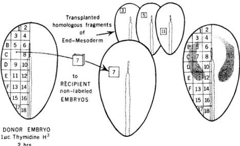
The method that is potentially most accurate for mapping the prospective embryonic fate of tissues—in contrast to the.r much broader competency—is that of transplanting small fragments from a radioactively labeled donor embryo to an unlabeled recipient (Rosenquist, 1966; Weston, 1967). The operation as we have em
ployed it is shown diagrammatically on the left side of Fig. 1. The donor embryo is cut according to some preconceived pattern. Each labeled fragment is then transferred to the matching site in an un
labeled recipient. The implanted tissue heals in rapidly. Its subsequent fate can be determined by autoradiographic analysis of serial sections of the host embryo at a later stage of development. Each such implant participates in the normal development of the host, fulfilling the devel
opmental task of the excised piece it replaces. By applying this tech
nique, in conjunction with particle-marking experiments and time- lapse cinematography, we have been able to localize the prospective heart cells and trace their movements from the stage 3+ (midstreak) embryo epiblast, through the primitive streak into the lateral plate mesoderm (at stage 5) and into the forming myocardial troughs and tubular heart at stage 12 (Rosenquist and DeHaan, 1966; Stalsberg and DeHaan, 1968).
By means of implants into the epiblast of stage 3+-5 embryos, prospective heart cells can be identified in paired regions about mid
way down the length of the streak, extending from the midline later
ally, approximately halfway to the edge of the pellucid area. Within these regions prospective conus cells He nearest the primitive streak.
Progressively more laterally on each side are—in sequence—prospec
tive ventricular, atrial, and sinus cells (Rosenquist and DeHaan, 1966).
As labeled cells move into the streak, the graft begins to lose its coherent structure. Graft tissue mixes with unlabeled cells from re
gions across the streak. Labeled cells enter the streak and are then scattered in the "primary mesenchyme" of the mesoblast (Trelstad et al.? 1967) moving rostrolaterally away. Implants made on one side of the epiblast-contribute labeled cells to both right and left sides of the heart, at roughly bilaterally symmetrical positions. The movements of preendocardial cells from epiblast to mesoderm are largely inde
pendent of those of the prospective myocardium.
The last prospective sinus cells reach the streak shortly before stage 5 and descend to mesoderm. After moving into the sheet of mesoderm, preheart cells move rostrolaterally to form the broad lateral wings of the horseshoe-shaped cardiogenic crescent (Mollier, 1906). In the embryo diagrammed in Fig. 1 (right side) the heavily shaded lateral limbs of the crescent contain both preendocardial and premyocardial cells. The rostromedial bridge of the crescent (shown with light stip
ple ) is formed by only a sparse scatter of prospective endocardial cells in some embryos; it is lacking altogether in others (Stalsberg and DeHaan, 1968).
Although the broadly separated regions of prospective myocardial cells within the heart-forming crescent at stage 5 are not recognizable histologically, there is suggestive evidence that they have attained a degree of biochemical differentiation by this stage. For example, embryonic heart tissue is rich in glycogen. Chiquoine (1957) localized cells of the splanchnic mesoderm that stained heavily with periodic- acid Schiff reagent in two areas of the mouse embryo corresponding in position to the lateral wings of the cardiogenic crescent. The preheart cells are also differentially susceptible to poisoning by oxidative in
hibitors such as sodium fluoride (Spratt, 1950) and antimycin A (Reporter and Ebert, 1965). When sodium fluoride was applied to the stage 5 chick embryo, it produced localized areas of necrosis and edema, again corresponding in position to the precardiac areas (Duf- fey and Ebert, 1957). Furthermore, at stage 5, Ebert (1953) found that immunologically reactive groups of cardiac proteins were restricted mainly to the same bilateral regions.
By making very small labeled implants of endoderm and mesoderm
FORM AND FUNCTION IN THE EMBRYONIC HEART 2 1 3
2 hrs.
FIG. 1. Transplantation mapping technique. Left: Fragments of endoderm- mesoderm are transplanted from a thymidine-3H-labeled donor embryo into nonlabeled recipient embryos. After development of the host to stage 12, the position of labeled cells in the heart is determined by autoradiographic analysis.
Right: Regions shaded on the embryo are those that contributed cells to the stage 12 heart. Dark shading = myocardium and endocardium; light stipple = endocardium only.
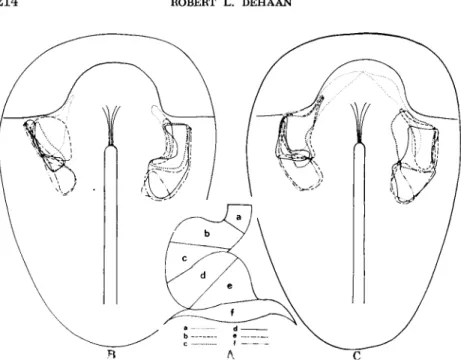
into the stage 5 embryo (Stalsberg and DeHaan, 1968), the outer boundaries and internal organization of the heart-forming regions could be mapped with considerable precision. To this end, the stage 12 tubular heart was first subdivided into a series of six arbitrarily defined regions (Fig. 2A, regions #-/), and the origin of each of these was mapped in the mesoderm of the stage 5 embryo. A smooth line was drawn through the center of each of the outermost implants which had contributed to region a, region b, and so on, yielding separate regional maps for myocardium (Fig. 2B) and endocardium
(Fig.2C).
The subdivisions of the precardiac areas, mapped in this way, show extensive overlap. Nonetheless, the distinct rostrocaudal sequence of regions which is apparent in both premyocardium and preendocardium is the same as the mediolateral array seen in the stage 3+ epiblast.
Preendocardial cells for each region tend to be localized somewhat more rostrally along the cardiogenic crescent than the premyocardial
FIG. 2. Inclusive regional map of the precardiac mesoderm. (A) Subdivisions (a-f) of stage 12 heart (B) Areas of stage 5 precardiac mesoderm contributing to each region of myocardium. ( C ) Areas of stage 5 precardiac mesoderm contributing to each region of endocardium. From Stalsberg and DeHaan (1968).
cells that form that region. But it is clear that preconal cells, medial in the stage 3+ epiblast, must have entered the primitive streak first to form the first portion of the cardiogenic crescent. These were followed by the next most lateral, preventricular cells, and in turn by the preatrial and presinus material, each contributing to the cres
cent in orderly sequence.
Labeled endocardial cells in the stage 12 heart, contributed by stage 5 grafts, were found either in more or less discrete regions, or scattered diffusely as singlets or small groups. The boundaries of the labeled region of endocardium were often difficult to distinguish because of the low ratio of labeled to unlabeled nuclei, suggesting some intermingling of preendocardial graft cells and host cells between stage 5 and stage 12, just as was found after stage 3+ grafts.
In sharp contrast, labeled myocardial cells in the stage 12 heart, contributed by stage 5 implants, were always found in well-defined areas, in the form of elongated bands running obliquely to the axis of
F O R M AND FUNCTION IN T H E EMBRYONIC HEART 2 1 5
the tubular organ. Labeled and unlabeled nuclei were uniformly dis
tributed throughout the labeled band, with no indication of inter
mingling of graft cells with those of the host. This suggested that the sheet of precardiac splanchnic mesoderm, or any small zone within it, maintains the integrity of its boundaries while it undergoes the topological changes—condensing, folding, and stretching—involved in cardiogenesis.
This correspondence of myocardial regions in stage 5 and stage 12 embryos, and the lack of random scatter of cells within these regions during this period, suggested that a myocardial map might be ob
tained of greater precision than that produced by simply outlining the areas within which regional implants lay (Fig. 2B). To this end, the stage 12 heart was further subdivided into 24 defined areas, and the origin of each of these was mapped.
A set of five longitudinal lines was used to divide the heart cylinder into longitudinal quadrants. Two lines ( dotted on the stage 12 heart in Fig. 3F ) were used to define the attachment of the heart to the dorsal mesocardium on each side. Line 3 represents the ventral line of fusion between left and right heart-forming regions (Stalsberg, 1968). In addition, a set of seven transverse lines used to delimit the same rostrocaudal sequence of regions (a-f) shown in Fig. 2. For purposes of mapping, the 42 intersections between these longitudinal and trans
verse lines were the main points of reference. Each of the stage 5 implants that contributed labeled cells to a point of intersection was so designated, and a geometric midpoint was calculated for each group of implants contributing to the same point of intersection. This process was repeated for each of the twenty-one intersections on each side, and these 21 averaged interseetional points were connected with smooth lines, yielding the more precise regional map of the stage 5 premyocardial mesoderm shown in Fig. 3A (for details of this mapping procedure, see Stalsberg and DeHaan, 1968).
Heart Tabulation
In order to understand how the flat sheet of precardiac mesoderm at stage 5 is converted into the contorted tube of the stage 12 heart, the configuration of the splanchnic mesoderm layer was examined in embryos at closely spaced intervals by making graphic reconstructions of the mesoderm, by microdissections of that layer in lightly fixed embryos, and by tracing the location of particulate markers placed
FIG. 3. Heart formation from stage 5 to stage 12. Only mesoderm is shown.
(A) The premyocardial subdivision map is superimposed on the mesoderm layer, and (B, C ) is appropriately distorted in shape to fit the forming myocardial troughs and elongating myocardial tube ( D - E ) ( F ) Subdivisions of the stage 12 heart from which division lines were obtained. From Stalsberg and DeHaan (1968).
on the forming myocardial troughs and early heart tube (Stalsberg and DeHaan, 1968).
At stage 4, electron micrographs of the cells of the forming lateral plate mesoderm show it to be a loose spongy layer of primary mesen- chyme (Trelstad et ah, 1966) in which the cells are apposed to one another across small close junctions and highly focal tight junctions.
These intercellular adhesions are thought to be transient in nature
FORM AND FUNCTION IN THE EMBRYONIC HEART 2 l 7
(Trelstad et al., 1967), and could well permit a degree of intermingling of cells from one region with those of another. Because of the open reticular structure of the mesenchyme, little of the surface of each cell is in contact with neighbors.
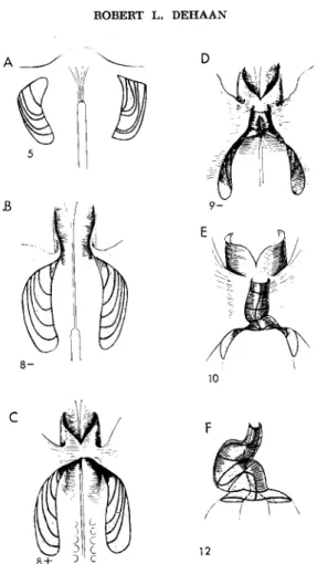
At stage 5-6 (Fig. 3A), the lateral mesoderm forms a thin flat sheet, now mesothelial in character. If the endoderm is dissected away, the mesoderm can be lifted as a coherent layer from the underlying ecto
derm, but there are no pronounced regions of local condensation. The layer has a distinct rostral border, which can usually be seen even in the intact living embryo. From this point on, the major morphogenetic movements in the formation of the myocardium are those of folding and anteriorward condensation.
The first indications of these processes are seen at stage 7, when the ectoderm and endoderm begin to fold to form, respectively, the early head-fold and shallow foregut. At this stage, the anteromesial portion of the mesoderm folds between those two layers, and is found enclos
ing the roof and lateral walls of the foregut ( Fig. 3B ). In these folded regions the mesoderm begins to thicken and condense, prior to split
ting into splanchnic and somatic layers, by increasing apposition of the cells with one another, and by their beginning conversion from flat mesothelial to columnar shapes. Although the cells are attached at their basal ends into a continuous mesothelial sheet, their free sur
faces are not in close apposition.
By the time the embryo has 4-5 somites (Fig. 3C), pronounced spaces, the amniocardiac vesicles, have formed between the separated layers of splanchnic and somatic mesoderm. The splanchnic layer has folded down to form the ventral edge of the head mesoderm, and a fold of this layer also pushes in medioventrally over the anterior in
testinal portal. The rostrolateral boundary of each of the mapped heart-forming regions forms the crest of this fold, and becomes the ventral edge of each myocardial trough. In the inner surface of this fold, i.e., the layer of splanchnic mesoderm in contact with the floor of the foregut, cellular condensation continues, and from this layer a few hemangioblasts or prospective endocardial cells begin to emigrate into the thin endomesodermal space (Sabin, 1920).
At stage 8+-9" ( Fig. 3D ) the crests of the ventromedial mesodermal folds meet in the midline, and the foregut is encircled by mesoderm.
The two folds of splanchnic mesoderm investing the ventral foregut have thickened further, and each has formed a slight concavity be-
tween itself and the endoderm. Within these concavities, clusters and bands of hemangioblasts begin to hollow out to produce the primitive endocardial plexus. As the ventral edges of the myocardial folds swing together and fuse, they establish the ventral midline of the heart tube, and carry the subdivision lines sequentially into transverse positions
(Fig.3D-F).
The process of cellular condensation in the forming walls of the heart has been analyzed recently with the electron microscope by Manasek (1968b). At about stage 8-8+, in the thickening regions of splanchnic mesoderm, the layer changes from being one cell in thick
ness to 3-4 cells thick. The continuous outermost layer of cells forms the free surface of the epithelium. These cells tend to be elongate, with their long axes roughly perpendicular to the layer, and are tightly bound to each other at their apices. The deeper cells of the layer are more mesenchyme-like, and are widely separated by large amounts of extracellular material. As the myocardium condenses for
ward to form the folding myocardial troughs, the basal layer of cells becomes more clearly defined as the cells establish more extensive lateral contacts with each other. By stage 10, the cellular architecture of the heart is strikingly more compact. Although substantial inter
cellular spaces still persist, each cell is in contact with neighbors over a major portion of its surface. Within the next few hours, intercellular spaces are further reduced, producing a compact myocardial layer 2-3 cells thick. Concomitant with this progressive loss of intercellular space and packing of the cells is a notable change in their orientation.
The myoblasts in the splanchnic mesoderm of the stage 8+ embryo have their long axes directed radially or perpendicular to the embry
onic axis. As the myocardial troughs form, there is a gradual flattening of the cells so that by stage 12 the myocardial cells have their long axes oriented circumferentially with respect to the heart tube.
It should be noted that during the period under consideration ( stage 5 to stage 12) the cardiogenic splanchnic mesoderm, the myocardial troughs, and the muscular heart tube are composed of an essentially pure population of myoblasts and myocytes (Manasek, 1968a). Neither epicardial cells nor fibroblasts are seen until much later in develop
ment. It is for this reason that we have referred here to myocardial troughs, instead of the more common term "epimyocardiaF troughs.
Essentially all myocardial cells begin to form myofilaments at about stage 9, and gradually complete the transition to myocytes—having
F O R M AND FUNCTION IN T H E EMBRYONIC HEART 2 1 9
well organized myofibrils which exhibit Z-bands—by stage 12 (Olivo et al, 1964; Manasek, 1968b).
In an earlier study, I observed the development of embryos growing in vitro, with the aid of time-lapse cinematography (DeHaan, 1963a,b), and reported seeing clusters of heart-forming cells migrating as inde
pendent entities. These clusters appeared between stage 4 and 5, silhouetted against the background tissue, and migrated, at first, in random directions. At about stage 6, however, their movements seemed to become organized, and from stage 6 to about stage 10 they ap
peared to migrate in parallel routes rostromedially up the cardiogenic crescent into the forming heart tube.
The impression of the process of cardiogenesis which emerges from the autoradiographic studies described here, and from the appearance of the precardiac mesoderm in the electron microscope, is not one of a loose mesenchymatous tissue in which cells migrate substantial dis
tances with respect to one another. The evidence is quite the contrary.
The splanchnic mesoderm is largely epithelial in character, rather than mesenchymal. Labeled regions in the stage 5 premyocardial mesoderm retain their discrete boundaries throughout development to stage 12, with no evidence of mixing of cells from labeled and unlabeled regions.
Moreover, the percentage of labeled cells within an implant does not change with time after implantation (Rosenquist and DeHaan, 1966), as would be expected if clusters of cells were moving independently.
From these data it is clear that the premyocardial mesoderm behaves as a coherent sheet in the formation of the tubular heart. It condenses, stretches, folds, and deforms, but it does not lose its integrity as a sheet, nor its continuity with the rest of the layer of splanchnic meso
derm. It does not break up into cells or cell clusters which migrate independently (Stalsberg and DeHaan, 1968).
There is a vast literature in support of the idea that embryonic cells exhibit differential adhesiveness toward different surfaces with which they come in contact, and that this property is involved in their capac
ity to reaggregate and sort out histotypically after dissociation ( Stein
berg, 1964; Roth and Weston, 1967; Curtis, 1967). However, as an important mechanism of morphogenesis in the intact embryo, evidence for different degrees of intercellular adhesion is weaker, and entirely circumstantial (DeHaan, 1963c; Gustaf son and Wolpert, 1963; Weston and Butler, 1966). Moreover, recent studies, in which labeled cells have been traced autoradiographically, have indicated that whatever
their state of adhesiveness, cells do not mix and sort out in the forma
tion of the embryonic limb bud (Searls, 1967) or in fused pairs of heart or liver fragments (Weston and Abercrombie, 1967); nor do they aggregate preferentially with like cells when injected into the intact embryo (Burdick, 1968). These results emphasize that most move
ments in the embryo—with a few notable exceptions, such as the neural crest cells (Weston, 1963)—involve folding or deformation of cell sheets, or change of shape or bulk of large masses of cells, rather than the individual-migration of single cells.
In recent reviews, Trinkaus (1965, 1967) has emphasized that changes in the surface properties of cells are often a prelude to the movements of the cell masses or sheets which those cells comprise.
The change in cell contacts and condensation of metanephrogenic mesenchyme cells during kidney tubulogenesis ( Wartiovaara, 1966);
the movements of the gastrodermal and epidermal tissue sheets dur
ing the steady-state growth of Hydra ( Campbell, 1968 ) ; the increased
"stickiness'' of cells of the pancreatic epithelium as the diverticulum evaginates (Wessells and Cohen, 1967); and the apparent increase in membrane activity of the cells of Fundulus blastoderm prior to epiboly (Trinkaus and Lentz, 1967) are examples recently provided. It does not seem unreasonable to speculate that a similar localized increase in intercellular adhesiveness in the anterior regions of the premyocardial mesoderm may underlie the condensation and thickening of that tissue, as described above; and may provide part of the motive force for the rostrad movement of that layer relative to the underlying endoderm between stages 5 and 8 (DeHaan, 1964; Rosenquist and DeHaan, 1966).
If differential adhesivity of cells plays any guiding role during car- diogenesis (DeHaan, 1963c), it would appear from the results de
scribed here that preendocardial cells are more likely candidates for such guidance than those of the premyocardium. In histological sec
tions hemangioblasts can be seen emigrating in small groups from the forming myocardial troughs. Moreover, cells from a discrete labeled region in the stage 5 mesoderm were often found as singlets or in small groups in the stage 12 endocardium. Nonetheless, despite this degree of intermixing, the pattern of organization of the preendo
cardial cells is roughly similar to that of the premyocardium (Fig.
2B, C). Thus it may well have been the movements of these cells that were recorded on cinema film in the earlier study (DeHaan, 1963b )s
F O R M AND FUNCTION IN T H E EMBRYONIC HEART 2 2 1
from which the anteroposterior organization of the cardiogenic ma
terial was first surmised.
The conoventricular portion of the heart tube, which forms between stage 9 and 10, consists of three components of the definitive heart.
The rostral end, which Davis (1927) designated the "aortic bulbar"
portion of the tube represents the precursor of the definitive truncus arteriosus. Caudal to that is the region generally termed the bulbus cordis, which forms the infundibulum of the prospective right ventri
cle. The caudalmostpart of the differentiated heart tube (at this stage) will become the anatomical right ventricle. The paired halves of the posterior ventricular region ( prospective left ventricle ) and the primi
tive atrium are still widespread as open myocardial troughs over the arch of the anterior intestinal portal.
Through stages 10 to 12, elongation of the heart tube results from continued condensation and fusion of progressively more posterior portions of the precardiac mesoderm which feed in over the ever- receding anterior intestinal portal; and from differential expansion of the already differentiated tube (Fig. 3D-F). When the embryo has 10-11 somite pairs, the prospective left ventricle has fused at the caudal end of the heart—emphasizing that the right and left ventricles arise in tandem ( DeVries and Saunders, 1962 ). The heart tube bulges distinctly to the embryo's right, in anticipation of the dextral looping of the ventricle. The differential elongation of the two sides of the heart, which appears to be responsible for this rightward looping can be seen in the distortion of the subdivision lines that occurs between stages 9 and 12. (Fig. 3D-F). At stage 13-14 the atria are established;
and by the time the embryo has 25-26 pairs of somites, sinoatrial tissue has differentiated. The experimental evidence in support of this mecha
nism of looping has recently been reviewed (DeHaan, 1968).
THE INITIATION OF FUNCTION
The chick heart begins beating at about the 10-somite stage, when only the conoventricular portion of the cardiac tube has differentiated (Johnstone, 1925; Patten and Kramer, 1933). As each new region of the heart forms, it brings to the organ tissues with different physio
logical properties. For example, the shape and characteristics of the action potentials recorded from cells in ventricular, atrial, and sinoatrial tissues are distinctly different in each of these regions, as they form in the heart tube (Fingl et al., 1952; Meda and Ferroni,
1959; Lieberman and Paes de Carvalho, 1965; Coraboeuf et al., 1965), just as they are in the adult heart (Paes de Carvalho et al., 1959;
Moore, 1965).
Each new region is also different in its intrinsic rate of contraction.
The beat starts in the right margin of the myocardium near the posterior end of the ventricle. Initially irregular and spasmodic, the heart soon develops a rhythmic slow rate of 30-40 beats per minute.
Gradually, as more posterior regions of the heart tube differentiate, the rate increases. By the time dextral looping of the ventricle has occurred and a distinct atrioventricular sulcus is observable (stage 13), the heart rate has increased to 80-90 B/M. By about 60 hours of incubation (stage 16), after the sinoatrial tissue has differentiated, the heart normally beats 110-120 times per minute.
This gradient of rhythmicity is clearly built into each portion of the heart tube. If the tube is cut transversely into three fragments at a stage when it is beating 120 B/M, the sinoatrial piece continues to beat at that rate, whereas each of the more rostral pieces reverts to its earlier rhythm. That is, the ventricular piece slows to about 70 B/M; the conoventricular portion takes on a rate of only 30-40 B/M (Barry, 1942; DeHaan, 1965b). In fact these rate differences must reside ultimately in the individual cells that comprise the heart.
Cells isolated in tissue culture from the embryonic ventricle beat more slowly, on the average, than those from the atria (Cavanaugh, 1955). Under these conditions with each cell isolated from contact with any neighbors, every beating cell necessarily determines its own pulsation rate (see below).
The gradual increase in rate of the entire heart tube as each new region forms, suggests that as each segment of the heart tube differentiates, with a higher intrinsic rate, it acts as pacemaker for the rest of the organ, driving the heart at its own rate. This has been confirmed recently by Van Mierop (1967), who determined the location of the pacemaker region in the tubular chick heart before and during the time of initiation of the beat, using both intracellular and surface exploring electrodes. He reported that when the first region of tissue began to contract in the right posterior portion of the ventricle, at stage 10, the electrical stimulus for that contraction arose from a more caudal point, about 100 milliseconds prior to each beat. Furthermore, even in the 9-somite and occasional 8-somite hearts, 3-6 hours before actual contractile activity begins, Van Mierop could record rhythmic action potentials. These findings indicate clearly
F O R M AND FUNCTION IN T H E EMBRYONIC HEART 2 2 3
that pacemaker activity is localized at the posterior end of the tubular heart, and suggest that cells in the myocardial troughs caudal to the formed heart tube at each stage, begin to function as pacemakers before they fuse in the midline and are themselves incorporated into the beating myocardium.
These results open the question whether the differentiation of pace
maker membrane activity could be stimulated, or regulated merely by the condensation of myocardial trough material prior to myo- fibrillar formation or other signs of histodifferentiation. Although we are dealing here with totally unfounded speculation, many investigators of embryonic cell movements and cell interactions have emphasized the importance of intercellular communication, both in terms of physical contact ( Abercrombie, 1967; Steinberg, 1964; Saxén and Wartiovaara, 1966), and short-range transmission of materials between cells (Grobstein, 1967).
It has recently been demonstrated that a variety of cell types, including mature cardiac muscle (Dewey and Barr, 1964) and all three germ layers of the chick embryo (Trelstad et al, 1967) exhibit inter
cellular contacts of the "zonula occludens" type, where the outer leaf
lets of the apposed cell membranes appear to fuse. These "tight" junc
tions have been associated with regions of low intercellular resistance (Loewenstein et al, 1965; Potter et al, 1966; Sheridan, 1968; Barr et al, 1965) across which ions (Woodbury and Crill, 1961; Weidmann, 1966) and perhaps even larger molecules (Loewenstein, 1966; Sheri
dan, 1968) can flow from cell to cell. The ubiquitous presence in embryonic blastomeres and tissues of such electrical coupling has led to much theorizing about its possible role in intercellular communica
tion (Loewenstein, 1968; Trelstad et al, 1967; Sheridan, 1968; Hay, 1968 ). In this vein, it is of interest to wonder whether increasing num
bers of tight junctions are established during condensation of the pre- myocardial mesoderm, and whether during this period, concomitant with the closer physical contact seen, the differentiating myocytes achieve the electrical continuity required for the genesis of electrical signals, and the later coordination of the heart beat, at stage 10. The fact that epiblast cells of the primitive streak embryo are electrically coupled (Sheridan, 1968) and that focal "tight" junctions may be seen in the lateral plate mesoderm in early somite stages (Trelstad et al, 1967) suggests that cell communication via this route may play an even more general role in early development of the chick embryo.
But no such role has yet been demonstrated.
Further confirmation that rapidly beating tissue can act as a pacemaker for regions with a slower intrinsic rate, derives from tissue culture studies. Heart fragments with different rates may be explanted close together, in pairs, on the surface of a nutrient medium. As cells from each fragment bridge the intervening gap, the two pieces synchronize, at or near the rate of the initially faster piece (Paff, 1935; Egorin and DeHaan, unpublished). Moreover, even individual cells, rapidly beating in culture, can act as pacemakers, influencing their slower neighbors to take on their own faster rate when random movements bring them into contact (Garofolini, 1927). Thus, that area of the heart which at any given stage has the highest intrinsic rate of pulsation, contains the cells that act as pacemakers for the entire heart.
There is good evidence that this gradient of rhythmicity—high rate caudally, low rostrally—is coded into the cells of the embryo well before the heart itself forms. The stage 6 embryo can be cut into fragments containing either the anterior, middle, or posterior portion of the cardiogenic crescent; that is, the regions destined to form, respectively, the conoventricular, ventricular, and sinoatrial parts of the heart. Such fragments isolated in culture medium, form vesicles of heart tissue that begin beating spontaneously at an average rate of 36, 65, and 115 beats per minute, anterior, middle, and posterior, respectively (DeHaan, 1963d). With the electron microscope these vesicles exhibit well-formed myofibrils with Z-bands and intercalated discs, characteristic of normal heart muscle. When they are impaled with intracellular microelectrodes, the slowly beating tissue shows action potentials typical of conus, the vesicles with intermediate rates have ventricle-like action potentials, and the fastest fragments exhibit pacemaker potentials characteristic of sinoatrial tissue (Le- Douarin et al., 1966). Thus not only prospective rate, but the physio
logical character of the beat, appear to be determined in the cells of the premyocardial mesoderm at early stages, although inductive interactions between mesoderm and endoderm cannot be excluded from functioning in such determination (Orts Llorca and Collado, 1967).
THE PROPERTIES OF HEART CELLS IN CULTURE
From this description of the development of the heart, we are left with a host of questions concerning underlying mechanisms.
F O R M AND FUNCTION IN T H E EMBRYONIC HEART 2 2 5
With regard to the morphogenetic events: what change in cell properties causes the lateral plate mesoderm to split into two layers;
how do all the precardiac cells find their way to the splanchnic layer;
what forces cause the thickening, folding, and forward condensation of that splanchnic mesoderm? If, as suggested, this condensation results from alterations in intercellular adhesiveness, what internal changes act as trigger; what environmental agents regulate or influence the increasing contact; why does the condensation begin along the rostral edge of the mesoderm and progress backward in an orderly fashion; is there a causal relation between condensation and differentiation?
In terms of the onset of function of the heart: are some precardiac mesoderm cells destined to formed nonspontaneous myocardial muscle, while others differentiate into the spontaneously active specialized tissues of the heart? If so, how many prepacemaker cells are there;
does the proportion increase or decrease with development; what are the parameters that determine whether a heart myoblast differentiates pacemaker or nonpacemaker properties; once differentiated, what factors determine whether pacemaker capacity is expressed? Why do pacemaker cells differentiate different intrinsic rates of electrical firing?
How is this rate related to their position in the cardiogenic crescent?
How is information regarding prospective rate coded into the pre
cardiac cell?
Since as already mentioned the developmental events one observes during cardiogenesis arise out of the characteristics and behavior of cells that comprise the precardiac tissue, most of the questions we wish to ask are not answerable by experiments—however ingenious—on the intact embryo. Therefore, we have devoted considerable attention to optimizing techniques of dissociation and in vitro culture of embryonic heart cells in order to investigate three of their fundamental properties: their spontaneity, mitotic activity, and contact relations with one another, under circumstances where (hopefully) one or a small number of environmental variables at a time can be manipu
lated. In this section I will summarize material already published (DeHaan, 1967a to c) and add some new information.
It has been apparent for 40 years or more that embryonic heart cells in culture can initiate their own pulsations and can act as pace
makers for neighbors with which they come into functional contact (Burrows, 1912; Garofolini, 1927). It has only been in the past few years, however, since relatively simple techniques have become
available for dissociating embryonic tissues into their component cells with proteolytic enzymes (Moscona, 1952; Steinberg, 1967) that it has been feasible to obtain reproducible cultures of healthy isolated heart cells, plated at any desired density. Cavanaugh (1955) first re
ported on such cultures from 5-day chick heart dissociated with trypsin. Within a few hours after a suspension of isolated cells was inoculated into a culture medium, the cells attached to the bottom of the dish, and as they did so, began (or resumed) beating. She confirmed that only a portion of the population was spontaneously active, that each beating cell had a different rate from its neighbors if they were not in contact, and that after some time in culture, groups or sheets of cells came into mutual contact and frequently began to beat in synchrony. More recently, Harary and Farley (1963) have shown that cells from postnatal mammalian hearts behave in much the same way.
In seeking a culture system, initially, to determine how many of the cells that comprise an embryonic heart are spontaneously active at any given stage, it became immediately apparent that that parameter of the cell population was dependent to a remarkable degree on the environmental conditions to which the cells were exposed (DeHaan, 1967b). In fact, most of the characteristics of these cells—their appearance, their spontaneity, their mitotic activity, and contact re
lations—were found to be influenced by, or completely under the control of, experimental parameters, including mode of tissue dis
sociation, density of cell inoculum, components of the culture medium, and age of embryo from which the cells were obtained.
Cell Morphology
The mature heart is composed of several cell types. Although the bulk are myocardial muscle cells, substantial amounts of fibroelastic and collagenous connective tissue, endocardial tissue, vascular en- dothelium, and the various components of the sinoventricular con
duction system are also present. In contrast, the embryonic heart, at least for the first few days, comprises an essentially pure population of myocardial myocytes, plus a small number of endocardial cells (Manasek, 1968a). Despite this apparent early homogeneity, cultures of embryonic heart cells contain two morphologically distinguishable types; myocytes, and those generally thought to be fibroblastic or endothelial cells (Stilwell, 1944; Mark and Strasser, 1966). Cells of the
F O R M AND FUNCTION IN THE EMBRYONIC HEART 227
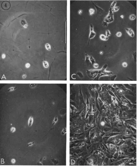
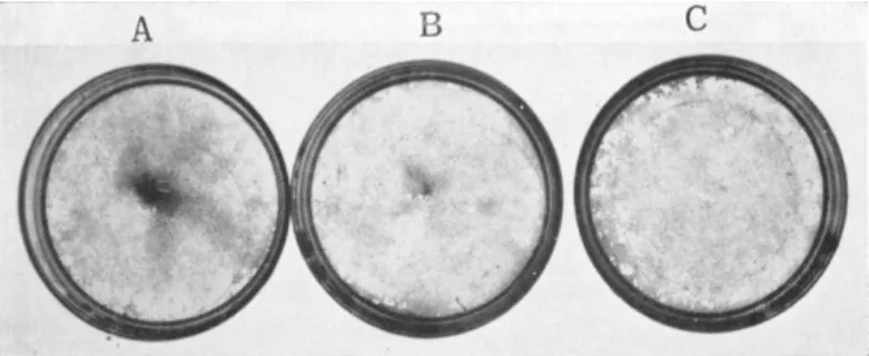
fibroblastic type, for which I have used the neutral term F-cells (DeHaan, 1967b), are usually very thin and well spread on the substratum; frequently triangular or stellate in shape (Fig. 4). Because of their thinness, they are transparent and poorly refractile under phrase optics. They normally contain a ring of granules or droplets
FIG. 4. Cells from 7-day hearts, after 24 hours in a nongrowth medium (629A). Plated at (A) 1 X 10⁵, (B) 2 X 10⁵, (C) 5 X 10\ and (D) 1 X 10e cells per plate. Photographed live, phase contrast, Polaroid type 108 film.
Scale = 100 microns. From DeHaan (1967b).
surrounding the single nucleus, which itself is clear and usually contains two or more nucleoli.
The second type resembles the myoblasts of skeletal muscle. I have termed these M-cells. They are thick, highly refractile cells, generally round or spindle-shaped, and usually uninucleate. Their cytoplasm is more granular than that of F-cells, and their nuclei often less distinct. The M-cell nucleus usually contains only a single large nucleolus.
I reported, earlier that M-cells comprise slightly more than 50%
of the population (DeHaan, 1967b). With subsequent improvements in medium we now obtain 60-75% M-cells routinely (DeHaan and Gottlieb, 1968). This figure corresponds well with that reported by Mark et al. (1967) for cultures of neonatal rat heart cells. The re
mainder have the F-cell morphology. We refer to the intranuclear phase-dense structures as nucleoli on the basis that, in both M- and F-cells, they are RNase but not DNase sensitive, they stain specifically with azure B, and all but a small percentage incorporate tritiated uridine ( I. Polinger, unpublished data ).
Other differences between M- and F-cells are evident. Time-lapse cinematographic studies have demonstrated that M-cells are generally nonmotile in culture, and rarely divide. F-cells move about very actively, and have a generation time of 13-18 hours. F-cells are apparently more adhesive to the plastic dish than M-cells. This im
pression is based upon their tendency to spread thinly on the sub
stratum, and on the fact that they attach more rapidly. In appro
priate media approximately 80% of M-cells beat spontaneously (20 experiments, 8300 cells counted; DeHaan and Gottlieb, 1968), where
as only 1-5% of F-cells do. Finally, rat myocardial cells are killed by high levels of oxygen; endothelioid cells are not (Mark et al., 1967).
I have confirmed this observation with M- and F-cells of the 7-day chick heart ( DeHaan, unpublished ).
Despite these apparent differences between M- and F-cells, which suggest strongly that they represent separate populations, another possibility must be entertained. These two morphological forms might represent different manifestations of a single heart cell type, respond
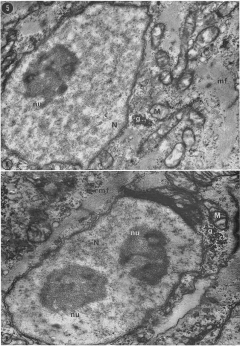
ing differently to the conditions of culture, or perhaps caught at two different times in the cell cycle by the processes of dissociation and cultivation. The fact that, in the intact heart, myocardial cells with myofibrils and with one and two nucleoli are seen side by side (Fig.
5) indicates that nucleolar number is not an adequate criterion for
FIG. 5. Ventricular cells from a section of an intact 7-day embryonic chick heart. Palade's fixative, pH 7.4, stained with lead citrate, g, glycogen; M, mitochondrion; mf, myofibrils; N, nucleus; nu, nucleolus. (A) One-nucleolate cell (X 14,100), (B) Neighboring cell with two nucleoli (X 19,800). Courtesy of I. Polinger.
229
distinguishing cell types (Manasek, 1968b; Polinger, unpublished).
In fact, nucleolar number is known to change as a result of nucleolar fusion in amphibian cells (Wallace, 1963), and varies with mitotic activity in rat liver (Swift, 1965; Mironescu and Dragomir, 1966).
With studies now in progress on cloning of heart cells, we hope to determine whether nucleolar number or other properties of the two morphological types are stable characteristics.
SPONTANEOUS ACTIVITY OF CULTURED CELLS
What proportion of the cells of an embryonic heart are pacemakers?
Does that proportion change with development? Before attempting to answer these questions we must clarify them by answering three prior ones: (a) how do we define a pacemaker, (b) from which population of cells do we measure the proportion of pacemakers? and (c) what are the conditions under which development is taking place?
Definition of a Pacemaker
We defined a pacemaker implicitly above as a region of heart tissue which drives another region at its own rate, or as a cell which causes a neighbor with which it comes in contact to synchronize with it. In traditional electrophysiological terms a pacemaker cell is recognized by the shape of its transmembrane action potential—that is, by the presence of a slow diastolic depolarization or "pacemaker potential"
before the action potential spike (Woodbury, 1962)—or by the re
sponse of the cell to hyperpolarizing or depolarizing currents passed across its membrane (Lehmkuhl and Sperelakis, 1967). Operationally, for our purposes, neither of these definitions is adequate. Although it has recently become possible to impale isolated heart cells in cultures with intracellular microelectrodes and record good action potentials, this technique is delicate and tedious, and not suitable for a statistical analysis of a large population (DeHaan and Gottlieb, 1968). The capacity of a cell to drive another is perhaps an ultimate test, but requires that the two cells be observed both before and after electrical contact is established, to know which is acting as pacemaker for the other. This is a rare event and, moreover, points up another difficulty.
If rapidly beating cell A joins cell B and the two synchronize at the initial rate of A, cell A is classified as the pacemaker. However, if B had happened to contact, not A, but a third cell with a still slower intrinsic rate, B would have acted as pacemaker in that pair. We may
F O R M AND FUNCTION IN T H E EMBRYONIC HEART 2 3 1
thus define as a pacemaker any single cell which is seen to beat rhythmically, since such a cell, completely isolated from contact with neighbors, must be initiating its own activity, and can be presumed capable of driving a responsive neighbor with which it makes elec
trical contact. Any isolated cell that is quiescent in culture must fall into one of five classes: (1) a noncontractile cell—from one of the fibroblastic or endothelial components of the heart; (2) a myocardial
"blast" cell—which at the time of observation had not yet reached a functionally differentiated state; (3) a nonpacemaker—a contractile myocardial cell which requires an external stimulus to beat; (4) a
"covert" pacemaker—a cell which has an electrically active pacemaker membrane but which cannot respond to its own stimuli due to de
ficiencies in its contractile elements or excitation-contraction coupling mechanism; (5) a latent pacemaker—a cell which is quiescent under the particular circumstances of observation but which, with only minor changes of environmental conditions or internal physiological state, manifests its pacemaker capacity by beginning to beat spontaneously.
Proportion of Spontaneously Active Cells
I have defined the percentage of beating cells {% BC) as the propor
tion of the total cell population derivable from a heart, which is spon
taneously active (DeHaan, 1967b). This is a value readily obtained from plates seeded to give a density of 20-50 cells per mm2. In these conditions 90% or more of the cells are singlets, i.e., not in contact with any neighbor (Fig. 4A). Counting the number of beating and non- beating cells in five to ten microscopic fields of such a plate (usually 200-300 cells per plate counted; duplicate plates per experiment), and excluding groups of two or more conjoint cells, gives a direct measure of % BC.
Another value which has been used (Mark et al., 1967) is the per
centage of myocardial cells that is spontaneously active. This may be equally valid, as long as the distinction is made clear. However, such an assay assumes that the investigator, counting hundreds of cells, is able to distinguish at a glance a myocardial cell from all other types, and that these categories of heart cell types are meaningful.
Environmental Effects on Spontaneity
Previous estimates of the proportion of pacemakers in the total cell population range from 1-50% (Wollenberger, 1964) to 80-90% (Lehm-
kuhl and Sperelakis, 1967) for embryonic chick heart, and from 2%
(Harary and Farley, 1963) to "all heart muscle cells" (Mark et al., 1967) for neonatal rat heart. My own results however, have demon
strated that the percentage of spontaneously active cells can be experi
mentally varied over the range of 1% BC to about 60% BC ( = about 80% of M-cells) merely by varying specific aspects of the dissociation procedure or components of the culture medium (DeHaan, 1967b).
The length of time the tissue is in contact with trypsin, and the con
centration of the enzyme can produce twofold differences in the per
centage of beating cells in the resultant cultures. Horse sera, obtained from different individual animals, can have equally dramatic effects, even when used at a concentration as low as 4% (by volume) in the medium.
The most potent agent for varying % BC in cultures, however, is the potassium-ion concentration in the medium. Employing dissociation procedures and media tested to give optimal results in terms of % BC ( DeHaan, 1967b ), I found that increasing concentrations of potassium cause pacemaker cells to become quiescent. At a K level of 1.3 mM 50-60% of the cells are spontaneously active. Adding potassium in small increments results in progressively more and more cells switch
ing off. At 12 mM K, for example, only 5-10% of the cells continue beating. This effect is reproducible, and over a short time period, is completely reversible.
Cells do not survive reduction in potassium much below 1 mM.
Although we have continued to test a large number of variables in
cluding sodium and calcium concentration, temperature, protein com
ponents of the medium, lipids, and the cardiac glycoside ouabain, we have been unable to obtain more than 60-65% BC in any culture of 7-day heart cells, and continue routinely to find a range of 50-60%
BC from one culture to the next. On the basis of this negative evidence, we cannot declare with confidence that 30-40% of the cells of a 7-day heart are fundamentally nonpacemakers. However, it seems equally inadvisable to conclude that the high proportion of spontaneously beating cells suggests "that all heart muscle cells" are pacemakers
(Mark et al, 1967).
The serum potassium concentration of a 7-day chick embryo is about 5 mM (Grabowski, 1967). On the basis of the pacemaker-inhibition curve published previously (DeHaan, 1967b), and on the further assumption that the behavior of heart cells in culture under the condi-
F O R M AND FUNCTION IN T H E EMBRYONIC HEART 233 tions employed is representative of those cells in the intact organ, we can predict that in the normal 7-day embryonic heart 20-25% of its cells are functional pacemakers; at least 35-40% are latent pacemakers;
and the remaining 30-35% may be nonpacemakers, presumably falling within the remaining four categories listed above.
Percentage of Beating Cells in Hearts of Different Ages
Does the proportion of pacemakers rise or fall with the age of the heart? With knowledge of a number of the conditions in the external environment that control the proportion of spontaneously active cells in culture, it is possible to approach this question in a more meaning
ful way. Hearts from embryos of different ages (4 days to 18 days) can be dissociated and cultured under circumstances designed to maximize
% BC. Under these conditions, it was reported that the highest per
centage of spontaneously active cells could be derived from the 7-day heart (DeHaan, 1967b). Hearts from older embryos yielded pro
gressively fewer pacemakers, to a minimum of 10-15% at 18 days.
Hearts from embryos younger than 7 days also showed lower fractions of beating cells.
The dissociation procedure can also be modified (by increasing the number of trypsinization cycles) to maximize the total cell yield, and special precautions can be taken to avoid losing cells during any of the dissociation steps. Aliquots of such cell suspensions counted on a hema- cytometer yield estimates of the total number of cells derivable from hearts at each age. Knowing the % BC at each age permits a calcula
tion of the total number of spontaneously active cells so obtainable.
The 4-day heart consists of about 200,000 cells, 44% of which (88,000), are capable of beating in culture (Table 1). Three days later, the total number of cells has increased about 9-fold, and the
TABLE 1
TOTAL NUMBER OF CELLS AND PACEMAKERS OBTAINABLE FROM H E A R T S OF D I F F E R E N T AGES
embryo Age (days)
4 7 12 18
Total cells per heart
(X106)
0.2 ± 0.05 1.7 ± 0 . 4 9.4 ± 0.1 26.9 ± 2.8
% B C
44.1 ± 1.4 57.1 ± 0.8 36.3 ± 3.0 13.7 + 0.7
Total BC per heart
(X1Q6)
0.088 0.97 3.4 3.7
number of spontaneously active cells has grown about 11-fold, to just under one million cells. During the ensuing 10 days, the heart continues to increase rapidly in total cell number (though not logarithmically), whereas the rate of increase of pacemakers declines markedly.
The gradual decline in rate of growth corresponds with the results of mitotic studies in this tissue. Grohmann (1961) found that mitotic index reached a maximal level in 4-day chick myocardium, and there
after declined slowly throughout development. Parallel results were obtained with -rat hearts (Rumyantsev, 1963). This finding is also consistent with the fact that the mature heart is composed of a popu
lation of stable, nondividing cells (Leblond et al., 1959; Spraragen et al, 1962; Pelc, 1964).
Indeed, it is not the decrease in rate of mitosis in the late embryonic heart which is notable, but rather the rapidity of growth in the early stages. Cultured cells from 7-day hearts in growth media pro
liferate rapidly, increasing by as much as 10-fold in 4 days. If suffi
ciently sparse cultures are prepared initially, cells do not form exten
sive contacts with neighbors for at least a few days, and the number of beating M-cells and nonbeating F-cells per unit area on the plate can be counted. In these conditions the number of beating M-cells remains constant or declines. Growth of the culture is due almost entirely to proliferation of nonbeating F-cells. Under continuous time- lapse cinematography beating M-cells have been seen to divide in rare cases, but never more than once in 4 days of culture. In the same microscopic fields, F-cells divide repeatedly with a generation time in the range of 12-18 hours. Mark and Strasser (1966) have demon
strated the division of beating rat heart cells, but also noted the much greater mitotic frequency of the endothelioid cells in their cultures.
Again, if cultured heart cells are representative of those in the intact organ, and beating myocardial cells only rarely divide, then the growth of the heart from 0.2 X 10e to 1.7 X 10⁶ cells between 4 days and 7 days (Table 1) must have resulted from proliferation mainly of the nonbeating complement. This would represent an increase of about 15-fold in 3 days, or a doubling time of about 17 hours. This is a great deal more rapid than the doubling time of 45 hours calculated for 4-day cells by Rumery and Rieke (1967). Even so, however, it would not account for the 11-fold increase in functional pacemakers, i.e., differentiated cells, during this time.
In the intact embryonic heart, cells within the myocardium incor-
F O R M AND FUNCTION IN T H E EMBRYONIC HEART 2 3 5
porate tritiated thymidine ( Sissman, 1966 ) and mitotic figures may be seen ( Grohmann, 1961 ). However, the ultrastructural study of Wain- rach and Sotelo (1961) suggested that the developing myocardium may contain a population of undifferentiated cells in which mitoses occur. On the other hand, Manasek (1968a) could find no evidence for such stem cells. He in fact records several dividing cells in which well- formed myofibrils are present, along with spindle fibers and a meta- phase plate.
A traditional controversy among embryologists concerns the stability of the differentiated state, and the capacity of cells, differentiated for some specific function, to proliferate. This topic has been reviewed extensively in recent years (Ebert and Kaighn, 1966; Abbott and Holtzer, 1966; Königsberg and Hauschka, 1965). All workers would probably agree with Herrmann et al. ( 1967, p. 306 ) that "the assump
tion of a complete and general mutual exclusion of DNA synthesis and cell replication on the one hand, and production of cell-specific pro
teins on the other, cannot be accepted without reservation. . . ." Any stem cell line, rapidly dividing but committed to the production of a specialized cell type, must, by its very commitment, exhibit some degree of differentiation (e.g., Marks and Kovach, 1966). The produc
tion of specific products of differentiation and histotypic cellular architecture in clonal populations of cells is equally convincing (Königsberg, 1963; Coon, 1966; Cahn and Cahn, 1966).
The process of commitment or determination is itself a gradual one, requiring apparently on the one hand nuclear replication, and on the other continued synthesis of specialized products (Hadorn, 1966).
Skeletal muscle myoblasts, for example, do not cease mitotic activity abruptly, and immediately begin synthesizing myofibrils. More prob
ably a series of specialized products is accumulated in blast cells as the Gi phase of the cell cycle gradually increases and proliferation stops
(Herrmann et al., 1967). Even after the fusion of myoblasts, when DNA synthesis and nuclear division normally cease, the nuclei incor
porated into the myotubes are still capable of resuming some level of DNA replication (as measured by thymidine-3H incorporation) under the stimulus of infection by Rous sarcoma virus (Lee et al., 1968).
How then might we obtain the pattern of growth of the heart seen in Table 1. There are at least two possibilities.
Hypothesis I. Once a cell has differentiated to the point of forming
myofibrils it is prevented from undergoing more than one further nuclear division. This would account for the rapid F-cell proliferation and occasional M-cell mitosis reported in culture, and the finding of cells containing both myofibrils and division figures in the intact heart.
It would also explain our inability to obtain extensive proliferation of beating M-cells in vitro. In this case we would postulate, as above, that the increase in total cells between 4 and 7 days, for example, must have resulted from the division of stem cells among the 56% of nonbeat
ing cells counted. Assuming that all those nonbeating cells were capable of dividing, four logarithmic doublings would produce the increase in total cells seen. If, between the fourth and fifth division, a differentia- tive trigger caused half of the progeny of that division to cease pro
liferation and convert to the synthesis of specialized products, the % BC obtained from 7-day heart cultures would also be accounted for. The prime difficulty with this hypothesis is that substantial numbers of undifferentiated stem cells have not been observed in the organ (Manasek, 1968).
Hypothesis II. All myocardial cells in the intact heart are capable of dividing, or at least the presence of myofibrils does not exclude them from that activity. This possibility would account for the rapid increase in cells capable of spontaneous contractility, despite the lack of an observed population of undifferentiated cells in the myocardium.
However, it leaves unexplained the derivation of F-cells in culture, and the apparent unwillingness of beating M-cells to divide.
If myocardial cells do divide in situ, but not under our conditions in culture, it would seem to contradict the frequent observation that cells are released from mitotic inhibition by dispersal in vitro (Chay- tor, 1962; Lefford, 1964; Abbott and Holtzer, 1966). However, it must be recalled that in order to distinguish pacemakers from nonpace- makers, our growth studies have been done with low-density cultures.
Although there is one report of formation of a beating clone of myo
cardial cells (Cahn, 1964), there is also ample evidence that, for a variety of primary cultures of cells, there is a minimal cell density below which growth does not occur. Above this level, at least within some range, mitotic activity is augmented with increasing plating densities (Eagle and Piez, 1962; Rubin, 1967). Some aspects of this population density effect are related to nutritional considerations. For example, serine, which is synthesized by HeLa cells, leaks out of the cells too fast to be maintained at the necessary intracellular concentra-
F O R M AND FUNCTION IN T H E EMBRYONIC HEART 2 3 7
tion unless the amino acid is supplied in the medium, or the popula
tion of cells is large enough to "condition" the medium with this metabolite before the cells suffer irreversible damage from its deple
tion (Eagle and Piez, 1962).
There is also evidence, however, that short-range or surface- modulated interactions underlie other aspects of the low-density effect on growth. For example, sister cells of a hamster line, observed with time-lapse cinematography, are more likely to divide simultaneously if they remain in close proximity, than if they wander away from each other (Froese, 1967). Moreover, chick embryo fibroblasts in a complete growth medium, divide more frequently when plated in a restricted area of a dish than when they are more widely dispersed, even though the total number of cells, and the volume of medium is the same in both cases ( Rein and Rubin, 1968 ).
This localized density effect is abolished by growing cells in a con
ditioned medium recovered from dense cultures (Königsberg, 1963;
Rein and Rubin, 1968). The component of such conditioned medium which absolves cells (at least skeletal muscle cells) from any depend
ency on neighbors appears to be a collagen-like substance which is deposited as a layer on the surface of the dish and acts as a physical substratum for the cells (Königsberg and Hauschka, 1965; Hauschka and Königsberg, 1966). It has been reported that embryonic chick heart fibroblasts synthesize collagen in culture (Kuwabara, 1959).
However, we do not yet know whether M-cells plated on a substratum of reconstituted rat-tail or fish swim bladder collagen, will divide with a greater frequency. Experiments of this nature are now in progress.
The alternative hypothesis ( I above ) was that M-cells do not divide in vivo (except occasionally), but that cells of a stem line at some point differentiate. Is it possible that by altering our culture conditions after some appropriate number of divisions of F-cells (presumably representing that stem-cell line), a cessation of replication can be produced leading to differentiation? The fact that we have failed to observe such behavior may again involve a problem of cell density.
There is ample evidence that the mitotic activity of cells can be influenced greatly by their compactness or density, that is, by the num
ber of cells per unit area or volume (Stoker, 1967; Tumanishvilli, 1967).
In general, growth rate is inversely related to cell density at higher densities. Some so-called density effects may be due merely to inade
quate nutrition. It is commonly found, for example, that cells grow