Mitotic Spindle
1SHINYA INOUÉ
Department of Cytology, Dartmouth Medical School, Hanover, Neiv Hampshire
Introduction
The occurrence of birefringence in contractile elements of living cells may have a deeper significance than the mere localization of oriented molecules or micelles. This assertion is supported by two gen- eralizations, the first made by Engelmann in 1875, stating that contrac- tility in living systems depended upon the presence of birefringence (also Engelmann, 1906; but see Schmidt, 1937, p. 251). Whether strictly true or not, the statement takes on added significance in the light of the second generalization made by Dr. Aaron Katchalaski at the February, 1963 Meeting of the Biophysical Society. On thermodynamic grounds, he argued that in order to convert chemical energy (a scalar quantity) directly into contractile mechanical force (a vectorial quantity), struc- tural anistotropy is required. In other words, the molecules giving rise to contractile force cannot be randomly arranged, but must form an organized structure whose properties vary in different directions. In general, structural anisotropy is associated with optical anisotropy, i.e., a different response of the material to light vibrating or traveling in different directions. When the velocity of light propagation in a material varies in different directions, the material is said to be biréfringent or to exhibit double refraction.
The birefringence of muscle fibers, for example, is strong enough to be seen with conventional polarizing microscopes. T h e mitotic spindle, on the other hand, exhibits a considerably lower retardation than do muscle fibers and can be detected only by the use of refined polarization microscopic techniques. The contrast due to weakly biréfringent speci- mens must be maximized by reducing all possible sources of stray light and by judicious use of a compensator (Schmidt, 1934; Swann and Mitchison, 1950; Inoué and Dan, 1951; Mitchison, 1953; Inoué, 1961).
Conventional polarizing microscopes are not satisfactory for the study of
1 Supported in part by grants from the National Science Foundation (G 19487) and National Cancer Institute, U.S. Public Health Service (CA 04552).
549
550 SHINYA INOUÉ
detailed structures of the mitotic spindle, not only because of excessive stray light, but also because of the presence of a troublesome diffraction anomaly which limits effective lateral resolution (Inoué and Kubota, 1958; Kubota and Inoué, 1958). Some years ago we developed the polari- zation rectifier which corrects both of these defects (Inoué and Hyde,
FIG. 1. The spindle of meiosis I as seen in fixed and stained preparation of a hybrid wheat pollen mother cell. (Courtesy, Dr. G. Östergren, University of Lund, Sweden.)
1957). T h e polarizing microscope used in these studies (Figs. 57, 58) has been described elsewhere (Inoué, 1961).
In this paper the term "mitotic apparatus" will be used in a broad sense, as Mazia and Dan (1952) defined it, to include astral rays, cen- trioles, chromosomal and continuous spindle fibers, and so on, i.e., all that which makes up the assembly that comes out as one piece when the
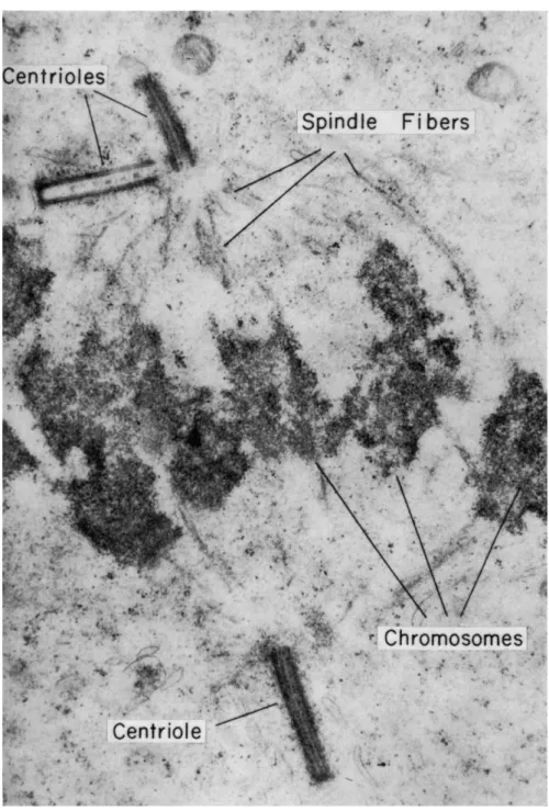
FIG. 2. Electron micrograph of a mitotic spindle from the spermatocyte of the domestic fowl. (From Bloom and Fawcett, 1962, p. 36.)
552 SHINYA INOUÉ
"spindle" is isolated from the cell in division (Mazia and Dan, 1952;
Kane, 1962).
The fibrous structures of the mitotic apparatus (Fig. 1) as generally depicted in textbooks of cytology have been observed in fixed and stained slides since the last century, as also recently by electron micros- copy (Fig. 2; also see article by Roth in this Symposium). T h e functions of these fibrous structures have been variously discussed (see summaries by Schräder, 1953; Mazia, 1961), but there have also been numerous debates as to whether the fibers really existed in living cells. The grounds for the debate have been chiefly that (1) with careful fixation the fibers sometimes could not be demonstrated (see Porter and Machado, 1960, as an example at the electron-microscopic level), whereas with poor fixation they appeared coarser and (2) chromosomes exhibited strange mitotic movements which could not be explained by the simple pulling or pushing by spindle fibers (Östergren, 1949; Schräder, 1953).
As to the reality of these fibers in living cells, I have proved over the years by the use of sensitive polarizing microscopes (Inoué, 1961) that fibers as depicted by the best classic work (e.g., Bêlar, 1929a,b) in fact do exist in a wide variety of healthy dividing cells (Inoué, 1953; Inoué movie, 1960b; Inoué and Bajer, 1961). Additional evidence along this line is also presented in this paper.
The main features which are stressed in this paper are the following:
(1) The spindle fibers are not static structures but exist in a dynamic state of flux; (2) the fibers are oriented and organized by "centers" (Bo- vert, 1888; Wilson, 1928) such as centrioles and kinetochores and other organelles; these will be referred to as "orienting centers"; (3) depend- ing on the activity of such centers and the physiological state of the cell, the spindle fibers can be readily built up, broken down, or reorganized.
The same material can, therefore, be made into one kind of fiber or another or transformed from one type of fiber to another, depending on which center happens to be active at that time.
Description of Spindle Birefringence
Very broadly speaking, there are two types of spindles, one with dis- crete centrioles and the other without (Fig. 3). Most commonly in animal cells one encounters the former type and in plant cells the latter.
In the animal cell spindle with centrioles the birefringence of the spindle fibers is stronger, and the fibers converge, toward the centrioles and toward the kinetochores. T h e biréfringent fibers between kineto- chores and the centrioles we shall call the "chromosomal fibers." T h e biréfringent fibers running from pole to pole we shall call "continuous fibers."
In spindles of plant cells, lacking centrioles, asters are generally miss- ing, and there may or may not be convergence toward the poles. Whether or not there is convergence, the birefringence of the chromosomal fibers is invariably weak at the pole. The birefringence is strongest adjacent to the kinetochores and gradually becomes weaker toward the poles.
Continuous fibers are generally present.
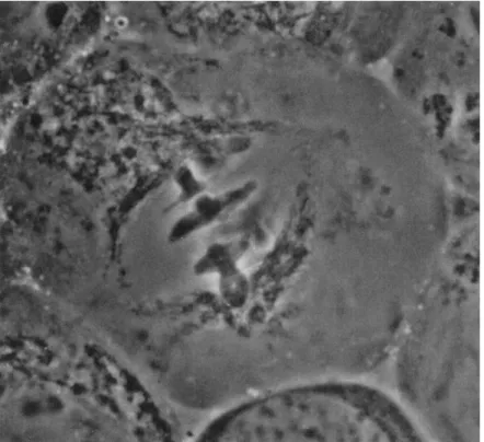
If a living cell, for example, a grasshopper spermatocyte (Fig. 4), is observed under a phase-contrast microscope, one may see the centrioles in an appropriate focus. The spindle region is outlined by mitochondria
FIG. 3. Schematic diagram of mitotic spindle (a) with centrioles and (b) without.
(Modified from Schräder, 1953.)
which are often elongated and surround the spindle as a sheath. T h e region which should show spindle fibers, however, generally appears structureless under the phase-contrast microscope. "Structureless" here means that there is not an adequate optical path difference (owing in this case to difference of refractive index between the fibers and their surroundings) to produce detectable contrast. It is significant that in living cells in general the spindle fibers do not exhibit such refractive index difference unless the cell is fixed, exposed to acid (Lewis, 1923), or otherwise maltreated. Exceptions to this generalization are found in the flagellate spindle (Cleveland, 1953, 1963), in mite eggs (Cooper, 1941), and occasionally in various other healthy cells just at the onset of anaphase.
554 SHINYA INOUÉ
Observing a living cell with a sensitive polarizing microscope, one can clearly see these fibers in regions which appear empty under the phase-contrast microscope. In Fig. 5 we see a plant cell, a pollen mother cell of the Easter lily, Lilium longiflorum, in anaphase. The spindle converges toward the poles somewhat, but the birefringence of the chromosomal fibers is strongest adjacent to the kinetochores and weaker
FIG. 4. Living grasshopper spermatocyte (Chloealtis genicularibus) as seen with a phase-contrast microscope. Notice early anaphase chromosomes still at equator and mitochondria outlining the spindle. Half-spindles appear empty, and continuous and chromosomal fibers are not visible. (Courtesy, Professor K. Shimakura, Faculty of Agri- culture, Hokkaido University, Sapporo, Japan.)
toward the poles. T h e continuous fibers of this stage are less biréfringent and not readily detectable. During anaphase movement, the birefring- ence of the chromosomal fibers adjacent to the kinetochore remains strong and the fibers become shorter but not detectably thicker. In late anaphase, continuous fibers can be seen again by their increased bire- fringence.
As an example of an animal cell, Fig. 6 shows the first maturation division spindle in a oocyte of a marine worm, Chaetopterus pergamen-
fringence adjacent to the helical chromosomes. (Original photo.)
FIG. 6. Biréfringent spindle fibers in a living oocyte of a marine worm, Chaetop- terus pergamentaceiis. Egg centrifuged to move away the biréfringent yolk granules.
(From Inoué, 1953.)
555
556 SHINYA INOUÉ
Ulceus. This cell has been centrifugée! to remove the strongly biréfring- ent yolk granules. Not only do the chromosomal fibers converge to the kinetochores of the chromosomes, but they also converge to the poles, where the birefringence of the spindle fibers and astral rays is strong.
Owing to the action of the compensator, the positive birefringence of the spindle fibers and astral rays is depicted brighter than the back- ground in one quadrant and darker in the opposite quadrant.
Changes in Birefringence during Mitosis
The appearance, disappearance, and general behavior of the spindle fibers and the change in their birefringence during mitosis are evidence for their dynamic nature. Figures 7 through 14 show a series of low- magnification photographs of the developing eggs of a West Coast jelly- fish, Halistaura cellularia. These eggs are so clear in ordinary light that one can distinctly see the individual sperm on the other side of the egg.
The contrast within the cytoplasm seen with polarized light is due to walls of vacuoles which fill most of the cytoplasm and the material in-between the vacuoles. The peripheral birefringence is apparently due to the cortical layer which without the birefringence is difficult to dif- ferentiate optically.
The nucleus is very difficult to see in interphase, but, as the cell pre- pares for prophase, it suddenly becomes clear for it swells and a biré- fringent (spindle) material appears around it. T h e breakdown of the nuclear membrane is accompanied by the development of a small spindle and asters which appear black or white, depending on the orien- tation of their fibers relative to the compensator axes. After the meta- phase plate is formed, the chromosomes are gradually pulled apart. The spindle length increases, and the over-all birefringence diminishes, es- pecially between the separating chromosomes. This diminution of bire- fringence was observed as early as 1937 by W. J . Schmidt in Giessen and later studied more in detail by Michael Swann in Edinburgh. Schmidt concluded in 1939, after he decided that the birefringence was due not to chromosomes but to spindle fibers, that the drop of birefringence in the "half-spindle" represented a folding of protein chains. Swann (1951) measured the distribution of birefringence in dividing sea urchin eggs and concluded that a disorienting substance was released from the chromosomes. We should notice, however, that under this magnification one is not able to resolve spindle fibers nor to determine the exact loca- tion of the chromosomes. We can, in fact, show with improved resolu- tion that Swann's postulate was based on erroneous assumptions con- cerning the microscopic structure of the spindle (Inoué and Bajer, 1961).
Spermatocytes of the grasshopper, Dissosteira Carolina, are shown in Figs. 15 to 26. By prometaphase one clearly sees the strongly biréfringent chromosomal fibers attached to the kinetochores, the convergence of the spindle fibers and the biréfringent asters at the poles, and weakly biré- fringent astral rays attached at the poles.
As seen in the time-lapse motion picture to be described below, it is important to note that these fibers are not simply static, but in meta- phase and especially prometaphase, their birefringence fluctuates as though one were observing northern lights. This fluctuation is a vivid expression of the dynamic equilibrium and presumably reflects the varia- tion in the amount of oriented material going in and out of the fibers all the time.
At anaphase (Figs. 18 to 21), we see the biréfringent chromosomal fibers, the chromosomes, and some sign of the continuous fibers in- between. The mitochondria which have lined up outside of the spindle also become strongly positively biréfringent in late anaphase (Figs. 19 to 24, 26).
Figures 27 to 30 show a pollen mother cell of Lilium longiflorum, previously centrifuged to move away the biréfringent light-scattering granules. In prometaphase, the biréfringent continuous fibers can be seen to run between the poles past the chromosomes (Inoué, 1953). As metaphase approaches, the chromosomal fibers take on a stronger bire- fringence, especially adjacent to the kinetochores; this obtains through- out division (Figs. 27 and 28). T h e continuous fibers, whose birefringence is very weak during early anaphase, become more strongly biréfringent again in late anaphase and eventually become transformed into phrag- moplast fibers in telophase (Figs. 29, 30). Within the phragmoplast thus formed, little vacuoles accumulate, oscillate parallel to the fibers, and finally merge to form the new cell plate (Fig. 30).
We thus see the transformation of the birefringence from continuous fibers to chromosomal fibers, back to continuous fibers again, and finally into phragmoplast fibers (Inoué, 1953).
In the African blood lily, Haemanthus katherinae,2 we see birefrin- gence around the nucleus in the so-called "clear zone" before the nuclear membrane breaks down (Fig. 31). As soon as the nuclear membrane breaks down, this birefringence cannot be distinguished from that now present within the nuclear area (Fig. 32). In other words, this whole area becomes biréfringent, occupying the same area and with the same magni- tude and positive sign as the spindle, showing longitudinal orientation
2 This part of the work was done in collaboration with Dr. Andrew Bajer of K r a k o w , Poland; see Inoué and Bajer, 1961.
SHINYA INOUÉ
FIGS. 7 to 14. Successive divisions in the egg of a jellyfish, Halistaura cellularia. Spindles and astral rays show positive bire- fringence, the cortex negative birefringence with respect to its tangent. The first cleavage of this egg was suppressed during telophase by cold. (Original photos.)
SHINYA INOUÉ
-Ό 'Ό Ο
•2 w !Τ cS ·2 3 Ω C
§ I
.S -S§ .s-â
* ^ -g
Χ ! ^ Ο
«ι ϋ «, _ 4_> CB —'
£ ~ Λ ο
* g
.a
ο S | £ eufaß g «u Ί3
•S ο g .s ο *g bc*G
Λ G η
Μ «g 'G S.
5
β .Sa
u .2 Ώ Ω
§ I I
I
•£ _5 Ä «»
Ο « 5 /3 υ s S 'S Ά ω u α ,Η -g ° ύ .§ -3 .2 S ? ο -SS
. 5 Ο *- >
? .§ 2 §
Λ S s -
g Q I ^
«Ε s-T 1(_ c
£ fr « S
.S Λ :^ Λ Λ
S s S
60 3s
- o ft SÎ
•S w c 3
ζΛ Ο Λ g
ÎC; S ω ο et Ϊ Ν
2 5 /3 s »
« « ö .SP
^ £ χ ^
«S d W 0
£ ,- -Β -S s
•2 c g
SHINYA INOUÉ
FIGS. 27 to 30. Single frames printed from a 16-mm time-lapse motion picture of division in the pollen mother cell of the Easter lily, Lilium longiflorum. Chromosomal fiber birefringence prominent in Figs. 27 and 28. Continuous fibers transforming into phragmoplast fibers and vacuoles merging to form the cell plate, Figs. 29 and 30. Short diameter of cell, 50 μ. (Original photos.)
SHINYA INOUÉ
FIGS. 31 to 40. Development of the biréfringent spindle fibers and anaphase movement in the endosperm cell of the African blood lily, Haemanthus katherinae. Pairs of photos are taken with opposite compensation. Same magnification as Figs. 41 to 48. See Fig. 31 for 10 μ scale. (From Inoué and Bajer, 1961.)
566 SHINYA INOUÉ
FIGS. 41 to 48. Chromosomes and biréfringent spindle fibers in flattened cells of Haemanthus katherinae. T h e strong birefringence of the chromosomal fibers adjacent to the kinetochore is indicated by arrows in Figs. 41 and 42. Continuous fibers visible
Figs. 45 and 46. Phragmoplast fibers visible in Figs. 47 and 48. Same magnification Figs. 31 to 40. A 10 μ scale is shown on Fig. 31. (From Inoué and Bajer, 1961.)
568 SHINYA INOUÉ
For legend see pages 566 and 567.
For legend see pages 566 and 567.
570 SHINYA INOUE
of positive biréfringent material in agreement with the postulate of Wada (1950). Continuous fibers thus appear, then chromosomal fibers, then gradually the chromosomes are moved up and down until the meta- phase plate is formed (Figs. 33 to 35). Here is another example of trans- formation of the clear-zone fiber to continuous spindle fibers and to chromosomal fibers (Inoué and Bajer, 1961).
Continuing the series (Figs. 36 to 40) throughout anaphase, the bire- fringence of the chromosomal fibers remains strongest adjacent to the kinetochore region. In these unflattened cells, the individual spindle fibers, although visible, are difficult to distinguish, but if we flatten the endosperm cells (Figs. 41 to 48) we clearly see the individual continuous fibers, the pairs of chromosomes, and their chromosomal fibers. T h e flattened endosperm cells continue to divide, and in the movie, the un- winding of the chromosome pairs and their pulling apart by the chromo- somal fibers can be clearly seen.
The continuous fibers in late anaphase (Fig. 45) become more strongly biréfringent in the mid-region and turn into phragmoplast fibers (Figs. 46 to 48); small vacuoles accumulate at the equator amidst the phragmoplast fibers and form the cell plate. As the cell continues to divide, the phragmoplast birefringence extends toward the cell periphery where the new cell plate is laid down.
Motion Picture of Living Cells in Division
A time-lapse motion picture film3 showing spindle fiber birefringence during division of various animal and plant cells was shown at this Symposium. The pictures taken with the sensitive polarizing microscope developed by the author demonstrate the following points.
1. In the eggs of the jellyfish, Halistaura cellalaria, flattening the cell places the spindle in a single plane of focus and reveals the syn- chrony of cell division. T h e nucleus normally lies at the periphery of the cell; flattening the cell may displace the nucleus and, therefore, the final position of the spindle. Regardless of which direction or where the spindle comes to lie, the cell divides at right angles to the axis of the
3 T h e film required exposure times of from 5 to 20 sec/frame because of the low brightness of the specimen. T h e interval between frames was identical with the exposure. This film is now available at cost (Inoué, 1960b).
FIGS. 49 and 50. First and second cleavage in the egg of a jellyfish, Aglantha digitale. T h e positive birefringence of the spindle fibers and the negative birefringence of the vacuolar walls provide contrast in these illustrations. T h e vacuolar walls are deformed and point to the astral centers at these stages. (Original photos.)
572 SHINYA INOUÉ
spindle, and the furrow begins to form at the surface nearest the spindle.
Comparison with the sequence of cleavage in a noncompressed egg (Figs. 7 to 14) demonstrates that the birefringence of the spindle and its relation to the cleavage furrow is not altered by compression.
2. In the eggs of another jellyfish, Aglantha digitale (Figs. 49 and 50), which are even more transparent than those of the previous species, as the spindle and asters develop, the cytoplasmic vacuoles become tear- shaped, their apices pointing toward the astral centers. Prior to the separation of the chromosomes, a striking oscillation (or "rocking") of the whole spindle of the kind reported in nematode eggs (Ziegler, 1895) and in chick fibroblasts (Hughes and Swann, 1948) is observed. In the Aglantha eggs, the vacuoles change back and forth between tear-drop and round shape as the spindle rocks, as if the astral rays were attached to them and exerting a pull. After mitosis, the vacuoles regain their spherical shape.
3. In spermatocytes of the grasshopper, Dissosteira Carolina (Figs. 15 to 26), the "northern lights" flickering of individual chromosomal fibers is clearly demonstrated in prometaphase (Figs. 15 to 19). Just prior to anaphase, the birefringence of the chromosomal fibers becomes stronger.
While the chromosomes are pulled apart and the chromosomal fibers shorten, their birefringence remains strong until late anaphase (Figs. 18 to 21). T h e birefringence of the astral rays (Figs. 15 to 22) and of the continuous fibers in the interzonal region (Figs. 19 to 21) is much weaker than that of the chromosomal fibers. T h e mitochondrial sheath acquires a very strong positive birefringence in telophase (Figs. 19 to 23 and 26) and often becomes twisted in late telophase (Fig. 24).
4. In pollen mother cells of Lilium longiflornm (Figs. 27 to 30), chromosomal fiber birefringence becomes strongest just at the onset of anaphase, the birefringence being strongest adjacent to the kinetochores (Fig. 27). The chromosomal fibers retain their strong birefringence as they lead the chromosomes to the poles (Fig. 28). Continuous fiber bire- fringence is initially weak but becomes stronger during the later stages of anaphase, especially near the equatorial region (Fig. 29) where it finally transforms into the phragmoplast (Fig. 30). At this time, vacuoles come together in the equator and merge to form the cell plate—a structure which is also biréfringent but with a different character.
5. Endosperm cells of Haemanthus katherinae, unlike most other types of plant cells, are not surrounded by conventional cell-wall material and may thus be flattened like an animal cell in tissue culture (see foot- note 2 on p. 557). In the flattened cells, the relatively large chromosomes are sufficiently dispersed (Figs. 41 to 48) so that the individual chromo- somes, their kinetochores, the chromosomal and continuous and the
4 Original report.
phragmoplast fibers are all clearly visible in polarized light. T h e chromo- somes are untwisted and pulled at their kinetochores by the chromosomal fibers.
As stated previously, the fibers in all these cells are not visible with phase-contrast microscopy in untreated dividing cells.
The motion picture of living cells in division shows not only the reality of the fibers but how the spindle fibers appear to transform from one type of fiber to another, depending on the sequence of events. T h e various spindle movements and the fluctuation in the fiber birefringence which also reflect the dynamic state of these fibers in the dividing cells are demonstrated.
Effect of Low Temperature4
Low-temperature experiments further demonstrate the dynamic state of the spindle fibers.
A pollen mother cell of L. longiflorum in early anaphase was chilled to 3°C (Fig. 51) resulting in complete disappearance of spindle bire- fringence. On raising the temperature to 27°C, the birefringence re- appeared, at first as continuous fibers; after about 8 min, both the con- tinuous and chromosomal fibers were fully reorganized. This process of spindle re-formation after chilling is faster than, but otherwise similar to, spindle formation at prometaphase in normal mitosis.
After re-establishment of the spindle birefringence and structural organization, the chromosomes began to move again and completed mitosis.
If we cool a cell at a later stage in anaphase (Fig. 52), by which time the chromosomes have become stretched but not yet separated, the chromosomes are found to "recoil" as the spindle birefringence disap- pears. In this case, when the temperature is raised, biréfringent chromo- somal fibers reappear first. Then the chromosomes become stretched, the kinetochore distance increases, and the cell completes mitosis, but only after the size and birefringence of the chromosomal fibers have fully recovered.
There thus exists a direct correlation between spindle birefringence and deformation and movement of the chromosomes (also see Inoué, 1952a on the effect of colchicine).
This temperature treatment can be performed on many types of cells over and over again during a single division. Thus one can interrupt the mitotic process experimentally without impeding the ability of the spindle fibers to become reorganized again.
574 SHINYA INOUÉ
Figure 53A shows the biréfringent spindle in a Chaetopterus perga- mentaceus egg. When this cell is chilled, the spindle becomes thin and then quickly loses its birefringence. Upon returning the cell to room temperature, a miniature spindle reappears in the original site, quickly migrates to the cell surface, and grows back again (Fig. 53B), first form-
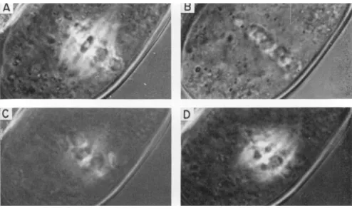
FIG. 51. Cold treatment of a pollen mother cell of Lilium longiflorum in early anaphase. T h e birefringence of the spindle fibers disappears at 3°C and returns rapidly at 27°C. A, Before treatment; B, after 6.5 min at 3°C; C, after 3.5 min at 27°C; D, after 8 min at 27°C. (Original photos.)
ing continuous fibers with chromosomes randomly arranged (Fig. 53C and D), and then eventually forming the metaphase plate and more prominent chromosomal fibers (Fig. 53E and F). This cycle takes about
15 min at 22°C. In the Chaetopterus egg which is in a metaphase arrest, such a cycling can be done on the same cell for as many as ten times without losing the spindle material (Inoué, 1952b).
Thermodynamic Studies
By measuring the equilibrium birefringence at various temperatures, one can gain insight into the mechanism of orientation of the molecules making up the fibers.
When the temperature of the cell is changed quickly, the bire- fringence of the spindle reaches an equilibrium value after a few minutes. T h e birefringence of the spindle fibers is then measured and can be plotted as a function of temperature, as illustrated in Fig. 54.
FIG. 52. Cold treatment and recovery of an anaphase Lilium cell. Same cell as shown in Fig. 51, but at a later stage. The chromosomes are extended again after spindle birefringence and organization are restored (F). A, Before treatment; B, after 4 min at 4°C; C, recovery after 1 min; D, recovery after 2 min; Ε, recovery after 4 min; F, recovery after 13 min. (Original photos.)
SHINYA INOUÉ
FIG. 53. Cold treatment and recovery of the metaphase spindle in the oocyte of Chaetopterus pergamentaceus. A, Before treat- ment; B, ca. 30 sec after treatment at 4°C; B-F, re-establishment of spindle birefringence and fiber organization. (Original photos.)
578 SHINYA INOUÉ
As shown in the figure, the birefringence increases as the temperature is raised, at least up to an optimum. This is, at first glance, contrary to intuitive thermodynamics, since one would expect randomization of the spindle elements with the increased temperature. But it does agree
Γ(π\μ)
with "viscosity" measurements on dividing cells where the gel structure is weakened either with high hydrostatic pressure or with low-tempera- ture treatment (Marsland et aL, 1960).
Figure 55 shows a thermodynamic plot of the data shown in Fig. 54. Assuming that the total concentration (A0) of orientable material is constant and that the birefringence (B) represents the con-
* n 0 °000 ΰ00 c=, ο
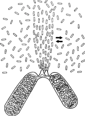
FIG. 56. Schematic diagram of orientation equilibrium of chromosomal fiber.
Fiber orientation is organized by kinetochores of chromosomes. T h e oriented material is in a temperature-sensitive equilibrium with the nonoriented material surrounding it. (Original.)
centration of material oriented at that temperature, one can draw an equation: A0 — B^±B. Plotting the log of the equilibrium constant k — (B/A0) — Β against the reciprocal of the absolute temperature, one would expect a linear relationship if one were dealing with an isolated equilibrium system in which the equilibrium between the oriented and non-oriented material is a function of temperature.
A straight-line relationship has in fact been observed and the change in free energy, the heat evolved per mole, and the entropy change have
580 SHINYA INOUÉ
been calculated by Dr. Manuel Morales (Inoué, 1959, see correction in 1960a).
The result of this analysis showing a great increase of entropy at higher temperatures is similar to that for globular to filamentous actin transformation (Asakura et al., 1960) and to that for tobacco mosaic virus A protein association (Lauffer, 1958; Ansevin and Lauffer, 1963) and may suggest a common mechanism (see Inoué, 1959; also Kauzmann,
1959, Singer, 1962, for pertinent reviews).
The orientation equilibrium of the spindle elements is schematically shown in Fig. 56. On the chromosomes, the kinetochores act as orienting centers which determine the localization of the oriented material. There is a pool of unoriented or perhaps slightly oriented material around the chromosomal fibers, depending on the stage, which can be transformed into oriented chromosomal fibers. Conversely, the chromosomal fiber material may become disoriented. This equilibrium is temperature- sensitive.
Orienting Centers—Ultraviolet Microbeam Experiments5 Experiments were done to test further the notion of orienting centers and orientation equilibrium. If we somehow stop the action of the orienting center, then the oriented material should go into the dis- oriented state. If we could locally disorient the material distal to the orienting center, then the material proximal to it should not be affected because the orienting center remains, but distally the orientation and the birefringence should disappear.
If we remove the disorienting influence, we should again recover the orientation by the incorporation of the nonoriented material into the oriented state. These predictions have been fulfilled by an experiment in which the spindle fibers were irradiated with a small spot of ultra- violet light.
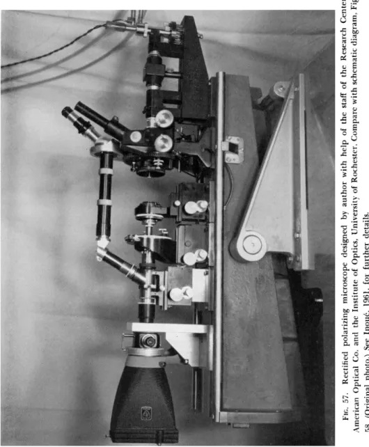
Figure 57 shows a part of the instrumentation, the high extinction polarizing microscope built onto a stable optical bench. As shown in the schematic diagram (Fig. 58), the visible light source is at the top of the instrument. The light coming through the polarizer, a compensator, and condenser, illuminates the object. Through the objective, analyzer, and eyepiece, we see the birefringence of the specimen. A mercury arc lamp (Osram, HBO-200) shines the ultraviolet light onto a small first surface mirror placed in front of the visible source diaphragm. T h e
5 This section reports original work carried out in collaboration with Dr. Hidemi Sato of the Department of Cytology, Dartmouth Medical School.
FIG. 57. Rectified polarizing microscope designed by author with help of the staff of the Research Center, American Optical Co. and the Institute of Optics, University of Rochester. Compare with schematic diagram, Fig. 58. (Original photo.) See Inouc, 1961, for further details.
582 SHINYA INOUÉ
image of the mirror is projected into the specimen plane and delineates the area of the specimen receiving the ultraviolet exposure.
Figure 59 shows a prophase Haemanthus endosperm cell in which a small spindlelike body spontaneously developed. This body was irradi- ated in its mid-region (Figs. 59 and 60), where the birefringence instantly disappeared. After irradiation, the two halves that remained biréfring- ent quickly came together (Fig. 61) and then merged to form another single spindle-shaped body (Fig. 62).
i Y >- LIGHT SOURCE
POLARIZER
RECTIFIED r^S <~
CONDENSER
UV- POLARIZER
REFLECTING CONDENSER 3 STAGE
RECTIFIED OBJECTIVE
ANALYZER
FIG. 58. Schematic diagram of (polarized) ultraviolet-microbeam irradiating system which was incorporated into the rectified polarizing microscope shown in Fig. 57.
(Original.)
When a cell in early anaphase is irradiated so that the ultraviolet beam covers the basal regions of some chromosomal fibers and their kinetochores, the whole length of the chromosomal fibers including the distal unirradiated part is lost and does not reappear for a long time (Figs. 63 and 64).
If instead we irradiate only the chromosomal fibers and avoid the kinetochores (Figs. 65 and 66), we find the birefringence has disappeared from the irradiated region as well as from the distal part, but is still intact between the kinetochores and the irradiated region (Fig. 67). In
the course of a few minutes the birefringence of the irradiated and distal regions returns (Fig. 68).
These experiments not only support the notion of the dynamic equilibrium and the activity of the organizing centers, but provide us with a means for establishing the locations of organizing centers in general.
For example, in the case of the phragmoplast, the birefringence is stronger next to the cell plate, but the presence of an organizing center in that region of the phragmoplast had never been suspected.
However, when we irradiated a narrow diagonal area of the phrag- moplast, the birefringence was lost from the full length of the phragmo- plast fibers where the cell plate itself was irradiated (Figs. 69 to 71). If only the distal parts of the fibers, and not the cell plate, were irradiated, the portions of the fibers nearer the cell plate remained biréfringent.
The birefringence of the distal parts eventually returned.
Figure 72 shows a late phragmoplast in which the birefringence is now confined to the peripheral regions; in the middle, the cell plate has already been laid down. When the center of one phragmoplast is irradi-
ated, as shown in Fig. 73, we find that all birefringence is gone from the phragmoplast fibers on that side (Fig. 74). Here then exists an unsus- pected orienting center, the activity of which moves progressively with
the phragmoplast as it moves toward the cell periphery.
As shown by these experiments, the phragmoplast fibers behave very much like chromosomal and continuous spindle fibers, not only in their birefringence but in terms of their response to ultraviolet irradiation.
This provides further support for the notion that the composition and molecular organization of the phragmoplast fibers is similar or identical to those of the chromosomal fibers, continuous fibers, and the clear-zone fibers.
Conclusion
In summary, then, we have seen in a wide variety of living, dividing cells "fibers" which could explain, at least topologically, the movement of chromosomes. This does not explain the molecular mechanism of chromosome movement yet, but at least we know there is an anisotropic distribution of material which could one way or another account for the development of mechanical forces required for pulling (or pushing) the chromosomes, most likely dependent on the shift of orientation equilib- rium.
It may well be that as material is removed from the chromosomal fibers, while the organizing centers are still active, the fibers shorten in length simply to reach a new orientation equilibrium. The continuous
584 SHINYA INOUÉ
FIGS. 59 to 6 2 . Irradiation of cytoplasmic spindle spontaneously formed in pro- phase of a Haemanthus endosperm cell. Figure 59, before irradiation; Fig. 6 0 , the dark bar indicates the area irradiated with ultraviolet; Fig. 6 1 , 1 min after irradiation
—the irradiated area lost birefringence, the nonirradiated parts have moved together to close the gap; Fig. 62, ca. 5 min after irradiation, the spindle has become reorganized into a single body again. (Original photos.)
586 SHINYA INOUÉ
FIGS. 63 and 64. Ultraviolet-microbeam irradiation of chromosomal fibers and kinetochores in one-half spindle of Haemanthus endosperm cells. The irradiating ultraviolet beam covered a square area between the tips of the arrows and the
nearby kinetochores. Figure 63, before, Fig. 64, after irradiation. No recovery of chromosomal fibers can be seen. (Original photos.)
588 SHINYA INOUÉ
FIGS. 65 to 68. Ultraviolet-microbeam irradiation of chromosomal fiber only.
When kinetochores are not irradiated (Figs. 65 and 66), birefringence disappears from the area irradiated and also the distal portions of the chromosomal fibers (Fig. 67).
They both rapidly grow back (Fig. 68) if the ultraviolet exposure is not too great.
{Original photos.)
590 SHINYA INOUÉ
Fies. 69 to 71. Ultraviolet-microbeam irradiation of phragmoplast in the endosperm cell of Haemanthus katherinae. The area irra- diated ran diagonally over the phragmoplast as shown by the shadow of the ultraviolet mirror in Fig. 70. Figure 69, before irradiation. Figure 71 shows no birefringence of the phragmoplast fibers where the cell plate was irradiated. (Original photos.)
FIGS. 72 to 74. Ultraviolet irradiation of phragmoplast to one side of Haemanthus endosperm cell. Figure 72, before irradiation; Fig. 73 shows where right cell plate is irradiated; Fig. 74, whole length of right phragmoplast fibers have lost birefringence. (Original photos.)
592 SHINYA INOUÉ
fibers could then acquire more material and support the compressive force imposed on the spindle by the chromosomal fibers. It is even pos- sible that by adding more material into the oriented state the fibers can be made to elongate as well. In any event, there is no question that in- side the living cells these biréfringent fibers are present; they are or- ganized by centers and exist in a highly dynamic state.
REFERENCES
Ansevin, A. T., and Lauffer, M. A. (1963). Polymerization-depolymerization of tobacco mosaic virus protein. I. Biophys. J. 3 (3), 239-251.
Asakura, S., Kasai, M., and Oosawa, F. (1960). T h e effect of temperature on the equi- librium state of actin solutions. / . Polymer Sei. 44, 35-49.
Bélaf, Κ. (1929a). Beiträge zur Kausalanalyse der Mitose. II. Untersuchungen an den Spermatocyten von Chorthippus (Stenobolhrus lineatus) Panz. Wilhelm Roux1
Arch. Entwicklungsmech. Organ. 118, 359-484.
Bélaf, Κ. (1929b). Beiträge zur Kausalanalyse der Mitose. III. Untersuchungen an den Staubfadenhaarzellen und Blattmeristemzellen von Tradescantia virginica. Z.
Zellforsch. Mikroskop. Anat. 10, 73-134.
Bloom, W., and Fawcett, D. W. (1962). A Textbook of Histology," 8th ed. Saunders, Philadelphia, Pennsylvania.
Boveri, T. (1888). "Zellen Studien." Fischer, Jena, Germany.
Cleveland, L. R. (1953). Studies on chromosomes and nuclear division. IV. Photomi- crographs of living cells during meiotic divisions. Trans. Am. Philos. Soc.
[N.S.] 43, 805-869.
Cleveland, L. R. (1963). Functions of Flagellate and Other Centrioles in Cell Re- production. In "The Cell in Mitosis" (L. Levine, ed.), pp. 3-30. Academic Press, New York.
Cooper, K. W. (1941). Visibility of the primary spindle fibers and the course of mitosis in the living blastomeres of the mite Pediculopsis graminum Reut. Proc. Natl.
Acad. Sei. (U.S.) 27, 480-483.
Engelmann, Th. W. (1875). Contractilität und Doppelbrechung. Arch. Ges. Physiol.
11, 432-464.
Engelmann, Th. W. (1906). Zur Theorie der Contractilität. Sitzher. Kgl. Preuss. Akad.
Wiss., pp. 694-724.
Hughes, A. F., and Swann, M. M. (1948). Anaphase movements in living cell. A study with phase contrast and polarized light on chick tissue culture. / . Exptl. Biol.
25, 45-70.
Inoué, S. (1952a). The effect of colchicine on the microscopic and submicroscopic structure of the mitotic spindle. Exptl. Cell Res. Suppl. 2, 305-318.
Inoué, S. (1952b). Effects of temperature on the birefringence of the mitotic spindle.
Biol. Bull. 103, 316.
Inoué, S. (1953). Polarization optical studies of the mitotic spindle. I. The demonstra- tion of spindle fibers in living cells. Chromosoma 5, 487-500.
Inoué, S. (1959). Motility of cilia and the mechanism of mitosis. Rev. Mod. Phys.
31, 402-408.
Inoué, S. (1960a). On the physical properties of the mitotic spindle. Ann. N.Y. Acad.
Sei. 90, Article 2, 529-530.
Inoué, S. (1960b). "Birefringence in Dividing Cells." Time-lapse motion picture. Availa- ble at cost from Geo. W. Colburn Laboratory, Inc., Chicago 6, Illinois.
Inoué, S. (1961). Polarizing microscope. Design for maximum sensitivity. In "Encyclo- pedia of Microscopy" (George Clark, ed.), pp. 480-485. Reinhold, New York.
Inoué, S., and Bajer, A. (1961). Birefringence in endosperm mitosis. Chromosoma 1 2 , 48-63.
Inoué, S., and Dan, K. (1951). Birefringence of the dividing cell. / . Morphol. 8 9 , 423-455.
Inoué, S., and Hyde, W. L. (1957). Studies on depolarization of light at microscope lens surfaces. II. T h e simultaneous realization of high resolution and high sen- sitivity with the polarizing microscope. / . Biophys. Biochem. Cytol. 3 , 831-838.
Inoué, S., and Kubota, H. (1958). Diffraction anomaly in polarizing microscopes.
Nature 1 8 2 , 1725-1726.
Kane, R. E. (1962). The mitotic apparatus: isolation by controlled pH. / . Cell Biol.
12, 47-56.
Katchalsky, A. (1963). Mechanochemistry of cell movement. In "Symposium on Non- Muscular Contractions in Biological Systems," 7th Annual Meeting of the Bio- physical Society.
Kauzmann, W. (1959). Some factors in the interpretation of protein denaturation.
Advan. Prot. Chem. 1 4 , 1-64.
Kubota, H., and Inoué, S. (1958). Diffraction image in the polarizing microscope.
/. Opt. Soc. Am. 4 9 , 191-198.
Lauffer, M. A. (1958). Polymerization-depolymerization of tobacco mosaic virus protein.
Nature 1 8 1 , 1338-1339.
Lewis, M. R. (1923). Reversible gelation in living cells. Bull. Johns Hopkins Hosp.
3 4 , 373-379.
Marsland, D., Zimmerman, A. H., and Auclair, W. (1960). Cell division: experimental induction of cleavage furrows in the eggs of Arbacia punctulata. Exptl. Cell Res.
2 1 , 179-196.
Mazia, D. (1961). Mitosis and the physiology of Cell Division. In "The Cell" (J.
Brächet and A. E. Mirsky, eds.), Vol. 3, pp. 77-412. Academic Press, New York.
Mazia, D., and Dan, K. (1952). T h e isolation and biochemical characterization of the mitotic apparatus of dividing cells. Proc. Natl. Acad. Sei. (U.S.) 3 8 , 826-838.
Mitchison, J . M. (1953). A polarized light analysis of the human red cell ghost.
/. Exptl. Biol. 3 0 , 379-432.
Östergren, G. (1949). Luzula and the mechanism of chromosomal movements. Here- ditas 3 5 , 445-468.
Porter, K. R., and Machado, R. D. (1960). Studies on the endoplasmic reticulum: IV.
Its form and distribution during mitosis in cells of onion root tip. / . Biophys.
Biochem. Cytol. 7 , 167-180.
Schmidt, W. J . (1934). Polarisationsoptische Analyse des submikroskopischen Baues von Zellen und Geweben. In "Handbuch der biologischen Arbeitsmethoden" (E.
Abderhalden, ed.), See. 5, Part 10, p. 435. Urban und Schwarzenberg, Berlin and Vienna.
Schmidt, W. J . (1937). "Die Doppelbrechung von Karyoplasma, Zytoplasma und Meta- plasma," Protoplasma-Monographien, Vol. 2. Gebrüder Borntraeger, Berlin.
Schmidt, W. J . (1939). Doppelbrechung der Kernspindel und Zugfasertheorie der Chro- mosomenbewegung. Chromosoma 1, 253-264.
Schräder, F. (1953). "Mitosis. T h e Movements of Chromosomes in Cell Division,"
2nd ed. Columbia Univ. Press, New York.
Singer, S. J . (1962). T h e properties of proteins in nonaqueous solvents. Advan. Prot.
Chem. 1 7 , 1-68.
594 SHINYA INOUÉ
Swann, M. M. (1951). Protoplasmic structure and mitosis. II. T h e nature and cause of birefringence changes in the sea-urchin egg at anaphase. / . Exptl. Biol. 28, 434-444.
Swann, M. M., and Mitchison, J . M. (1950). Refinements in polarized light microscopy.
/. Exptl. Biol. 27, 226-237.
Wada, B. (1950). T h e mechanism of mitosis based on studies of the submicroscopic structure and of living state of the Tradescantia cell. Cytologia 16, 1-26.
Wilson, Ε. B. (1928). "The Cell in Development and Heredity," 3rd ed. Macmillan, New York.
Ziegler, H. E. (1895). Untersuchungen über die ersten Entwicklungsvorgänge der Nema- toden. Ζ. Wiss. Zool. 60, 351-410.
DISCUSSION
DR. TERU HAYASHI: DO you get the same equilibrium birefringence values when you lower the temperature as when you raise it, or do you come down a different path?
DR. INOUÉ: Within the limit of experimental error, the equilibrium birefringence seems to fall on the same curve. There is, however, a hysteresis in approaching the equilibrium value.
DR. ALLEN: I would like to pursue the point you made at the beginning and ask whether it really is necessary to have optical anisotropy to have contraction. It is a common laboratory occurrence that supposedly isotropic gels of gelatin con- tract; of course, when they contract, they do so isodiametrically.
DR. INOUÉ: When gelatin contracts isodiametrically, we do know there are fila- ments and that the filaments actually shorten. T h e structural anisotropy is already built in. It depends whether the filaments were randomly arranged or preferentially arranged whether an anisodiametric or isodiametric contraction occurs. Kunitz showed in the 1920's that you can take gelatin or agar and line up the molecules anisotropically; in this case, the responses to swelling and contraction will also be anisodiametric. This was published in J. Gen. Physiol. 13, 565-606.
DR. ALLEN: I brought this up after thinking about Dr. Wolpert's results. He isolates a system of proteins (supposedly contractile) from the ameba at a low tem- perature, lets it warm up in the presence of adenosine triphosphate, and finds stream- ing apparently based on contractile processes. If birefringence is a prerequisite for contraction, then one must assume that the proteins somehow line themselves up as an anisotropic array. If so, this must constitute a self-organizing system.
DR. INOUÉ: Well, if one has a large number of filaments, as I think you yourself well know, they tend to line up as they precipitate out. This is well known in to- bacco mosaic virus. Also, if you take a solution of G-actin and give it conditions that will form F-actin with very long filaments, it will line up spontaneously. So I think simply by increasing the concentration to the point where the form or precipitate comes out, one can expect some degree of alignment, unless there is a constraint that makes them not line up.
DR. ALLEN: By his method of preparation, I would guess the concentration of this material could not be greater than 3 - 4 % by weight. What role do you think the centriole plays in lining up spindle fiber elements?
DR. INOUÉ: I think the plants which generally show no centriole have answered this question for us. In the clear-zone material, simply by having anisodiametric con- densation, one gets a spontaneous alignment, and very often it is true that in plant cells
the orientation of the spindle filaments takes on the orientation or spatial distribution imposed by the nucleus or the cytoplasm.
DR. REBHUN: It appears from your pictures that neither the shape nor the bire- fringence of the chromosomal fibers change, at least during the early part of ana- phase. Is this correct?
DR. INOUÉ: Yes, that is correct.
DR. REBHUN: About how long does this last? In other words, how long can the chromosomal fibers move without changing either of these parameters?
DR. INOUÉ: T h e length is very difficult to judge. In Haemanthus, we have not been able to measure the length because the birefringence tapers off into an imper- ceptible value. In the case of Lilium, one can, by frame-by-frame analysis, measure the position of the spindle poles. In this case, the apparent change of position of the tip is illusory, and I can hardly detect any length change of the over-all spindle in Lilium until very late anaphase or telophase.
DR. REBHUN: HOW about the chromosomal fibers?
DR. INOUÉ: T h e chromosomal fibers obviously must be shorter, and they do not go beyond the poles.
DR. REBHUN: And they do not change birefringence?
DR. INOUÉ: Near the kinetochore, as far as I can tell, they do not change to any measurable extent. W e do not have the exact numbers.
DR. MARSLAND: There appears to be a remarkable similarity between the be- havior of this material and other gel structures. As you probably know, we can cause a dissolution of the spindle with pressure, and it reconstitutes itself after decompres- sion. The type of gel with which we are dealing becomes more disorganized with lowered temperatures and more highly organized with increased temperatures. I would like to point out, also, that perhaps a gel strand is a structure which can contract without changing its diameter. As a gel contracts, it necessarily loses ma- terial by syneresis. This type of contraction is probably one example where you can get contraction of a structure without any change in the diameter of the fiber.
DR. INOUÉ: Well, perhaps some of the chemists would also like to comment on your remark. If syneresis is taking place, simply expelling water without loss of protein matrix, then the refractive index should go up if the mass is not changing.
DR. MARSLAND: There might be quite a residuum of protein material which has not entered into gel structure which might be expressed in syneresis.
DR. INOUÉ: I am sorry; I did not understand.
DR. MARSLAND: When gelation occurs, it does not necessarily involve all of the available material, and part of the fluid which presumably is trapped in the frame- work of the gel may still have quite a residuum of protein material. So that the change in birefringence might be very small.
DR. INOUÉ: I did not mean in birefringence; I meant in total refractility. You are saying that there may be contraction.
DR. MARSLAND: I also meant total refractility. As you are losing one thing, aren't you condensing another? In other words, the syneretic exudate may have a high pro- tein content and huge refractility.
DR. INOUÉ: Does it sound likely?
DR. MARSLAND: Maybe I am wrong, but I do not think so.
CHAIRMAN BISHOP: It sounds like an argument for the Free Discussion. Any other questions?
DR. KAUZMANN: IS this form or intrinsic birefringence, or are they the same thing?