CHAPTER 1
Botulinum Toxin
D A N I E L A . BOROFF AND BIBHUTI R . D A S G U P T A
I. T h e D i s e a s e 1 II. History 4 III. Requirements for Growth and Toxin Production 7
IV. Purification of the Toxin 9 V. Nature of the Crystalline Toxin T y p e A 15
V I . Nature of Toxins of Other T y p e s 24
V I I . Spore Toxin 28 V I I I . Hemagglutinins of Clostridium botulinum 30
IX. Activation Phenomenon 33 X. Effect of Proteolytic Enzymes on the Toxin 37
X I . Specific Chemical Groupings Involved in Toxicity 37
X I I . Site of Action 45 X I I I . Mode of Action of Botulinum Toxin 48
X I V . Botulism as an Infection 49 X V . Toxin as Antigen 52 X V I . Effects of Chemical and Physical Agents on the T o x i n 55
of CI. botulinum
X V I I . Is Botulinum Toxin an Exotoxin? 57 X V I I I . Role of the Toxin in CI. botulinum 59
X I X . Laboratory Detection of Botulinum T o x i n 60
X X . Conclusions 61 References 62
I. T h e D i s e a s e
It would be appropriate to introduce the topic of the toxin of Clostridium botulinum with a brief discussion of the disease, which we know as botulism. Botulism is primarily a result of food poisoning that affects a great variety of animal species in all parts of the world. However, although human outbreaks are relatively rare, the dramatic and often tragic manifestations of botulism have left a deep impression in human history. The disease is a result of the ingestion of food contaminated with the preformed toxin produced by Clostridium botulinum, an anaerobic, sporeforming, gram-positive (in the early stage of growth) bacillus. This toxin is one of the most powerful neuroparalytic poisons. In the purified form, 1 fig of this substance contains about 200,000 minimal lethal doses (MLD) for a 20-gm white mouse. While the human MLD is not known, it is suspected that not much more than 1 /xg of the toxin may be fatal for man (Morton, 1961).
Beside the high death rate among its victims, the disease is made more tragic by its sudden onset, swiftness of course, and the fact that because
1
2 D . A. BOROFF A N D B. R. D A S G U P T A
the central nervous system does not seem to be affected, the victim re
tains consciousness to the last. While definite botulism symptoms exist, such as difficulty in swallowing and breathing, inability to focus one's eyes, and paralysis of the extremities, the disease is difficult to diagnose because at the onset the manifestations of botulism are often confused with symptoms of other diseases and because very few physicians are familiar with its diagnostic aspects. By the time the nature of the illness becomes apparent, it is usually too late for therapy. In botulism the only therapy known is the early administration of the type-specific antiserum, which means that not only must the nature of the disease be established but that the type of toxin (of which at present six serologically distinct entities are known) must be determined. These organisms producing the toxin are ubiquitously spread over all parts of the earth. Their natural hab
itat is in the soil in the form of spores which are extremely resistant to adverse conditions. Upon culturing, the spores develop into motile rods.
Different types of CI. botulinum show distinctive cell morphology. Type A organisms are short and when sporulating present a spindle-like appear
ance. In other types, the spores are subterminal, noticeably distending the portion of the cell in this region.
Although there is a prevalence of certain types of CI. botulinum food poisonings in various localities, this does not rule out the presence of spores of other types in the same locality (K. F. Meyer and Eddie, 1950).
Surveys along the west as well as the east coast of the American continent disclosed the presence of toxigenic strains of CI. botulinum (through the examination of soil, shrimp, and fish) most frequently of types A and C, and occasionally of types B and E (Carroll et al., 1966; Ward et al.y 1967).
Surveys in England and Scotland resulted in the isolation only of type B from many samples collected. Nevertheless, an outbreak which wiped out a whole family in Loch Maree, Scotland, in 1922, was derived from duck paste contaminated with type A toxin. Subsequent investigation by Leigh ton and Buxton (1926) and Heines (1942) demonstrated the pres
ence of botulinum spores of both types A and B in Scottish and English soils. According to Kravchenko and Shishulina (1966), examination of over 4000 samples of soil and waters from five geographic zones of the USSR revealed the presence of CI. botulinum types A, B, C, D, and E, with no correlation between type of outbreak and population density.
In spite of the wide distribution of botulinum spores in nature, the prev
alence of one or the other type of botulinum poisoning in a given locality is apparently attributable to the dietary habits of its human inhabitants.
While human outbreaks are fortunately not too numerous and usually small, the disease among small and large animals, as well as birds, is quite prevalent (Tables I and II). Thousands of migrating birds die yearly
1. B O T U L I N U M T O X I N 3 as they rest and feed on the shallow lakes of bird sanctuaries in Colorado and Utah. Swans, chickens, and pheasants are also highly susceptible.
The disease is a scourge of mink, and, in South Africa, botulism among cattle is of serious economic importance.
T A B L E I
H U M A N I N C I D E N C E , F A T A L I T Y R A T E S , A N D T Y P E O F B O T U L I S M "
Country
Out
breaks Cases Deaths
Fatality rate(%)
T y p e
A B E ?
United States 4 7 7 1281 833 6 5 . 0 121 27 3 328
( 1 8 9 9 - 1 9 4 9 )
Soviet U n i o n 163 1283 4 5 9 35.8 (Mostly A ) 1 ?
( 1 8 1 8 - 1 9 3 9 )
Germany 4 3 4 1294 179 13.8 (Mostly B ) — ?
( 1 8 9 8 - 1 9 4 8 )
France 5 0 0 > 1 0 0 0 15 1.5 3 202 — 195
( 1 9 4 0 - 1 9 4 4 )
1574 4 8 5 8 1486 30.6
aReprinted by permission from Dolman, (1964).
T A B L E II
H U M A N I N C I D E N C E , F A T A L I T Y R A T E S , A N D T Y P E O F B O T U L I S M "
Country Out
C a s e s D e a t h s Fatality T y p e Country
breaks rate(%) A B E F ?
Japan
( 1 9 3 0 - 1 9 6 4 ) 62 347 97 28.0 — 1 4 6 — 15
Canada
( 1 9 1 9 - 1 9 6 4 ) 36 MO 62 56.4 6 3 12 — 15
Denmark
( 1 9 0 1 - 1 9 6 4 ) 12 34 14 41.2 1 — 3 1 7
N o r w a y
( 1 9 3 4 - 1 9 6 4 ) 13 63 1 1.6 — 8 1 — 4
Sweden
( 1 9 3 2 - 1 9 6 4 ) 7 16 2 12.5 — — 3 _ 4
British Isles
( 1 9 2 2 - 1 9 6 4 ) 11 21 7 6 76.2 3 1
- -
7141 591 192 32.5 10 13 65 1 52
a Reprinted by permission from D o l m a n (1964).
4 D. A. BOROFF AND B. R. DASGUPTA I I . H i s t o r y
The disease is not of recent origin, for it has been recorded in medical history over many centuries. The relation between the ingestion of sau
sage and fatal food poisoning had been established as long as 1000 years ago to the extent that Emperor Leo VI, who reigned in Byzantium in the years 886 to 911 A.D., forbade the preparation and eating of blood sausage (K. F. Meyer, 1928). Centuries later, in 1793 in Wurttemburg, because of an outbreak that affected thirteen persons, six of whom died, and which was traced to the consumption of blood sausage, the preparation of this food came under strict government regulation. In 1820 Justinus Kerner, a poet turned physician and medical officer for the Duchy of Wurt
temburg, published two monographs on data collected from 230 cases of sausage poisoning. Since it was still the predawn of the discovery of bacteria, Kerner attributed the disease to ptomaine or corpse acid. As a result of Kernels work, sausage poisoning syndrome was described in 1870 by Muller who named this disease botulism.
Another tragic outbreak 60 years later in the Belgian village of Elle- zelles, attributed to ham eaten by the members of a musical society who had just performed at a funeral, aroused the interest of van Ermengem of the University of Ghent. From the remnants of the ham and from the liver of one of the victims van Ermengem isolated an anaerobic sporebearing bacillus whose culture filtrates when injected into laboratory animals pro
duced symptoms of paralysis and death typically observed in humans who have eaten the contaminated food. With Muller's description in mind, van Ermengem proposed to call the isolated organisms Bacillus botu
linum', we at present know it as Clostridium botulinum (van Ermengem, 1897).
Van Ermengem's report described the demonstration of a powerful neuroparalytic toxin which the organisms elaborated in the culture me
dium. According to him, the organisms themselves were harmless sapro
phytes and did not produce toxin in the body of the animal. He observed and recorded differences in the susceptibility of various animal species to the toxin and described the behavior of the toxin under various physical and chemical conditions. Finally, he recognized that the disease might be conveyed not only by sausage and preserved meats, but also by fish and other proteins of animal origin. The latter fact was soon substantiated in Russia where Konstansov (1914) isolated a sporebearing toxigenic an
aerobe from smoked sturgeon implicated in food poisoning in Astrakhan.
He named the organism Bacillus ichthyism but conceded that it might be identical to B. botulinus.
It is remarkable how an erroneous notion will persist in spite of the evi-
1. B O T U L I N U M T O X I N 5 dence to the contrary. Since the isolation and identification of the causa
tive agent of botulism by van Ermengem, the prevalent belief was that the disease is transmitted only by sausage or products containing animal pro
teins (Landmann, 1904; Wilbur and Ophiils, 1914). This notion was dis
pelled some three decades later by a statistical study compiled by K. F.
Meyer and Eddie (1950) on 83 outbreaks of botulism in the United States.
Most of the outbreaks were due to contaminated vegetables and fruits, half of them commercially canned.
In all cases, whether the causative agent was found in meats, animal proteins, or vegetables, the disease syndrome appears as a paralysis af
fecting the eyes, the muscles of the neck and throat, and respiration; there was no apparent effect mediated by the central nervous system. In spite of the similarity of symptoms, the toxin of the organisms upon isolation did not always prove to be serologically the same. The toxins isolated from food by K. F. Meyer and Eddie (1950) were identified as serologically dis
tinguishable types A and B of CI. botulinum. These differences in serolog
ical reactivities had been established by Leuchs (1910) for types A and B, and by Bengston (1922) for CI. botulinum type C, of which there are two subtypes, Ca and Cp, with the latter being described by Seddon (1922). Cp cross-reacted serologically with type D antitoxin isolated in the Union of South Africa and described by Theiler and Robinson (1927). An
other serological type of CI. botulinum, the toxin of which at that time could not be neutralized by any specific antisera, was isolated from stur
geon in Russia by Kushnir et al. (1937) and was later classified as type E by Gunnison et al. (1935). More recently, Moeller and Scheibel (1960) succeeded in demonstrating still another type of CI. botulinum, the toxin of which was isolated from homemade liver paste; it did not react with any of the known antisera (Table III). These authors concluded that they had discovered a new type of CI. botulinum and designated it type F.
There appears to be a very interesting distribution in animal species selectivity of the various types of toxin. Until recently, the types isolated from human outbreaks in the United States were mainly types A and B, but several recent outbreaks showed that type E, which was known to be a causative agent of botulism in Japan, Russia, and far northern regions of Canada, had invaded this country and proved to be pathogenic for man (Dolman and Iida, 1963). Type C has long been recognized as the scourge of numerous species of birds (ducks, geese, pheasants) as well as small mammals (mink) and horses, but not humans. There have been a few re
ports of human intoxication with type C, but they have not been well doc
umented. Type D seems to be confined to cattle and sheep of South Af
rica with no human cases reported. The reasons for susceptibility of certain species to particular toxin types are still not entirely clear.
TABLE III MAIN FEATURE OF THE DIFFERENT TYPES OF C. botulinum" Differentiated Species mainly Commonest Highest geographic Type by affected vehicles incidence A Leuchs (1910); Burke (1919) Man; chickens Home-canned vegetables Western United States, ("limberneck") and fruits; meat and fish Soviet Ukraine B Leuchs (1910); Burke (1919) Man, horses, cattle Prepared meats, especially pork products France, Norway, Eastern United States c a Bengston (1922) Aquatic wild birds Fly larvae (Lucilia caesar); Western United States and ("western duck sickness") rotting vegetation of alkaline ponds Canada, South America, South Africa, Australia Q Seddon (1922) Cattle ("Midland cattle Toxic forage; carrion, Australia, South Africa, disease"), horses ("forage pork liver Europe, North America poisoning"), mink D Theiler and Robinson (1927); Meyer and Gunnison (1929) Cattle ("lamziekte") Carrion South Africa, Australia E Gunnison et al. (1936), Man Uncooked products of fish Northern Japan, British Kushnir et al. (1937) and marine mammals Columbia, Labrador, Alaska, Great Lakes region, Sweden, Denmark, USSR F Moeller and Scheibel (1960); Dolman and Murakami (1961) Man Home-made liver paste Denmark "Reproduced by permission from Dolman (1964).
6 D. A. BOROFF AND B. R. DASGUPTA
1. BOTULINUM TOXIN 7
I I I . R e q u i r e m e n t s f o r G r o w t h a n d T o x i n P r o d u c t i o n
The isolation of CI. botulinum as the causative agent of b o t u l i s m and demonstration of the toxin in the culture of these organisms by van Er
mengem stimulated numerous investigators to undertake studies of growth and toxin production. A variety of different media subjected to varying pH, temperature, and other growth conditions were employed. Surpris
ingly, media consisting of infusions of animal tissues, although capable of supporting abundant growth of t h e Clostridia, did not y i e l d as much t o x i n
as was obtainable in other types of media (Prevot and Brygoo, 1953).
Excellent growth and toxin production of CI. botulinum type A was ob
tained by Burrows (1932) in media consisting of casein hydrolyzate.
Abrams et al. (1946) achieved high titers of toxicity of crude culture (3.7 X 10⁵ MLD/mg N) with type A organism by growing it in mixtures of 2%
tryptic digest of casein, 0.75% corn steep liquor, and tap water with 0.5%
sterile dextrose added. Lewis and Hill (1947) described a medium in which they succeeded in producing high yields of toxin. This media con
sisted of commercial casein or 2% powdered skimmed milk and corn steep liquor (0.2-0.4% concentration of solids). The Hall strain of CI.
botulinum type A grown in this media produced a titer as high as 1 X 10⁶ MLD/ml for mice. Similarly, Matveev (1959) employed a mixture of acid hydrolyzates of fish and cornmeal and reported formation of over a mil
lion MLD/ml in cultures. Meisel and Rybicka (1953) and Zelevinskaia et al. (1955) obtained comparable titers of type A toxin in media consisting of corn steep liquor, powdered milk, and glucose. The concentration of glucose appeared to be critical for toxin production. Lamanna and Glass- man (1947) devised a medium in which toxicity produced by CI. botu
linum types A and B was as much as 1 X 10⁶ MLD/ml. This medium con
sisted of 1 % technical grade casein, 1 % alkaline-treated corn steep liquor (about 50% solids), and 0.5% of sterile glucose, the latter added after the medium was autoclaved. Over the years these media have undergone many modifications by leaving out or adding ingredients (Duff et al.,
1957a; Cardella et al., 1960). In general, however, all these media sup
ported production of good yields of toxin with titers of about 1 X 10⁶ MLD/ml for mice. We have obtained results which compare favorably with those previously reported by growing different strains of CI. botu
linum types A, B, C, and D in media composed of 7.5% corn steep liquor, 1% calcium chloride, and 0.5% glycerol. For culturing these organisms we introduced a modification of a method employed for similar purpose by Sterne and Wentzel (1950). This modification consisted of immersing dialyzing tubing containing saline into the medium and then seeding the saline with organisms. The advantages of this method were twofold; be-
8 D . A. B O R O F F A N D B . R. D A S G U P T A
sides yielding concentrations of toxin higher than that achieved in cul
tures without the dialysis tubing, the toxin obtained was free of proteins and other large molecular weight substances contained in the medium and much of the dialyzable products of bacterial metabolism (Boroff, 1955).
On purification, toxin obtained by this method contained much less con
taminating material and was therefore of higher specific activity (Boroff et al., 1968).
Clostridium botulinum can be grown and toxin produced in media con
sisting only of amino acids, glucose, salts, and vitamins. Early attempts by Burrows (1932, 1933) demonstrated the indispensable role of some of the amino acids. No growth or toxin production occurred in the absence of tryptophan. While arginine did not appear of importance for the growth of the organisms, cysteine, leucine, and proline were essential for growth of CI. botulinum type A and B. Similar studies by Elberg and Meyer (1939) showed that glycine, serine, alanine, arginine, lysine, histidine, and trypto
phan were essential for the growth and toxin production of their strains.
These discrepancies might perhaps be attributable to strain differences or perhaps to the impurity of the amino acid preparations of that time. Such nutritional differences in strains of the same type of CI. botulinum were noted by Mager et al. (1954).
Extensive work by Mager et al. (1954) established the basic require
ments for growth and toxigenicity of eight strains of CI. botulinum type A and record nephelometer estimates of culture density (Tables IV and V).
Five of the eight strains required for optimum growth biotin, p-aminoben- zoic acid, and thiamine. One strain also needed nicotinic acid and pyri- doxine. Two nontoxigenic strains required biotin and /7-aminobenzoic acid. The essential amino acids were tryptophan, serine (or glycine), va
line, leucine, isoleucine, methionine, arginine, phenylalanine, tyrosine, cystine, and histidine (Kindler et al., 1956). Glucose, phosphate, and magnesium were also essential. One interesting observation (about which more will be said later) was that in defined medium ten times as much tryptophan was needed for toxin production as was required for optimum growth. We found that either tyrosine or phenylalanine may be left out of the medium if one or the other of these amino acids is present in higher amounts. At best, the synthetic media produce only about one-tenth of the toxin concentration obtainable with complete medium. The results of Mager et al. (1954) were confirmed in our laboratories and we also found that type D (South African strain) could be grown in a defined medium.
Gullmar and Molin (1967) succeeded in growing CI. botulinum type E in a defined medium of somewhat modified amino acid composition with cho
line chloride added. The addition of the latter prevented the appearance of deformed cells as well as chain formation of the strain of type E of the organisms.
1. B O T U L I N U M T O X I N 9
T A B L E I V
A M I N O A C I D R E Q U I R E M E N T S O F CI. parabotulinum T Y P E AA
Nitrogen source
Percent transmission after 2 4 hours6
19 amino a c i d sc 23
9 amino acids'* 32
A m i n o acids omitted from mixture of 9
L-Arginine 100
DL-Phenylalanine 88
L-Tyrosine 100
DL-Valine 90
DL-Leucine 55
DL-Isoleucine 76
L-Tryptophan 9 4
DL-Threonine 84
DL-Methionine 80
DL-Leucine and DL-isoleucine 100
"Reprinted by permission from Mager et al. (1954). Representative values for strains 1-8. Basal medium with biotin, thiamine, and p-AB added.
&Growth of organisms estimated by measurement of turbidity at 4 5 0 m/x and expressed in terms of percent transmission.
c19 amino acids (mg/10 ml): L-arginine 3 0 , DL-phenylalanine 2 0 , L-tyrosine 2 . 5 , D L - valine 2 0 , DL-leucine 15, DL-isoleucine 5, L-tryptophan 0.5, DL-threonine 10, DL-methionine 6, L-proline 4.5, L-hydroxyproline 1, L-histidine 2, DL-glutamic acid 10, DL-aspartic acid 9, DL-lysine 12, DL-alanine 4.2, DL-serine 10, L-cysteine 2.5, glycine 1.
d9 amino acids (mg/10 ml): L-arginine 3 0 , DL-phenylalanine 2 0 , L-tyrosine 2 . 5 , D L - valine 2 0 , DL-leucine 15, DL-isoleucine 5, L-tryptophan 0 . 5 , DL-threonine 10, DL-methio
nine 6.
Growth was initiated and toxin was produced only when the pH of the medium, before seeding, was adjusted to neutrality or slightly above (pH 7-7.3). Reports in the literature on the temperature of incubation required for CI. botulinum vary greatly both within and across types. Early findings with freshly isolated organisms appear to yield good growth and toxin at temperatures ranging from 18 to 30°C (Romer, 1900; Schumacher, 1913;
Dickson, 1918; Graham and Brueckner, 1919). Other authors obtained best results at temperatures ranging from 30 to 38°C (Duff et al., 1957a;
Cardella et al., 1960; Boroff, 1955). It is generally agreed, however, that CI. botulinum type E grows best at 25 to 28°C.
I V . P u r i f i c a t i o n o f t h e T o x i n
The ease with which the cultures of CI. botulinum were grown and toxin produced stimulated, in turn, attempts to isolate and purify the toxin. Brieger and Kempner (1897) obtained the toxin from cultures of CI.
botulinum type A by calcium chloride precipitation. The redissolved pre-
10 D . A. B O R O F F A N D B . R. D A S G U P T A T A B L E V
E F F E C T O F V I T A M I N S O N G R O W T H O F CI. botulinum T Y P E AA
Transmission6 after hours Vitamins added to
Nitrogen source basal medium 2 0
of incubation:
38 4 6 84 C. H. ( D i f c o )c, 2.5% w/v Vitamins ( 1 6 )d 55 32 45 82
Vitamins ( 6 )e 9 0 3 4 37 6 0
Vitamins ( 5 / 92 9 0 87 85
Biotin, thiamine, p - A B 71 34 37 61
Biotin, thiamine 92 72 6 0 4 0
Biotin, p - A B 92 90 9 0 84
Biotin, calcium pantothenate 98 9 0 86 84
Thiamine, p - A B 100 100 100 100
Biotin 98 85 82 82
C H S0 Biotin 97 7 0
Biotin, thiamine 96 — — 65
Biotin, thiamine, p - A B 35 - - 87
Amino acid mixture71 Biotin 9 0 85
Biotin, thiamine 94 — — 80
Biotin, thiamine, p - A B 37 — — 82
Biotin, p - A B 92 - 84
"Reprinted by permission from Mager et al (1954).
^Growth of organisms estimated by measurement of turbidity at 4 5 0 m/x and expressed in terms of percent transmission.
CC . H. (Difco) = Casamino acids Difco "vitamin free."
rfSixteen vitamins (/xg/10 ml): biotin 0.005, thiamine 4, folic acid 0.1, choline 25, calcium pantothenate 10, pyridoxine 5, pyridoxamine 5, pyridoxal hydrochloride 5, pyridoxal phos
phate 5, nicotinic acid 10, nicotinamide 10, riboflavin 5, inositol 4 0 0 , citrovorum factor 0.1, p - A B 0.1, B12 0.5.
eS i x vitamins (Atg/10 ml): biotin 0.005, thiamine 4, folic acid 0.1, choline 25, calcium pantothenate 10, pyridoxine 5.
T i v e vitamins (/btg/10 ml): p - A B 0.1, nicotinamide 10, riboflavin 5, yeast nucleic acid 200, biotin 0.005.
^CHS = sulfuric acid hydrolyzate of casein, Norit adsorbed (17.5 mg N / 1 0 ml).
''Amino acid mixture (mg/10 ml): L-arginine 30, DL-phenylalanine 2 0 , L-tyrosine 2.5, DL-valine 20, DL-leucine 15, DL-isoleucine 5, L-tryptophan 0.5, DL-threonine 10, D L - methionine 6.
cipitate was, however, no more toxic than the original culture filtrate.
Subsequent attempts by other investigators to purify the toxin by the same or similar means seem not to have improved the toxin yield. Tani (1933) and Sommer(1936) isolated a much higher potency toxin by using hydrochloric acid precipitation. Their purified toxin contained 108 MLD for mice per gram of wet precipitate. The recovery was about 50% of the original amount of toxin in the culture. An even better titer could be ob-
1. B O T U L I N U M T O X I N 11 tained by reprecipitating the toxin dissolved in acetate buffer with 1.0 N HC1. This method yielded a toxin with 40 times the specific activity ob
tained in the first precipitate. A far greater degree of purification was achieved by Lamanna et al. (1946). These investigators employed Som- mer's method to precipitate the toxin in a culture grown in casein hydroly- zate, glucose, and corn steep liquor by acidification to pH 3.5. The precip
itate was dissolved in a small amount of distilled water and again precipitated with ammonium sulfate. To the dissolved precipitate was added chloroform, and the mixture was shaken in an atmosphere of car
bon dioxide. The mixture separated into three layers. The top, clear layer contained the toxin. On repeated fractionation with ammonium sulfate, a product was obtained which contained 240 X 106 MLD/mg N for mice.
This product, acidified to pH 3.5 and dialyzed in the cold against 0.1 M ( N H4)2S 04, crystallized when the concentration of the salt in the dialyz- ing sack reached 15%. The redissolved crystals were as toxic as the mother liquor. This crystallization has been successful so far only with type A toxin. Some basic data concerning this preparation are summa
rized in Table VI.
A somewhat different method of purification and crystallization of botulinum toxin type A was employed by Abrams et al. (1946). The Hall
T A B L E V I
S O M E B A S I C D A T A FOR P U R I F I E D CI. botulinum T Y P E A T O X I N "
Batch of toxin
Observation C 4 2 E Crystalline
Nature of materials Amorphous C 3 8
Electrophoretic data6 (pH 4.44, acetate buffer)
N u m b e r of components 1 1
Mobility ( VD = 1 05 c m3/ V / s e c o n d ) 3.2 3.4
M L D / m g Nc 120.7 x 10⁴
-
L D5 0/ m g Nd 198.5 x 10⁶ 239.9 x 10⁶
Mg N / M L D 8.3 x 10"9 —
Mg N / L D5 0e 5 x 10~9 4.2 x 10~!
Adamkiewicz test (tryptophan) Positive Positive
Molisch test Negative Negative
"Reprinted by permission from Lamanna et al. (1946).
"Data obtained by Lt. G. Kegeles.
CM L D is defined as the smallest amount of material injected IP which will kill 6 out of 6 18- to 20-gm white mice within 4 days.
dL D50 calculated by R. A . Tiede (1935) by the method of Bliss.
ePickett et al. (1945) have reported the isolation of tetanus toxin giving 2 . 3 - 5 . 7 X 10~8
mg N2/ m o u s e M L D . This is the most potent toxic material known to us that has been previously reported.
12 D. A. BOROFF AND B. R. DASGUPTA
strain of CI. botulinum was used in toxin preparation. The medium con
sisted of 2% tryptic digest of casein, 0.75% corn steep liquor, and 0.5%
dextrose. The cultures were grown for 4 days at 33-34°C. The toxin was purified by extracting the whole culture acidified to pH 3.5 with 1% so
dium acetate solution. This was followed by fractionation of the toxin with ethyl alcohol at 4°C. The final product was crystallized with the aid of 10-30% ( N H4)2S 04 (saturated), the toxin forming into small needlelike crystals. This toxin was found to contain 220 x 106 MLD/mg N and was not noticeably different from the toxin obtained by Lamanna et al. (1946).
Lamanna and Glassman (1947) also cultivated CI. botulinum type B, and by a series of acid precipitations obtained material containing 160 X 106 LD5o/mg N, a value not much below that for type A toxin. The type B toxin was, however, not crystallized.
A somewhat simpler method for the production and purification of types A and B toxins was described by Duff et al. (1957 a,b). The orga
nisms were grown in media consisting of 2.0% pancreatic digest of casein, 0.5% autolyzed yeast, and 0.5% glucose at pH 7.2. On the fourth day of incubation at 35°C, the toxin in the culture filtrate was precipitated by acidification to pH 3.5. The toxin was extracted from the precipitate with 0.075 M or 0.05 M CaCl2 and reprecipitated from the extract by acidifica
tion to pH 3.7. The precipitate was dissolved in phosphate buffer at pH 6.8 and again precipitated, but this time with 15% ethanol at 1°C. This fraction could be crystallized by dialysis against ammonium sulfate. The crystalline toxin had a specific activity of 269 x 106 (mouse, IP) LD5 ( )/mg N for type A toxin and 262 x 106 for type B toxin. On the basis of the data obtained from the behavior of the purified toxins in the ultracentrifuge at pH 5.5-6.0, where only one component for each of the types was ob
served, Duff et al. (1957 a,b) concluded that the purified fractions of types A and B toxin were homogeneous.
Boroff et al. (1952) partially purified type C toxin. The toxin was pro
duced in veal liver infusion broth with the strain of organisms isolated in an outbreak of botulism in horses. The organisms were first mistakenly identified as type D because of their strong serological reactions with this type of antiserum, but they were later found to be type Cp which cross reacts with antitype D serum. The basic medium was supplemented with 1 % glucose; the pH was adjusted to 7. Cultures were incubated at 33°C for 48 hours, and the toxin of the whole culture was extracted with 1 M NaCl. Two seemingly distinct toxic fractions were obtained and desig
nated N and S. Fraction S was 47 X and fraction N 6.7 X the specific ac
tivity of the original culture supernate. Toxin N, which appeared to be in a colloidal form, could be solubilized by the action of ultrasonic vibration;
the toxin released into solution in all respects resembled the toxic fraction
1. B O T U L I N U M T O X I N 13 S. Both fractions could be neutralized by homologous as well as type D antisera. Toxin N did not precipitate, however, upon the addition of anti
serum to fraction S (Table VII). Brygoo (1953a) used metaphosphoric acid to precipitate the toxin obtained from type C organisms by sodium chloride extraction, which upon purification yielded 6 X 10⁷ LD5 0/mg N.
The same results were obtained by Raynaud et al. (1955), who, after re
moving the toxin from the extracted culture of the same strain of CI. botu
linum with metaphosphoric acid, fractionated the precipitate with mono- and diphosphates. With the starting material containing 1 X 10⁵ MLD/ml, these authors reported a concentrate of type C toxin of 3 x 10⁷ MLD/mg of protein nitrogen.
T A B L E V I I
T O X I C I T Y O F C U L T U R E B R O T H FROM 4 8 - H O U R C U L T U R E O F CI. botulinum A N D V A R I O U S F R A C T I O N S O F T H E S E O R G A N I S M S "
Toxicity M g o f N L D50 per mg Increase in tox
Fraction per ml Total toxicity per per ml o f N icity per mg of N Toxic culture broth 2 x 10⁴ 1000 m l 2 x 10⁷ 5.7 3.5 x 1 03
_
Crude extract 2 x 10⁶ 100 m l 2 x 10⁸ 6.5 3 x 10⁵ —
Toxin S 1 x 10⁶ 50 ml 5 x 10⁷ 0.14 1.4 x 10⁷ 47 x
Toxin N 1 x 10⁶ 50 ml 5 x 10⁷ 0.70 2 x 10⁶ 6.7 X
"Reprinted by permission from Boroff et al. (1952).
Cardella et al. (1958) described purification of the toxin of CI. botu
linum type Ca grown in a medium consisting of 4.0% proteose peptone, 2% casein digest (N-Z-Amine-type B), 2% yeast extract, and 1% glu
cose. The cultures were grown for 5 days at 33°C. At the end of the incu
bation period, the toxicity of the culture filtrate was 800,000 (mouse, IP) LD5 0/ml. Purification of the toxin was accomplished either by the method of Duff et al. (1957a,b) employed in types A and B purification or by ini
tially isolating the crude toxin from the culture by 40% ammonium sul
fate precipitation, followed by calcium chloride extraction of the aqueous solution of the precipitate, and, finally, precipitation with 2 5 % ethanol at -5°C. The toxicity of the final product was 22 X 106 L D5 0/ m g N .
On purification of CI. botulinum type C obtained from cultures grown in veal infusion medium in cellophane sacks, Vinet (1966) obtained a toxic fraction which he designated heavy toxin. This fraction could be dissociated into a toxic protein (light toxin) and ribonucleic acid. The nu- cleoprotein precipitated on dialysis against distilled water adjusted to pH 5.55-6.5. The precipitate was soluble in 0.2 M phosphate buffer, pH 6.5, but not in 0.2 M citrate-hydrochloric acid buffer, pH 2. In the latter buffer this toxin dissociated into insoluble nontoxic ribonucleic acid and toxic
14 D . A. BOROFF A N D B . R. D A S G U P T A
protein soluble at pH 2. The two components recombined at pH 2 upon addition of 8.5% NaCl acid buffer. Similarly purified toxins of types A, B, and D also precipitated on dialysis against distilled water, but at pH 5.0-5.55. The analysis of the heavy toxin of types A, B, and D revealed the presence of ribonucleic acid in all respects similar to that obtained from type C botulinum toxin. The author points out that the significance of this observation is the indication that in the organisms the toxin may be in the form of a nucleoprotein. That nucleic acid, however, plays no part in the activity of the toxin was previously proven by both Buehler et al. (1947) and Schantz (1964).
Purification studies of type D toxin were carried out by Wentzel et al.
(1950). The cultures were grown in cellophane dialyzing sacks containing saline and immersed in corn steep medium. To the contents of the sack, freed of the organisms by centrifugation, was added ammonium sulfate to 40% saturation at pH 5.8. The slight precipitate, which appeared upon addition of the salt, contained most of the activity of the culture. Repeated reprecipitation of the material resulted in an electrophoretically homoge
neous substance. The toxicity of this preparation, as reported by the au
thors, was 4 x 10¹² MLD/mg N, or 20,000 times the toxicity of Laman- na's preparation of crystalline type A toxin. Such a high toxicity of type D toxin was never again obtained either by Wentzel et al. (1950) nor by any other workers. Cardella et al. (1960) partially purified type D toxin, the specific activity of which was approximately 5 x 10⁸ LD5 0/mg N. Rou
tinely, the titers achieved on purification of type D toxin are in the range obtained with types A and C (Duff et al., 1957a; Cardella et al., 1960) where the toxin yield in either casein hydrolyzate or corn steep liquor medium reaches 1-3 x 10⁶ MLD/ml.
Attempts at purification of type E toxin resulted in a very interesting phenomenon (Sakaguchi and Sakaguchi, 1959). Cultures of CI. botulinum type E invariably produce very low concentrations of toxin (about 1 x 10³ or less MLD/ml), but when these organisms were grown in a mixed cul
ture w i t h some proteolytic Clostridia, t h e t o x i n y i e l d was from 100 to 1000 times higher. Duff et al. (1957b) had shown that by adding trypsin to the culture filtrate and incubating the mixture for 60 minutes at 37°C, they could achieve the same results. The conclusion reached was that the toxin in the culture of type E was in the form of a precursor which was activated by the addition of the proteolytic enzyme. The activation phenomenon will be further discussed below.
Gordon et al. (1957) purified the toxin from cultures of CI. botulinum type E grown in 2% proteose pepticase, 2% yeast extract, and 1% dex
trose and incubated at about pH 7 for 5 days at 30°C. The toxicity of the culture supernate fluid after incubation was about 5000 (mouse, IP)
1. B O T U L I N U M T O X I N 15 LD5o/ml. Activation with trypsin increased the toxicity to 200,000 L D50 /ml. The toxin in the culture filtrate was removed by ethanol precipitation in the cold followed by extraction with 1.0 M CaCl2. Two more ethanol precipitations of the activated toxin resulted in the increase in specific ac
tivity of the purified toxin to 8 X 10⁷ LD5 0/mg N.
Purification of the type E toxin was also described by Gerwing et al.
(1964) who isolated the toxin from culture filtrates of the organisms with 60% saturated ammonium sulfate. The precipitate obtained on standing for 24-48 hours in the cold contained all the toxicity of the original cul
ture. Further purification was achieved by chromatography of the precipi
tate dissolved in 0.01 M acetate buffer, pH 5.55, on DEAE-cellulose col
umns. The toxin in the eluate was deemed homogeneous. As calculated from ultracentrifuge data, this protein was of remarkably low molecular weight (18,600). Essentially the same method used in the purification of types A and B toxins yielded toxins of molecular weight 12,200 for type A (Gerwing et al, 1965a) and 9000-10,000 for type B (Gerwing et al,
1966).
V . N a t u r e o f t h e C r y s t a l l i n e T o x i n T y p e A
During World War II the remarkable potency of botulinum toxin at
tracted the attention of the military as to the possibility of its use in biolog
ical warfare. This interest stimulated vigorous studies of the toxin by a group of investigators, resulting in the isolation of a crystalline prepara
tion of a then unheard of activity (Abrams et al., 1946; Lamanna et al, 1946). Since the ultraviolet absorption maximum of the toxin was at 278 m^i, and since both Biuret and Millon tests were positive, it was con
cluded that the crystals were protein. The molecular weight of the crystal
line toxin was established by Putnam et al. (1946) from the sedimentation and diffusion characteristics of the crystals dissolved in 0.1 M sodium acetate at pH 4.38. In the ultracentrifuge the toxin sedimented as a single boundary with a s2 0,w of 17.3. The diffusion constant £>2 0,w was shown to be 0.63. Calculated from £ >2 0, w and s2 0,w values, the molecular weight of the substance appeared to be 900,000.
Another factor which convinced these investigators of the homogeneity of the isolated toxin was the behavior of the toxin in a quantitative toxin-antitoxin precipitation reaction. It was found that at the equiva
lence zone —the zone at which the concentration of the antigen (the toxin) and the antibody (the specific antiserum) react to precipitate out of solu
tion to leave neither the antigen nor the antibody in the supernate —the ratio of the concentration of the two reactants appeared constant. This was taken to indicate a single antigen-antibody system and, thus, homo-
16 D . A. BOROFF A N D B . R. D A S G U P T A
geneity. Later, the same authors (Lamanna et al., 1946) found that while both toxin and antitoxin were completely removed from the solution at the equivalence zone, in the zone of antigen excess and at different tem
peratures there was a considerable variation in the amounts of precipitate formed. The seeming purity and homogeneity of crystalline toxin stimu
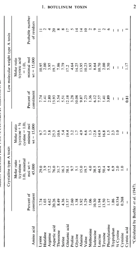
lated further studies of its chemistry. The amino acid composition of type A toxin was established by Buehler et al. (1947) by microbial bioassay of acid hydrolyzates of crystalline toxin and, wherever possible, by chemical means. By these methods, 19 known amino acids were identified with surprisingly high values of aspartic acid, tyrosine, and threonine residues (Buehler et al., 1947). The amino acid composition and the methods by which they were assayed are detailed in Table VIII. The total nitrogen content was calculated as 16.29%. No lipids, polysaccharides, or unusual amino acids which would explain the toxin's remarkable potency were found associated with the toxin. Recently, amino acid analysis of one batch of crystalline botulinum toxin type A was performed by Stefanye et al. (1964) with the use of an amino acid analyzer. These workers con
cluded that their results substantially agreed with those reported by Bueh
ler et al. (1947), except for the nitrogen, sulfur, and phosphorous concen
tration which were 16.08%, 0.69%, and 0.05%, respectively. Boroff and DasGupta (unpublished data) also performed similar analyses on several different crystalline toxin preparations and found that while the results of amino acid analysis of some of the crystalline preparations were in good agreement, others showed dissimilarities in amino acid contents from those recorded by Stefanye et al. (1964). This appears to imply that differ
ences in chemical composition may exist between different crystalline preparations.
From additional evidence supplied by Putnam et al. (1946) that in the ultracentrifuge at pH 4.38 this toxin sedimented as one component, the authors came to the conclusion that crystalline toxin was a homogeneous protein. The admixture of nucleic acids demonstrated in the course of pu
rification could be removed practically completely without any observa
ble decrease in toxicity. On further examination the homogeneity and the molecular weight determined for the toxin were not supported by other evidence. It was discovered that the toxin possessed an additional activity in that it agglutinated suspensions of erythrocytes of various animal spe
cies (Lamanna and Putnam, 1948). These hemagglutinins could be sepa
rated from the toxin by adsorption on erythrocytes without in any way impairing the activity of the toxin (Lamanna and Lowenthal, 1951; Low- enthal and Lamanna, 1951). These results led the authors to conclude that hemagglutination and toxicity may be associated with separate particles and that under certain conditions the hemagglutinin and toxin form stable
TABLE VIII AMINO ACID COMPOSITION OF CRYSTALLINE TOXIN OF CI. botulinum TYPE A" Determination Constituent Method of determination Percent of constituent Percent acid residue Percent N calcu lated
Percent S calcu lated 1 Lysine L. mesenteroides 7.74 6.78 1.48 — 2 Histidine L. mesenteroides, S.faecalis 1.03 0.91 0.28
-
3 Arginine L. casei, S.faecalis 4.62 4.14 1.49-
4 Tyrosine L. casei, photometric 13.50 12.18 1.04 — 5 Phenylalanine L. delbrueckii, L. casei 1.17 1.04 0.10-
6 Tryptophan L. casei, photometric, S.faecalis 1.86 1.69 0.25-
7 Valine L. casei, S.faecalis 5.29 4.45 0.63 - 8 Leucine S.faecalis, L. arabinosus 10.30 8.91 1.10-
9 Isoleucine S.faecalis, L. arabinosus 11.94 10.33 1.28-
10 Glutamic acid L. arabinosus 15.57 13.67 1.48-
11 (Aspartic acid) L. delbrueckii, L. mesenteroides (20.26) lla = Asparagine 20.10 17.34 4.26-
11 + 30 12 Serine L. delbrueckii 4.36 3.60 0.58-
13 Threonine S.faecalis, oxidation, L. arabinosus 8.49 7.19 0.99-
14 Cysteine Photometric 0.268 0.23 0.03 0.066 15 Half cystine Photometric 0.534 0.45 0.06 0.142 16 Methionine L. arabinosus, iodometric, S.faecalis 1.06 0.93 0.10 0.228 17 Proline L. mesenteroides 2.60 2.19 0.32-
18 Glycine L. mesenteroides 1.38 1.05 0.26 - 19 Alanine S.faecalis 3.92 3.12 0.61 — 20 Total 115.73 100.20 16.34 0.436 21 = 20 Water taken 15.53 (Column 4 up during — Column 5) hydrolysis "Reprinted by permission from Buehler et al. (1947).w o H G r G H O 2
18 D . A. B O R O F F A N D B . R. D A S G U P T A
complexes or combinations acting as homogeneous materials. The subject of hemagglutinins will be discussed more fully below. The observation of Putnam et al. (1946) on the behavior of the toxin in the ultracentrifuge where the crystalline toxin at acid pH 4.8 sedimented as one component was reexamined by Wagman and Bateman (1951, 1953) and Wagman (1954, 1963) at pH 6-9.2 in various buffers.
By bringing the toxin preparation to pH 7.2 and the buffer to ionic strength of 0.13, they were able to show by ultracentrifugation that the toxin exists in the form of two polydispersed components. Partial separa
tion of the slowly sedimenting component was achieved with the aid of a preparative rotor. This component was judged to be about 20% of the original toxin and was free from hemagglutinins. Its molecular weight was estimated as 71,000 and s2 0,w was 4.7. The toxicity of this component per milligram of nitrogen was at least as great as that of the parent substance.
This toxin was tentatively identified as the ultimate toxic unit of botu
linum type A toxin (Wagman and Bateman, 1953).
Additional studies by Wagman (1954) showed that in buffers of pH range 6.5-8 and ionic strength of 0.13 and above, crystalline toxin dissociated into a slowly sedimenting component with a £2o , w of 6.5 and a molecular weight from 40,000-100,000. The extent of dissociation in
creased with the increase of ionic strength. In phosphate buffer at pH 7.5 and ionic strength of 1.0, the fraction consisting of the faster-sedimenting, relatively intact particles is precipitated, leaving only the slowly sedi
menting fraction in solution. This low molecular weight fraction had a specific toxicity two to three times that of the original sample. Raising the pH to 9.2 irreversibly dissociated crystalline toxin with no loss of toxicity. Of the two components, one had a sedimentation constant of 7 S and molecular weight of 158,000 and the other a component of 14 S (Wagman, 1963). On the basis of tyrosine content, the slow compo
nent (4.5%) appeared dissimilar from crystalline toxin (10.9%). While crystalline toxin was resistant to peptic digestion, the 7 S component was readily hydrolyzed by the enzyme into fractions of decreasing
^2o,w values with some toxicity loss. The peptic digest dialyzed against five volumes of 0.05 M acetate buffer, pH 3.8, for 14 hours in the cold yielded a toxic filtrate. The toxin in the filtrate was estimated to have a molecular weight of about 3800. No specific toxicity could be calculated because of the very low toxicity of the filtrate. On the basis of these observations, Wagman suggests that molecules of intact toxin contain toxic and non
toxic subunits which can be separated without loss of toxicity. The dialyz- able fractions of the toxin were deemed to be the toxic peptide fragments which prior to cleavage were situated between the free amino terminal ends of the chains. It was assumed that these also were the fragments which escape through the intestinal wall into the circulation.
1. B O T U L I N U M T O X I N 19 That the toxin of CI. botulinum type A need not be of small molecular weight has been demonstrated by May and Whaler (1958) who discov
ered the presence of the toxin in the lymph of orally intoxicated rats.
Heckly et al. (1960) investigated this toxin and found that its particle size was equal to that of proteins. In a glycerol density gradient separation of the toxin in the lymph, the toxin tended to separate in the same way as the bulk of the protein in the lymph. Figure 1 shows two such separations.
Evidence obtained by the latter investigators from the ultracentrifuge data suggested the toxin to be of mean s20,w v^ ue about 7 S. This was consist
ent with the value of the toxic moiety described by Wagman (1954), from which the molecular weight of the component could be calculated as larger than 100,000 but smaller than that of crystalline toxin with s2 0,w °f 17.3 and molecular weight of 900,000.
The unlikelihood of a protein of even 100,000 molecular weight to be one peptide chain, as well as Wagman's report (1954) of the dissociation of crystalline toxin into small molecular weight subunits, prompted at
tempts to dissociate the toxin by means other than ultracentrifugation.
Schantz et al. (1960) employed a 0.9 x 10 cm column of DEAE-cellulose in an attempt to fractionate the crystalline toxin by ion exchange chroma
tography. In this investigation, they used potassium phosphate buffer pH 6.5 and stepwise gradients of potassium chloride, whereby a number of peaks were eluted. The toxicity of the eluate was, however, no greater
0-60% 0-30%
I I I I L
0 1 2 3 4
Cm down from meniscus
F I G . 1. Fractionation of toxic lymph after 4 hours centrifugation at 3 9 , 4 6 0 rpm through density gradients. T h e lymph was collected from rats that had received 10 ml of a partially purified botulinum type A toxin containing 5 x 1 06 mouse L D50 per milliliter. T h e gradients were 1 0 - 2 0 - 4 0 - 6 0 % and 5 - 1 0 - 2 0 - 3 0 % glycerol. T h e fractions, 0.4 ml each, were collected at the rate of 0.2 ml per minute. Reprinted from Heckly et al. (1960).