Tetanus Toxin
W . E. VAN HEYNINGEN AND J A N E MELLANBY
I. Introduction 69
II. Toxicity 0 7
A. Factors Affecting Toxicity 70 B. A s s a y of Toxicity 71 C. Toxicity of Purified Toxin 7 4 III. Production and Purification 75
A. Production 75 B. Purification 77 IV. Nature 79
V. Synthesis 82 V I . Mode of Action 85
A. Molecular Level 86 B. Physiological Level 92 VII. Immunology and Immunochemistry 102
V I I I . Pathogenesis 104 References 105
I. I n t r o d u c t i o n
Tetanus toxin was the second bacterial toxin to be discovered, the first having been diphtheria toxin. Nicolaier had noted that the organism (as yet not identified) causing tetanus was not distributed throughout the body but confined to the wound of entry. Simpson had already observed in 1854 that the symptoms of tetanus were similar to those of strychnine poisoning, and in 1885 Nicolaier suggested that the organism brought about its pathological effects by producing a strychnine-like poison. In 1889 Kitasato isolated the tetanus bacillus, and in 1890 Faber justified Nicolaier's suggestion by showing that it was possible to reproduce the spastic symptoms of tetanus in experimental animals by injecting them with sterile filtrates of cultures of the bacillus. The toxin in the culture fil
trates producing this effect, sometimes known as tetanospasmin, is the subject of this chapter. Since tetanus toxin is wholly implicated in the dis
ease of tetanus, and since the disease is so easily preventable by immuni
zation against the toxin (see below), the toxin has been in commercial production in many countries for many years. Consequently, the produc
tion and properties of the toxin have been the subjects of much study. [Be
sides tetanospasmin, the tetanus bacillus also produces an oxygen- labile hemolysin, tetanolysin, that contributes little or nothing to the tox
icity of Clostridium tetani filtrates and has not been much studied. It may be responsible for occasional attributions of hemolytic activity to tetano-
69
spasmin (see Hardegree, 1965). Feigen et al. (1963) reported the presence in crude preparations of tetanus toxin of a nonspasmogenic substance that could be separated from tetanospasmin, but no physiological activity in the whole animal has been ascribed to it (see Section VI,B,3).]
I I . T o x i c i t y
A. FACTORS A F F E C T I N G TOXICITY
Considerations of the toxicity of tetanus toxin (and there are different ways of considering it) are affected by several factors —inactivation of the toxin; differences in the susceptibility of different animals to the toxin and variations in these differences; and possible potentiation of the toxin.
In assaying the toxicity of tetanus toxin, very dilute solutions are used since the toxin is so toxic; a solution of toxin containing 2 mouse LD5 0/ml would contain about 40 pg of toxin/ml. In dilute solution, the toxin would denature or be adsorbed on the surfaces of the containing vessels, and this is prevented by adding a protective protein to the diluent (e.g., gelatin or peptone). However, Pillemer (1946) confirmed an early observation by Mutermilch et al. (1934) that when toxin is dissolved in saline containing 1 % peptone it will tend to be reversibly adsorbed onto glass, but this does not take place when saline alone is used. Nowadays, 0.2% gelatin is usually used when making serial dilutions of toxin, and a fresh pipette is used for each dilution. Another complication is that highly purified toxin in solution may lose activity (but not antigenicity) by spontaneous toxoid- ing (Pillemer et al, 1948).
Animals vary in their susceptibility to tetanus toxin (as to all bacterial toxins). Of the animals tested, the guinea pig is one of the most suscepti
ble and the pigeon one of the least (Table I). The frog is highly resistant at its natural ambient temperature (10-15°C), but its susceptibility increases with increasing temperature, although it does not approach that of mam
mals (Rowson, 1961). Smith (1942-1943) found that the ratio of toxicity for various species of animal differed from one toxic culture filtrate to another. Thus, the ratio for rabbits compared with guinea pigs varied from 29 to 354, while that for mice and guinea pigs varied only from 6.25 to 8.
From these and similar earlier observations, the inference was drawn that the different culture filtrates contained different proportions of two or more toxins. However, there has in fact never been any toxicological, immunological, kinetic, or electrophoretic evidence for the existence of more than one spasmogenic component in CI. tetani cultures (Largier,
1956b; Hardegree and Wannamaker, 1965).
T A B L E I
R E L A T I V E M I N I M U M L E T H A L D O S E S O F T E T A N U S T O X I N FOR V A R I O U S S P E C I E S OF M A M M A L S A N D B I R D S ( O N A N E Q U A L W E I G H T BASIS)"
M L D according to the following investigators:
Friede- von
Fildes Abel Bardelli Ipsen mann Smith Behring D a v i e s Shumacker et al. et al. et al. et al.
(1929) (1935) (1927) (1940- (1941) ( 1 9 4 2 - (1912) (1955) (1939) Species 1941) 1943)
Horse 1 0.5
Guinea pig 1 1 1 1 1 1 1 1
Monkey 2 4
Sheep 2
Mouse 2 4 2 - 6 4 - 2 0 3 - 6 6 6
Goat 12
Rabbit 12 4 - 5 0 0 2 4 - 7 2 0 5 0 - 3 2 0 3 - 3 5 0 9 0 0
D o g 3 0 0 4 8 0
Cat 1,200 9 6 0
G o o s e 6,000
Pigeon 2 4 , 0 0 0 6 , 0 0 0
Hen 180,000
"Adapted from G. P. Wright (1955).
It is possible that the variations in the ratios of susceptibilities to differ
ent samples of crude toxin might be connected with the phenomenon of potentiation. For example, Traub et al. (1946) observed a 64-fold poten
tiation of some samples of crude tetanus toxin when these were diluted with serum, and this was a genuine potentiation, not just a protection of the toxin against denaturation (Zuger et al., 1939). However, potentiation was observed only with toxin injection into mice and guinea pigs and not with cats and rabbits. Thus, it seems that the toxicity of tetanus toxin may be influenced by concomitant substances and that this influence is mani
fest to different degrees in different animals.
B . ASSAY OF TOXICITY
No in vitro assay has been devised for tetanus toxin since no simple in vitro action of the toxin (e.g., hemolysis) has been found. Nor is there any skin reaction as is used to assay (among others) diphtheria toxin. Conse
quently, tetanus toxin has usually been assayed by observing its lethal effect on the whole animal. However, if the toxin is injected intramuscu
larly into a limb, it will produce typical spastic paralysis in that limb, and this action has been used in assaying the toxin (see below).
T A B L E I I
R E L A T I V E M I N I M U M L E T H A L D O S E S O F T E T A N U S T O X I N W H E N A D M I N I S T E R E D BY D I F F E R E N T R O U T E S O F I N J E C T I O N "
Relative M L D following injection:
Refer Intra- Subcut- Intra Intra- Spinal Medulla Cerebrum Intra
Species e n c e6 venous aneous muscular neural cord oblongata ventricular
Rabbit a 1 1 V 1 0 - V 2 0
b 1 V20-V100
c 1 VlO
d 1 Viooo V100
e 1 V25
Guinea pig a 1 1 1 1
b 1 I- V 3
c 1 1
e 1 1
Hutia g 1 V7-V13
Cat f 1 7 3 0 0
D o g f 1 Vso
Monkey f 1 V4
Pigeon h 1 Vs
"Adapted from G. P. Wright (1955).
^References: (a) van den H o v e n van Genderen (1933); (b) Friedemann and Hollander (1943); (c) Sawamura (1909); (d) E . A. Wright (1953); (e) Roux and Borrel (1898); (f) Shu- macker et al. (1939); (g) Angulo (1943); (h) D a v i e s et al. (1955).
The lethality of the toxin depends on the route by which it is introduced into the animal. It can be seen that the toxin is most potent when intro
duced into the central nervous system rather than peripherally or into the blood stream (Table II). Contrary to previous belief, the toxin is also toxic by mouth, but the toxicity by this route is 1 / 2 0 0 , 0 0 0 - 1 / 1 , 2 0 0 , 0 0 0
that of the parenteral route (Lamanna, 1 9 6 0 ) . Intramuscular injection is the most convenient route for assaying tetanus toxin, and the animal most frequently used is the mouse.
1. MINIMAL LETHAL D O S E
Unfortunately, authors do not always state what they mean by minimal lethal dose (MLD), since although it is defined as the least dose that will kill the animals injected, this could mean that one, some, or all the injected animals should be killed. In our experience the least dose that would kill all the mice injected (the L D1 0 0) was 1.4 times the dose that would kill 5 0 % of the mice injected (the LD5 0), and the L D50 was 1.7 times the maxi
mum dose that would fail to kill any of the mice injected (the LD0). Of these three doses, the L D50 is the most meaningful, since it lies on the steepest part of the dose-response curve. Since the dose-response curve
of tetanus toxin is steep, in practice if a series of twofold dilutions of teta
nus toxin is injected, it is common to find that with the higher of two adja
cent doses, all the mice will be killed, and with the lower, none. The defi
nition of L D50 must include the time within which the animals die (in the case of tetanus toxin, usually 4-7 days).
2. D E A T H T I M E ASSAYS
Some workers assay tetanus toxin by determining the time it takes for a given dose to kill mice. Ipsen (1941) expressed the relationship between the MLD and the death time by the formula:
( Z W - l )0 5x ( 7 7 8 - 1 ) = 10.3
where D = dose, d = MLD, and T = death time. Sheff et al. (1965) have introduced the notion of the minimum saturation dose of tetanus toxin, the determination of which is claimed to be less time-consuming than that of the minimal lethal dose. The minimum saturation dose (equivalent to about 40,000 MLD) is the dose beyond which the death time is not fur
ther reduced. No data have yet been provided to enable a judgment to be made of the reliability of this assay.
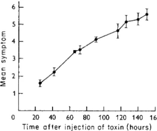
3. M E A N SYMPTOM ASSAY
If tetanus toxin is injected intramuscularly (0.5 ml) into the hind limb of a mouse, characteristic and clearly definable symptoms of increasing se
verity are produced in a given time by increasing doses of the toxin. Mel- lanby et al. (1968a) have assigned numerical values to these symptoms (Table III), and Fig. 1 shows the progress of the mean symptom produced
T A B L E I I I
V A L U E S A S S I G N E D T O S Y M P T O M S O F L O C A L T E T A N U S I N D U C E D IN M I C E BY I N J E C T I N G T O X I N I N T R A M U S C U L A R L Y I N T O
A H I N D L I M B
Symptom Value
assigned
N o symptoms 0
Slight stiffness in injected limb visible only
when mouse suspended by tail 1
An obvious limp in injected limb, still
used effectively in walking 2
Injected limb still movable, but not functional 3
Injected limb rigid; toes immovable 4
Animal convulsing; very ill 5
D e a d 6
I I I I I I I I I
0 20 40 60 80 100 120 140 160 Time after injection of toxin (hours)
F I G . 1. T h e progression of tetanus symptoms in a group of 2 0 mice injected with 2 L D5 ()
tetanus toxin. S e e Table III for assessment of symptoms. T h e vertical lines indicate the standard deviations of the values.
in a group of 20 mice injected with 2 L D50 of tetanus toxin. The figure shows that the standard deviation is greater when the animals are near death, where extraneous factors such as inability to get at water and food and cannibalism play a greater part. The intermediate effects, up to mean symptom 3 are more meaningful than the final effect (death), which is the only effect observed in the customary determinations of lethal dose or survival time. An amount of toxin which produces symptom 3 by day 3 is approximately equivalent to 1 L D5 0.
C. TOXICITY OF P U R I F I E D T O X I N
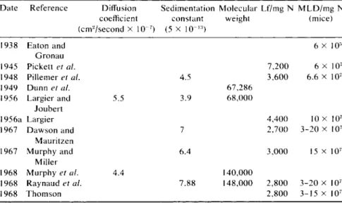
Table IV shows the properties, including the toxicity, of preparations of purified tetanus toxin as obtained by several different workers in differ
ent countries. Most of the postwar preparations have conformed with at least some of the acceptable criteria of purity, and the fact that they are all of the same degree of toxicity (bearing in mind the limitations of assay) suggests that if these preparations are not approaching a state of complete purity, at least they are approaching a state of uniformity that must have some significance in the economy of the parent bacillus. It will be seen that the greatest potency observed in pure toxin preparations is 200 mil
lion mouse MLD/mg N, or about 30 million mouse MLD/mg protein.
Tetanus toxin is thus one of the most poisonous substances known, only botulinus toxin (Vol. IIA, Chap. 1) and dysentery toxin (Vol. IIA, Chap.
6) having comparable toxicity.
I I I . P r o d u c t i o n a n d P u r i f i c a t i o n
A. PRODUCTION
Practically all the tetanus toxin that has been produced in laboratories all over the world since the last war (Pillemer et al, 1948; Largier, 1956a;
Thomson, 1957; Dawson and Mauritzen, 1967; Murphy and Miller, 1967) has been produced by the Harvard strain of CI. tetani (Mueller and Miller, 1945) grown on the Mueller-Miller medium (see Taylor, 1945;
Mueller and Miller, 1954; C. Latham, et al, 1962; Table V). The only notable exception has been the Institut Pasteur at Garches, where Ray
naud, et al (1960) (see also Raynaud, 1951) have used a French strain, as well as the Harvard strain, and the medium of Prevot and Boorsma (1938) which consists of a peptic and papain digest of beef. The final medium of Mueller and Miller (1954) yielded 130-150 Lf/ml or 1.3-1.5 million mouse MLD/ml. The variant medium of Latham et al. (1962) avoided the use of the beef heart infusion and substituted ferric chloride for reduced iron powder, at a cost of rather lower yields of toxin (70-90 Lf/ml). It is important that gases should be allowed to escape freely from the culture, and, for this reason, Mueller and Miller (1954) used wide-mouthed ves
sels (cylinders holding 6 liters of medium). Thomson (1957) grew 70-liter cultures in 80-liter tanks with continuous stirring. In converting to pro-
TABLE IV
P R O P E R T I E S O F V A R I O U S P R E P A R A T I O N S O F P U R I F I E D T E T A N U S T O X I N Date Reference Diffusion Sedimentation Molecular Lf/mg N M L D / m g N
coefficient constant weight (mice) (cm2/second x 1(T7) (5 x 10"1 3)
1938 Eaton and 6 X 10⁵
Gronau
1945 Pickett et al. 7,200 6 X 10⁷
1948 Pillemer et al. 4.5 3,600 6.6 X 10⁷
1949 Dunn et al. 6 7 , 2 8 6
1956 Largier and 5.5 3.9 6 8 , 0 0 0
Joubert
1956a Largier 4,400 10 x 107
1967 D a w s o n and 7 2,700 3 - 2 0 x 107
Mauritzen
1967 Murphy and 6.4 3,000 15 x 107
Miller
1968 Murphy et al. 4.4 140,000
1968 Raynaud et al. 7.88 148,000 2,800 3 - 2 0 x 1 07
1968 Thomson 2,800 3 - 1 5 x 1 07
T A B L E V
C U L T U R E M E D I A FOR T E T A N U S T O X I N P R O D U C T I O N
Quantity per liter
Mueller and Miller C. Latham et al.
Constituents" (1954) (1962)
Digest of casein 22.5 gm 25 gm
Beef heart infusion6 50 ml 0
G l u c o s e 11 gm 8 gm
Sodium chloride 2.5 gm 2.5 gm
Sodium phosphate ( N a2H P 04) 2 gm 0
Potassium phosphate ( K H2P 04) 0.15 gm 0
Magnesium sulfate 0.15 gm O.lOgm
Cystine 0.25 gm 0 . 1 2 5 g m
Tyrosine 0.5 gm 0
Calcium pantothenate 1.0 mg 1.0 mg
Uracil 2.5 mg 1.25 mg
Nicotinic acid 0 0.25 mg
Thiamine 0.25 mg 0.25 mg
Riboflavin 0.25 mg 0.25 mg
Pyridoxine 0.25 mg 0.25 mg
Biotin 2.5/xg 2.5/ng
Vitamin B12 0 0.05
Reduced iron powder 0.5 gm 0
Ferric chloride ( F e C l3 • 6 H20 ) 0 32 mg
Lf/ml 1 3 0 - 1 4 0 7 0 - 9 0
"Medium adjusted to pH 7 . 0 - 7 . 2 , sterilized by autoclaving at 10 pounds for 2 0 minutes.
6O n e kilogram of minced beef heart suspended in 1 liter tapwater, brought rapidly to active boiling, strained through cheesecloth, and filtered through paper.
duction on such a large scale, an important factor is the need to avoid ov- erautoclaving the medium with consequent poor toxin production. How
ever, C. Latham et al. (1962) found that some heating was necessary, apart from sterilizing, because it brought about toxigenic changes in the medium, presumably involving cysteine.
Previously, cultures were grown for 4 or 5 days until the organisms autolyzed, and the toxin was discharged into the medium. Thus, for exam
ple, Thomson (1957) would grow the cultures for 48 hours in stirred tanks and then transfer the tank harvests to 10-liter bottles where the cultures were kept for a further 48 hours at 35°C. During this time, there was no further growth, but autolysis took place. The culture would then be centri- fuged in a continuous centrifuge, and the toxin was recovered from the culture filtrate. However, since Raynaud (1951) developed his method of extracting the toxin from the intact organisms before autolysis, several laboratories grow the cultures for a relatively short time (72 hours) and harvest the organisms by centrifugation before they autolyze (see below).
B . PURIFICATION 1. FROM FILTRATES OF AUTOLYZED CULTURES
Since World War II tetanus toxin has been obtained in a state of purity higher by two orders of magnitude than the best prewar prepara
tions (Table IV). Pickett et al. (1945), without the help of modern meth
ods of protein purification, obtained by precipitation with cadmium salts from filtrates of autolyzed cultures a preparation which compares very favorably in toxicity with the best preparations reported more than 20 years later, although these workers made no claim that their preparation was approaching purity. (Their high values for the Lf/mg may have been due to the use of the prewar American Unit of antitoxin which had half the value of the present International Unit.)
Pillemer (1946) was the first (and only) worker to crystallize tetanus toxin. He used filtrates of cultures grown on Mueller-Miller medium and purified the toxin by a series of precipitations with methanol in the cold with precise control of the pH, protein concentration, and the ionic strength of the toxin solutions (Pillemer et al., 1948; Table IV). Crystalli
zation of the toxin was difficult; traces of nucleoprotein prevented crystal
lization and so did the spontaneous detoxification of the toxin which tended to take place during the purification process. Crystallization was achieved by adjusting the purified preparation as follows: 1% protein concentration, pH 6.0, methanol concentration 20%, ionic strength 0.02, and temperature —5°C. Crystallization occurred in a few days and was complete after several weeks. No other workers have since reported crys
tallization of the toxin. The preparation of Pillemer et al. (1948) gave sin
gle, apparently symmetrical peaks on free electrophoresis and ultracen- trifugation (but on standing for 10 days at 0°C, in addition to the peak of 4.5 S, a second peak of 7.0 S would appear, apparently due to the forma
tion of a nontoxic dimer, see below). The solubility curve for the fresh crystalline toxin was practically congruent with the theoretical curve for a single-component preparation.
The preparation of Largier (1956a; Table IV) was obtained by his pro
cess of "multimembrane electrodecantation" from the filtrates of auto
lyzed cultures grown in cellophane sacs suspended in Mueller-Miller medium. The final preparation behaved homogeneously upon electropho
resis and ultracentrifugation. Dawson and Mauritzen (1967; Table IV) may well be the last to have used filtrates of autolyzed cultures as a source of toxin. They precipitated the toxin at — 15°C with methanol (final con
centration 40%) and then processed the material on a column of DEAE- cellulose. On immunoelectrophoresis, their final product appeared to be homogeneous, but, on ultracentrifugation, there was a major component with a sedimentation constant of about 7 S (see below) and a minor com-
ponent with a constant of about 10 S. At pH 5.5 a minor component of 2 S was also observed.
2. FROM EXTRACTS OF CELLS
In 1947, Raynaud (1951) made an important discovery which has great
ly simplified the preparation and purification of tetanus toxin. He showed that in young cultures (1-3 days) of the tetanus bacillus, before there was appreciable soluble toxin in the culture medium, the toxin could be ex
tracted in high yield from the cells toward the end of the exponential phase of growth. The cells were harvested by centrifugation and washed with distilled water, and the toxin was extracted by suspending the cells in hypertonic saline (1 M NaCl, 0.1 M Na citrate) for 5 days in the cold.
After centrifugation, the toxin concentration in the extract was as high as 500 Lf/ml. Extraction for more than 5 days did not yield more toxin, but it extracted more protein and thus toxin with a lower specific activity. Shak
ing during incubation also caused the extraction of more nontoxic protein but not more toxin.
This method of preparing the toxin is advantageous in that (1) the toxin is concentrated easily and quantitatively simply by harvesting the cells by centrifugation, and (2) the proteins and other large molecular constituents of the culture medium (which may have contributed to toxin production) are easily eliminated by separating and washing the cells. The extract con
tains only a relatively small (7-8) number of antigenic constituents.
Using this simple method of preparing concentrated crude toxin, Ray
naud and his colleagues (Raynaud et al., 1960; Raynaud and Turpin, 1956; Turpin and Raynaud, 1959; Table IV) purified the toxin in the fol
lowing stages: (1) precipitation of the toxin with metaphosphoric acid at - 15°C, pH 3.8-4.0, with 23 gm NaCl/liter; (2) salting out by the addition of 3.5 M potassium phosphate buffer to concentrations between 1.15 M and 1.75 M; (3) removal of nucleic acid by precipitation with methanol at final concentration of 7.5% at pH 4.0 and—5°C; (4) chromatography on DEAE-cellulose equilibrated with 0.01 M EDTA (necessary to prevent toxoiding). The final product gave only a single precipitin line on immuno
diffusion against complex antiserum, and antisera prepared against the preparation gave only a single line with crude toxin. It also appeared to be homogeneous on filtration through G-100 and G-200 Sephadex in EDTA solution, and on ultracentrifugation.
Thomson (1957, 1968) also prepared the toxin from hypertonic saline extracts of cells. Murphy and Miller (1967; Murphy et al., 1968; Table IV) grew the organism in the modified Mueller-Miller medium of C. La
tham et al. (1962) for 72 hours at 33°C in 30-100-liter lots distributed in
8-liter portions in 9-liter bottles. The cells were extracted at 4°C for 5 days with 0.1 culture volume of M NaCl, 0.1 M Na citrate. The toxin was purified from these extracts by the following steps: (1) precipitating with 470 gm (NH4)2S04/liter, dissolving in 0.09 extract volume tris buffer, pH 7.5, and desalting on Sephadex 50; (2) processing on DEAE-cellulose; (3) precipitating with 300 gm (NH4)2S04/liter; (4) processing on G-100 Se
phadex. The final product appeared to be homogeneous on Immunoelec
trophoresis and double diffusion, ultracentrifugation, electrophoresis in polyacrylamide gel, and passage through a G-100 Sephadex column.
I V . N a t u r e
Tetanus toxin is a simple protein containing 15.7% nitrogen, 0.065%
phosphorus, and 1.04% sulfur. It contains no carbohydrate (Pillemer et al, 1948) and no lipid (Mellanby and van Heyningen, unpublished).
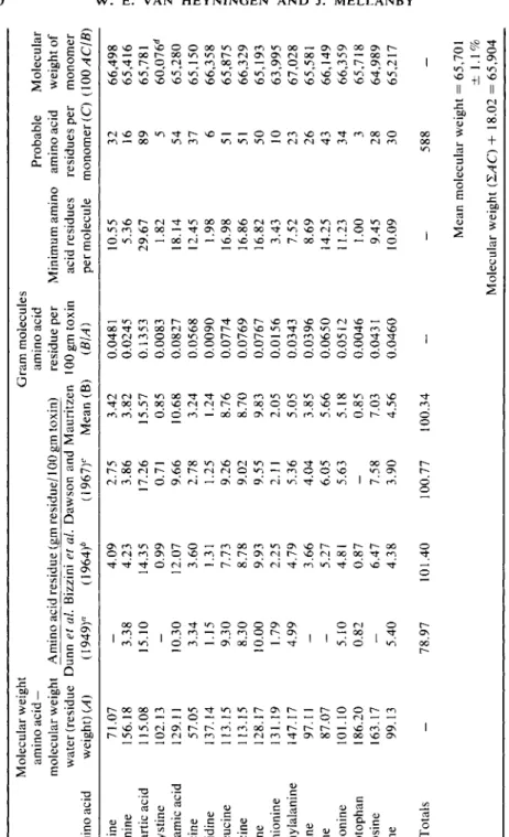
Amino acid analyses of the protein are shown in Table VI. The iso
electric point is between 5.0 and 5.2. The toxin begins to precipitate from solution and to lose activity when the pH is reduced to 6, but it is stable to pH 5 in the presence of glycine (Pillemer et al, 1948).
Bizzini et al. (1963) found by amperometric and spectrophotometric techniques that the toxin contained, for a molecular weight of 150,000, 9.3 free sulfydryl groups, 4.8 of which were directly accessible and played no part in the biological activity of the molecule. The remaining 4.5 sul
fydryl groups were masked. By blocking the free sulfydryl groups with p- chloromercuribenzoate and reducing the toxin, they showed that the mol
ecule contained 2.63 to 2.88 disulfide bridges. Murphy et al. (1968) found, for a molecular weight of 140,000, 10 half-cystine residues but only 2 di
sulfide bonds. No free sulfhydryl was found by alkylation of the native molecule, but 4 residues were obtained after denaturation in urea, and 2 additional residues after denaturation with urea in the presence of 8-hy- droxyquinoline-5-sulfonic acid. Gel filtration and chemical studies on the toxin denatured in 8 M urea indicated that the molecule was a single poly
peptide chain without a free N-terminal amino group.
It will be seen in Table IV that different values have been reported for the sedimentation constant and the molecular weight of tetanus toxin. The sedimentation constant of 4.55 S observed by Pillemer et al. (1948; Pille
mer and Moore, 1948) suggested a molecular weight of 66,000-74,000 (see Dunn et al, 1949). The sedimentation constant of 3.9 S and the diffu
sion constant of 5.5 observed by Largier and Joubert (1956) indicated a molecular weight of 68,000.
But evidently there are forms of tetanus toxin of greater molecular
TABLE VI AMINO ACID ANALYSES OF VARIOUS PREPARATIONS OF PURIFIED TETANUS TOXIN Molecular weight Gram molecules amino acid — amino acid Probable Molecular molecular weight Amino acid residue (gm residue/100 gm toxin) residue per Minimum amino amino acid weight of water (residue Dunn et al. Bizzini et al. Dawson and Mauritzen 100 gm toxin acid residues residues per monomer Amino acid weight) (A) (1949)° (1964)6 (1967)c Mean (B) (BIA) per molecule monomer (C) (100ACIB) Alanine 71.07
_
4.09 2.75 3.42 0.0481 10.55 32 66,498 Arginine 156.18 3.38 4.23 3.86 3.82 0.0245 5.36 16 65,416 Aspartic acid 115.08 15.10 14.35 17.26 15.57 0.1353 29.67 89 65,781 V2 Cystine 102.13 — 0.99 0.71 0.85 0.0083 1.82 5 60,076d Glutamic acid 129.11 10.30 12.07 9.66 10.68 0.0827 18.14 54 65,280 Glycine 57.05 3.34 3.60 2.78 3.24 0.0568 12.45 37 65,150 Histidine 137.14 1.15 1.31 1.25 1.24 0.0090 1.98 6 66,358 Isoleucine 113.15 9.30 7.73 9.26 8.76 0.0774 16.98 51 65,875 Leucine 113.15 8.30 8.78 9.02 8.70 0.0769 16.86 51 66,329 Lysine 128.17 10.00 9.93 9.55 9.83 0.0767 16.82 50 65,193 Methionine 131.19 1.79 2.25 2.11 2.05 0.0156 3.43 10 63,995 Phenylalanine 147.17 4.99 4.79 5.36 5.05 0.0343 7.52 23 67,028 Proline 97.11 — 3.66 4.04 3.85 0.0396 8.69 26 65,581 Serine 87.07 — 5.27 6.05 5.66 0.0650 14.25 43 66,149 Threonine 101.10 5.10 4.81 5.63 5.18 0.0512 11.23 34 66,359 Tryptophan 186.20 0.82 0.87 — 0.85 0.0046 1.00 3 65,718 Tyrosine 163.17-
6.47 7.58 7.03 0.0431 9.45 28 64,989 Valine 99.13 5.40 4.38 3.90 4.56 0.0460 10.09 30 65,217 Totals _ 78.97 101.40 100.77 100.34 _ 588 _ Mean molecular weight = 65,701 ± 1.1% Molecular weight (SAC) + 18.02 = 65,904 G Mean of 2 determinations. c Mean of 2 determinations. 6 Mean of 8 determinations. ^Ignored.weight. Pillemer et al. (1948; Pillemer and Moore, 1948) found that when solutions of pure tetanus toxin in neutral isotonic saline were allowed to stand 10 days at 0°C, 75% of the toxicity was lost and the toxin prepara
tion became inhomogeneous. Still, 40-45% of the material had a sedimen
tation constant of 4.5 S, but the remainder had a sedimentation constant of 7 S, corresponding to a molecule of twice the molecular weight. The 7 S material was no longer toxic but still flocculated readily with tetanus anti
toxin. This suggested that the pure toxin spontaneously condensed to form a nontoxic dimer. Largier and Joubert (1956), similarly, found a toxic monomer with a sedimentation constant of 3.9 and a nontoxic dim
mer of 7.6 S.
Since 1956, it seems that sedimentation constants corresponding to a molecular weight of about 68,000 have not been observed. Raynaud et al.
(1960; Mangalo et al., 1968) observed sedimentation constants of 6.1 and 7.1 S; Dawson and Mauritzen (1967) 6.1 to 6.7 S; Murphy and Miller (1967) 6.4 S. Mangalo et al. (1968) stated that with 20 different samples of purified toxin, they had never observed a sedimentation constant of 4.5 S, except when the toxin was treated with cysteine followed by monoiodoac- etate by the method of Raynaud et al. (1960). Under these circumstances an active toxin was obtained with a sedimentation constant of 4.5 S, and this suggested that depolymerization due to rupture of disulfide bonds had taken place.
It therefore appears that tetanus toxin can exist in an active state (and in a nontoxic but still antigenic state) in two forms — a monomer of molec
ular weight of about 68,000 and a dimer of about twice that molecular weight. It is possibly significant that the monomeric toxin was isolated by earlier workers from culture filtrates of autolyzed organisms, whereas the dimeric toxin has been isolated by later workers from cell extracts. An exception is the preparation of Dawson and Mauritzen (1967), with sedi
mentation constants from 6.1 to 6.7 S, isolated from toxic culture filtrates.
Mangalo et al. (1968) showed that the sedimentation constant of teta
nus toxin depended on the concentration of the toxin and found a value of 7.88 S after extrapolation to zero concentration. However, this depen
dence of S on concentration is unlikely to account for the difference be
tween about 4.5 and about 7 S, since extrapolation of the data of Mangalo et al. (1968) suggest that their preparation might have had an apparent S of 5 only at a concentration as high as about 35 mg protein/ml, which would hardly have been used by any of the earlier workers. When the molecular weight of the toxin was determined by Mangalo et al. (1968) by the method of Yphantis (1960), a value of 150,000 ± 8 % was obtained.
This value was independent of rotor speed or toxin concentration. Mur
phy et al. (1968) found a sedimentation constant of 6.4 S and diffusion
constants of 4.2 and 4.4 by chromatographic and immunodiffusion tech
niques, respectively. These values yielded an average molecular weight of 140,000.
In Table VI, values for the molecular weight of a probable monomer are calculated in two ways from the means of 12 amino acid analyses of the toxin by three groups of workers. A molecular weight of nearly 66,000 is obtained. It is likely, therefore, that the molecular weight of the dimer is 132,000.
V . S y n t h e s i s
Tetanus toxin is one of the three most poisonous substances known; its biological activity is such that 1 ml of a solution made by dissolving 1 mg of toxin in half a million liters will produce signs of paralysis in a mouse.
Yet the physiology of the production of the toxin by the bacillus is a mys
tery. The toxin appears to be of no use to the bacillus; it does not destroy any structures to assist the organism in invading animal tissues. The ac
tion of the toxin appears to be confined to nervous tissue, a tissue with which the bacillus is unlikely to come in contact in the course of infecting an animal. Yet it is hardly credible that the bacillus would produce such a complex substance as a protein in such amount (5-10% of the bacterial weight) without its being of importance to the organism.
One approach to the study of the bacterial physiology of toxin produc
tion is to determine the nature of any essential toxigenic substances in the medium. This subject has been studied by Mueller and Miller (1955,
1956), using their medium (Table V) as a base. The main component of the medium, a pancreatic digest of casein (which could not be replaced by an acid digest) was separated by means of reversible resin columns into three main fractions —acidic, neutral, and basic —all of which were essen
tial for toxin production. In all three fractions the active material ap
peared to be of a peptide nature. The peptides in the acid fraction con
tained glutamic acid, isoleucine, serine, and phosphoserine. The large and complex neutral fraction contained many amino acids and peptides, par
ticularly lysine and glutamic acid peptides. It seemed possible that a pep
tide of glutamic acid was a toxigenic substance since glutamic acid was essential for growth of the organism, although not present in the free state in the pancreatic digest of casein; yet, if the free L-amino acid was added to the complete medium toxin production was inhibited (see below).
The basic fraction contained free histidine, arginine, and lysine, and many histidine peptides. These histidine peptides could not be replaced by free histidine, but they could be replaced by certain synthetic histidine peptides, particularly glycylhistidine. Acetylhistidine and carnosine
(alanylhistidine) were equally effective if added in high concentration; glu- tamylhistidine was half as effective; anserine (A^-methylcarnosine) and methylhistidine were ineffective.
Free histidine was utilized by the organism for growth, but even in large amounts it could not replace histidine peptide in toxin production. Ki- hara and Snell (1955) suggested that the superior effect of the peptides over the free amino acids might have been due to a more rapid microbial degradation of free amino acids and to a greater cell permeability to pep
tides, but the specificity of the histidine in peptide linkage must also be taken into account.
Miller et al. (1960) found that when CI. tetani was grown on a toxigenic medium, the cells contained an enzyme which hydrolyzed histidine pep
tides. The peptides most suitable for toxin production were those which were most readily attacked by the peptidase, and it was not possible to induce peptidase formation without toxin formation; however, peptidase activity and toxic activity appeared to reside in different proteins.
Iron, in concentration higher than certain maxima, has been shown to be inhibitory to toxin production by a number of organisms, including the tetanus bacillus, but this effect depends on the medium. Mueller and Miller (1943) found that with a "peptone-free" medium, the concentration of iron was highly critical for toxin production; media containing too little iron failed to support growth, while with increasing concentration of iron, growth and toxigenesis increased up to a concentration of 0.05 mg Fe/liter; with a higher iron concentration, growth increased further but toxin production fell off sharply. However, on the "peptone-free" me
dium toxin production was never very high, and when it was replaced by a more complex medium (Mueller and Miller, 1954), there was no longer an optimum iron concentration. Indeed, the more iron that was added, the greater the production of toxin. [C. Latham et al. (1962) variant of the Mueller-Miller medium contains 6.6 mg Fe/liter.]
A phenomenon that could be of importance in elucidating a function for tetanus toxin in the parent organism is the specific suppression of the pro
duction of the toxin by glutamic acid. Mueller and Miller (1949) showed that under certain conditions glutamine increased toxin formation, and several considerations suggested that peptides containing glutamic acid or glutamine were important in the nutrition of the organism during toxin production: (1) glutamic acid was necessary for the growth of the orga
nism; (2) it was not present in the free state in the Mueller-Miller medi
um, but only in the form of peptides; and (3) the addition of free glutamic acid to an otherwise toxigenic medium resulted in a suppression of toxin formation (from 40 to 1 Lf/ml). Tsunashima et al. (1964a,b) found that the addition of glutamic acid ( 1 % final concentration) to the culture medium
suppressed toxin production without affecting the final yield of organisms.
Glutamine, aspartic acid, asparagine, and histidine had similar effects.
Tetanus toxin was the only antigen which was suppressed by 1 % glutamic acid. They were able to remove nonspecific flocculating antibodies from pepsinized tetanus antitoxic horse serum by absorbing the serum with a concentrated filtrate of a culture of a toxigenic strain of CI. tetani grown on a medium containing 1% glutamic acid. After 2 or 3 absorptions, no precipitating antibodies were detectable in the antiserum by immuno-dif- fusion except the antitoxin, and this was present in 90% yield. The ab
sorbed serum gave a single flocculation zone with any crude preparation of tetanus toxin, and a precipitation curve characteristic of a single antigen-antibody system.
Mellanby (1968) confirmed the specific effect of glutamic acid on toxin production with a more toxigenic strain of CI. tetani and showed that sup
pression of toxin production was associated with a marked effect on the life-span of the organism. This suggested that toxin production has some important function in the physiology of the organism. Mellanby found that a culture grown on a variant of the Mueller-Miller medium supplemented with 1 % sodium L-glutamate grew more rapidly than a culture without added glutamate for the first 24 hours after inoculation and then started to autolyze, whereas the ordinary culture continued to grow for a further 30 hours and produced 30% more dry weight of organisms. At 36 hours, a glutamate culture (176,000 LD5 0/ml) had twice the toxin content of an ordinary culture (77,000 LD5 0/ml), and most of the toxin was extracellu
lar, probably due to autolysis; at 89 hours the glutamate culture (522,000 LD5 0/ml) had 1/5 of the toxin of the ordinary culture (2,650,000 LD5 0/ml), which by now had also autolyzed. At 36 hours, the glutamate culture supernate contained twice as much protein (1.65 mg protein/ml) as the normal culture (0.85 mg protein/ml), but this did not increase, whereas the protein content of the normal culture supernate doubled in the next 53 hours. By 89 hours, both culture supernates had the same protein content (1.80 and 1.77 mg protein/ml) and both contained the same antigens as revealed by immunoelectrophoresis. The suppression of toxin production by glutamate was not due to toxoiding, since L D50 values were paralleled by L+ values. In cultures grown on ordinary medium, the major production of toxin takes place after the time that autolysis begins in a glutamate culture. Thus, in a particular culture, 92% of toxin production took place between the 37th and the 62nd hours, at which time 96% of the toxin was still intracellular; by the 86th hour autolysis had taken place and practically all the toxin was in the superna
tant fluid. By the 157th hour the toxin content had fallen to 1/5 due to tox
oiding. Glutamate thus appears to suppress toxin formation by causing
the organisms to autolyze at a time by which they have produced prac
tically all the protein and practically all the antigens, but before they have produced the toxin. The toxin appears to differ from the other anti
gens in being the only one that is formed at a later, stationary, phase of growth.
The glutamate cultures removed 90% of the added glutamate from the medium within 15 hours, but since autolysis took place 24 hours after this, it is unlikely that these cultures autolyzed early because they had developed a dependence on glutamate.
There is evidence that tetanus toxin is produced within the cell in the first instance as an inactive and possibly insoluble protoxin (Seki et al., 1954, 1957; Lettl et al., 1966a,b). Lettl et al. (1966b) showed that when a culture of CI. tetani was made to lyse immediately after the logarithmic growth phase by the addition of penicillin, the titer of toxin in the super
natant fluid continued to increase, suggesting activation of a protoxin. The toxin content of these penicillin-suppressed cultures was only about one- tenth that of a normal culture. Mellanby (1968) obtained comparable re
sults with glutamate cultures; autolysis was induced prematurely and the toxicity of the supernatant fluid increased threefold after lysis (see above).
In a normal culture, there was no increase in toxicity after lysis. This sug
gests that if protoxin is produced in a normal culture, it is activated before autolysis.
V I . M o d e o f A c t i o n
The majority of cases of tetanus in man fall into one or another of two forms: (1) "general tetanus," in which, after an ill-defined prodromal stage, intoxication is manifested by an increasing spasticity of the jaw muscles, followed by similar changes in the muscles of the trunk and the limbs, and finally, generalized convulsions; and (2) "local tetanus," in which the muscles of the region infected (generally a limb) become at first painful and then spastic. Experimentally, these two forms can readily be reproduced in small animals, general tetanus by intravenous and local tet
anus by intramuscular injection of the toxin.
The action of 1 MLD, and of half a million MLD, of highly purified tet
anus toxin injected into the gluteal muscles to the right at the base of the tail of the mouse has been described as follows by Pillemer and Wartman (1947):
The injection of one M L D of crystalline toxin resulted in the appearance of signs in 20 to 30 hours. T h o s e manifestations gradually increased in severity until the classical signs of tetanus were present in 48 to 72 hours with death usually ensuing in 96 hours. After 24 hours there was some roughing of the fur and mild stiffness of
the tail accompanied by slight paralysis of both hind legs which caused an awk
ward, waddling gait. After 48 hours there was constant spastic paralysis of the right hind limb with the paw outstretched and turned upwards. The tail was stiff and curved to the right. Moderate scoliosis with convexity to the left side was also pres
ent. The righting reflex was impaired so that the animals when placed on their backs were able to right themselves only with great difficulty. Urinary incontinence was often present. These signs increased in severity, and after 72 hours pro
nounced respiratory difficulty and cyanosis due to partial fixation of the muscles of the thorax and abdomen appeared. The right hind limb could not be flexed and the left hind limb and both fore limbs were rigid, causing the animals to move with a clumsy gait. Many animals showed peculiar eye changes at this time. The eyelids were either partially or completely closed and there was marked lacrimation and crusting on the margin of the eyelids. The faces had a pinched expression. Intermit
tent convulsions occurred and a few animals died at this stage. H o w e v e r , some of the mice were able to move about and take food and water in spite of the severity of the disease. Care was taken not to disturb the animals because many were ex
tremely sensitive to noise or exercise and handling might shorten the time before death. The majority of the animals receiving one M L D died within 96 hours. At this time the signs described above were extreme, and the mice appeared to die from a combination of exhaustion and asphyxia. Injection of 5 0 0 , 0 0 0 M L D of toxin produced a clinical picture quite different from that described above for small doses of toxin. The first detectable signs appeared in about 30 minutes and con
sisted of moderate fixation of the muscles of thorax and abdomen with increased and labored respiration. Within 45 minutes the abdominal muscles and diaphragm were markedly retracted and resulted in an hour-glass appearance of the animals. Gener
alized tetanic convulsions occurred, accompanied by some paralysis of all extremi
ties. Stiffness of the limbs produced a bouncing gait. Complete paralysis soon fol
lowed and death invariably occurred within 60 to 70 minutes after injection of toxin. Stiffness of tail, constant spastic paralysis of hind limbs, loss of righting re
flex and eye signs were not observed.
A . MOLECULAR LEVEL
Very little is known of the actions of the toxin at the molecular level which result in the manifestations described above. Suggestions have been made (see Firor et al, 1940; Pillemer and Wartman, 1947; Janoff,
1964) that the toxin itself is not directly toxic but produces a toxic sub
stance in the tissues of the host, but these suggestions have not been sub
stantiated. Nor has there been any substantiation of the several sugges
tions that tetanus toxin affects cholinesterase or choline acetylase activity (for references, see Laurence and Webster, 1963).
Tetanus toxin does not act on any isolated cells or fluids of the body, such as red or white blood cells or plasma; it does not produce any erythe
ma, edema, or hemorrhage when injected intracutaneously; and even when injected into mice in such massive doses as 500,000 MLD, it appar
ently produces no pathological lesions. Occasionally, some changes in some tissue are reported, but these are never consistently observed, and
have not been reported after the injection of pure toxin. If such changes are produced with crude toxin preparations there is no evidence that they are not due to enzymes or other substances accompanying the toxin.
Since there are no pathological lesions after tetanus intoxication, there are no clues to the mode of action of the toxin at the histological, let alone the chemical or molecular, levels. All that is known about the site of ac
tion of the toxin is the physiological evidence that it acts on the nervous system (see Section VI,B).
1. FIXATION OF TETANUS TOXIN BY NERVOUS TISSUE
A phenomenon that might possibly provide a clue to the molecular ba
sis of the action of tetanus toxin is the fixation of the toxin by nervous tis
sue, first observed by Wassermann and Takaki (1898). They found that when a tetanus culture filtrate was mixed with an emulsion of guinea pig brain and filtered, toxicity was reduced or abolished. Injection of brain emulsion gave some protection to mice injected with tetanus toxin 24 hours later. The opalescent supernatant fluid obtained after centrifuging brain emulsion, or cerebrospinal fluid, had no detoxifying effect. Liver, spleen, bone marrow, and normal serum had no effect either. These obser
vations were taken up and confirmed by many workers.
Metchnikoff (1898) stated that the brain of mammals was more capable of fixing toxin than that of tetanus-resistant animals such as the hen, the frog, and the turtle. This finding was confirmed with respect to the frog in later years (Rowson, 1961), but not with respect to the hen (van Heynin- gen, 1959a). Danysz (1899) showed that after being heated for 20 minutes at 100°C, the toxin-fixing capacity of guinea pig brain was not reduced, but somewhat enhanced. Fulthorpe (1956) found that the capacity of dried horse brain to fix tetanus toxin was slightly reduced if the tissue was first saturated with heated tetanus toxoid and markedly reduced after pre- treatment with diphtheria toxoid. Pons (1938) and Fulthorpe (1956) found that tetanus toxoid was apparently not fixed by brain emulsion.
Coleman (1924) showed that brain tissue did not fix botulinum or diphthe
ria toxins, and Fulthorpe (1956) showed that diphtheria, Clostridium oed- ematiens a, Clostridium welchii a, /3, €, and /c, and staphylococcus a tox
ins were not fixed. Thus, the evidence so far accumulated suggested that the Wassermann-Takaki phenomenon was specific in that (1) of a number of toxins tested, only tetanus toxin was fixed, (2) tetanus toxoid was not fixed, and (3) of a number of tissues tested, only nervous tissue fixed teta
nus toxin.
Progress has been made in identifying the cellular component responsi
ble for the fixation of tetanus toxin by nervous tissue. Wassermann and Takaki (1898) had found that spinal cord was less effective than brain in
fixing toxin, and Marie (1898) had found that gray matter of brain had a greater toxin-fixing capacity than white matter. Mellanby et al. (1964) studied the fixation of toxin by subcellular fractions of brain and showed that fractions with a high content of synaptosomes (nipped-off nerve endings) had a greater toxin-fixing capacity than relatively pure mito
chondrial or microsomal fractions. Mellanby and Whittaker (1967) dis
rupted brain synaptosomes by suspending them in hypo-osmotic media and carried out a further fractionation. Synaptic vesicles had little toxin- fixing capacity, but a fraction believed, on the basis of morphological ap
pearance and enzymatic composition, to be rich in synaptic membranes had a toxin-fixing capacity about 10 times that of synaptic vesicles and twice that of the original brain homogenate.
2. FIXATION OF TOXIN BY GANGLIOSIDES
The substance in nervous tissue that is responsible for the fixation of tetanus toxin has been shown to be a ganglioside. The gangliosides are water-soluble and chloroform-soluble mucolipids — ceramidyloli- gosaccharides containing residues of stearic acid, sphingosine, glucose, galactose, ^-acetylgalactosamine, and sialic acid (7V-acetylneuraminic acid) (see Fig. 2). Their concentration is greater in gray matter than in white. Landsteiner and Botteri (1906; see also Takaki, 1908) had found that the "protagon" component (now known to be a mixture of cerebro- sides and other sphingolipids) of nervous tissue had the greatest toxin- fixing capacity, but they were puzzled by the fact that the cerebroside content of gray matter was less than that of white matter, whereas the toxin-fixing capacity of gray matter was greater than that of white matter.
The cerebrosides are water-insoluble ceramidylmonosaccharides (see Fig. 2); van Heyningen (1959a,b,c; van Heyningen and Miller, 1961) showed that pure cerebroside in fact does not fix toxin, but, in "prota- gon," it is complexed with ganglioside, and it is the ganglioside that fixes the toxin. Ganglioside itself is water-soluble, but it cannot be extracted from mammalian brain with water, probably because it is complexed with water-insoluble substances such as cerebrosides. "Protagon" fixed teta
nus toxoid, but only to a small extent compared with tetanus toxin; it did not fix diphtheria, dysentery, CI. welchii types A, B, C, and D toxins, nor trypsin, pepsin, serum albumin, or serum a- and /3-globulins. Although it fixed the basic enzyme lysozyme in the absence of salt, when the salt con
centration was raised to the physiological level this fixation was abol
ished. The fixation of tetanus toxin is independent of salt concentration.
Ganglioside in solution will inactivate a number of toxins, including tet
anus, diphtheria, and staphylococcal toxins, especially on incubation at 37°C in the absence of protective protein. This is a nonspecific phenome-
N- Acetyl galactosamine Cerebroside Oligosaccharide Ceramide FIG. 2. Structure of gangliosides showing also ceramide, cerebroside, and oligosaccharide moieties. Adapted from Wiegandt (1968). X, Y = H Monosialoganglioside, GI (Kuhn and Wiegandt, 1963) X = sialic acid; Y = H Disialoganglioside, Gil X = H; Y = sialic acid Disialoganglioside, GUI X, Y = sialic acid Trisialoganglioside, GIV