IV. 2. MECHANISM OF ACTION OF ALCOHOL DEHYDROGENASES FROM YEAST AND LIVER
AND ß-GALACTOSIDASE OF E. COU Kurt Wallenfels, Horst Sund, Marie Luise Zarnitz,
Om Prakash Malhotra, and Jürgen Fischer
Chemisches Laboratorium, Universität Freiburg im Breisgau, Germany
I. Introduction 215 II. Yeast Alcohol Dehydrogenase Studies 216
1. Relation of SH Groups to Activity 216 2. Maximum Number of Free SH Groups 217 3. SH Groups in the Presence of Substrate 218
4. Number of Active Sites 218 5. Increase in Number of Active Sites 221
III. Horse Liver Alcohol Dehydrogenase Studies 225
IV. 0-Galactosidase Studies 227 1. Preparation and Properties of ß-Galactosidase 227
2. Specificity > 232
3. pH and Activity 234 4. Influence of Cations 235 5. Possible Reaction Mechanism 239
I. Introduction
In 1894 Emil Fischer, lecturing in Berlin, expressed the aim of modern enzymology by saying (1) : "The specificity of the effect which enzymes have on substrates could be explained if it is assumed that only in case of similar geometry the molecules can approach each other sufficiently close to make a chemical reaction possible." The same idea is fundamental to the theory of Michaelis and Menten (β), which was advanced some 20 years later, and according to which the intermediate formation of an enzyme-substrate complex is necessary for any enzymatic reaction. The further development of the kinetic theory of enzyme reactions, satisfac- tory as it may be for the physical chemist, did not solve the problem E. Fischer brought up 63 years ago and the organic chemist still remains unsatisfied. The chemical structure of the enzyme-substrate complex, and how its formation can be explained from the structure of both enzyme and substrate, are still in question. We will be able to understand the
215
2 1 6 KURT WALLENFELS ET AL.
catalytic activity of an enzyme only when the reaction between it and its substrate can be traced back to some reaction which is well known in the range of small organic molecules.
Structures responsible for the specific approach between enzyme and substrate appear to be mainly side chains of the amino acids which form the peptide chain of the enzyme. They are responsible for the chemical re- activity of the protein surface. Together with carboxyl and amino groups, which at a given pH form a certain charge pattern essential for optimum activity of the enzyme, SH groups are of special importance for the con- tact between enzyme and substrate, as we know from many examples.
Besides, these functional groups are also important for maintaining the over-all structure of the protein, and often it will be difficult to decide between inactivation through loss of a group's capacity to combine with the substrate and inactivation by the general change of protein structure which is called denaturation.
We want to discuss the reactivity of SH groups and its relationship to enzymatic activity, which we have investigated in recent years. In these investigations it has been attempted to separate the special function of substrate binding from the general one of maintaining protein structure.
II. Yeast Alcohol Dehydrogenase Studies
1. RELATION OF SH GROUPS TO ACTIVITY
Alcohol dehydrogenase from yeast (ADH) is one of the most impres- sive examples of free SH groups being a condition sine qua non for en- zymatic activity. We have crystallized the enzyme in numerous prepara- tions and recrystallized it to constant activity (8). However, this activity varied from one preparation to another and we did not get a specific value characteristic of pure ADH-protein although each preparation consisted of homogenous, native protein. The only chemical difference we could de- tect was a varying content of free SH groups (Fig. 1).
Also, if a highly active enzyme preparation is kept as a suspension of crystals, activity decreases. Recrystallization leads to only a slight in- crease in activity because denatured protein is removed. Recrystallized enzyme still has a lower activity than the original one and also it contains less free SH groups (8).
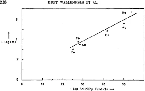
This stoichiometric relationship between activity and free SH groups corresponds to a stoichiometric relation between inhibition of the enzyme and heavy metal ions added to it (Fig. 2 ) . The order of inhibiting power of the heavy metal ions is closely related to the solubility products of their sulfides. Heavy metal ions behave towards A D H exactly as they do towards SH-containing complexing agents (4) as for instance mercapto-
A C T I O N O F A D H A N D ß - G A L A C T O S I D A S E 217
S H - 6 r o u p| o, per Molecu le
15
1 0
1 0 0 0 0 20000 Turnover Number -
30000
FIG. 1. Dependence of the activity on the number of free SH groups of crystalline yeast-ADH. (Estimation of activity in pyrophosphate-semicarbazide-buffer pH 8.6;
estimation of SH groups according to Boyer (14) ·)
ethyliminodiacetic acid described by Schwarzenbach and co-workers (5a) (Fig. 3 ) .
2. M A X I M U M N U M B E R OF FREE SH GROUPS
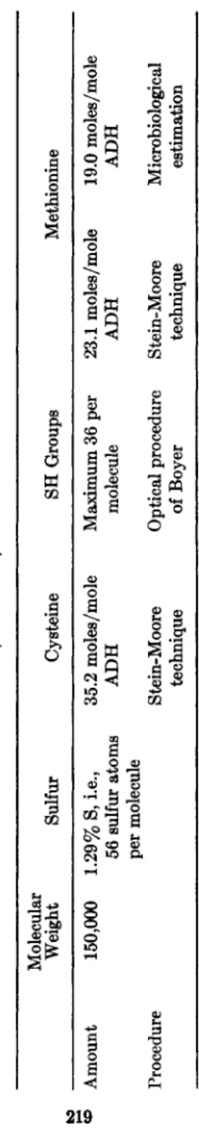
Tables I and II show the elementary analysis as well as the amino acid composition of A D H (8). The sum of methionine and cysteine rather
700 80 SO
ο -s
PCMB' Pb++
V \ \\ \ Ν \ \ V
1 1
Λ
-8 - 7 -ε -s
log \Inhibifon\—
-3
FIG. 2. Inhibition of yeast-ADH with various heavy metal ions in 0.05 M tris buffer, pH 7.6, without incubation, at 20°. D P N+ = 4.5 Χ 10"4 M, alcohol = 0.12 M, A D H =s 2.1 χ 10"* M (turnover number 21,200). [(Α/Αο· 100 = relative activity in per cent. A is the activity in the presence of the inhibitor, Ao, in its absence.]
CO ,
35
3 0
t 2 5
218 KURT WALLENFELS ET AL.
Hg ο
6
- loa [Ml log [Ml
Z n ο
2
0
0 1 0 20 30 50
log Solubility Products —>
FIG. 3. Relation between the solubility products of metal sulfides and the concen- tration of metal ions sufficient for complete inhibition of yeast-ADH. [ M ] = metal ion concentration.
closely equals the total amount of sulfur although allowance has to be made for the experimental error of elementary analysis. Cysteine as de- termined by the procedure of Stein and Moore equals exactly the number
of 3 6 free SH groups, which we found in our most active enzyme prepara- tion. With 4 . 5 X 1 0 ~4 M D P N + this preparation had a turnover number of 3 1 , 7 0 0 , with 7 . 5 X 1 0 ~3 M D P N + the turnover number was 5 9 , 0 0 0 . If free S H groups are the only chemical function at the enzymatically active site, it should then be impossible to obtain a preparation of yeast- A D H with an activity much higher than that.
3 . SH GROUPS I N THE PRESENCE OF SUBSTRATE
Barron and Levine found that the number of free SH groups as de- termined by amperometric titration with silver nitrate decreases in the presence of D P N + and alcohol (5b). The same is true for reduced D P N and acetaldehyde, as we found (Table I I I ) . Apparently each of the two substrates is bound by different SH groups because the sum of SH groups reacting with each substrate alone equals the decrease obtained in the presence of an excess of both substrates together.
4 . NUMBER OF ACTIVE SITES
If reduced D P N and acetaldehyde are bound by different SH groups and together consume 1 0 . 3 of these groups, then the number of active sites
TABLE I ESTIMATION OF SULFUR, CYSTEINE, AND METHIONINE IN YEAST-ADH Molecular Weight Sulfur Cysteine SH Groups Methionine Amount 150,000 1.29% S, i.e., 35.2 moles/mole Maximum 36 per 23.1 moles/mole 19.0 moles/mole 56 sulfur atoms ADH molecule ADH ADH per molecule Procedure Stein-Moore Optical procedure Stein-Moore Microbiological technique of Boyer technique estimation
219
220 KURT WALLENFELS ET AL.
Moles amino acid/Mole A D H
Amino Acid
Estimation by Stein-Moore
Technique" Microbiological Estimation5
Alanine 131.7
—
Arginine 34.3
—
Aspartic Acid 125.2 —
Cysteine 35.2 —
Glutamic Acid 122.6 —
Glycine 200.2 —
Histidine 42.3
—
Isoleucine 86.5 —
Leucine 104.0 —
Lysine 103.2 106.6
Methionine 23.1 19.0
Proline 56.0 —
Serine 77.8 —
Threonine 62.5 —
Tyrosine 51.7 —
Valine 146.9 —
° Unpublished data of Prof. Dr. M. Brenner and Dr. H. Büchner, Basel, Switzerland.
b Analysis carried out by Dr. G. Holz and G. Limberg, Tutzing, Germany.
should be about half as great, that is 5-6. In fact this is the maximum number of D P N molecules which can be bound by A D H . Figure 4 shows the number of bound D P N + molecules as determined by us with the method of Klotz (6), and Hayes and Velick (7). Extrapolation towards
T A B L E III
AMPEROMETRIC TITRATION OF S H GROUPS OF Y E A S T - A D H IN THE PRESENCE OF ACETALDEHYDE AND D P N HA
SH groups per molecule % decrease
6.3 mg. ADH 18.0 —
6.3 mg. ADH
1 mg. D P N H 11.4 37
6.3 mg. A D H
5 mg. acetaldehyde 13.0 28
6.3 mg. ADH 1 mg. D P N H
5 mg. acetaldehyde 7.7 57
° Titrations in NH4NOe-buffer pH 9.2 with 0.1 Ν AgNOg, T A B L E II
AMINO ACID COMPOSITION OF ALCOHOL DEHYDROGENASE FROM BAKERS' YEAST
ACTION OF ADH AND ß-GALACTOSIDASE 221
9 6.35 Ί
16 6.10 I 5.90 21,600 Recrystallized once with
17 5.50, 6.00, 5 . 5 5 J * ( N H4)2S 04
19 5.25, 5.20 5.22 19,700
20 4.45, 4.95 4.70 24,800 Recrystallized thrice with ( N H4)2S 04
22 33.6 1 34.7 24,800 Zinc-ADH, 32.5 SH Groups 23 35.0, 35.4/
26 4.80, 4.96 4.88 19,700 Zinc-ADH dialyzed against EDTA
27 25.6, 24.1, 25.2 25.0 19,700 Zinc-ADH, 26.5 SH groups 28 10.8, 10.5 10.65 19,700 Zinc-ADH
recrystallized once with ( N H4)2S 04 without Zn-addition 29 28.3, 27.7 28.0 26,400 Zinc-ADH, 33.5 SH groups
18 11.5 — — Known sample (11.7 zinc)
a 9-28: X-ray fluorescence; 29: Titration with EDTA.
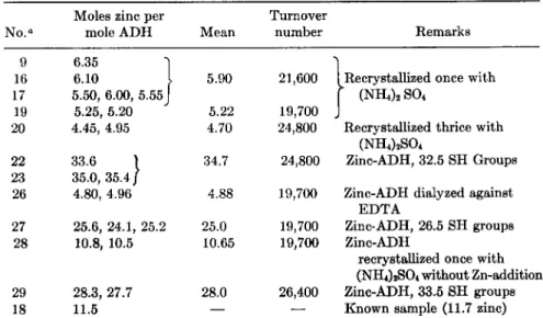
Table IV shows the amount of zinc in zinc-ADH compared to the num- ber of SH groups in the A D H used as starting material. It can be seen that both groups of values parallel each other to a certain extent. How-
ever, there is a difference between the 5 zinc atoms of A D H and the
* Vallee and Hoch (10a) reported about 4 zinc atoms per mole ADH in their best preparations. Pfleiderer et al. (10b), however, found about 5 atoms by the dithizone method.
infinitely large D P N + concentration leads to 5.5 molecules D P N + per molecule A D H (8).
5. INCREASE OF N U M B E R OF ACTIVE SITES
Vallee and Hoch were the first to show that A D H from yeast contains a number of very tightly bound zinc atoms which corresponds roughly to the number of binding sites for D P N (9). We were able to confirm this by the X-ray fluorescence method and found 5 atoms zinc/molecule A D H of the molecular weight 150,000 (10) * When A D H is crystallized from an ammonium sulfate solution in the presence of zinc sulfate different crystals are obtained of a protein which we call zinc-ADH and which even after days of dialysis still contains several times as much zinc as did the origi- nal A D H , which served as starting material (10).
TABLE IV
ESTIMATION OF ZINC IN DIFFERENT CRYSTALLINE Y E A S T - A D H AND Z I N C - A D H PREPARATIONS
Moles zinc per Turnover
N o .a mole ADH Mean number Remarks
222 KURT WALLENFELS ET AL.
6
FIG. 4 . The binding of D P N+ by yeast-ADH; r = 5.5 (r = number of binding sites).
additional 20-30 atoms in zinc-ADH regarding the stability of the zinc- protein bond. A D H can be dialyzed against E D T A for days without losing zinc to any appreciable amount. Zinc-ADH under the same conditions entirely loses its additional zinc atoms. This unusual stability of the zinc- protein bond in A D H leads to the assumption that at least one sulfur par- ticipates in it, because there are no other functional groups in proteins of which binding constants of this magnitude are known. In zinc-ADH it is mainly the 42 histidine residues together with SH groups which may be responsible for the additional zinc binding capacity.
2 5
ο ι 1 1 1- 1 1 0 25 50 7 5 100 1 2 5
r / t D P Nf+r e e] — »
FIG. 5 . The binding of D P N+ by Z i n c - A D H ; r = 2 3 (r = number of binding sites).
A (upper line) = calculated for the total number of binding sites; Β (lower line) = calculated for the additional binding sites.
A C T I O N O F A D H A N D ß - G A L A C T O S I D A S E 223
90
FIG. 6. Relative activity of yeast-ADH and zinc-ADH. ADH = 2.5 χ 10~9 Af, zinc- ADH = 3 X 10"· Af. Ao = activity at D P N+ = 4.5 χ 10"* M.
creased number of binding sites leads to an increased turnover number of zinc-ADH for which the maximum of 80,000 can be extrapolated (10).
This increase in activity, however, is not proportional to the increase in number of D P N+ binding sites. Active site and binding site therefore are not necessarily identical.
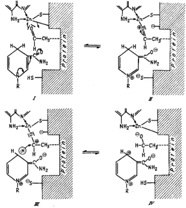
From the experiments described above it can be said that the enzymat- ically active sites of yeast-ADH probably consist of free SH groups and zinc atoms sterically arranged in such a manner that D P N + and alcohol are bound as a mixed zinc complex, within which hydrogen can be trans- ferred as hydride ion (8) (Fig. 7). The mechanism of hydrogen transfer
Combining A D H with additional zinc atoms increases the number of binding sites for D P N (8). This can be seen from Fig. 5. Extrapolation towards infinitely large D P N + concentration shows 5.5 originally present binding sites (Fig. 4) and 23 additional ones (Fig. 5) giving a total of 28.5 binding sites. However, D P N + is less tightly attached to these addi- tional binding sites than to the sites in normal A D H , which can be seen from the different slopes of the curves. This corresponds exactly to the relation between turnover number and D P N + concentration of zinc-ADH (Fig. 6 ) . At low concentrations of D P N + , A D H and zinc-ADH exhibit the same activities, and only at very high D P N + concentration the in-
224 KURT WALLENFELS ET AL.
FIG. 7. Postulated intermediate compound of ADH with D P N+ and alcohol.
FIG. 8. Postulated mechanism of action of yeast-ADH. R = adenosinediphosphate- ribose.
A C T I O N O F A D H A N D ß - G A L A C T O S I D A S E 225
with A D H and D P N would therefore be that of a Meerwein-Ponndorf- Verley reaction with zinc substituting for aluminium (Fig. 8 ) . This mech- anism was proposed by us in a short communication i n 1955 (11) and was later substantiated by Mahler and Douglas with deuterated alcohol
(12).
III. Horse Liver Alcohol Dehydrogenase Studies
Determined by amperometric titration liver-ADH has 8 free SH groups (18) ; the photometric method of Boyer (14) leads to 125 SH groups/molecule of a molecular weight of 83,300 (15, 15a). The rate- constant for the second order reaction between P C M B and A D H is
3.4 X 1 04 liters/moles X minutes at 20°
and
10.5 X 1 04 liters/moles X minutes at 35°
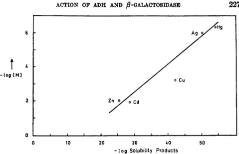
The activation energy for this reaction is 13.3 kcal./mole. The neces- sity of free SH groups for enzymatic activity follows from inhibition of the enzyme by heavy metals (Fig. 9 ) , the inhibiting power decreasing in the order H g + + , Ag+, P C M B , Cd+ + , C u + + , Zn++. As with yeast-ADH there is a straight line relationship between the solubility product of a metal sulfide and the smallest metal ion concentration which entirely in- hibits enzymatic activity (Fig. 10). Inhibition by P C M B starts at a con- centration of 1 0 ~7M and depends on time of incubation, temperature
(Fig. 11) and pH. Determined by enzymatic reaction the rate-constant is 6.4 X 1 04 liters/moles X minutes at 20°
and
18.7 X 1 04 liters/moles X minutes at 35°
for the second order reaction between P C M B and liver-ADH, the activa- tion energy is 12.8 kcal./mole. As with yeast-ADH (8) cysteine does not reverse the Hg-inhibition; in the presence of Z n S 04 and glutathione, how- ever, partial reactivation occurs.
N o t only in the reactivity of SH groups but also in its behavior to- wards weak and strong complex-forming agents, is liver-ADH analogous to yeast-ADH. This analogy points to a reaction mechanism in which, as postulated for yeast-ADH also (8), the reversible hydrogen transfer takes place in a ternary protein-DPN-substrate complex with zinc as the center of complexing.
-9 -8 -7 -6 -5 -4 -3 -2 tog [Inhibitor] • FIG. 9. Inhibition of liver-ADH with various heavy metal ions in 0.05 M tris buffer, pH 7.6, with incubation (5 minutes) at 20°. DPN+ = 3 X 10"* M, alcohol = 0.4 M, ADH = 2.7-6.0 Χ 10"8 M.
226
Ο ι é ι ι i I I I I 1 1 1— — J Ο 10 20 30 LQ 50
- l o g Solubility Products
FIG. 10. Relation between the solubility products of metal sulfides and the con- centrations of metal ions for complete inhibition of liver-ADH. [M] = metal ion con- centration.
IV. ί-Galactosidase Studies (76) 1. PREPARATION AND PROPERTIES OF ^-GALACTOSIDASE
Many investigations have been reported on the specificity of glycosi- dases, however no pure and homogeneous enzyme preparation has pre- viously been obtained. ί-Galactosidase of E. coli is the first really pure and crystalline enzyme of this class (17). Table V shows the purification procedure. The enzymatic activity was determined photometrically (405
FIG. 11. Inhibition of liver-ADH by PCMB in tris buffer, pH 7.6, at 20° and 35°.
PCMB = ΙΟ"6 Μ, ADH = 1.78 X 10"8 M, D P N+ = 3 X 10"4 M.
2 2 8 KURT WALLENFELS ET AL.
T A B L E V
PURIFICATION PROCEDURE FOR JS-GALACTOSIDASE FROM Ε. coli M L 309°
cell-free extract specific activity 42,000
Mn(Ac)2
supernatant specific activity 75,500
precipitate discarded protamine
precipitate discarded
precipitate specific activity 175,000
supernatant specific activity 83,000
alcohol \ 0-50%
supernatant discarded
4:
supernatant discarded
(NH4)2SO<
precipitate specific activity 360,000
alcohol-ether precipitate
specific activity 450,000
supernatant discarded crystallization
specific activity 500,000 over-all yield 45%
recrystallization
i
specific activity 630,000 over-all yield 11%
β Specific activity is expressed as units per mg. protein; a unit of enzyme activity has been defined as the amount of enzyme required to hydrolyze 1 Mg. O N P G in 5 ml.
phosphate buffer (ikf/30; pH 6.8) in 15 minutes at 40°, in the presence of Mn(Ac)*
( Ι Ο "8 M).
ACTION OF ADH AND ß-GALACTOSIDASE 229
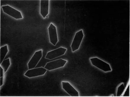
FIG. 12. Crystals of ß-galactosidase ; magnification 1:200.
tryptophan and 3.6% tyrosine can be calculated.* Special attention has been paid to the homogeneity of the enzyme. Ultracentrifuge studiesf have shown that our most active specimen contained 97% of a single component, the molecular weight of which was estimated (18) to be 365,000. Under certain conditions of pH and electrolyte concentration, this component dissociates or undergoes association to bigger units.
T A B L E V I
ENZYME CONTENTS OF E. coli AND CALF INTESTINE POWDERS
Amounts per gm. dried powder E. coli Calf intestine Enzyme activity units 1.2 X 10⁷ 1.5 X 10³
mg. protein 285 146
* We thank Dipl. Chem. A. Arens for these values.
t We are grateful to Prof. E. Patat, Munich, and Dr. E. Wiedemann, Basel, for ultracentrifuge studies.
ΐΎΐμ) as the rate of hydrolysis of o-nitrophenyl-ß-D-galactoside (OPNG).
The crystal shape of ß-galactosidase is shown in Fig. 12. Figure 13 gives the ultraviolet absorption spectrum of ß-galactosidase. From the ultra-
violet absorption spectrum in 0.1 Ν NaOH, an amino acid content of 6.8%
230 KURT WALLENFELS ET AL.
Especially high yields of ß-galactosidase are obtained from the strain E. coli ML 3 0 9 4 On the basis of activity of the pure enzyme the bacteria were found to contain 20.3 mg. ß-galactosidase protein per gm. dry weight.
This high content of a single enzyme is better appreciated when compared
«ι 0.8 I 0.7 0.6 0.5
0.4
ο
0.3 0.2
0.1 h
1.75
1.5
1.0
I 0.75 rp
£
CSI ε
0.5 ο
0.25
FIG. 1 3 . Ultraviolet absorption spectrum of ß-galactosidase in phosphate buffer (M/ZO; pH6.8).
with ß-galactosidase content of calf intestine (Table V I ) . Purification of the enzyme from this latter source has also been attempted as is shown in Table VII. In this case we have not yet been able to crystallize the enzyme. To obtain 100 mg. of our purest preparation about 1 mile of calf intestine had to be processed.
t We express our thanks to Dr. J. Monod for supplying the bacterial strain.
ACTION OF ADH AND ß-GALACTOSIDASE T A B L E VTI
PURIFICATION PROCEDURE FOR 0-GALACTOSIDASE FROM CALF INTESTINE0
cell-free e x t r a c t specific a c t i v i t y 8
s u p e r n a t a n t specific a c t i v i t y
11
M n C l2, >
p r o t a m i n e
p r e c i p i t a t e d i s c a r d e d
a l c o h o l
s u p e r n a t a n t discarded
p r e c i p i t a t e specific a c t i v i t y
17
a c e t i c a c i d
p r e c i p i t a t e discarded
p r e c i p i t a t e specific a c t i v i t y
2 0 0
s u p e r n a t a n t specific a c t i v i t y
1 2 6
s u p e r n a t a n t d i s c a r d e d
p r e c i p i t a t e specific a c t i v i t y
1,520
' ( N H4)2S 04 \ 4- a l c o h o l
s u p e r n a t a n t d i s c a r d e d
p r e c i p i t a t e specific a c t i v i t y
1 7 , 0 0 0
s u p e r n a t a n t d i s c a r d e d
over-all y i e l d 3 2 % purification 2,100 χ
a Specific activity is expressed as in Table V (see footnote).
2 3 2 KURT WALLENFELS ET AL.
2 . SPECIFICITY
In order to understand fully the mechanism of hydrolysis of lactose we should know: ( 1 ) the geometry and common structural features of all com- pounds which are substrates of ß-galactosidase, and ( 2 ) the nature of specific structures existing at the active sites of the enzyme, which are responsible for its biological activity.
The rates of hydrolysis of several substrates were determined quanti- tatively as shown in Tables VIII and IX. For some substrates hydrolysis
T A B L E V I I I
ACTIVITY OF 0-GALACTOSIDASE AT 40°
Substrate
Minole/mg. protein X minutes
Substrate
E. coli enzyme crystallized
Calf intestine enzyme (purified) o-Nitrophenylgalactoside 630.0 11.5 p-Nitrophenylgalactoside 171.5 3.2
Lactose 33.9 57.4
rates were estimated semiquantitatively by means of paper chromatog- raphy (Table X ) . Both enzymes (from E. coli and calf intestine) hydro- lyze 1 - 3 , 1 - 4 , 1 - 5 , and 1 - 6 galactosides but at different rates. The calf intestine enzyme hydrolyzes the 1 - 3 bond most rapidly while hydrolysis of the 1 - 6 bond is slowest. For the E. coli enzyme, however, this order is re-
T A B L E I X
ACTIVITY OF /3-GALACTOSIDASE FROM E. coli AT 2 0 °
Substrate /xmole/mg. protein X minutes o-Nitrophenylgalactoside 152.0
Phenylgalactoside 5.5 Ethylgalactoside 1.2
versed, which shows that differences in the rates of hydrolysis cannot be related to differences in heat of hydrolysis of the galactosides. A perfect analog of the hydrolytic specificity is the specificity of group transfer.* It was found that calf intestine ß-galactosidase could be distinguished from other lactases in that it synthesized large quantities of a galactosidoglucose which moved faster than lactose on the chromatogram. This fast moving synthetic product was identified as the galactosido-l-3-glucose by compar-
*Our thanks are due to Dipl. Chem. J. Lehmann for these results.
A C T I O N O F A D H A N D ß - G A L A C T O S I D A S E 233 TABLE X
SPECIFICITY OF J8-GALACTOSIDASES FROM Ε. coli AND CALF INTESTINE
Hydrolysis with
Substrate E. coli enzyme Calf intestine enzyme
Galactosido-l-3-glucose
+ + +
Galactosido-l-3-glucosamine
+ + +
Galactosido-l-3-glucosazone 0b 0
Galactosido- 1-3-fructose
+ +
Galactosido-l-4-glucose
+ + + + +
Galactosido-l-4-glucosamine
+
Galactosido-l-4-glucosazone 0 0
Galactosido-l-5-arabinose
++ +
Galactosido-l-6-glucose
+++
Galactosido-l-6-glucosamine
++ +
Galactosido-l-6-glucosazone
+
0Ethyl-£-D-galactoside
+++ +
Ethyl-jS-D-fucoside
+ ++
Nitrophenyl-j8-D-galactoside
++++ ++
Nitrophenyl-a-L-arabinoside
+
° + , hydrolyzed.
b 0, no hydrolysis observed.
ison with an authentic sample synthesized by R. Kuhn (19, 20). In con- trast the E. coli enzyme yielded mainly 1-6-disaccharide as the synthetic product. This shows that, contrary to expectation, the sugar that is most readily hydrolyzed is also predominantly synthesized. This observation is of importance for the elucidation of the mechanisms of hydrolysis and transglycosidation.
With respect to the geometry of the substrate, certain structural fea- tures seem to be essential for a sufficiently close approach of the molecules to the active centres in both enzymes. These are: (1) the arrangement of hydroxy groups on C-atoms 3 and 4 as it occurs in D-galactose and L-arabinose, and (2) the ß-glycosidic arrangement of substituents at posi- tion 2. The following points may also be mentioned: (1) Replacement of the C H2O H group on C-atom 5 by C H3 group or Η-atom decreases the rate of hydrolysis. (2) The nature of the agluconic part of the substrate has an apparent, though smaller, influence on the rate of hydrolysis. (3) Hydrolysis and synthesis through group transfer are brought about by the same enzyme. Further, during group transfer the steric arrangement at the anomeric C-atom remains unchanged; one ß-galactoside is converted into another /?-galactoside. (4) The rate determining step of hydrolysis and synthesis is the rupture of the C ( l ) - 0 bond.
The elucidation of the relationship between catalytic action and geom-
2 3 4 KURT WALLENFELS ET AL.
etry of the enzyme presents serious difficulties. Which groups of the en- zyme molecule are necessary for binding the substrate? Some light is shed on this question from studies of the change in enzymatic activity with changes in the pH of the medium and from inhibition and activation re- actions.
3 . P H AND ACTIVITY
Figure 1 4 shows the relationship between enzyme activity and pH of the reaction medium at 4 0 ° using ONPG as a substrate. Comparison of
5 6 7 8 p H — >
FIG. 14. Activity and stability of ß-galactosidase from E. coli at different pH val- ues; substrate = 3.33 X 10"8M OPNG, NaCl = 5 XlO"2M, in modified Veronal buffer, at 4 0 ° . Enzyme protein ( 2 5 0 7 ) was incubated in modified Veronal buffer at different pH values for 3 0 minutes at 4 0 ° . The activity was then measured at pH 6.8 in M / 3 0 phosphate buffer against ONPG ( 1 3 3 X 10~8) at 4 0 ° .
the two curves shows that within the pH range of stability of the enzyme there are at least two groups with definite charges, which are necessary for activity. The pK values of these two groups lie between 6.5 and 8.5.
That there are at least two different groups, and not just one, is indicated
ACTION OF ADH AND ß-GALACTOSIDASE 235
I I I I ι I l_J
-6 -4 -2 0 Log [Activator] —>
FIG. 15. Activation of j8-galactosidase from E. coli with I, NaCl, II, NH*C1, and III, KCl in 0.05 M tris buffer, pH 7.6, at 2 0 ° ; enzyme = 0.15 7 / m L ; substrate = 3.33 X 1 0SM ONPG.
against E D T A and distilled water for three days. It still contained a very small amount of sodium and was still active even in the absence of N a + . The activation of ß-galactosidase with sodium chloride was found to be a slow process as shown in Fig. 16 which is drawn from the data ob- tained with a Cary self-recording spectrophotometer at 280 τημ using phenylgalactoside as a substrate. Moreover there seems to be a hydrogen atom present on the site of activation by NaCl. This is shown by the fact that sodium activation proceeds at a slower rate in heavy water than in light water, while the activity in the absence of sodium chloride is the same in both media. p-Chloromercuribenzoate ( P C M B ) , heavy metal ions, and B e+ + have a strong inhibitory effect on ß-galactosidase as shown in Fig. 17. An interesting, but as yet not very clear, linear rela- tionship seems to exist between the inhibitory power of the heavy metal ions and the solubility products and heats of formation of the correspond- ing sulfides, as indicated in Fig. 18. This may be taken to suggest that ß-galactosidase is a sulfur enzyme requiring free — S H or — S ~ groups by the fact that the activity decreases very rapidly on both sides of the maximum.
4. INFLUENCE OF CATIONS
The presence of cations exerts a significant influence on the activity of the enzyme; alkali and ammonium ions activate while heavy metal ions have an inhibitory effect. Sodium ions possess the strongest activating action (Fig. 15). The enzyme used for these experiments was dialyzed
236 K U R T W A L L E N F E L S E T A L .
Time (Minutes) —»
FIG. 16. Activation of j8-galactosidase from Ε. coli with NaCl in light and heavy water; I, in H20 and D20 in the absence of NaCl; II, in H20 in the presence of NaCl (5 X 10"* M) ; III, in D*0 in the presence of NaCl (5 χ ÎO'2 M) ; enzyme = 39.5 7 / m l . ; substrate = 8 XlO"8 M phenyl-galactoside in 0.05 M tris buffer, pH 7.6, at 20°. A slight curvature in line II (compared with dotted straight line) shows a slow activating ac- tion of NaCl. This seems to proceed more slowly in heavy water (line III).
100 •«
80
1 \ \
t 60 Relative
Activity IPCMB \Ag θ λ \ ζ η \ B e 40 - 1
20
0 \ \ \
- 9 - 7 - 5 . 3
Log [inhibitor] — *
FIG. 17. Inhibition of 0-galactosidase from E. coli. Enzyme (0.12 7/ml.) was incu- bated with inhibitor (5 minutes at 20° for heavy metallic ions; 10 minutes at 40° for B e++ and PCMB) in tris buffer (0.05 M ; pH 7.6) and the reaction started with ONPG (333 χ 10"sAf in test). In the case of B e+ +, 5 X 10- 2Af NaCl was also present in test.
A C T I O N O F A D H A N D ß - G A L A C T O S I D A S E 237
70
î
( Δ Η ) \ 60 -Log (Solubility Products)
54
Hg0
-
-
- o Zn
1 ι ι ι ι
A 5 6 7 - L o g [inhibitor] — *
FIG. 18. Relationship of the solubility products and heats of formation of metal sulfides with concentration of metal ions required for 50% inhibition of /3-galactosidase from E. coli.
SH Groups
reocted
20 40 Time (Minutes)
FIG. 19. Interaction of PCMB with 0-galactosidase from E.coli; I, Inhibition ( • ) and SH estimation ( # ) at 40°; II, Inhibition at 20°; III, SH estimation at 20°; PCMB concentration, 10"4 Af, in 0.05 M tris buffer, pH 7.6, substrate = (I) 3.33 X 10"3 M ONPG at 40°, (II) 8.0 X 10"3 M phenylgalactoside at 20°. The SH reaction was fol- lowed optically at 253.7 m/z according to Boyer (14) :
% inhibition = A · 100
First order rate constants for inhibition reaction were 0.80 X 10~3 sec."1 and 1.48 X 10~3 sec."1 respectively at 20° and 40°.
2 3 8 KURT W A L L E N F E L S E T A L .
I ι ι ι ι ι 1—I
20 40 60 Time (Minutes) —»
FIG. 20. Inhibition of 0-galactosidase from Ε. coli with B e+ + in light and heavy water;
B e+ + concentration, 10~4 M, substrate = 3.33 Χ 10~8 M ONPG, NaCl = 5.10""* M, at 40°, in 0.05 M tris buffer, pH 7.6, enzyme = 0.15τ/πι1.
for its activity. Figure 19 shows the relationship between the number of SH groups (per molecular weight 100,000) that react with P C M B and the inhibition resulting therefrom at 20° and 40°. The large number of sulfhy- dryl groups involved in the rate of enzyme action leads us to the conclu- sion that ß-galactosidase possesses a large number of binding, and perhaps active, sites in a molecule of molecular weight 365,000. At 40° there is one sulfhydryl group per molecular weight 10,000 of the enzyme.
Beryllium ion strongly inhibits enzymatic activity. However, it is not to be presumed that this inhibition also results from reaction with sulfhy- dryl groups. Perhaps hydroxy groups from tyrosine or serine residues in the protein are involved. The difference between beryllium inhibition and that brought about by heavy metals is clearly demonstrated by experi-
100F
Relative Activity
20 40 60 Time (Minutes)-*
FIG. 21. Inhibition of ß-galaetosidase from E. coli with A g+i n light and heavy water;
Ag+ concentration, ΙΟ"8 M, substrate = 3.33 X 10~8 M ONPG, NaCl = 5 X 10"« M, at 20°, in 0.05 M tris buffer, pH 7.6, enzyme = Ο.ΐδγ/ml.
A C T I O N O F A D H A N D ß - G A L A C T O S I D A S E 239 ments in heavy water. Deuterium has a much stronger effect on the B e + + - enzyme interaction than on Ag+-enzyme interaction, as shown by Figs.
20 and 21.
5 . POSSIBLE REACTION MECHANISM
Let us turn once again to Emil Fischer's question about the geometry of substrate and enzyme which makes possible a sufficiently close approach of the reactants. The chemical constitution of the intermediate enzyme- substrate complex may be similar to the structure shown in Fig. 22. The
FIG. 22. A possible structure of the ß-galactosidase-substrate complex.
substrate, in its most stable conformation, C-l, according to Reeves {21), is bound to the enzyme protein through the hydroxy groups at posi- tions 2, 3, and 4. Additional binding at the C-6-hydroxy group facilitates the reaction but is not absolutely essential. It may be conjectured that the other reactants are the C = 0 groups on the enzyme sites and that bind- ing takes place through hydrogen bonds. As already shown hydroxy and sulfhydryl groups can be taken to be ultimately responsible for the rupture of the C ( l ) — Ο bond. This attack can take place in two possible ways, which have been dealt with by Koshland in detail {22). They are: (1) two SN 2 reactions involving two Waiden inversions or (2) a single frontside at- tack without inversion. The double displacement mechanism (1) is favored by Koshland. The enzyme-galactoside intermediate in this case should have the a-galactoside configuration. This is known to be more stable than the ^-configuration. If the enzyme happened to react through its SH groups, the intermediate would be a thiogalactoside, which would further increase its stability. Therefore both these intermediates seem to be unlikely. A possibility for maintaining the double displacement theory would be that
240 KURT WALLENFELS ET AL.
the stability of the α-configurated intermediate is reduced by an energy- rich conformation of the enzyme-galactoside.
One m a y as well keep in mind a third possibility of the reaction mech- anism through an intermediate ring opening as is anticipated by Shafizadeh and Thompson (28) for the H+-catalyzed hydrolysis of glycosides.
In the enzymatic process the intermediate could have the structure of a mixed O-S-acetal. This type of sugar derivative was prepared some years ago by Wolfrom and co-workers (24, 25). These compounds are reported to be very sensitive to acid hydrolysis. If the galactosyl residue were transferred to the acceptor from such a ring-opened intermediate, of course no inversion would take place. In any case, elucidation of the func- tion of the enzymatic SH groups will contribute considerably to our knowl- edge of the mechanism of glycosidic cleavage.
ACKNOWLEDGMENT
Our thanks are due to Deutsche Forschungsgemeinschaft, Fonds der Chemie der Deutschen Chemischen Industrie, and Research Foundation, New York for financial assistance and to the Government of India, Ministry of Education, for a scholarship to one of us (O.P.M.). We express our gratitude to Dipl. Chem. H. Grünewald for the translation.
REFERENCES 1. E. Fischer, Ber. 27, 2985 (1894).
2. L. Michaelis and M. Menten, Biochem. Z. 49, 335 (1913).
8. K. Wallenfels and H. Sund, Biochem. Z. 329, 17 (1957).
4. H. Sund, Doctoral Dissertation, Universität Freiburg im Breisgau, Germany, 1957.
δα. G. Schwarzenbach, G. Anderegg, W. Schneider, and H. Senn, Helv. Chim. Acta 38, 1147 (1955).
o~b. E. S. G. Barron and S. Levine, Arch. Biochem. Biophys. 41, 175 (1952).
6. I. M. Klotz, Arch. Biochem. 9, 109 (1946).
7. J. E. Hayes, Jr. and S. F. Velick, J. Biol. Chem. 207, 225 (1954).
8. Κ. Wallenfels and Η. Sund, Biochem. Ζ. 329, 59 (1957).
9. B. L. Vallée and F. L. Hoch, J. Am. Chem. Soc. 77, 821 (1955).
10. K. Wallenfels, H. Sund, A. Faessler, and W. Burchard, Biochem. Z. 329, 31 (1957).
10a. B. L. Vallée and F. L. Hoch, Proc. Natl. Acad. Sei. U. S. 41, 327 (1955).
10b. G. Pfleiderer, D . Jeckel, and Th. Wieland, Biochem. Z. 330, 296 (1958).
11. K. Wallenfels and H. Sund, Angew. Chem. 67, 517 (1955).
12. H. R. Mahler and J. Douglas, J. Am. Chem. Soc. 79, 1159 (1957).
18. R. Bonnichsen, in "Biologie und Wirkung der Fermente," p. 151. Springer, Berlin, 1953.
14. P. D . Boyer, J. Am. Chem. Soc. 76, 4331 (1954).
16. K. Wallenfels and H. Sund, unpublished data (1958).
lôa. A. Ehrenberg and Κ. Dalziel, Acta Chem. Scand. 12, 465 (1958).
16. K. Wallenfels and co-workers, unpublished data (1958), 17. K. Wallenfels and M. L. Zarnitz, Angew. Chem. 69, 482 (1957).
A C T I O N O F A D H A N D ß - G A L A C T O S I D A S E 241 18. J. Hengstenberg in "Die Physik der Hochpolymeren" (H. A. Stuart, ed.), Vol. 2,
p. 414. Springer, Berlin, 1953.
19. R. Kuhn and H. H. Baer, Ber. 87, 1560 (1954).
20. R. Kuhn, H. H. Baer, and A. Gauhe, Ber. 88, 1713 (1955).
21. R. E. Reeves, J. Am. Chem. Soc. 72, 1499 (1950).
22. D . E. Koshland, Jr., Discussions Faraday Soc. 20, 142 (1955).
23. F. Shafizadeh and A. Thompson, / . Org. Chem. 21, 1059 (1956).
24. M. L. Wolfrom and D . I. Weisblat, J. Am. Chem. Soc. 62, 878 (1940).
25. M. L. Wolfrom, D . I. Weisblat, and A. R. Hanze, J. Am. Chem. Soc. 66, 2065 (1944).
Discussion
VELICK: Dr. Wallenfels, I was interested in the inhibition in heavy water. I won- der what pH you used, and what pH means in heavy water.
WALLENFELS: We measured this pH with a glass electrode and adjusted to the same value in these two series of experiments.
VELICK: What was the pH?
WALLENFELS: It was 7.6 at 20°, that is the optimum pH for 0-galactosidase.
KOLTHOFF: YOU had a slide where you showed some relation between the activity of the metals and the solubility product. Is there any reason why there should be a relation? I think you had the solubility product of the sulfides. I was wondering for two reasons why there should be any relation at all. One is that you are dealing with the sulfides and the other is that you are dealing with the stability of mercaptides and not with the solubility.
WALLENFELS : We plot on the one side the concentration which we need for a cer- tain per cent inhibition. On the other side, the solubility products which are known for metallic sulfides. We see that these two are related. We should like to say from these experiments that probably the inhibition by heavy metal ions has to do with the formation of metal-sulfur bonds. This is the same relationship as Schwarzenbach demonstrated between the solubility products of the metal sulfides and the stability constants of complexes which he got from the same heavy metal ions with SH containing complexing agents, e.g., mercaptoethyliminodiacetic acid. So that one can say that in these complexes of very high stability the formation of the metal-sulfur bond contributes most to the stability of these complexes.
KOLTHOFF: Yes, but the thing that puzzles me is this. If you add a certain amount of metal ion, say you add equivalent amounts, then it is the concentration of the free sulfhydryl group left which determines the activity, and what you remove is a metal mercaptide, and that mercaptide is inactive. There should not be any relation at all even between the stability constants of the mercaptides, let alone the solubility prod- uct of the sulfides.
BOYER : Dr. Wallenfels, do you have to add more than a stoichiometric amount in order to get complete inactivation?
WALLENFELS : Yes, because if we add only stoichiometric amounts, we have to in- cubate for a very long time so as to attain measurable inhibition and during this period the enzyme may suffer a major loss of activity. It is then very difficult to clear up what the loss in activity corresponds to, the loss without anything or the loss by this metallic interaction.
BOYER: Then it is the concentration of metal that you have to add which is related.
WALLENFELS: Yes.
242 KURT WALLENFELS ET AL.
KOLTHOFF: Then he is measuring the rate and not the stability. If I understand what you say, with all these metals, if you wait long enough, there is an increasing inactivation, and there is a relation between the rate of inactivation and the solubility product of the sulfides.
WALLENFELS: In the reaction of protein S H groups one has always to wait for some time. With ADH it is amazing how rapid this reaction with these SH groups is. As you have seen, in the j8-galactosidase, the SH groups are not so reactive as in ADH with respect to their combination with metallic ions.
HOCH: I should like to comment about the zinc content of yeast alcohol dehy- drogenase, which Dr. Wallenfels reports as 5 atoms per molecule. Actually, examina- tion of the data on the zinc content of his preparations shows that the once-crystal- lized enzyme contains 5.90 or 5.22 atoms, while a 3-times crystallized enzyme contains 4.70 atoms of zinc. Since the metal content was decreasing with increasing purifica- tion of the enzyme, further crystallization might be expected to decrease these values even further, to correspond to the 4 atoms per molecule, reported originally. That the yeast-ADH molecule does contain 4 zinc atoms is further demonstrated by recent experiments in our laboratory (B. L. Vallée, J. H. R. Kagi, and F. L. Hoch, Federa- tion Proc. 17, 1292, 1 9 5 8 ) , in which bakers' yeast has been grown in the presence of Zn8 5. The metal is biosynthetically incorporated into yeast-ADH, and is not exchange- able with contaminating inactive zinc, as shown by the observed isotope enrichment during the procedure for isolation of the enzyme. Measurement of total zinc, Zn6 5, protein, and enzyme activity all give values for zinc content which are almost exactly 4 gram atoms per mole.
A second point has to do with Dr. Wallenfels* data indicating that there may be as many as 3 6 enzymatic sites in the yeast-ADH molecule, when he adds zinc to the enzyme.
WALLENFELS: Not enzymatic; binding sites.
HOCH : We have repeated these experiments with added zinc, and do not find any evidence of activation of yeast-ADH. On the contrary, excess zinc inhibits the en- zyme, as has been reported previously. This inhibition is more marked when activity is measured in the presence of low D P N concentrations, but is also present when D P N concentration is high. The plot of data purporting to demonstrate yeast-ADH acti- vation by excess zinc, as shown by Dr. Wallenfels, presents relative enzyme activities : that is, the numerator of his activity parameter is the observed enzymatic rate, and the denominator is the rate measured in the presence of 4.5 X 10"* D P N . This is a low concentration of D P N . The relative activity is an erroneous way of presenting these data, since it transforms an observed inhibition into an apparent activation;
this is accomplished by a decrease in the denominator of the activity parameter, rather than by an increase in activity, the numerator.
WALLENFELS: YOU have different concentrations of protein in these two experi- ments.
HOCH: No, the concentrations of protein are identical in all these experiments.
WALLENFELS : In our case this curve lies higher.
HOCH : Since we can reproduce your apparent results by plotting them as you do, I do not believe that this is an experimental difference, but an artifact produced by your calculations.
WALLENFELS : I think zinc ions do inhibit in the test, as you have seen in Fig. 2 , but if we crystallize in the presence of ZnSO* ( 4 0 moles per mole ADH) we obtain a preparation (containing 2 5 bound zinc atoms per mole A D H ; see preparation No. 2 7 , Table IV) of higher activity with a larger number of binding sites. The concentration
A C T I O N O F A D H A N D ß - G A L A C T O S I D A S E 243 of bound zinc in the final test solution was about 10"7 M, while for the onset of in- hibition we use 10"*Af zinc ion concentration (cf. Figs. 2, 5, and 6).
EDSALL: I had a question about the slower rate of reaction in deuterium. I think you suggested this might be due to the difference in rate of dissociation of the SH and OH groups.
WALLENFELS: Yes.
EDSALL : This was a reaction that took of the order of many minutes to occur.
WALLENFELS: Yes.
EDSALL: I think one would expect the ionization to go much faster than that at this pH. One might perhaps get evidence of what the relative rates might be from nuclear magnetic resonance studies.