Original Article
Electron dose rate and oxygen depletion protect zebrafish embryos from radiation damage
Jörg Pawelke
a,b, Michael Brand
c, Stefan Hans
c, Katalin Hideghéty
d,e, Leonhard Karsch
a,b,
Elisabeth Lessmann
f, Steffen Löck
b,g,h, Michael Schürer
b,i, Emília Rita Szabó
d, Elke Beyreuther
b,f,⇑aHelmholtz-Zentrum Dresden – Rossendorf (HZDR), Institute of Radiooncology – OncoRay;bOncoRay – National Center for Radiation Research in Oncology, Faculty of Medicine and University Hospital Carl Gustav Carus, Technische Universität Dresden, Helmholtz-Zentrum Dresden – Rossendorf;cCenter for Regenerative Therapies TU Dresden (CRTD), and Cluster of Excellence ’Physics of Life’, Technische Universität Dresden, Germany;dELI-ALPS, ELI-HU Non-Profit Ltd.;eOncotherapy Department, University of Szeged, Szeged, Hungary;
fHelmholtz-Zentrum Dresden – Rossendorf, Institute of Radiation Physics;gDepartment of Radiotherapy and Radiation Oncology, Faculty of Medicine and University Hospital Carl Gustav Carus, Technische Universität Dresden;hGerman Cancer Consortium (DKTK), Partner Site Dresden, and German Cancer Research Center (DKFZ), Heidelberg; andiNational Center for Tumor Diseases (NCT), Partner Site Dresden, German Cancer Research Center (DKFZ), Heidelberg, Helmholtz-Zentrum Dresden – Rossendorf, Germany
a r t i c l e i n f o
Article history:
Received 28 October 2020
Received in revised form 29 January 2021 Accepted 1 February 2021
Available online 13 February 2021
Keywords:
Electron flash effect Oxygen depletion Normal tissue toxicity Zebrafish embryo
a b s t r a c t
Background and purpose: In consequence of a previous study, where no protecting proton Flash effect was found for zebrafish embryos, potential reasons and requirements for inducing a Flash effect should be investigated with higher pulse dose rate and partial oxygen pressure (pO2) as relevant parameters.
Materials and methods: The experiments were performed at the research electron accelerator ELBE, whose variable pulse structure enables dose delivery as electron Flash and quasi-continuously (reference irradi- ation). Zebrafish embryos were irradiated with~26 Gy either continuously at a dose rate of~6.7 Gy/min (reference) or by 1441 electron pulses within 111ms at a pulse dose rate of 109Gy/s and a mean dose rate of 105Gy/s, respectively. Using the OxyLite system to measure the pO2a low- (pO25 mmHg) and a high- pO2group were defined on basis of the oxygen depletion kinetics in sealed embryo samples.
Results: A protective Flash effect was seen for most endpoints ranging from 4 % less reduction in embryo length to about 20–25% less embryos with spinal curvature and pericardial edema, relative to reference irradiation. The reduction of pO2below atmospheric levels (148 mmHg) resulted in higher protection, which was however more pronounced in the low-pO2group.
Conclusion: The Flash experiment at ELBE showed that the zebrafish embryo model is appropriate for studying the radiobiological response of high dose rate irradiation. The applied high pulse dose rate was confirmed as important beam parameter as well as the pivotal role of pO2during irradiation.
Ó2021 Elsevier B.V. All rights reserved. Radiotherapy and Oncology 158 (2021) 7–12
Radiotherapy (RT) dose delivery techniques were continuously improved during the last decades with respect to tumor confor- mity. However, the still unavoidable exposure of normal tissue holds the risk of severe side effects, which negatively affect patient’s quality of life. Strict dose constraints, but also the applica- tion of charged particles with its beneficial inverse dose profile are existing options to protect normal tissue. Alternatively, the recently described Flash effect[1], i.e. the delivery of therapeutic doses at high dose rates within maximal 500 ms, promises better normal tissue protection but similar tumor treatment efficacy compared to conventional, continuous dose delivery over minutes.
The protective effect of very high dose rates was primarily shown for electron Flash, and in the following verified for electrons and photons revealing less normal tissue side effects in different spe-
cies[2-6]. Summarising the published Flash studies, a recipe for Flash-RT[7] was formulated recommending mean dose rates of 100 Gy/s, pulse dose rates of~106Gy/s, and minimum doses per pulse (>1 Gy) and fraction (>10 Gy). Moreover, it is assumed that the oxygen concentration of the irradiated tissue is important for the presence of a Flash effect[5,7-9].
Several attempts were made at clinical proton facilities[10-12]
to verify a protecting proton Flash effect. Exemplarily, Diffenderfer et al.[12] have shown that high proton dose rates significantly reduce the radiation damage in the intestine of mice, whereas flank tumors of these mice were treated efficiently. By contrast, the irra- diation of zebrafish embryos at the same type of cyclotron failed to reveal an influence of proton dose rate on radiation induced mor- phological alterations [11]. In the discussion of this experiment, three potential explanations were identified for the missing Flash effect: 1) the zebrafish model itself; 2) a pulse-time-regime that did not fulfil the recommendation by [7] and 3) an uncertain
https://doi.org/10.1016/j.radonc.2021.02.003 0167-8140/Ó2021 Elsevier B.V. All rights reserved.
⇑Corresponding author at: Bautzner Landstraße 400, 01328 Dresden, Germany.
E-mail address:E.Beyreuther@hzdr.de(E. Beyreuther).
Contents lists available atScienceDirect
Radiotherapy and Oncology
j o u r n a l h o m e p a g e : w w w . t h e g r e e n j o u r n a l . c o m
oxygen level during irradiation that might mask a high dose rate effect[5,7,9,13,14].
In order to study the influence of 1) – 3) an experiment was scheduled at the research accelerator ELBE (Electron Beam of high Brilliance and low Emittance,[15]) at HZDR. ELBE provides an elec- tron beam of highly variable pulse structure [16,17] that was deployed in the present study to mimic the conventional, quasi- continuous dose delivery of a clinical Linac (‘‘reference”) and to generate a ~0.1 ms long electron pulse of maximum pulse dose rate of 109Gy/s. At this ultra-high pulse dose rate a Flash effect should be observable referring to a previous study, where a Flash effect was induced in zebrafish embryos by 105Gy/s electron pulse dose rate[2]. The influence of oxygen was studied by irradiation of embryos under controlled conditions at high or low partial oxygen pressure (pO2).
Materials and methods
Experimental setup and control of dose delivery at the ELBE accelerator are described in theSupplement.For the present exper- iment, a previously established setup[16-18]was complemented by additional measurement devices for online control of dose delivery.
Embryo handling and oxygen measurements
Zebrafish embryos were treated as described previously (Sup- plement, [19]), except irradiation and oxygen deprivation in 0.5 ml Eppendorf tubes. The tubes were filled with 200 ml low- melting agarose (Fig. S2b, UltraPureÒ Agarose, Invitrogen, Ger- many) to assure comparable sample height and to protect embryos from shear forces in the narrow tip of the tube. For irradiation, about 30 embryos (25–40) each were placed in the tubes, which were filled with E3 embryo medium [20] and sealed. The term
‘‘sample” hereafter referred to one Eppendorf tube including about 30 embryos.
The partial oxygen pressure was determined in E3 surrounding the embryos in the closed Eppendorf tube using the OxyLiteTM (Oxford Optronix Ltd, Abingdon, UK;[21]) system. Oxygen deple- tion kinetics were measured by inserting the sensor in a tube with a small hole in the cap, which was sealed with Parafilm to avoid gas exchange during measurement. In consequence of previously measured kinetics the embryos were treated in two groups: a low-pO2 group, which was maintained in sealed tubes one hour prior irradiation, and a high-pO2group, treated as fast as possible after sealing. Actual kinetics were proven by daily measurements in parallel to sample irradiation.
To assess potential adverse effects of volume restriction, tempo- ral hypoxia and experiment conditions control samples remain in an open vessel (6.5 cm petri dish) in the laboratory until half of the samples were irradiated.
Three independent experiment replications were performed on consecutive days using embryos of different breeding pairs, but keeping the experimental timing, room temperatures and proce- dures constant for all days. Irradiation starts every day with embryos at an age of 24 hours post-fertilization (hpf). However, the consecutive irradiation of ten runs each comprising four to six samples and the necessary changing time at the ELBE accelera- tor need several hours. In consequence, the actual irradiation time per sample was recorded and considered as surrogate marker for embryo age, relative to 24 hpf as starting point. To avoid influences of embryo age, Flash and reference samples were treated in each run.
Endpoints
Applying similar observation periods for all samples, embryonic survival and morphological alterations, like pericardial edema (pe) and curved spines (sc) were assessed daily over the four-day follow up period. Morphological alterations were related to the actual numbers of surviving embryos, whereas the survival itself was related to the number of living embryos at irradiation day. In addi- tion to that, the severity of pericardial edema (SVmean,PE) and spine curvature (SVmean,SC) was assessed from pictures taken at the 4th day post-irradiation (dpi)[19]by staging from 1 (normal appear- ance) to 4 (most severe damage). Embryo body length and diame- ters of eye and yolk sac were also determined in these pictures (Fig. S2c) using the software ZEN (Version 2.6, Zeiss, Germany).
The severities as well as the length and diameters were determined as mean values per sample, i.e., averaged over all surviving embryos.
Statistical analyses
Two-sample two-sided t-tests of independent samples were performed to compare the endpoints between reference and Flash irradiation and between Flash subgroups (irradiation before vs.
after reference). Correlations between the endpoints and the most important experimental parameters (applied dose, sealing time (i.e. pO2), irradiation time, experiment repetition (day, one-hot- encoded)) were evaluated using the Pearson correlation coefficient R to check for dependencies. To confirm that the impact of Flash irradiation was independent of these experimental parameters, multivariable linear regression was additionally performed. Every endpoint that showed a significant difference between reference and Flash in the t-test was individually considered as the depen- dent variable, while applied irradiation (reference or Flash) and the four experimental parameters were simultaneously included as independent variables. All analyses were performed with SPSS 25 (IBM Corporation, Armonk, NY, USA) and p-values below 0.05 were considered as statistically significant. Because of the explo- rative character of the present study, no multiple testing correction was applied.
Results
Comparability of experiment parameters, like irradiation and sealing time, for reference and Flash regime was confirmed by the non-significant results of the t-test (p > 0.45). Likewise, a dose homogeneity better than 90 % over the irradiation field (width 6.5 mm, height 3 mm) and an uncertainty of 10 % for the absolute dose, mainly caused by the uncertainty of radiochromic film cali- bration, was achieved for samples irradiated at both regimes. A sta- tistically significant difference (p = 0.013) was, however, found for the mean doses (±uncertainty,Suppl. S2) of 26.2 ± 1.4 Gy for refer- ence and 26.7 ± 1.6 Gy for Flash irradiation, respectively, which corresponds to a dose difference of 2 %. Considering the treatment dose, this small difference was of no (radiobiological) consequence, but confirms the reproducibility of dose delivery over all experi- mental days. Moreover, adverse effects of sample handling during irradiation were excluded by comparing the control samples run- ning in parallel to the treatment and those remaining in the lab (Table S1).
The daily measured oxygen data were summarized in a com- mon oxygen deprivation kinetics (fit procedure described inSuppl.
S4), which was applied to estimate the actual pO2prevailing at irradiation time in the embryo medium on basis of the sealing time before treatment (Fig. 1, Table S2). Independent on treatment group, the potential influence of varying sealing times, i.e. actual
pO2 at irradiation, was considered in the further analysis as one experimental parameter.
From the 98 samples (62 Flash, 36 reference, each sample com- prising about 30 embryos) irradiated at ELBE, two Flash treated samples had to be excluded due to accidental overdosage. The Flash regime turned out to be beneficial for most of the endpoints (two-samplet-test;Table 1), except yolk sac diameter and embry- onic survival observed at the 4th dpi. Indeed, the survival was higher after reference irradiation, but since the overall embryonic survival after both treatments was not distinguishable from the controls (96.06 ± 4.00) this endpoint was excluded from further analysis. Moreover, contrary to later time points, no significant dif- ferences between the two regimes were observed at the 2nd dpi.
This might indicate the temporal development of morphological malformations, since almost all embryos were hatched and clearly visible at that time. For the other endpoints, a significant protect-
ing Flash effect was found ranging from almost 25% less embryos with spinal curvature to 4% longer embryos and larger eyes at 4 dpi. Notably, stable reductions of about 20 % and 25 % were seen in the number of embryos with pericardial edema and spinal cur- vature from the 3rd dpi on.
On endpoint level, no significant correlation to radiation dose (|
R| < 0.2) was found by the Pearson correlation coefficient R (Table S3). In addition, moderate correlations to sealing and irradi- ation time and a strong correlation to experiment day were revealed for most of the endpoints.
Taking into account these influences, a protecting effect of high electron dose rate was still revealed by multivariable linear regres- sion (Table 2). Both, the number and the severity of morphologic malformations were significantly reduced after Flash compared to reference irradiation (p < 0.05). Likewise, the embryos were longer (Fig. 2a) and the eye diameter was larger in Flash samples (p < 0.05). Irradiation and sealing time as analogies to embryo age and remaining pO2level followed in general the same depen- dencies: the older the embryo and the longer the sealing the less damage was induced and the stronger the protecting effect by Flash (Fig. 2b, c). Surprisingly, most endpoints were also strongly related to the irradiation day, exemplarily shown in Fig. 2d for the endpoint embryo length, although independent of the protect- ing Flash effect. The broadening of the sealing time window (Fig. 1) and the consequential broader pO2 distribution for the high-pO2
group as well as the increased workload in the lab due to follow up in parallel to experiment are potential sources of this effect.
The protecting effect of high dose rate electron irradiation becomes evident under all circumstances, but most pronounced for the low- pO2group (Fig. 2c).
Two-sample t-tests for all endpoints between the two Flash subgroups were finally applied to check for any bias from the fixed sample order (Flash – reference – Flash) within each run that results in different sealing times and consequentially different pO2(Table S4). Significant differences were found for the embryo length, for the spinal curvature measured at the 3rd and 4th dpi and for the severity of the induced morphological malformations.
Thereby, less damage was indicated for those samples treated after the reference-sample, most likely due to the further reduction in pO2.
Discussion
The zebrafish embryo model as alternative vertebrate model is more and more applied in radiobiology and preclinical research in general[19,22]. Intermediate betweenin vitroculture and rodent, the model was used to prove the protecting effect of electron Flash [2]and to study the formation of reactive oxygen species as one of its mechanisms [5]. In distinction from these previous studies, where embryos were applied at 4 hpf[2,5], the embryos in the pre- sent work were irradiated at an age of at least 24 hpf. Zebrafish embryos become more radioresistant with age[23], for what rea- son a dose of 26 Gy, instead of 8 Gy like in the electron studies, was applied. This dose was necessary to induce measurable mor- phological alterations but did not significantly reduce embryonic survival. Moreover, the influence of embryo aging during the 6–8 hour overall beam time was considered by alternating irradiation of both regimes and is visible by the increasing embryo length with increasing age (Fig. 2b). The distribution of embryo age was com- parable for both groups and does not alter the outcome of the pre- sent study (Table 2). Consequently, the zebrafish embryo model is applicable to study the Flash effect on a whole organism level pro- vided that the age is considered and the required doses are achieved.
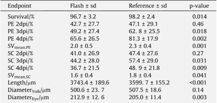
Table 1
Comparison of the mean values ± standard deviation (sd) of the different endpoints analysed in zebrafish embryos irradiated with Flash and reference electron regime. P- values of the t-test are given in the last column. Embryo length, diameters of yolk sac and eye as well as the severities of PE and SC were determined on basis of the pictures taken from all embryos at the 4th dpi.
Endpoint Flash ± sd Reference ± sd p-value
Survival/% 96.7 ± 3.2 98.2 ± 2.4 0.014
PE 2dpi/% 42.7 ± 27.7 47.1 ± 29.1 0.46
PE 3dpi/% 49.2 ± 27.4 62. 8 ± 25.5 0.018
PE 4dpi/% 65.6 ± 26.5 81.3 ± 17.9 0.002
SVmean,PE 2.0 ± 0.5 2.3 ± 0.4 0.001
SC 2dpi/% 41.0 ± 26.9 47.4 ± 27.6 0.27
SC 3dpi/% 44.2 ± 28.0 57.4 ± 29.0 0.031
SC 4dpi/% 36.7 ± 21.5 48. 9 ± 21.8 0.009
SVmean,SC 1.6 ± 0.4 1.8 ± 0.4 0.041
Length/mm 3743.4 ± 189.6 3599. 7 ± 155.2 <0.001 DiameterYolk/mm 500.6 ± 23. 7 507.5 ± 18.6 0.14 DiameterEye/mm 212.9 ± 12. 6 205.0 ± 11.4 0.003 dpi. . .day post irradiation, PE. . .pericardial edema, SC. . .spinal curvature, SV..severity.
Fig. 1.Decline of pO2after sealing of the embryos in an 0.5 ml Eppendorf tube.
Assuming exponential decay the daily measured pO2values (black dots) were fitted to pO2kinetics (grey line, pO2= 147.04exp( tsealed/12.26) + 3.26,Supplement).
The 95% confidence interval is given in light grey. Mean values as well as the range of sealing times per group are indicated by vertical lines and line segments, respectively, the level of radiobiological hypoxia (5 mmHg) is indicated by the horizontal, dashed line. The corresponding mean pO2-values as well as the extrema for each group and day were calculated on basis of the measured kinetics and were summarized inSupplement Table S2.
By careful tuning of the ELBE electron beam, pulse dose rates of 109Gy/s and time averaged dose rates of 105Gy/s were applied in the present work that clearly exceed the parameters recommended for Flash[7]. Relative to the quasi-continuous reference irradiation a protective Flash effect was seen for the majority of endpoints ranging from 4 % less reduction in embryo length and eye diameter to about 20–25% less embryo with spinal curvature and pericardial edema at final observation day. In contrast to the present finding, in previous in vitro studies at ELBE the radiation response was not altered by pulse dose rates of 109Gy/s[16,17], which indicates that this factor alone is not sufficient for an improved response by electron Flash. Likewise, in most of thein vitroexperiments with laser-driven particles no influence of the inherent ultra-high pulse dose rates on cellular outcome could be revealed [24-29]. Most probably, low pulse doses[7,8], the focus on tumor cells and the atmospheric partial oxygen level in vitro are responsible for the missing dose rate effect.
The partial oxygen level of irradiated tissue as a critical factor for ultra-high dose rate effects was already mentioned decades ago [30-33] and also pointed out in the context of Flash-RT [5,7-9,13]. Flash or any other high dose rate effects seem to require physoxia (20–50 mmHg) or hypoxia (<5 mmHg) in the irradiated sample[34]and a sufficient pulse dose to deplete enough oxygen in the irradiated volume[7,8,13,32]. In a simulation study[8], it
was shown that pulse doses of 10 Gy and higher are necessary to alter the radiation response in physoxic samples. Under atmo- spheric oxygen pressure (150 mmHg) the required pulse doses for oxygen depletion approach few 100 Gy[8], which is incompat- ible with cellular or animal survival. In the present work, the oxy- gen consumption was characterized and its kinetic (Fig. 1) was utilized to treat the embryos at different pO2. For most of the end- points, the Flash regime was protective under physoxia (high-pO2) as well as radiobiological hypoxia (low-pO2). The pronounced effect in the low-pO2group (Fig. 2c) seem to be incompatible with the idea, that hypoxia would prevent the protecting Flash effect by already low oxygen levels[7]and raises the question if the Flash effect also occurs in tumors. However, since direct pO2measure- ment in the embryos was not possible without killing them, the surrounding medium was used as surrogate keeping in mind that the chorion of the embryos might act as a buffer for quite some time. During the experiment, no oxygen deficiency effects were observed in the controls for sealing times100 minutes, which somehow supports this hypothesis.
From the present study, but also from literature, it is not clear if oxygen depletion is the only biological factor that determines the Flash effect. Other factors like the influence of the immune system [35]and an altered response of stem cell niches[13]are also dis- cussed. However, to study these issues more complex organisms Table 2
Fit parameters and coefficient of determination (R) returned from the multivariable linear regression of different endpoints and the respective experimental parameters. For each endpoint the fitted parameters (upper row), their standard deviations (second row), test statistic (T, third row) and the significance (p-values, lower row) are given. Significant parameters are bold labelled.
R Flash (0:Ref, 1:Flash) Dose/Gy Irradiation time/min Sealing time/min 2nd exp day 3rd exp day Constant
(baseline: 1st day) PE 3d pi/%
0.84 13.00 0.60 0.05 0.31 20.26 46.97 47.51
3.39 2.00 0.02 0.05 4.53 3.96 53.78
3.83 0.30 2.5 6.2 4.47 11.86 0.88
<0.001 0.76 0.007 <0.001 <0.001 <0.001 0.38
PE 4 dpi/%
0.87 15.34 0.40 0.06 0.26 17.22 41.43 74.95
2.79 1.65 0.02 0.04 3.73 3.26 44.28
5.50 0.24 3.00 6.50 6.62 12.71 1.69
<0.001 0.81 <0.001 <0.001 <0.001 <0.001 0.094
SVmean,PE
0.90 0.33 0.004 0.001 0.01 0.29 0.79 2.36
0.05 0.03 0.0001 0.001 0.07 0.06 0.79
6.60 0.13 10.0 10.0 4.14 13.17 2.99
<0.001 0.89 <0.001 <0.001 <0.001 <0.001 0.004
SC 3 dpi/%
0.84 11.18 0.53 0.03 0.24 20.00 53.58 62.67
3.62 2.14 0.02 0.06 4.84 4.23 57.51
3.09 0.25 1.50 4.00 4.13 12.67 1.09
0.003 0.81 0.077 <0.001 <0.001 <0.001 0.28
SC 4 dpi/%
0.86 12.61 0.93 0.10 0.25 12.32 26.26 93.51
2.62 1.55 0.01 0.04 3.50 3.06 41.63
4.81 0.60 10.0 6.25 3.52 8.58 2.25
<0.001 0.55 <0.001 <0.001 0.001 <0.001 0.027
SVmean,SC
0.85 0.18 0.03 0.002 0.005 0.23 0.50 2.83
0.05 0.03 0.001 0.001 0.07 0.06 0.80
3.6 1.0 2.0 5.0 3.29 8.33 3.54
<0.001 0.38 <0.001 <0.001 0.001 <0.001 0.001
Length/mm
0.91 141.71 1.95 0.64 1.62 144.43 321.23 3607.25
17.75 10.49 0.09 0.28 23.70 20.71 281.58
7.98 0.19 7.11 5.79 6.09 15.51 12.81
<0.001 0.85 <0.001 <0.001 <0.001 <0.001 <0.001
Deye/mm
0.76 8.58 2.53 0.001 0.12 0.68 15.98 271.95
1.87 1.11 0.01 0.03 2.50 2.18 29.67
4.59 2.28 0.1 4.0 0.27 7.33 9.17
<0.001 0.024 0.91 <0.001 0.79 <0.001 <0.001
dpi. . .day post irradiation, PE. . .pericardial edema, SC. . .spinal curvature, SV..severity, Deye.. eye diameter, Ref. . .Reference.
are required that allow for long-term follow up and detailed radio- biological studies on individual organs. For the parameters consid- ered in the present study (Fig. 2) an influence on the radiation response in general was seen, which has however no impact on the protecting effect of high dose rate electron treatment. The sur- prisingly observed day-to-day variation of the radiation effect could not be clarified, but indicates statistical variations in the radiosensitivity of different embryo batches or the influence of workload on individual experimental days. More important, these variations pointed out the general necessity of repetitive experi- ments on several days.
In conclusion, the Flash experiment at ELBE showed that the zebrafish embryo model is appropriate for the investigation of the radiobiological response of high dose rate irradiation. The high doses required to induce a Flash effect in 24 hpf embryos might be challenging for some accelerators, whereas the study of individual organs might be tricky from a technical point of view. The impor- tance of partial oxygen pressure in the irradiated sample was con- firmed by a clear Flash effect below atmospheric pO2. Therewith, the present study indicates that an inappropriate partial oxygen level could explain the missing proton Flash effect in[11], which could be further investigated by a repetition of this experiment under more controlled pO2-conditions. In addition to this, the con- sequences of different beam pulse structures, like the MHz and the GHz pulse frequencies of proton cyclotron and electron Linac on the dose rate effect should be investigated in more detail.
Declaration of Competing Interest
The authors declare that they have no known competing finan- cial interests or personal relationships that could have appeared to influence the work reported in this paper.
Acknowledgements
This research was carried out at ELBE at the Helmholtz-Zentrum Dresden – Rossendorf e. V., a member of the Helmholtz Associa- tion. We would like to thank Pavel Evtushenko, Rico Schurig, Ulf Lehnert and Peter Michel from the ELBE crew for support and their ongoing interest in our high-dose rate electron experiments. We thank Sandra Spieß and Johanna Stucke for help with zebrafish embryo transfer and Marika Fischer, Sylvio Kunadt and Daniela Mögel from the animal facility for dedicated zebrafish care. MB is supported by the European Union (ERC Advanced Grant Zf- BrainReg). The ELI-ALPS project (GINOP-2.3.6-15-2015-00001) is supported by the European Union and co-financed by the European Regional Development Fund. KH and ERS has received funding from the European Union’s Horizon 2020 research and innovation programme under grant agreement no 871124 Laserlab-Europe.
Appendix A. Supplementary data
Supplementary data to this article can be found online at https://doi.org/10.1016/j.radonc.2021.02.003.
References
[1] Favaudon V, Caplier L, Monceau V, et al. Ultrahigh dose-rate FLASH irradiation increases the differential response between normal and tumor tissue in mice.
Sci. Transl. Med. 2014;6(245):245ra93. https://doi.org/10.1126/
scitranslmed.3008973.
[2] Vozenin M-C, Hendry JH, Limoli CL. Biological benefits of ultra-high dose rate FLASH radiotherapy: sleeping beauty awoken. Clin Oncol 2019;31(7):407–15.
https://doi.org/10.1016/j.clon.2019.04.001.
[3] Montay-Gruel P, Petersson K, Jaccard M, et al. Irradiation in a flash: Unique sparing of memory in mice after whole brain irradiation with dose rates above Fig. 2.Subclassification of the treatment groups reference (black circle), Flash (light grey diamond) and control (dark grey square) to elucidate the influence of the different experiment parameters. The mean lengths and 95% confidence intervals are given for whole treatment groups (a) and the subgroups formed according to irradiation time and consequential embryo age (for better representation, the embryos are grouped in three time slots of 2 hpf each) (b), partial oxygen pressure at treatment (c) and experiment repetition (d).
100 Gy/s. Radiother Oncol 2017;124(3):365–9. https://doi.org/10.1016/j.
radonc.2017.05.003.
[4] Vozenin M-C, De Fornel P, Petersson K, et al. The advantage of FLASH radiotherapy confirmed in mini-pig and cat-cancer patients. Clin Cancer Res 2019;25(1):35–42.https://doi.org/10.1158/1078-0432.CCR-17-3375.
[5] Montay-Gruel P, Acharya MM, Petersson K, et al. Long-term neurocognitive benefits of FLASH radiotherapy driven by reduced reactive oxygen species.
Proc Natl Acad Sci USA 2019;116(22):10943–51. https://doi.org/10.1073/
pnas.1901777116.
[6] Montay-Gruel P, Bouchet A, Jaccard M, et al. X-rays can trigger the FLASH effect: Ultra-high dose-rate synchrotron light source prevents normal brain injury after whole brain irradiation in mice. Radiother Oncol 2018;129 (3):582–8.https://doi.org/10.1016/j.radonc.2018.08.016.
[7] Wilson JD, Hammond EM, Higgins GS, Petersson K. Ultra-high dose rate (FLASH) radiotherapy: silver bullet or fool’s gold?. Front Oncol 2020;9.https://
doi.org/10.3389/fonc.2019.01563.
[8] Petersson K, Adrian G, Butterworth K, McMahon SJ. A quantitative analysis of the role of oxygen tension in FLASH radiation therapy. Int J Radiat Oncol Biol Phys 2020;107(3):539–47.https://doi.org/10.1016/j.ijrobp.2020.02.634.
[9] Adrian G, Konradsson E, Lempart M, et al. The FLASH effect depends on oxygen concentration. BJR 2020;93(1106):20190702. https://doi.org/10.1259/
bjr.20190702.
[10] Patriarca A, Fouillade C, Auger M, et al. Experimental set-up for FLASH proton irradiation of small animals using a clinical system. Int J Radiat Oncol Biol Phys 2018;102(3):619–26.https://doi.org/10.1016/j.ijrobp.2018.06.403.
[11] Beyreuther E, Brand M, Hans S, et al. Feasibility of proton FLASH effect tested by zebrafish embryo irradiation. Radiother Oncol 2019;139:46–50.https://doi.
org/10.1016/j.radonc.2019.06.024.
[12] Diffenderfer ES, Verginadis II, Kim MM, et al. Design, implementation, and in vivo validation of a novel proton FLASH radiation therapy system. Int J Radiat Oncol Biol Phys 2020;106(2):440–8. https://doi.org/10.1016/j.
ijrobp.2019.10.049.
[13] Pratx G, Kapp DS. A computational model of radiolytic oxygen depletion during FLASH irradiation and its effect on the oxygen enhancement ratio. Phys Med Biol 2019;64:185005.https://doi.org/10.1088/1361-6560/ab3769.
[14] Spitz DR, Buettner GR, Petronek MS, et al. An integrated physico-chemical approach for explaining the differential impact of FLASH versus conventional dose rate irradiation on cancer and normal tissue responses. Radiother Oncol 2019;139:23–7.https://doi.org/10.1016/j.radonc.2019.03.028.
[15] Gabriel F, Gippner P, Grosse E, et al. The Rossendorf radiation source ELBE and its FEL projects. Nucl Instrum Methods Phys Res Sect B 2000;161-163:1143–7.
https://doi.org/10.1016/S0168-583X(99)00909-X.
[16] Beyreuther E, Karsch L, Laschinsky L, et al. Radiobiological response to ultra- short pulsed megavoltage electron beams of ultra-high pulse dose rate. Int J Radiat Biol 2015;91(8):643–52. https://doi.org/10.3109/
09553002.2015.1043755.
[17] Laschinsky L, Karsch L, Leßmann E, et al. Radiobiological influence of megavoltage electron pulses of ultra-high pulse dose rate on normal tissue cells. Radiat Environ Biophys 2016;55(3):381–91. https://doi.org/10.1007/
s00411-016-0652-7.
[18] Zeil K, Beyreuther E, Lessmann E, et al. Cell irradiation setup and dosimetry for radiobiological studies at ELBE. Nucl Instrum Methods Phys Res, Sect B 2009;267(14):2403–10.https://doi.org/10.1016/j.nimb.2009.04.015.
[19] Szabó ER, Brand M, Hans S, et al. Radiobiological effects and proton RBE determined by wildtype zebrafish embryos. PLoS One 2018;13.https://doi.org/
10.1371/journal.pone.0206879. e0206879.
[20]Brand M, Granato M, Nüsslein-Vollhard C. Keeping and raising zebrafish. In:
Zebrafish a practical approach. Oxford: Oxford University Press; 2002. p. 7–37.
[21] Griffiths JR, Robinson SP. The OxyLite: a fibre-optic oxygen sensor.. BJR 1999;72(859):627–30.https://doi.org/10.1259/bjr.72.859.10624317.
[22] Rasooly RS, Henken D, Freeman N, et al. Genetic and genomic tools for zebrafish research: The NIH zebrafish initiative. Dev. Dyn. 2003;228(3):490–6.
https://doi.org/10.1002/dvdy.10366.
[23] Praveen Kumar MK, Shyama SK, Kashif S, et al. Effects of gamma radiation on the early developmental stages of Zebrafish (Danio rerio). Ecotoxicol Environ Saf 2017;142:95–101.https://doi.org/10.1016/j.ecoenv.2017.03.054.
[24] Zeil K, Baumann M, Beyreuther E, et al. Dose-controlled irradiation of cancer cells with laser-accelerated proton pulses. Appl. Phys. B 2013;110(4):437–44.
https://doi.org/10.1007/s00340-012-5275-3.
[25] Raschke S, Spickermann S, Toncian T, et al. Ultra-short laser-accelerated proton pulses have similar DNA-damaging effectiveness but produce less immediate nitroxidative stress than conventional proton beams. Sci Rep 2016;6(1).https://doi.org/10.1038/srep32441.
[26] Bin J, Allinger K, Assmann W, et al. A laser-driven nanosecond proton source for radiobiological studies. Appl. Phys. Lett. 2012;101(24):243701.https://doi.
org/10.1063/1.4769372.
[27]Laschinsky L, Baumann M, Beyreuther E, et al. Radiobiological effectiveness of laser accelerated electrons in comparison to electron beams from a conventional linear accelerator. J Radiat Res 2012;53:395–403.
[28] Bayart E, Flacco A, Delmas O, et al. Fast dose fractionation using ultra-short laser accelerated proton pulses can increase cancer cell mortality, which relies on functional PARP1 protein. Sci Rep 2019;9(1). https://doi.org/10.1038/
s41598-019-46512-1.
[29] Hanton F, Chaudhary P, Doria D, et al. DNA DSB repair dynamics following irradiation with laser-driven protons at ultra-high dose rates. Sci Rep 2019;9 (1).https://doi.org/10.1038/s41598-019-40339-6.
[30] Epp ER, Weiss H, Santomasso A. The oxygen effect in bacterial cells irradiated with high-intensity pulsed electrons. Radiat Res 1968;34(2):320.https://doi.
org/10.2307/3572557.
[31] Weiss H, Epp ER, Heslin JM, et al. Oxygen depletion in cells irradiated at ultra- high dose-rates and at conventional dose-rates. Int J Radiat Biol Relat Stud Phys Chem Med 1974;26(1):17–29. https://doi.org/10.1080/
09553007414550901.
[32] Berry RJ, Hall EJ, Forster DW, et al. Survival of mammalian cells exposed to X rays at ultra-high dose-rates. BJR 1969;42(494):102–7. https://doi.org/
10.1259/0007-1285-42-494-102.
[33] Hendry JH, Moore JV, Hodgson BW, Keene JP. The constant low oxygen concentration in all the target cells for mouse tail radionecrosis. Radiat Res 1982;92(1):172.https://doi.org/10.2307/3575852.
[34] McKeown SR. Defining normoxia, physoxia and hypoxia in tumours—
implications for treatment response. BJR 2014;87(1035):20130676.https://
doi.org/10.1259/bjr.20130676.
[35] Jin J-Y, Gu A, Wang W, Oleinick NL, Machtay M, (Spring) Kong F-M. Ultra-high dose rate effect on circulating immune cells: A potential mechanism for FLASH effect? Radiother Oncol 2020;149:55–62. https://doi.org/10.1016/j.
radonc.2020.04.054.