Systematic Review
Efficacy and safety of stenting and additional oncological
treatment versus stenting alone in unresectable esophageal cancer:
A meta-analysis and systematic review
Benedek Tinusz
a,b, Alexandra Soós
c, Péter Hegyi
c, Patrícia Sarlós
d, László Szapáry
c, Adrienn Er} os
c, Donáta Feczák
c, Zsolt Szakács
c, Katalin Márta
c, Viktória Venglovecz
e, Bálint Er} oss
c,⇑aInstitute for Translational Medicine, Medical School, Szentágothai Research Centre;b1st Department of Internal Medicine, Medical School;cInstitute for Translational Medicine, Medical School, Szentágothai Research Centre, University of Pécs, Hungary;dDepartment of Gastroenterology, 1st Department of Medicine, Medical School, University of Pécs, Hungary;eDepartment of Pharmacology and Pharmacotherapy, University of Szeged, Hungary
a r t i c l e i n f o
Article history:
Received 24 July 2019
Received in revised form 30 April 2020 Accepted 11 May 2020
Available online 15 May 2020 Keywords:
Esophageal cancer Malignant stricture Esophageal stent Irradiation stent Palliation
a b s t r a c t
Aim:To compare the efficacy and safety of stent insertion alone to stent insertion combined with any active oncological treatment in the palliative care of esophageal cancer.
Methods:A meta-analysis and systematic review were performed according to the PRISMA Statement.
Comparative studies with patients receiving stent insertion alone (control group) were compared to patients receiving oncological therapy in addition to stent placement (intervention group). For mean dys- phagia grade before stenting, weighted mean differences (WMD), for the complications of stenting, risk ratios (RR) were calculated, both were interpreted with 95% confidence intervals (CI). Whenever possible, subgroup analyses were performed for studies with irradiation stents as intervention. Survival, late dys- phagia, esophageal perforation and medical costs were analyzed via systematic review. The protocol of the study was registered prior on PROSPERO.
Results: 17 studies with 1177 esophageal cancer patients were included in the final analysis, with 629 and 548 in the control and intervention groups, respectively. We found no significant difference in any complications of stenting between the two groups. 13 studies reported mean or median survival, and 8 found that combined therapy resulted in a significantly longer life expectancy. In the other 5 studies, there was no difference in survival between the two groups. Furthermore, additional treatment may be more effective in the long-term relief of dysphagia than stenting alone.
Conclusions: Irradiation stents may prolong survival, and stenting combined with oncological treatment does not increase the risk of complications as compared to stenting alone. However, further studies are warranted.
Core tip: Esophageal cancer is the eighth most common type of malignancy worldwide, and its prognosis is very poor. This suggests that palliative treatment modalities are paramount in its treatment. Self- expanding metal stents play an important role in the management of dysphagia caused by the tumor.
However, it is unclear whether any additional oncological therapy should be administered to patients besides stenting. In this meta-analysis and systematic review, we evaluated the safety and efficacy of additional oncological therapies alongside stenting versus stenting alone in case of unresectable esopha- geal cancer.
Ó2020 Published by Elsevier B.V. Radiotherapy and Oncology 147 (2020) 169–177
Esophageal cancer is the eighth most common cancer world- wide with an estimated annual incidence and mortality above 572,000 and 508,000, respectively[1]. This makes it the sixth lead- ing cause for cancer-related mortality[2]. Its five-year survival rate is estimated to be as low as 14%[3]and patients with metastatic
disease have a median survival of less than one year if treated with chemotherapy[4].
Low survival rates are also explained by the fact that the major- ity of tumors are unresectable at the time of diagnosis[3]. In most cases the diagnosis is made after the onset of dysphagia, which indicates at least locally advanced cancer[5]. The most common symptom is dysphagia in 74% of patients at the time of diagnosis [6]. Absence of early symptoms and the lack of precancerous states make screening procedures difficult to organize effectively [3],
https://doi.org/10.1016/j.radonc.2020.05.015 0167-8140/Ó2020 Published by Elsevier B.V.
⇑ Corresponding author at: Institute for Translational Medicine, Medical School, University of Pécs, Szigeti út 12, Pécs 7624, Hungary.
E-mail address:eross.balint@pte.hu(B. Er}oss).
Contents lists available atScienceDirect
Radiotherapy and Oncology
j o u r n a l h o m e p a g e : w w w . t h e g r e e n j o u r n a l . c o m
with the exception of Barrett’s esophagus, where surveillance is recommended[7].
Such high mortality rates underline the importance of palliative treatment options. Endoscopic stenting with metal stents plays an important role in the management of malignant dysphagia to achieve immediate dysphagia relief and quality of life improve- ment[8].
Currently, there are no clear guidelines on whether additional oncological treatment is required besides stenting in the palliative care of esophageal cancers[9]. Stenting may be combined with various types on oncological treatment, including photodynamic therapy, brachytherapy (irradiation stents), radiotherapy, chemotherapy, or chemo-radiotherapy. At present, the decision lies in the hands of individual clinicians, and is usually based on patient characteristics, such as performance status, presence of metastases, age and expected survival time[9].
Our study aims to quantitatively and qualitatively analyze the potential benefits, drawbacks and safety measures of oncological treatment administered in addition to palliative stenting for incur- able esophageal cancer.
Methods
We conducted a meta-analysis and systematic review following the Preferred Reporting Items for Systematic Reviews and Meta- Analyses (PRISMA) Statement [10]. Our work was performed in accordance with the Cochrane Handbook for Systematic Reviews of Interventions Version 5.1.0 [11]. The protocol was registered in PROSPERO under registration number CRD42018093921[12].
Search
Two independent authors (BT and LS) carried out a comprehen- sive search using six electronic databases (PubMed, EMBASE, the Cochrane Library, Web of Science, clinicaltrials.gov, and the WHO Global Health Library) with the purpose of gathering all relevant articles on the topic of palliative stent therapy in esophageal cancer patients from inception until 10 February 2020.
Our PICO items were as follows: we looked for studies on patients with incurable esophageal cancer (P) that compare two palliative treatment modalities: stent insertion alongside any addi- tional active oncological therapy (I) or stent insertion alone (C). The primary outcomes were mean survival time after stent insertion and the relief of dysphagia. Secondary outcomes were the compli- cations of stenting (such as hemorrhage, deaths due to hemor- rhage, chest pain requiring opiate analgesics, fever, stent migration, restenosis or obstruction, tracheoesophageal fistula for- mation, aspirational pneumonia and esophageal perforation) and the cost of the medical treatment (O).
The query ‘‘(esophagus OR oesophagus) AND ((malignan* OR cancer* OR carcinoma) AND (stricture OR stenosis OR obstruction OR blockage OR dysphagia)) AND (stent OR onco* OR radio* OR chemo* OR beam OR best supportive OR palliat*)” was used in all six databases. For a draft of our search strategy, seeSupplementary 1.
The ‘‘human” filter was applied when searching in PubMed, EMBASE and the WHO Global Health Library. The ‘‘trials” and
‘‘completed” filters were used in case of the Cochrane Library and clincaltrials.gov, respectively. We imposed no language restriction to our search.
Inclusion and exclusion criteria
We included both observational and interventional studies except for case studies, case reports, editorials, comments, letters and reviews. Conference abstracts were included if they contained
sufficient data for analysis. We included studies with either prospective or retrospective data collection, regardless of their pri- mary objectives. In case of multiple publications on the same group of patients, the most recent record was chosen.
The inclusion criteria required patients to be over 18 years of age, with a diagnosis of incurable, late-stage esophageal cancer (of any histological subtype). An intervention and a control group both had to be present in the same study in order to provide com- parability. The control group was defined as patients receiving any type of metallic stent implantation for palliative purposes, without the concurrent addition of other active treatment modality. The indication for stenting was malignant dysphagia. The intervention group consisted of those patients who received a metallic esopha- geal stent of any kind in combination with any type of active oncotherapy (radio-, chemo-, or photodynamic therapy) for pallia- tive purposes. Insertion of irradiation stents was considered an intervention, as brachytherapy delivered by the stent is an addi- tional active oncological treatment modality.
Screening and selection
Articles yielded by the initial search were imported into a refer- ence management program (EndNote X7, Clarivate Analytics, Philadelphia, PA, USA). The same software was utilized for the removal of duplicates by searching for articles with overlapping publication year, authors or title.
After duplicate removal, two independent researchers (BT and LS) simultaneously screened all remaining articles against the pre-defined eligibility criteria first by title, abstract and then full text in order to find studies for inclusion. Reference lists of selected articles were searched to identify studies potentially missed by the electronic search. Any disagreements were resolved by arbitration by a third investigator (BE).
Data extraction
Two investigators separately extracted data from studies included and manually entered them on a Microsoft Excel 2016 sheet (Office 365, Microsoft, Redmond, WA, USA). Data were col- lected on first author, publication year, study type, geographical location, definition of control and intervention groups, demograph- ical characteristics of patients included, and histological subtype of the tumors. Finally, data were collected on the aforementioned outcomes of interest. Differences in the data sheets were resolved by consensus.
Quality assessment and quality of evidence
We used a modified version of the Newcastle-Ottawa Scale (NOS) for the quality assessment of cohort studies included in our analysis[13].Supplementary 2shows the NOS Quality Assess- ment Form for Cohort Studies modified to fit the study design of the articles included. The Cochrane Risk of Bias Tool was used to assess the quality of the randomized-controlled trials[14]. The Grading of Recommendations Assessment, Development and Eval- uation (GRADE) methodology was used to rate the quality of evi- dence as high (level A), moderate (level B), low (level C) or very low (level D)[15].
Data synthesis and analysis
In case of the survival time and the dysphagia grade weighted mean differences (WMD) were calculated with their 95% confi- dence intervals (CI). In other comparisons, risk ratios (RRs) with 95% CI were calculated from the raw data extracted. Subgroup analyses were also performed by treatment type. Pooled estimates
were calculated with random effects model by using DerSimonian–
Laird method [16]. Results of the meta-analysis were displayed graphically using forest plots. Heterogeneity was tested by using the Cochrane’sQand the I2 statistics, where I2 = 100%(Q df)/
Q, and represents the magnitude of the heterogeneity (moderate:
30–60%, substantial: 50–90%, considerable: 75–100%) [11]. All meta-analytical calculations were performed by Stata v15.1 soft- ware (Stata Corp LLC, College Station, TX, USA).
Results
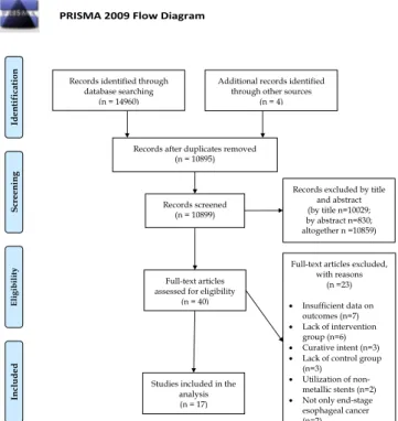
Our search yielded a total of 14,960 articles, 1226 in PubMed, 9505 in EMBASE, 355 in the Cochrane Library, 1503 in Web of Science, 2371 in the WHO Global Health Library, and 0 in clinical- trials.gov. Four articles were identified by cross-referencing. After duplicate removal and three-step selection, a total of 40 articles were assessed for eligibility by their full text. Out of these studies, 23 were excluded for the following reasons: lack of intervention group (6 studies, [17–22]), lack of a control group (3 studies, [23–25]), insufficient outcome data (7 studies, [26–32]), use of non-metallic stents (2 studies,[33,34]), patients with curable can- cer (2 studies,[35,36]) and curative intent (3 studies,[37–39]). For a summary of our search and selection, seeFig. 1: PRISMA flow- chart of the study selection process.
Finally, 17 studies were deemed eligible for either qualitative or quantitative synthesis[40–56]. These included a total of 1177 sub- jects, out of which 629 and 548 patients received stent therapy alone (control group) or stent therapy supplemented by additional oncological treatment (intervention group), respectively. In case of 234 patients, brachytherapy with I125-coated irradiation stents was the intervention method of choice, which provided basis for subgroup-analysis. Characteristics of studies included are shown inTable 1.
Tables 2 and 3contain data collected on outcomes included in the meta-analysis, andTable 4 shows a summary of the results of the quantitative synthesis.
Complications of stenting. We found no significant association between additional oncological treatment and complications of the stenting procedure, such as chest pain requiring opiate anal- gesics (RR, 1.03; 95% CI, 0.84–1.26; I2 0.0%; Supplementary 3), hemorrhage (RR, 1.32; 95% CI, 0.89–1.98;I20.0%;Supplementary 4) and deaths due to hemorrhage (RR, 1.19; 95% CI, 0.67–2.11;I2 0.0%;Supplementary 5). Neither increased risk of stent migration (RR, 0.96; 95% CI, 0.51–1.78;I20.0%;Supplementary 6), nor stent restenosis or obstruction (RR, 0.62; 95% CI, 0.36–1.09; I221.6%;
Supplementary 7) were associated with additional treatment. Fis- tula formation and development of aspirational pneumonia and development of fever as complications of stenting also did not show association with additional active oncotherapy (RR, 1.62;
95% CI, 0.68–3.87; I2 0.0%; RR, 0.76; 95% CI, 0.40–1.45;I2 0.0%;
RR, 1.24; 95% CI, 0.61–2.50; I2 0.0%; Supplementary 8–10, respectively).
Subgroup-analyses of studies where irradiation stents were used[40,42,44,45,48,54,55]as intervention were performed when- ever possible, showing no association with any of the complica- tions examined (seeSupplementary 3, 4, 6, 7, 8).
Dysphagia score before stenting and within 3 days of stenting.We found no significant difference between the two groups in dyspha- gia scores prior to and within 3 days of the stenting procedure (WMD, 0.03; 95% CI, 0.11–0.05; I2 0.0%; WMD, 0.08; 95% CI, 0.01–0.17; I20.0%; seeSupplementary 11 and 12, respectively).
Table 5demonstrates data extracted on outcomes analyzed in the systematic review section of our study.
Survival after stenting.13 studies reported this outcome as mean or median survival time after stent insertion[40–46,48–51,54,55].
Out of the 13 articles, 12 compared the survival of patients in the two groups via log rank test[40–45,48–51,54,55]. These 12 articles included a total of 894 patients, out of which 473 and 421 belonged to the control and intervention group, respectively. The use of additional oncological treatment was associated with prolonged survival in 8 of these 12 studies[40,41,43,45,48,49,54,55]compar- ing a total of 542 patients, with 281 in the control group and 261 in the intervention group, respectively. The remaining 4 studies (352 total patients; 212 and 140 patients in the control and intervention group) found no significant difference between survival in the two groups [[42,44,50,51]]. No study showed significantly reduced sur- vival time in patients that received stenting and additional onco- logical therapy as compared to stenting alone.
Regarding the sub-group of 7 articles (539 total patients, 254 and 285 patients in the control and intervention groups) where iodine-coated stents were used as intervention [40,42,44,45,48,54,55], 5 (345 total patients; 182 versus 163 patients in the control and intervention group) reported that that irradiation stents significantly prolong survival as compared to regular stents[40,45,48,54,55]. At the same time, 2 articles (194 patients, 72 in the control and 122 in the intervention group) found no significant difference between the two groups[42,44]. No arti- cles suggested that irradiation stents are associated with reduced survival as compared to regular stents. Detailed results of this out- come in each study can be found inTable 6.
Late dysphagia.The studies contained insufficient data on dys- phagia scores more than 3 days after the stenting procedure, there- fore only quantitative synthesis was carried out in case of this outcome. 5 studies[40,42,48,49,57]of the included 17 contained data on the long-term improvement of dysphagia. Guo et al. found that dysphagia was equally well palliated in both groups up until the second month after treatment, where the intervention group had a significantly lower dysphagia score (exact dysphagia score values were not specified in the article,p< 0.05)[40]. Liu et al. con- cluded that there was no significant difference between dysphagia scores of patients in the control and intervention groups at 1 month after stenting (mean dysphagia score at 1 month 1.6 and 1.7 in the Fig. 1.PRISMA flowchart for the study selection process.
control and intervention group, respectively,p= 0.91). At 3 months, however, the difference between the two groups became signifi- cant (mean dysphagia scores 2.6 vs 2.1 in the control and interven- tion groups, respectively,p= 0.03)[42]. In case of the article by Zhu et al., dysphagia was significantly lower in the intervention group as compared to the control group starting from 1 month after the procedure to the end of the follow-up[48]. Javed et al. states that the mean dysphagia score was comparable between the two groups up until 3 months post-treatment. Thereafter, dysphagia scores at 5 and 7 months were significantly lower in the interven- tion group [49]. Zhong et al. found that stenting dramatically improved dysphagia of patients, however patients in the control group had an increasing tendency of dysphagia grades 9 months after stent implantation (mean dysphagia in the control and inter- vention groups at 9 months after stenting; 1.75(±0.35) 4 and 1.58 (±38) respectively; statistical analysis not reported).
Perforation.5 studies[42,45,47,49,53]on a total of 312 patients included data concerning esophageal perforation (or a lack thereof) as a complication of stenting. Perforation only occurred in two studies [42,53], with a combined rate of 6.1% (4/66) and 9.4%
(6/64) in the control and intervention group, respectively. Out of the total 182 patients (83 in the control group and 99 in the inter- vention group) of the other 3 studies[45,47,49], no perforation was observed. None of the articles found a significant difference between the two groups (p= 0.672 in case Liu et al.).
Medical costs.Only one article out of 17[44]assessed medical costs as an outcome. They reported that regular stent treatment received by the control group to be significantly cheaper than treatment with irradiation stent in the intervention group (7000 and 26,000 Chinese yuan, approximately 1000 and 3800 USD respectively,p< 0.01)[44].
Supplementary 16 and 17represent the results of the quality assessment of the studies included.
Applying the GRADE approach to each of the outcomes assessed above resulted in a very low (D) level of evidence for every outcome.
Discussion
It is established knowledge that self-expanding metal stent insertion provides an acute relief of malignant dysphagia with an immediate success rate of 90–100% [57,58]. At the same time, recurring dysphagia within 4 to 10 weeks after stenting occurs in around 50% of patients[38,59]. On the other hand, radiotherapy exceeds at relieving long-term dysphagia [60], but the onset of its beneficial effects is slow, and may even worsen dysphagia early on due to radiation-induced swelling[49].
A randomized-controlled trial in 2005 demonstrated longer ongoing dysphagia relief with radiotherapy than stent alone[37].
Table 1
Characteristics of studies included.
Author (publication year)
Country Design Group definitions (C: Control; I:
Intervention)
Patient Number (n)
Male (n)
Age (years, mean, SD)
Follow-up (months, mean)
Histology AC/SCC (n) Bakheet (2019) South
Korea
Retrospective cohort
C: SEMS alone 41 38 67.7(11)
I: SEMS + chemotherapy 64 60 66.7(10.7)
Tian (2016) China Cohort C: SEMS alone 91 67 66.3 (9.4) 4 (median) 0/90
I: I125-coated Irradiation stent 40 30 66.9 (8.6) 4 (median) 0/41
Zhao (2016) China RCT C: SEMS alone 25 0/25
I: I125-coated Irradiation stent 18 0/18
Kim (2015) South Korea
Retrospective cohort
C: SEMS alone 45 7/10
I: SEMS + multiple modalities of oncological treatment
44 3/9
Liu (2014) China Cohort C: Conventional SEMS 32 20 61.2 2.7 5/27
I: I125-coated Irradiation stent 31 16 59.6 3.4 6/25
Zhu (2014) China RCT C: SEMS alone 75 53 71(median) 4.1(median) 14/61
I: I125-coated irradiation stent 73 61 71(median) 5.7(median) 8/65 Zhongmin
(2012)
China Cohort C: Covered stent alone 30 18 68.8(6.9) 12/18
I: I125-coated Irradiation stent 28 19 65(7.9) 8/20
Xu (2011) China RCT C: SEMS alone 17
I: I125-coated Irradiation stent 15 Zhao (2011) China Cohort C: SEMS alone I: I125-coated Irradiation
stent
25 18 Burstow (2009) Australia Retrospective
cohort
C: SEMS alone I: Adjuvant chemotherapy, radiotherapy, or both
67 23
Guo (2008) China RCT C: Conventional covered stent 27 20 69.54 (8.68) 3.3 6/20
I: I125-coated irradiation stent 26 19 72.19 (8.71) 7.2 5/22
Zhang (2005) China Cohort C: Metal stent only 34 25 62.04 2/31
I: Endoprothesis and external radiotherapy
33 26 60.17 2/30
Fu (2004) China RCT C: SEMS alone 27 16 64.2 (14) 2/23
I: Stent + external radiotherapy and/or chemotherapy
26 23 59.4 (9.6) 2/24
Javed (2004) India RCT C: SEMS alone 37 27 58.1 (12.44) 6/31
I: Stent + external beam radiotherapy 42 29 58.6 (12.13) 7/35
Zhong (2003) China Cohort C: SEMS alone 18 13 64.6 0/18
I: SEMS + external radiotherapy and/or chemotherapy
16 12 61.5 0/16
Ludwig (1998) Germany Cohort C: Nitinol stent or wallstent 17 13 67 (median) 7/10
I: stent combined with radiochemotherapy
12 11 57 (median) 3/9
Raijman (1997) USA Retrospective cohort
C: Coated expandable wallstent alone 21 10 66.8 6.4 8/13
I: Stent + chemo/radio/both 39 29 64.7 5.9 12/14
Table 2
Data extracted on outcomes synthetized via meta-analysis, part 1.
Author (year of publication)
Group (C: control; I:
intervention)
Patient Number (n)
Severe chest pain (n)
Hemorrhage (n)
Deaths due to hemorrhage (n)
Stent migration (n)
Restenosis/stent obstruction (n)
Bakheet (2019) C 41 2
I 64 8
Tian (2016) C 91 16 6 6 5 4
I 40 12 1 1 2 2
Zhao (2016) C 25 3 0 0 0 3
I 18 2 0 0 0 2
Kim (2015) C 45
I 44
Liu (2014) C 32 9 7 3 4
I 31 8 11 4 3
Zhu (2014) C 75 15 5 5 0
I 73 17 5 6 0
Zhongmin (2012) C 30 24 2 2
I 28 15 1 1
Xu (2011) C 17 4 4 7
I 15 3 4 3
Zhao (2011) C 25 4 0 0
I 18 3 0 0
Burstow (2009) C 67
I 23
Guo (2008) C 27 7 7 7 3 6
I 26 8 9 9 2 8
Zhang (2005) C 34 3 3 7
I 33 7 2 1
Fu (2004) C 27 4 1 9
I 26 7 2 1
Javed (2004) C 37 0 9
I 42 1 6
Zhong (2003) C 18 1 2 1
I 16 0 3 0
Ludwig (1998) C 17
I 12
Raijman (1997) C 21 4 2
I 39 9 2
Table 3
Data extracted on outcomes synthetized via meta-analysis, part 2.
Author (year of publication)
Group (C: control; I:
intervention)
Patient Number (n)
Mean dysphagia grade before stentinga
Mean dysphagia grade within 3 days of stentinga
Fistula formation (n)
Aspirational pneumonia (n)
Bakheet (2019) C 41 3.15 1.17
I 64 3.17 1.14
Tian (2016) C 91 3.27 0.20
I 40 3.28 0.38
Zhao (2016) C 25
I 18
Kim (2015) C 45
I 44
Liu (2014) C 32 3.03 1 3
I 31 3.10 2 2
Zhu (2014) C 75 3.40 1.30 5 14
I 73 3.30 1.40 6 11
Zhongmin (2012) C 30 3.40
I 28 3.43
Xu (2011) C 17
I 15
Zhao (2011) C 25
I 18
Burstow (2009) C 67
I 23
Guo (2008) C 27 3.12 1.04 0 2
I 26 3.22 1.07 1 1
Zhang (2005) C 34 2.15 1
I 33 2.18 3
Fu (2004) C 27
I 26
Javed (2004) C 37 3.22 0
I 42 3.10 0
Zhong (2003) C 18 3.11
I 16 3.06
Ludwig (1998) C 17
I 12
Raijman (1997) C 21 3.60 1.40
I 39 3.50 1.40
a Measured on a 4-grade scale.
The complementary effects of the immediate relief from the stent insertion and the long term effect of brachytherapy make the com- bination of palliative stenting and radiotherapy a rational decision.
Several individual studies suggest that brachytherapy using irradi- ation stents is associated with prolonged survival, in addition to the effective treatment of dysphagia (both acutely and on the long-term)[56,61].
Our meta-analysis did not find a significant difference in any assessed complications of stenting between the two groups, which suggests that active oncological therapy combined with stenting is non-inferior to stenting alone in terms of its safety. Our analysis did not cover the potential differences in quality of life between the two groups; however we believe this to be an important parameter to monitor, which should be taken into consideration when designing future trials.
Limitations and explanation of heterogeneity
While conducting the meta-analysis, we came across several limitations that may potentially impair the strength of our findings.
Regarding differences in patient populations, the tumor stage of involved patients varied from study to study. The articles reported this characteristic inconsistently, by describing the mean tumor stage[44,48,50], the number of patients with metastatic cancer [40,42,49,50], or the ECOG performance scale of the patients[48].
Methodological differences between studies include that the modality of active oncological treatment was not the same in every article. Most studies utilized I125-coated irradiation stents in the
intervention group[40,42,44,45,48,54,55]. In other cases, external radiotherapy, chemotherapy or both were applied in addition to stenting as intervention[41,43,46,47,49,50].
Statistical methods did not reveal the causes for heterogeneity among the included studies. The major confounding factors accounting for the heterogeneity are likely the differences between the populations (age, gender ratio, histological type of cancer) in the individual studies, the differences between the standard of pal- liative care. Chronological bias may be another important con- founder as studies spread between 1997 and 2019.
Another methodological difference is the inconsistency of follow-up periods. Only 5 studies out of the analyzed 17 included data on the follow-up time of patients[40,42,44,47,48].
In case of two articles, patients were allowed to receive differ- ent treatment in addition to the treatment modalities of their assigned group. In Zhao et al., the use of alternative medicine and chemotherapy was allowed before, concurrently with, or after the assigned treatment[45]. In the study conducted by Guo et al., some patients received traditional Chinese medicine as well as their assigned treatment[40].
With regards to the outcomes assessed, we found slight differ- ences in the definition of dysphagia between studies. Although every article that assessed dysphagia did so as a 0–4 scale, the def- inition of each value varied slightly.
Generalizability of the findings is questionable as the majority of studies are from China, some, from the same center, with squa- mous cell cancer as the predominant type of esophageal cancer. As environmental and genetic factors may be very different, the results above may not be reproducible in Western populations.
Table 4
Results of the meta-analysis.
N of studies RR or WMD (95% CI) I2(%)
Chest pain requiring opioids
All studiesa 12 1.03 (0.84–1.26) 0.0
Irradiation stent only 8 0.96 (0.84–1.26) 5.2
Other oncological treatment 4 1.03 (0.63–4.21) 0.0
Hemorrhage
All studiesa 11 1.32 (0.89–1.98) 0.0
Irradiation stent only 7 1.25 (0.79–1.97) 0.0
Other oncological treatment 4 1.62 (0.71–3.71) 0.0
Deaths due to hemorrhage
Irradiation stent onlyc 5 1.19 (0.67–2.11) 0.0
Stent migration
All studiesa 11 0.96 (0.51–1.78) 0.0
Irradiation stent only 7 0.78 (0.35–1.71) 0.0
Other oncological treatment 4 1.34 (0.49–3.68) 0.0
Stent restenosis
All studiesa 8 0.62 (0.36–1.09) 21.6
Irradiation stent only 5 0.92 (0.51–1.66) 0.0
Other oncological treatment 3 0.29 (0.09–0.93) 39.7
Fistula formation
All studiesa 5 1.62 (0.68–3.87) 0.0
Irradiation stent only 3 1.47 (0.55–3.91) 0.0
Other oncological treatment 2 2.35 (0.34–16.10) 0.0
Aspirational pneumonia
All studiesa 3 0.76 (0.40–1.45) 0.0
Fever
All studiesa 3 1.24 (0.61–2.50) 0.0
Dysphagia grade before stenting
All studiesa 7 0.03b (-0.11–0.05) 0.0
Dysphagia grade after stenting within 3 days
All studiesa 4 0.08b (-0.01–0.17) 0.0
OR: odds ratio; WMD: weighted mean difference.
aAll studies where the corresponding outcome was reported.
b Weighted mean difference.
c Only studies with irradiation stents contained this outcome.
Our meta-analysis found that stenting with additional active oncological therapy does not increase the risk for complications of stenting in case of patients diagnosed with unresectable esopha- geal cancer as compared to stenting alone. Furthermore, our sys- tematic review strongly suggests that additional oncological therapy may prolong the survival of patients after stenting, and irradiation stents may be more effective in the relief of late dys- phagia when compared to stenting alone.
However, due to the differences in study design, definition of outcomes and patient characteristics of the studies included, the quality of evidence remains very low. We believe that further large-scale, randomized-controlled trials are warranted to assess the effectiveness and safety of palliative treatment modalities in esophageal cancer.
Guarantor of the article Bálint Er}oss MD
Specific author contributions
Er}oss B and Hegyi P conceptualized and designed the study in cooperation with Sarlós P; Tinusz B and Szakács Z constructed the search query and carried out the search process; Tinusz B, Szapáry L and Feczák D screened the articles for eligibility; Tinusz
B and Szapáry L performed the data extraction; Tinusz B and Er}os A conducted the quality assessment; Tinusz B and Er}oss B wrote the article; Soós A carried out the statistical analysis; Márta K, Ven- glovecz V and Hegyi P provided valuable feedback after critically reviewing the first drafts of the manuscript. All authors reviewed and approved the final manuscript for publication.
PRISMA 2009 Checklist statement
The authors have read the PRISMA 2009 Checklist, and the manuscript was prepared and revised accordingly.
Statement of interests
1. Authors’ declaration of personal interests:
The authors declare no competing interests.
2. Declaration of funding interests:
This work was supported by an Economic Development and Innovation Operative Programme Grant, GINOP 2.3.2-15- 2016-00048 to PH; a Human Resources Development Opera- tional Programme of the European Union and the Hungarian Government EFOP-3.6.2-16-2017-0006 to PH. Funding was also received from the ÚNKP-18-2-I New National Excellence Pro- gram within Hungary’s Ministry of Human Capacities.
Table 5
Data extracted on outcomes synthetized via systematic reviewameasured on a 4-grade scale,bmeasure of effect not specified in the article.
Author (year of publication)
Group (C:
control; I:
intervention)
Patient Number (n)
Mean dysphagia grade after stenting at 1 montha
Mean dysphagia grade after stenting at 3 monthsa
Mean dysphagia grade after stenting at 5 monthsa
Fever (n)
Medical costsb (Chinese yuan)
Perforation (n) Bakheet
(2019)
C 41
I 64
Tian (2016) C 40 7000
I 91 26,000
Zhao (2016) C 25 0
I 18 0
Kim (2015) C 45
I 44
Liu (2014) C 32 1.60 2.60 4 3
I 31 1.70 2.10 6 2
Zhu (2014) C 75 1.76 2.56 2.65
I 73 1.74 1.85 1.87
Zhongmin (2012)
C 30
I 28
Xu (2011) C 17 5
I 15 6
Zhao (2011) C 25
I 18
Burstow (2009)
C 67
I 23
Guo (2008) C 27 1.17 3
I 26 1.22 1
Zhang (2005) C 34 1
I 33 4
Fu (2004) C 27
I 26
Javed (2004) C 37 1.27 2.55 0
I 42 1.27 1.45 0
Zhong (2003) C 18 1.36
I 16 1.29
Ludwig (1998)
C 17
I 12
Raijman (1997)
C 21 0
I 39 0
ORCID numbers
Benedek Tinusz (0000-0001-6187-526); Alexandra Soós (0000- 0001-9305-5251); Péter Hegyi (0000-0003-0399-7259); Patrícia Sarlós (0000-0002-5086-9455); László Szapáry (0000-0003-2056- 0825); Adrienn Er}os (0000-0001-6494-2708); Donáta Feczák (0000-0003-3946-2993); Zsolt Szakács (0000-0002-7035-941X);
Katalin Márta (0000-0002-2213-4865); Viktória Venglovecz (0000-0002-2316-7247); Bálint Er}oss (0000-0003-3658-842).
Appendix A. Supplementary data
Supplementary data to this article can be found online at https://doi.org/10.1016/j.radonc.2020.05.015.
References
[1]Bray F, Ferlay J, Soerjomataram I, Siegel RL, Torre LA, Jemal A. Global cancer statistics 2018: GLOBOCAN estimates of incidence and mortality worldwide for 36 cancers in 185 countries. CA Cancer J Clin 2018;68:394–424.
[2]Ferlay J, Soerjomataram I, Dikshit R, Eser S, Mathers C, Rebelo M, et al. Cancer incidence and mortality worldwide: sources, methods and major patterns in GLOBOCAN 2012. Int J Cancer 2015;136:E359–86.
[3]Enzinger PC, Mayer RJ. Esophageal cancer. N Engl J Med 2003;349:2241–52.
[4]Enzinger PC, Ilson DH, Kelsen DP. Chemotherapy in esophageal cancer. Semin Oncol 1999;26(5 Suppl 15):12–20.
[5]Huang FL, Yu SJ. Esophageal cancer: risk factors, genetic association, and treatment. Asian J Surg 2018;41:210–5.
[6] Daly JM, Fry WA, Little AG, Winchester DP, McKee RF, Stewart AK, et al.
Esophageal cancer: results of an American College of Surgeons Patient Care Evaluation Study. J Am Coll Surg, 2000;190(5):562–72; discussion 72–3.
[7]Sampliner RE. Practice guidelines on the diagnosis, surveillance, and therapy of Barrett’s esophagus. The Practice Parameters Committee of the American College of Gastroenterology. Am J Gastroenterol 1998;93:1028–32.
[8]Hindy P, Hong J, Lam-Tsai Y, Gress F. A comprehensive review of esophageal stents. Gastroenterol Hepatol 2012;8:526–34.
[9] Steyerberg EW, Homs MY, Stokvis A, Essink-Bot ML, Siersema PD, Group SS.
Stent placement or brachytherapy for palliation of dysphagia from esophageal cancer: a prognostic model to guide treatment selection. Gastrointest Endosc, 2005;62(3):333–40.
[10] Moher D, Liberati A, Tetzlaff J, Altman DG, Group P. Preferred reporting items for systematic reviews and meta-analyses: the PRISMA statement. Ann Intern Med, 2009;151(4):264-9, W64.
[11]Chandler J, Hopewell S. Cochrane methods–twenty years experience in developing systematic review methods. Syst Rev 2013;2:76.
[12]Booth A, Clarke M, Dooley G, Ghersi D, Moher D, Petticrew M, et al. PROSPERO at one year: an evaluation of its utility. Syst Rev 2013;2:4.
[13]Lo CK, Mertz D, Loeb M. Newcastle-Ottawa Scale: comparing reviewers’ to authors’ assessments. BMC Med Res Method 2014;14:45.
[14]Higgins JP, Altman DG, Gotzsche PC, Juni P, Moher D, Oxman AD, et al. The Cochrane Collaboration’s tool for assessing risk of bias in randomised trials.
BMJ 2011;343:d5928.
[15]Iorio A, Spencer FA, Falavigna M, Alba C, Lang E, Burnand B, et al. Use of GRADE for assessment of evidence about prognosis: rating confidence in estimates of event rates in broad categories of patients. BMJ 2015;350:h870.
[16]DerSimonian R, Laird N. Meta-analysis in clinical trials. Control Clin Trials 1986;7:177–88.
[17]Debevec M. Results of nonoperative treatment for esophageal cancer. Radiol Oncol 1994;28:129–33.
[18] Dimofte G, Crumpei F, Trifina L, Nicolescu S, Leanca D, Fu J-H, et al. Cost- effectiveness of endoscopically placed stents in the palliation of locally advanced esophageal carcinoma [Treatment of unresectable esophageal carcinoma by stenting with or without radiochemotherapy]. Rom J Gastroenterol. 2004;13:17–22.
[19]Antonello A, Realdon S, Diamantis G, Vecchiato M, Bocus P, Giacomini F, et al.
Cervical esophageal stent placement: a 15-year experience. Digest Liver Dis 2014;46:S18.
[20]Bertschinger P, Hacki WH. Endoprostheses in palliative treatment of malignant esophageal stenosis. Schweizerische Medizinische Wochenschrift 1989;119:803–7.
[21]Homs MYV, Steyerberg EW, Eijkenboom WMH, Tilanus HW, Stalpers LJA, Bartelsman JFWM, et al. Palliative treatment of oesophageal cancer with dysphagia: More favourable outcome from single-dose internal brachytherapy than from the placement of a self-expanding stent; a multicentre randomised study. Ned Tijdschr Geneeskd 2005;149:2800–6.
[22]Shenfine J, McNamee P, Steen N, Bond J, Griffin SM. A randomized controlled clinical trial of palliative therapies for patients with inoperable esophageal cancer. Am J Gastroenterol 2009;104:1674–85.
[23]Mao AW, Gao ZD, Xu JY, Yang RJ, Xiao XS, Jiang TH, et al. Treatment of malignant digestive tract obstruction by combined intraluminal stent installation and intra-arterial drug infusion. World J Gastroenterol 2001;7:587–92.
[24]Sehgal CM, Sharma RR, Kapoor R, Goel DR, Patel FD, Sharma SC, et al. Role of combined approach with radiotherapy and metallic stent in palliation of advanced cases of carcinoma esophagus - a pilot study. JK Sci 2002;4:130–5.
[25]Touchefeu Y, Archambeaud I, Landi B, Lievre A, Lepere C, Rougier P, et al.
Chemotherapy versus self-expanding metal stent as primary treatment of Table 6
Detailed data on the survival outcome of our systematic review;1mean,2median3confidence interval,4standard deviation,5range.
Author (year of publication)
Design Group definitions (C: Control; I:
Intervention)
Patient number (n)
Survival time (months)
pvalue of the long-rank test
Bakheet (2019) Retrospective cohort
C 41 4.41(3.23–5.57)3 0.592
I 64 5.21(4.4–6.0)3
Tian (2016) Cohort C 40 4.21(2.8)4 0.752
I 91 4.41(2.4)4
Zhao (2016) RCT C 25 4.81(3.9)4 <0.01
I 18 9.81(4.3)4
Kim (2015) Retrospective
cohort
C 45 3,21(3.0)4 <0.001
I 44 5,61(4.2)4
Liu (2014) Cohort C 32 3.11(2.6–3.6)3 0.064
I 31 3.71(3.1–4.3)3
Zhu (2014) RCT C 75 4.92(5.1–6.7)5 0.0046
I 73 5.92(4.1–5.7)5
Zhongmin (2012) Cohort C 30 4.92(1–12)5 <0.001
I 28 112(3–18)5
Zhao (2011) Cohort C 25 4.81(4.83–8.77)3 <0.01
I 18 9.81(9.43–12.83)3
Guo (2008) RCT C 27 3.51(2.72–4.16)3 <0.001
I 26 8.31(6.36–10.21)3
Fu (2004) RCT C 27 8.171(5.47–10.83)3 0.813
I 26 8.731(6.97–11.5)3
Javed (2004) RCT C 37 42 0.009
I 42 62
Ludwig (1998) Cohort C 17 4.62(0.85–25.7)5 <0.05
I 12 8.02(0.3–10.57)5
severe dysphagia from unresectable oesophageal or gastro-oesophageal junction cancer. Dig Liver Dis 2014;46:283–6.
[26]Techagumpuch A, Chanswangphuvana P, Pungpapong SU, Udomsawaengsup S, Navicharern P. Tharavej C. Compare result of definite chemoradiation combine with stent insertion and stent alone in advance esophageal carcinoma: a single institute, case series study, Thailand. Gastrointest Endosc 2014;79:AB416–7.
[27] Toyokawa T, Horii J, Fujita I. Esophageal radiotherapy and metallic stenting are effective for the stenosis of advanced esophageal cancer; compared to stenting for patients without radiotherapy. Gastrointest Endosc. 2019;89:AB185.
[28]Xie H, Zhang H, Wu K, Fan D. A comparison between radioactive and traditional stent in patients with advanced esophageal carcinoma. J Gastroenterol Hepatol 2013;28:673.
[29]Park HS, Do YS, Suh SW, Choo SW, Lim HK, Kim SH, et al. Upper gastrointestinal tract malignant obstruction: Initial results of palliation with a flexible covered stent. Radiology 1999;210:865–70.
[30]Balázs A, Kokas P, Lukovich P, Kupcsulik P. Palliative management of malignant oesophageal strictures with endoprosthesis implantation – 25 years experience. Magyar sebészet 2011;64:267–76.
[31]Cwikiel M, Cwikiel W, Albertsson M. Palliation of dysphagia in patients with malignant esophageal strictures. Comparison of results of radiotherapy, chemotherapy and esophageal stent treatment. Acta Oncol 1996;35:75–9.
[32]Rodrigues-Pinto E, Pereira P, Baron TH, Macedo G. Self-expandable metal stents are a valid option in patients with long-term survival from advanced esophageal cancer. United Eur Gastroenterol J 2017;5:A708.
[33]Rueth NM, Shaw D, D’Cunha J, Cho C, Maddaus MA, Andrade RS. Esophageal stenting and radiotherapy: a multimodal approach for the palliation of symptomatic malignant dysphagia. Ann Surg Oncol 2012;19:4223–8.
[34]Reed CE, Marsh WH, Carlson LS, Seymore CH, Kratz JM. Prospective, randomized trial of palliative treatment for unresectable cancer of the esophagus. Ann Thorac Surg 1991;51(4):552–6.
[35]Han Y-T, Peng L, Fang Q, Li Q. Value of radiotherapy and chemotherapy after SEMS implantation operation in patients with malignant esophageal stricture.
Ai Zheng 2004;23:682–4.
[36]Zhang Y, Zhou M, Bai L, Han R, Lv K, Wang Z. Radiofrequency ablation combined with esophageal stent in the treatment of malignant esophageal stenosis: a single-center prospective study. Oncol Lett 2018;16:3157–61.
[37]Homs MY, Steyerberg EW, Eijkenboom WM, Tilanus HW, Stalpers LJ, Bartelsman JF, et al. Single-dose brachytherapy versus metal stent placement for the palliation of dysphagia from oesophageal cancer:
multicentre randomised trial. Lancet 2004;364:1497–504.
[38]Jiang XJ, Song MQ, Xin YN, Gao YQ, Niu ZY, Tian ZB. Endoscopic stenting and concurrent chemoradiotherapy for advanced esophageal cancer: A case- control study. World J Gastroenterol 2012;18:1404–9.
[39]Lecleire S, Di Fiore F, Ben-Soussan E, Antonietti M, Hellot MF, Paillot B, et al.
Prior chemoradiotherapy is associated with a higher life-threatening complication rate after palliative insertion of metal stents in patients with oesophageal cancer. Aliment Pharmacol Ther 2006;23:1693–702.
[40]Guo JH, Teng GJ, Zhu GY, He SC, Fang W, Deng G, et al. Self-expandable esophageal stent loaded with I-125 seeds: initial experience in patients with advanced esophageal cancer. Radiology 2008;247:574–81.
[41]Kim JY, Kim SG, Lim JH, Im JP, Kim JS, Jung HC. Clinical outcomes of esophageal stents in patients with malignant esophageal obstruction according to palliative additional treatment. J Digest Dis 2015;16:575–84.
[42]Liu N, Liu SG, Xiang C, Cong N, Wang B, Zhou B, et al. Radioactive self- expanding stents give superior palliation in patients with unresectable cancer of the esophagus but should be used with caution if they have had prior radiotherapy. Ann Thorac Surg 2014;98:521–6.
[43]Ludwig D, Dehne A, Burmester E, Wiedemann GJ, Stange EF. Treatment of unresectable carcinoma of the esophagus or the gastroesophageal junction by mesh stents with or without radiochemotherapy. Int J Oncol 1998;13:583–8.
[44]Tian D, Wen H, Fu M. Comparative study of self-expanding metal stent and intraluminal radioactive stent for inoperable esophageal squamous cell carcinoma. World J Surg Oncol 2016;14:18.
[45]Zhao P, Zhang M-Q, Zhang Y-L, Guo Y-C, Zhao Y-L, Wu Q-W, et al. Application of esophageal irradiation stents coated with 125I particles in advanced esophageal cancer. J Buon 2017;22(1):265–9.
[46]Burstow M, Kelly T, Panchani S, Khan IM, Meek D, Memon B, et al. Outcome of palliative esophageal stenting for malignant dysphagia: a retrospective analysis. Dis Esophagus 2009;22:519–25.
[47]Raijman I, Siddique I, Lynch P. Does chemoradiation therapy increase the incidence of complications with self-expanding coated stents in the management of malignant esophageal strictures?. Am J Gastroenterol 1997;92:2192–6.
[48]Zhu HD, Guo JH, Mao AW, Lv WF, Ji JS, Wang WH, et al. Conventional stents versus stents loaded with (125)iodine seeds for the treatment of unresectable oesophageal cancer: a multicentre, randomised phase 3 trial. Lancet Oncol 2014;15:612–9.
[49]Javed A, Pal S, Dash NR, Ahuja V, Mohanti BK, Vishnubhatla S, et al. Palliative stenting with or without radiotherapy for inoperable esophageal carcinoma: a randomized trial. J Gastrointest Cancer. 2012;43:63–9.
[50] Fu JH, Rong TH, Li XD, Yu H, Ma GW, Min HQ. Treatment of unresectable esophageal carcinoma by stenting with or without radiochemotherapy.
Zhonghua Zhong Liu Za Zhi 2004;26:109–11.
[51]Bakheet N, Hu HT, Park JH, Jeon JY, Yoon SH, Kim KY, et al. Clinical effectiveness and safety of self-expanding metal stent placement following palliative chemotherapy in patients with advanced esophageal cancer. Abdom Radiol 2020;45:563–70.
[52]Xu XW, Di HT, Zhu J, Shi J. Complications related to conventional self- expandable metal stent insertion and internal irradiation stent insertion in patients with advanced esophageal carcinoma: An analysis of 32 cases. J Interv Radiol 2011;20:452–4.
[53] Zhang S, Gao Y, Zhang J. Clinical research on treatment of esophageal carcinoma in advanced stage by metal stent in combination with radiotherapy. Chin J Clin Oncol, 2005;32(6):344-5+8.
[54]Zhao P, Cui HK, Yang RM, Zhang XZ. The implantation of esophageal stent with radioactive 125I particles for advanced esophageal carcinomas: observation of therapeutic results. J Interv Radiol 2011;20:448–51.
[55]Zhongmin W, Xunbo H, Jun C, Gang H, Kemin C, Yu L, et al. Intraluminal radioactive stent compared with covered stent alone for the treatment of malignant esophageal stricture. Cardiovasc Intervent Radiol 2012;35:351–8.
[56]Zhong J, Wu Y, Xu Z, Liu X, Xu B, Zhai Z. Treatment of medium and late stage esophageal carcinoma with combined endoscopic metal stenting and radiotherapy. Chin Med J (Engl) 2003;116:24–8.
[57]Dua KS. Stents for palliating malignant dysphagia and fistula: is the paradigm shifting?. Gastrointest Endosc 2007;65(1):77–81.
[58]Homann N, Noftz MR, Klingenberg-Noftz RD, Ludwig D. Delayed complications after placement of self-expanding stents in malignant esophageal obstruction:
treatment strategies and survival rate. Dig Dis Sci 2008;53:334–40.
[59]Kozarek RA, Ball TJ, Brandabur JJ, Patterson DJ, Low D, Hill L, et al. Expandable versus conventional esophageal prostheses: easier insertion may not preclude subsequent stent-related problems. Gastrointest Endosc 1996;43:204–8.
[60] Bown SG. Palliation of malignant dysphagia: surgery, radiotherapy, laser, intubation alone or in combination?. Gut 1991;32:841–4.
[61]Song HY, Deok HL, Seo TS, Kim SB, Jung HY, Kim JH, et al. Retrievable covered nitinol stents: Experiences in 108 patients with malignant esophageal strictures. J Vasc Interv Radiol 2002;13:285–92.