ROLE OF GENETIC FACTORS IN THE
DEVELOPMENT OF IN-STENT RESTENOSIS AFTER PERCUTANEUS CORONARY INTERVENTION
Ph.D. Thesis Zsolt István Bagyura
Doctoral School of Basic Medicine Semmelweis University
Supervisor: Prof. Béla Merkely, MD, D.Sc.
Official reviewers: András Vorobcsuk, M.D., Ph.D.
László Cervenák, M.D., Ph.D.
Head of the Final Examination Committee:
Prof. Katalin Darvas, M.D., Ph.D.
Members of the Final Examination Committee:
László Kőhidai, M.D., Ph.D. med. habil.
Tibor Szűk, M.D., Ph.D.
Budapest 2017
1. INTRODUCTION
Atherosclerosis is a systemic disease. Its local manifestation in the heart is coronary sclerosis and ischemic heart disease. The developing plaque causes increasing stenosis with the progression of the disease, which may appear in various clinical manifestations (angina pectoris, acute myocardial infarction). The percutaneous coronary intervention (PCI) is the contemporary therapy of coronary atherosclerosis. During this process, a guide is positioned to the stenotic segment and the plaque is destroyed with high pressure balloon inflation.
Coronary interventions revolutionized the treatment of acute and stable forms of coronary artery disease. However, after balloon angioplasty restenosis of the target segment of the vessel occurs in 40%. Coronary stents which are basically implantable metal meshes were designed to lower the rate of restenosis.
1.1 Coronary stents
The first types of stents were made of stainless steel. These stents have been in use for the longest time and these have the lowest price.
Newer stents are made of chrome-cobalt. These stents are offering greater biocompatibility therefore lower restenosis rate. The materials and the design of the stents are continuously evolving.
These stents do not have drug coating therefore commonly called bare metal stents (BMS).
Coronary stents were developed to lower the rate of early restenosis.
However, ISR still occurs in 10-30% of the interventions with deployment of bare metal stents, therefore still forms a clinically important problem. Although the new generation of stents – called drug eluting stents (DES) - further reduced ISR rate and the need for repeated revascularization, BMSs are still widely used.
The advantage of DES is based on that these stents are coated with anti-proliferative drugs that are released locally. These stents are designed to lower the ratio of endothel proliferation, therefore
restenosis ratio could be further reduced. However, indication of these stents is less wide and the cost of these stents is significantly higher. BMS could be used in many clinical situations when the use of DES is contraindicated, for instance when the double antithrombotic therapy is contraindicated, therefore BMSs are still widely used.
1.2 The in-stent restenosis
In-stent restenosis can be classified to focal (<10 mm) and diffuse (>10 mm) groups based on the results of the invasive coronarography. The former type is determined by local and procedural factors, and could be treated well with balloon dilatation, while the latter shows a significant relationship with general, patient- related factors.
Risk factors in relationship with ISR could be classified into two groups:
1) Patient-dependent or systemic factors. Risk for restenosis is particularly high among patients with diabetes mellitus, this may be associated with metabolic alterations that promote endothelial dysfunction, accelerate intimal hyperplasia, and increased platelet aggregability and thrombogenicity. There is evidence that gender itself (female) predisposes to restenosis, and some patients may have a genetically higher risk. Genetic polymorphisms could be associated with high risk for restenosis.
2) The other group consists of procedural-dependent or local factors as diameter of vessel, length of lesion or stent, minimal lumen diameter before and after stenting, ostial lesions, stent fracture, total occlusions. These factors are related to focal in-stent restenosis.
1.3 Development of ISR
During the PCI, mechanical forces strip the endothelium from vascular wall and crack the media and the lamina elastica interna.
Various mitogenic factors and cytokines are released from the
endothel cells and platelets for instance thromboxane A2, serotonin, platelet derived growth factors. These cytokines are affecting the proliferation and migration of vascular smooth muscle cells (VSMCs). Moreover, the phenotype of these cells is changing from contractile to synthetizing and some promitotic genes are upregulating. VSMCs are producing chemotactic proteins helping the migration of other cell types. The intensified matrix metalloprotease production facilitates the remodulation of the extracellular matrix.
The result of this cascade is the uncontrolled proliferation of the VSMCs and the deployment of ECM elements around the intima and development of neointima.
1.4 The role of genetic polymorphisms
Single-nucleotide polymorphism, often abbreviated to SNP is a variation in a single nucleotide that occurs at a specific position in the genome, where each variation is present to some appreciable degree within a population (every 100-300. nucleotide base, minimum 1%).
The SNP-s do not cause diseases, but variations in the DNA sequences could affect how we develop diseases and respond to pathogens and reactions to pathophysiologic processes, therefore predispose or protect. There are a large number of candidate genes that could affect development of in-stent restenosis, these are generally coding atherosclerosis associated proteins.
1.5 MBL (mannose-binding lectin)
Mannose-binding lectin (MBL) is an acute phase protein produced by the liver as part of the innate immune system. It also has a direct opsonisation effect by binding to cell-surface receptors on phagocytes. It is present in the blood serum forming a complex with serine proteases. MBL protein is encoded from the MBL2 gene on chromosome 10. Single nucleotide polymorphisms (SNP) in exon 1 concerning the promoter region of the MBL2 gene are known to reduce the amount of functional MBL subunits 5- to 10-fold,
resulting in lower serum levels of MBL: at codon 52 (arginine to cysteine, allele D), codon 54 (glycine to aspartic acid, allele B,) and codon 57 (glycine to glutamic acid, allele C). According to the results of prior studies, a low level of MBL is related to the rapid progression of atherosclerosis, severe coronary artery disease and/or increased carotid plaque formation and graft occlusion after bypass surgery.
1.6 VEGF (vascular endothelial growth factor)
VEGF family comprises five members in mammals: VEGF-A, placenta growth factor (PIGF), VEGF-B, VEGF-C and VEGF-D.
The latter ones were discovered later than VEGF-A, and, before their discovery, VEGF-A was called just VEGF. Following PCI, a process similar to wound healing takes place in the affected coronary artery.
VEGF has an important role directly in the progress of that endothelization process and also, it has an indirect effect on the inflammation cascade and VSMC proliferation and migration.
Accordingly, the endothel stimulating effect of VEGF is essential for restoring the integrity of the vessel wall but it can also become the cause of restenosis through neointima hyperplasia. Association have been identified between VEGF polymorphisms and the risk of coronary artery disease, the development of collateral circulation in individuals with coronary artery disease. VEGF contributes to mediating neovascularization of atherosclerotic plaques. The VEGF gene’s G405C (rs2010963) and C2578A (rs699947) single nucleotide polymorphisms are located in the 5′UTR region of the VEGF gene affecting the transcription and expression of the protein.
The rs699947 polymorphism of the VEGF gene is known to be associated with a higher risk of developing certain neoplastic diseases and with the development of coronary collaterals in patients with coronary artery disease or with susceptibility to coronary heart disease. Polymorphism rs2010963 was demonstrated to be associated with several disorders, such as diabetic retinopathy, diabetic nephropathy, metabolic syndrome and myocardial infarction.
2. OBJECTIVES
The aim of my work was to investigate the association of the MBL2 and the VEGF genes’ polymorphisms and the development of ISR following BMS implantation in patients of the Semmelweis University Heart and Vascular Centre. During my work, specific goals were:
1) I investigated the frequency of MBL2 gene’s R52C - rs5030737; G54D - rs1800450 and G57E - rs1800451 poly- morphisms and their association with the development of in-stent restenosis.
2) I investigated the frequency of VEGF gene’s G405C ‒ rs2010963 polymorphism and its association with the development of in-stent restenosis.
3) I investigated the frequency of C2578A ‒ rs699947 poly- morphism and its association with the development of in-stent restenosis.
3. METHODS 3.1. Patients
205 patients with a history of percutaneous coronary intervention (PCI) and BMS implantation in native coronary, who presented with non-acute or acute cardiac symptoms which warranted a repeat coronary angiogram were prospectively enrolled between 2011 and 2013 in the Heart and Vascular Centre, Semmelweis University. All patients received standard therapy according to the actual guidelines.
ISR have been evaluated by experienced clinicians according to Mehran’s classification and patients have been categorized to the diffuse restenosis group (ISR group) and control group (without diffuse restenosis).
3.2. Clinical definitions
In-stent restenosis was considered significant if the lumen was narrowed by at least 50% according to the coronarography. Smoking in anamnesis was positive if patient was active smoker or quit smoking in one year. Diagnosis of diabetes mellitus was based on medical history of diabetes treated with oral antidiabetics, insulin or diet. Diagnosis of hypertension was based on previous medication.
Hyperlipidaemia was defined as current anti-lipidemic medication or elevated blood lipid levels in current laboratory results. BMI was calculated from height and weight, obesity was defined as BMI > 25.
3.3. Sample collection, storage and DNA extraction
Genomic DNA was extracted from whole peripheral blood with a protease based technique (Flexigene DNA System, Qiagen, Hilden, Germany). Samples (1 ml) were added to a lysis buffer and were thoroughly mixed and centrifuged. After discarding the supernatant, samples were denaturized, DNA was ethanol precipitated and reconstituted in the provided buffer. Samples were stored at -80°C.
Estimation of the DNA yield and quality control was done by spectrophotometry and determination of the 260/280 absorption ratio (Nanodrop-2000, Thermo Scientific, Wilmington, USA), the average yield was 96.0 μg (range 25–370 μg).
3.3.1. Genotyping
All assays were performed in 96-well arrays, and each plate contained controls. Genotyping of 10% of the samples was performed for quality control, with complete congruence. Genetic analysis was performed blinded to patient data, with the provided software. We established the genotypes for all patients.
Determination of the alleles of the MBL2 gene at codons 52 (D - rs5030737), 54 (B - rs1800450), and 57 (C - rs1800451) were performed by polymerase chain reaction using sequence-specific priming with LightCycler (Roche GmbH, Penzberg, Germany) real- time PCR (RT-PCR). The primers were (forward, reverse): 5-′GCA- AAG-ATG-GGC-GTG-ATGA-3′, 3′-GGG-CTG-GCA-AGA-CAA- CTA-TTA-5′; 5′-AGT-CGA-CCC-AGA-TTG-TAG-GAC-AGAG- 3′, 3′-ACC-TGG-TTC-CCC-CTT-TTC-TT-5′, 5′-AGT-CGA-CCC- AGA-TTG-TAG-GAC-AGAG-3′, 3′-CTC-CCT-TGG-TGC-CAT- CACA-5′.
The PCR mix contained 1 μL DNA, 5 μM primer and probe, 1 μL LightCycler FastStart DNA Master HybProbe kit (Roche GmbH, Penzberg, Germany), and 2.5 mM MgCl2. The following protocol was used: 10 minutes denaturation at 95°C, 35 cycles: 95°C - 10 sec, 52–56–60°C - 15 sec, 72°C - 10 sec.
The common designation for these variant alleles is O, whereas the normal allele has been named A.
Determination of the alleles of the VEGF gene C2578A (rs699947) was performed with quantitative real-time PCR (RT-qPCR) (StepOne Plus, Applied Biosystems). Pre-designed primers were provided by Applied Biosystems (kit number: c__8311602_10).
Reactions were performed according to the manufacturer’s protocol and for each run parallel samples with positive controls were used.
Genotyping of G405C (rs2010963) was performed by RT-qPCR and melting curve analysis using LightCycler (Roche GmbH, Penzberg, Germany). The following primers were used: 5′-CCAGAAACC- -TGAAATGAAGG-3′ in the forward direction and 5′-GGGCTCGGTGATTTAGC-3’ in the reverse direction, the probe sequences were 5′-LC640-TGG AAT TGG ATT CGC CAT TTT ATT TTT CTT gC-3′ and 5′-GAC CCA GCA CGG TCC CTC-FL.
The PCR mix contained 1 μL of the genomic DNA, 5 μM of primers and probes, 1 μL of LightCycler FastStart DNA Master HybProbe kit (Roche), and 2.5 mM MgCl2. The initial 10 min denaturation at 95°C was followed by 35 cycles – denaturation (95°C; 10 s),
annealing (52–56–60°C; 15 s) and extension (72°C; 10 s) – on LightCycler. Melting curve analysis was performed following the PCR and the Tm of the products was determined. The melting points (Tm) were 59°C for C and 67°C for G alleles.
3.3.2. Statistical analysis
Data were collected in Microsoft Excel 2003 and were analysed with SPSS 13.0 for Windows (SPSS, Chicago, USA) software. Data are presented as mean ± SD for continuous variables, or n (%) for categorical variables. Comparisons between two groups were performed using Student’s t-test. Categorical values were compared by using the chi-square test. Multivariate logistic regression has been performed with adjustment for known clinical variables or clinical variables that reached p value of <0.3 when comparing patients with and without significant diffuse ISR. The genotype frequency was tested for deviation from Hardy–Weinberg equilibrium by using Pearson’s chi-square test. All analyses were performed two-tailed, and p < 0.05 was considered as significant.
4. RESULTS
The total number of 225 patients treated in Heart and Vascular Centre, Semmelweis University between 2011 and 2013 were involved in our study and categorized into diffuse restenosis and control group. The diffuse restenosis group (n=117) included patients with significant diffuse ISR at recoronarography, control group (n=108) included patients with no or only focal restenosis at recoronarography in the target bare metal stent.
The average follow-up time was 2.7±2.5 years in the control group and 1.0±1.4 years in the restenosis group (p<0.0001). According to this, diffuse restenosis occurred and caused symptoms earlier while controls had a longer asymptomatic period before re-coronarography was performed.
The mean age (control: 66.4 ±10.8 years vs. restenosis: 65.7 ±9.9 years; p=0.632) and distribution of genders (control: 76.9% male vs.
restenosis 70.9% male; p=0.314) did not differ significantly in the two groups.
The presence of risk factors such as hypertension, diabetes, hyperlipidaemia, obesity, multivessel disease did not differ significantly in the groups (Table 1). Total number of deployed stents was significantly higher in restenosis group 1.37 ±0.54 vs 1.69
±1.04 (p=0.005). There were only a few patients (n=29, 13%) who had multiple branches stented for the first intervention, 10 (9.4%) in the control group and 19 (16.2%) in the restenosis group (p=0.131).
4.1. The role of MBL2 polymorphisms in development of in-stent restenosis
Allele frequencies were similar between genders (67.7/31.1/1.2 in men and 67.2/35.6/1.7 in women, p=0.778). The genotype distribution did not deviate from Hardy–Weinberg equilibrium (p=0.08). Proportion of the MBL variant genotype (A/O + O/O) was 26.8% (29 vs. 79 normal homozygous) in the control group and 39.3% (46 vs. 71 normal homozygous) in the restenosis group (OR:
1.784, p=0.04)
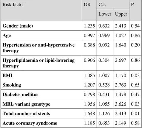
Univariate analysis revealed that MBL variant genotype (A/O + O/O) (OR: 1.784, p=0.047) and total number of implanted stents (OR: 0.519, p=0.034) influence the development of in-stent restenosis. We performed multivariate adjustment for known risk factors (gender, age, BMI, hypertension, dyslipidaemia, diabetes mellitus, smoking, acute coronary disease and total number of implanted stents) and MBL variant genotype (A/O + O/O) as covariates. The regression analysis revealed that MBL variant genotype (OR: 1.95, p=0.03), BMI (OR: 1.08, p=0.03) and total number of implanted stents (OR: 1.64, p=0.01) are independently associated with the development of in-stent restenosis (Table 2).
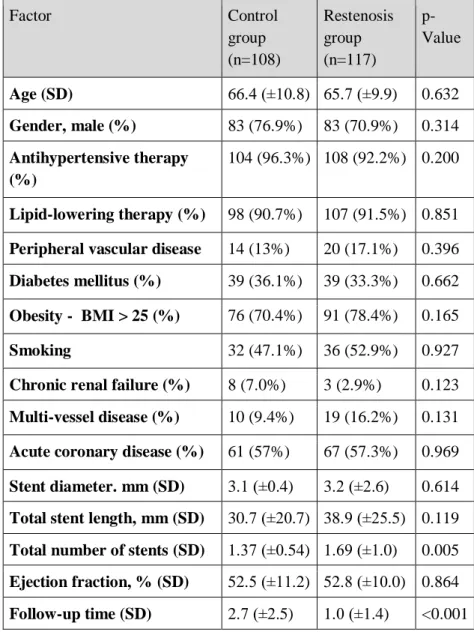
Table 1. - Patient characteristics and ISR risk factors in the control and restenosis groups. Data is presented as mean (SD) and n (%).
Factor Control
group (n=108)
Restenosis group (n=117)
p- Value
Age (SD) 66.4 (±10.8) 65.7 (±9.9) 0.632 Gender, male (%) 83 (76.9%) 83 (70.9%) 0.314 Antihypertensive therapy
(%)
104 (96.3%) 108 (92.2%) 0.200
Lipid-lowering therapy (%) 98 (90.7%) 107 (91.5%) 0.851 Peripheral vascular disease 14 (13%) 20 (17.1%) 0.396 Diabetes mellitus (%) 39 (36.1%) 39 (33.3%) 0.662 Obesity - BMI > 25 (%) 76 (70.4%) 91 (78.4%) 0.165 Smoking 32 (47.1%) 36 (52.9%) 0.927 Chronic renal failure (%) 8 (7.0%) 3 (2.9%) 0.123 Multi-vessel disease (%) 10 (9.4%) 19 (16.2%) 0.131 Acute coronary disease (%) 61 (57%) 67 (57.3%) 0.969 Stent diameter. mm (SD) 3.1 (±0.4) 3.2 (±2.6) 0.614 Total stent length, mm (SD) 30.7 (±20.7) 38.9 (±25.5) 0.119 Total number of stents (SD) 1.37 (±0.54) 1.69 (±1.0) 0.005 Ejection fraction, % (SD) 52.5 (±11.2) 52.8 (±10.0) 0.864 Follow-up time (SD) 2.7 (±2.5) 1.0 (±1.4) <0.001
Table 2. – Results of the multivariate logistic regression analysis with adjustment for generally known risk factors and MBL variant genotype (A/O + O/O) with in-stent restenosis as a dependent variable. Hosmer Lemeshow test p=0.477. OR – odds-ratio, C.I. – confidence interval, BMI – body mass index, MBL – mannose- binding lectin.
Risk factor OR C.I. P
Lower Upper Gender (male) 1.235 0.632 2.413 0.54
Age 0.997 0.969 1.027 0.86
Hypertension or anti-hypertensive therapy
0.388 0.092 1.640 0.20 Hyperlipidaemia or lipid-lowering
therapy
0.906 0.304 2.697 0.86
BMI 1.085 1.007 1.170 0.03
Smoking 1.207 0.528 2.763 0.65
Diabetes mellitus 0.798 0.431 1.478 0.47 MBL variant genotype 1.956 1.055 3.626 0.03 Total number of stents 1.648 1.126 2.413 0.01 Acute coronary syndrome 1.185 0.653 2.149 0.58
4.2. The role of VEGF polymorphisms in development of in-stent restenosis
In case of the VEGF polymorphisms (rs2010963 and rs699947) the genotyping procedure was successfully performed for 205 patients.
Allele frequencies were similar between genders (rs2010963: 50.1%
/ 41.7% / 9.3% in men and 40.7% / 50% / 9.3% in women, (G/G, G/C, C/C) p=0.454; rs699947: 25.5% / 48.3% / 26.2% in men and 15.4% / 48.1% / 36.5% in women, (A/A, A/C, C/C) p=0.209). The genotype distribution conformed to Hardy–Weinberg equilibrium (p
=0.45 and p=0.65 respectively).
Table 3. – Genotype distribution of control versus ISR group, Khi- square.
Polymorphism Genotype Control group n (%)
ISR group n (%)
p-value
rs2010963 Homozygous normal (G/G)
41 (41.0%)
58 (55.2%)
0.041 Variant
genotype (G/C + C/C)
59 (59.0%)
47 (44.8%) rs699947 Homozygous
normal (A/A) 21 (21.0%)
28 (26.7%)
0.342 Variant
genotype (A/C + C/C)
79 (79.0%)
77 (73.7%)
Comparison of the genotype distribution of the control group to the diffuse in-stent restenosis group shown that diffuse in stent restenosis was significantly less frequent in C/G and C/C genotypes (variant carrier) of rs2010963 polymorphism versus individuals with the G/G (homozygous normal) genotype (OR 0.56, p=0.04). Restenosis frequency did not differ between the two groups for rs699947 polymorphism (Table 3.).
Multivariate logistic regression has been performed with adjustment for clinical variables that reached p value of <0.3 when comparing patients with and without significant diffuse ISR (Table 4.).
Table 4. – Results of the multivariate logistic regression analyses with adjustment for risk factors and VEGF rs2010963 variant genotype (G/C + C/C) with in-stent restenosis as a dependent variable. Hosmer Lemeshow test p=0.139. OR – odds-ratio, C.I. – confidence interval, BMI – body mass index, VEGF – vascular endothelial growth factor.
Factor OR C.I. p
Lower Upper
Hypertension 0,450 0,043 4,692 0,503
BMI 1.05 0.906 1.235 0.478
Smoking 0.493 0.088 2.757 0.42
Chronic renal failure 0.754 0.034 16.807 0.859 Multi-branch stented 0.982 0.217 14.368 0.596 Total number of stents 1.441 0. 421 4.939 0.561 Average time to repeated
angiogram
0.226 0.756 0.413 0.004 Total stent length 1.002 0.958 1.049 0.992
VEGF rs2010963 0.754 0.034 0.535 0.003
Multivariate analysis adjusted for clinical variables (BMI, hypertension, smoking, chronic renal failure, average time to repeated angiogram, multiple branch stent deployment, total stent length and total number of implanted stents) revealed that the homozygous normal (A/A) genotype of rs2010963 is related to higher risk for diffuse ISR. The rs699947 polymorphism of VEGF gene is not associated with a risk of diffuse ISR
5. CONCLUSIONS
In-stent restenosis followed by percutaneous coronary intervention still forms a clinically important problem. Based on the findings of our studies, certain polymorphisms are associated with the development of in-stent restenosis.
1) MBL2 gene’s rs5030737, rs1800450 and rs1800451 polymorphisms are associated with the development of diffuse in-stent restenosis after BMS stent implantation. ISR occurs significantly more frequent in variant allele carriers, therefore patients with these genotypes have higher risk for ISR.
2) We have not found association between VEGF gene’s rs699947 polymorphism and the development of diffuse ISR after BMS implantation.
3) In the case of VEGF rs2010963 polymorphism homozygous normal genotype was associated with higher rate of diffuse in-stent restenosis after BMS implantation. The carriage of the variant allele is a protecting factor against the development of coronary in-stent restenosis.
In summary, polymorphism rs2010963 of VEGF gene is independent protecting factor and polymorphisms of MBL2 gene are independent risk factors in the development of diffuse in-stent restenosis.
6. Publications
List of publications related to the doctoral work
1. Bagyura Z, Kiss L, Hirschberg K, Berta B, Széplaki G, Lux A, Szelid Z, Soós P, Merkely B. (2017) Association between VEGF Gene Polymorphisms and In-Stent Restenosis after Coronary Intervention Treated with Bare Metal Stent. Dis Markers, 2017:1-7. IF 2,137
2. Bagyura Z, Kiss L, Berta B, Szilágyi A, Hirschberg K, Széplaki G, Lux A, Szelid Z, Soós P, Merkely B. (2017) High rate of in- stent restenosis after coronary intervention in carriers of the mutant mannose-binding lectin allele. BMC Cardiovasc Disord, 17: 1-4. IF 1,916
Other publications
1. Kiss LZ, Bagyura Z, Vadas R, Polgár L, Lux Á, Édes E, Szenczi O, Soós P, Szelid Z, Becker D, Jermendy G, Merkely B.
(2017) Signs of subclinical atherosclerosis in asymptomatic patients at increased risk of type 2 diabetes mellitus. J Diabetes Complications, 31: 1293-1298. IF 2,734
2. Kovács K, Szakmár E, Méder Ü, Kolossváry M, Bagyura Z, Lamboy L, Élő Z, Szabó A, Szabó M, Jermendy Á. (2017) Hypothermia treatment in asphyxiated neonates - a single center experience in Hungary. Orv Hetil, 158: 331-339. IF 0,291 3. Bagyura Z, Kolossvary M, Merkely B, Maurovich-Horvat P.
(2017) A coronariarendszer komputertomográfiás vizsgálata - Országos Plakk Regiszter és Adatbázis (OPeRA). Orv Hetil, 158: 106-110. IF 0,291
4. Boros AM, Szeplaki G, Perge P, Jenei Z, Bagyura Z, Zima E, Molnar L, Apor A, Becker D, Geller L, Prohaszka Z, Merkely B.
(2016) The ratio of the neutrophil leucocytes to the lymphocytes predicts the outcome after cardiac resynchronization therapy.
Europace, 18: 747-754. IF 4,021
5. Szilveszter B, Elzomor H, Karolyi M, Kolossvary M, Raaijmakers R, Benke K, Celeng C, Bartykowszki A, Bagyura Z, Lux A, Merkely B, Maurovich-Horvat P. (2016) The effect of iterative model reconstruction on coronary artery calcium quantification. Int J Cardiovasc Imaging, 32: 153-160. IF 1,880 6. Bagyura Zs, Kolossváry M, Merkely B, Maurovich-Horvat P.
(2015) Személyre szabott kardiovaszkuláris rizikóbecslés koronária CT-vel Strukturált leletezés és az OPeRA (Országos Plaque Regiszter és Adatbázis) Projekt. Informatika és Management az Egészségügyben, 14: 19-23.
7. Kelloniemi A, Szabo Z, Serpi R, Napankangas J, Ohukainen P, Tenhunen O, Kaikkonen L, Koivisto E, Bagyura Z, Kerkela R, Leosdottir M, Hedner T, Melander O, Ruskoaho H, Rysa J.
(2015) The Early-Onset Myocardial Infarction Associated PHACTR1 Gene Regulates Skeletal and Cardiac Alpha-Actin Gene Expression. PLoS One, 10: p. e0130502. IF 3,057
8. Merkely B, Gara E, Lendvai Z, Skopal J, Leja T, Zhou W, Kosztin A, Varady G, Mioulane M, Bagyura Z, Nemeth T, Harding SE, Foldes G. (2015) Signalling via PI3K/FOXO1A Pathway Modulates Formation and Survival of Human Embryonic Stem Cell-Derived Endothelial Cells. Stem Cells Dev, 24: 869-878. IF 3,777
9. Moilanen AM, Rysa J, Kaikkonen L, Karvonen T, Mustonen E, Serpi R, Szabo Z, Tenhunen O, Bagyura Z, Napankangas J, Ohukainen P, Tavi P, Kerkela R, Leosdottir M, Wahlstrand B, Hedner T, Melander O, Ruskoaho H. (2015) WDR12, a Member of Nucleolar PeBoW-Complex, Is Up-Regulated in Failing
Hearts and Causes Deterioration of Cardiac Function. PLoS One, 10: p. e0124907. IF 3,057
10. Szelid Z, Lux A, Kolossvary M, Toth A, Vago H, Lendvai Z, Kiss L, Maurovich-Horvat P, Bagyura Z, Merkely B. (2015) Right Ventricular Adaptation Is Associated with the Glu298Asp Variant of the NOS3 Gene in Elite Athletes. PLoS One, 10:
e0141680. IF 3,057
11. Bagyura Z, Kiss L, Edes E, Lux A, Polgar L, Soos P, Szenczi O, Szelid Z, Vadas R, Jozan P, Bagdy G, Merkely B. (2014) Cardiovascularis szűrőprogram a közép-magyarországi régióban.
Budakalász Vizsgálat. Orv Hetil, 155: 1344-1352.
12. Reed DM, Foldes G, Gatheral T, Paschalaki KE, Lendvai Z, Bagyura Z, Nemeth T, Skopal J, Merkely B, Telcian AG, Gogsadze L, Edwards MR, Gough PJ, Bertin J, Johnston SL, Harding SE, Mitchell JA. (2014) Pathogen Sensing Pathways in Human Embryonic Stem Cell Derived-Endothelial Cells: Role of NOD1 Receptors. PLoS One, 9: e91119. IF 3,234
13. Bagyura Z, Szelid Z, Soós P, Szenczi O, Maurovich-Horvát P, Édes E, Lux Á, Polgár L, Andrási Z, Tátrai A, Józan P, Merkely B. (2012) Magyarországi primer prevenciós populációs felmérés:
Budakalász Epidemiológiai Vizsgálat előzetes eredmények.
Orvosképzés, 87: 102-108.
14. Kalman M, Mahalek J, Adorjan A, Adorjan I, Pocsai K, Bagyura Z, Sadeghian S. (2011) Alterations of the perivascular dystrophin-dystroglycan complex following brain lesions. An immunohistochemical study in rats. Histol Histopathol, 26:
1435-1452. IF 2,480
15. Soós P, Schmack B, Istók R, Polgár L, Bagyura Zs, Veres G, Merkely B, Szabó G, Weymann A. (2011) Szövetépítés egész szíven. Cardiologia Hungarica, 41: 373-378.
16. Szelid Zs, Soós P, Bagyura Zs, Merkely B. (2011) Személyre szabott medicina a kardiológiában. Orvosképzés, 86: 111-114.
17. Bagyura Z, Pocsai K, Kalman M. (2010) Distribution of components of basal lamina and dystrophin-dystroglycan complex in the rat pineal gland: differences from the brain tissue and between the subdivisions of the gland. Histol Histopathol, 25: 1-14. IF 2,502
18. Pocsai K, Bagyura Z, Kalman M. (2010) Components of the basal lamina and dystrophin-dystroglycan complex in the neurointermediate lobe of rat pituitary gland: different localizations of beta-dystroglycan, dystrobrevins, alpha1- syntrophin, and aquaporin-4. J Histochem Cytochem, 58: 463- 479. IF 2,381